tmd disease: treatment module three - dental learning · the academy of dental learning and osha...
TRANSCRIPT
TMD Disease: Treatment Module Three
The Academy of Dental Learning and OSHA Training, LLC, designates this
activity for 4 continuing education credits (4 CEs).
William D. Bellavia, DDS, MAGD, FAANaOS-L, FABDMS-C
Health Science Editor: Megan Wright, RDH, MS
Publication Date: June 2013
Updated Date: August 2017
Expiration Date: September 2020
The Academy of Dental Learning and OSHA Training, LLC is an ADA CERP Recognized
Provider. ADA CERP is a service of the American Dental Association to assist dental
professionals in identifying quality providers of continuing dental education. ADA CERP does not
approve or endorse individual courses or instructors, nor does it imply acceptance of credit hours
by boards of dentistry. Concerns or complaints about a CE provider may be directed to the
provider or to the Commission for Continuing Education Provider Recognition at ADA.org/CERP.
Conflict of Interest Disclosure: ADL does not accept promotional or commercial funding in
association with its courses. In order to promote quality and scientific integrity, ADL's evidence-
based course content is developed independent of commercial interests. Refund Policy: If you
are dissatisfied with the course for any reason, prior to taking the test and receiving your
certificate, return the printed materials within 15 days of purchase and we will refund your full
tuition. Shipping charges are nonrefundable.
California Registered Provider Number: RP5631

1
Answer Sheet: TMD Disease: Module Three
1. _______
2. _______
3. _______
4. _______
5. _______
6. _______
7. _______
8. _______
9. _______
10. _______
11. _______
12. _______
13. _______
14. _______
15. _______
16. _______
17. _______
18. _______
19. _______
20. _______
21. _____
22. _____
23. _____
24. _____
25. _____
Name: ________________________________________ Profession: _________________________
License State: ____________ License Number: ________________ Expiration Date
Address
City: ____________________________________ State: __________ Zip Code:
Telephone:________________________________ Fax: ____________________________________
E-mail:
If you have downloaded the course and printed the answer sheet from the Internet please enter
payment information below.
Card type: ___________________ Card Number:___________________________________________
Exp. Date: _______________ Name as it appears on card: __________________________________
*To enter your answers online you MUST return to our website www.dentallearning.org.
Return answer sheet:
Via fax: 518.514.1103
Via email: [email protected]
Postal Mail: ADL, PO Box 14585, Albany, NY 12212
***PLEASE PRINT CLEARLY; ILLEGIBLE ANSWER SHEETS WILL NOT BE
PROCESSED.
Notes:

2
Course Evaluation
Please place an X in the box to rate these
statements:
Poor Fair Good Very
Good
Excellent
The content fulfills the overall purpose of the course.
The content fulfills each of the course objectives.
The course subject matter is accurate.
The material presented is understandable.
The teaching/learning method is effective.
The answers to the test questions are appropriately
covered in the course.
How would you rate this course overall?
Time to complete the entire course and the test? Hours: _________ Minutes: _______
Other Search Engine
Friend/Coworker
Other
Do you have any suggestions about how we can improve this course? If so please note them on a
separate sheet of paper and send it in with your answer sheet.
If you studied the course online, did all the links work? If not please note the page and link on a separate
sheet of paper and send it in with your answer sheet so we can fix it.

3
Instructions
1. Review the Objectives: Objectives provide an overview of the entire course.
2. Read the course material.
3. Complete the test:
a. Return to our website: www.dentallearning.org, click on Take the Exam,
enter your answers, register, if you are new customer (existing customers
login), pay for the course, click Grade Test. Your test will be graded
immediately. If required, complete the course evaluation. Your certificate
will display for you to print.
b. If you would rather, you may return your completed answer sheet and
course evaluation to us via the options listed below.
To successfully complete the course you must score 80% or above on the test. If you
do not score 80% you may retake the test one more time free of charge. If you fail a
second time you must purchase a new course and test.
If you’ve downloaded this coursebook off the Internet you can:
Return to our website (www.dentallearning.org) to take the test online (only if you have not purchased the coursebook separately). You will need to provide credit card information at the time you submit your test online for scoring.
Write your answers on the one-page answer sheet included in this book, complete the credit card payment information, and return the form to the address below, fax, or email address below. Or, you may send a check or money order to the address below with your answer sheet.
Academy of Dental Learning and OSHA Training, LLC (ADL)
P.O. Box 14585
Albany, NY 12212
Fax: 518-514-1103
Email: [email protected]
Answer sheets received without payment will not be processed.
We grade all tests in a timely manner; if you do not receive your certificate within five
days, please email ([email protected]) or call us: 518-209-9540.
There is no time limit for return of your answer sheet. Completion dates are taken from
the envelope postmark or the finish date recorded in the computer when you do an
online exam. Tests MUST be completed in the licensing cycle you wish to use the
credits.
If you are dissatisfied with the course for any reason, prior to taking the test and
receiving your certificate, return the printed materials within 15 days of purchase and we
will refund your full tuition. Shipping charges are nonrefundable.
4
If someone else would like to use this material after you are done, he or she may register with us and take advantage of a “sharing discount”. Courses downloaded from the Internet can be shared at the same tuition rate as currently available on our website. Please call us if you need an extra answer sheet or download one from our website. There is no “sharing discount” for online exams. The author and ADL have made every effort to include information in this course that is
factual and conforms to accepted standards of care. This course is not to be used as a
sole reference for treatment decisions. It is your responsibility to understand your legal
obligations and license requirements when treating patients. ADL is not responsible for
the misuse of information presented in this course. The material in this course cannot
be reproduced or transmitted in any way without the written consent of ADL.

5
Table of Contents
Answer Sheet 1
Evaluation 2
Instructions 3
Table of Contents 5
Educational Objectives 6
The Principle Goals and Philosophy 6
Phase I 6
Oral Medications 7
Nutrition Counseling 8
Hydrocollator 8
Transcutaneous Electrical Nerve Stimulations (TENS Unit) 9
Splint Therapy 10
Maxillary Splints 12
Mandibular Splints – History 13
New in Temporary Splints 14
Diagnosis MPD - Vapo-Collant Sprays 16
Diagnosis MPD – Electro/Ultrasound Therapy 16
Botox Injections 17
Continued Evaluation 18
Diagnosis Temporal Tendonitis/Stylomandibular Ligamentitis 19
Conclusion Phase I 21
Phase II 21
Diagnosis MPD – Occlusal Equilibration 21
Referrals 22
Conclusion Phase II 22
Phase III Treatment 22
Time Interval One Year After Phase I 23
Review and Detailed Treatment of Disorders Inclusive in TMD 23
Greater and Lesser Occipital Nerve INTIngement 25
Dental Disease/Differential Diagnosis 27
2017 New Alternative Treatments/Less Invasive 28
Conclusion 29
References 29
Course Test 31
6
Educational Objectives
Upon completion of the course, the student will:
Understand the basic treatment options for TMJ. Identify the differing methods of treating TMJ. List the steps to follow in treating TMJ. Identify other diseases associated with TMJ.
The Principal Goals and Philosophy
The principal goals of treatment are:
• minimal treatment
• reversible treatment
• do the patient no harm
Once the diagnosis has been made it is time to home in on specific treatment.
In the management of this disease I found it useful to break treatment into three phases.
Phase I
Phase I can also be referred to as treatment of the acute stage. During this phase the
chief complaint is the focus. Once that has been treated successfully other maladies
may surface. Rather than charge a separate fee and begin the process anew, I found it
advantageous for both the patient and the clinician to approach TMD with a global fee
for Phase I and a time limit of six weeks. Six weeks seemed an adequate time to treat
the chief complaint and determine any contributing underlying maladies.
The rationale for a global fee was similar; it allowed the clinician the freedom to treat
muscle spasm, nerve impingements, sprains, etc. without having to revisit fees. This is
by no means set in stone; it is the method I felt best suited this syndrome.
Moreover, since most patients have a myriad of problems in this anatomical region, it
aids the patient in that the treatment is not limited to one diagnosis. It benefited the
clinician in that he/she is able to determine the best modalities to treat each contributing
condition.
I would explain to the patient that the six week treatment is like ‘peeling an onion’. There are several layers or conditions which contribute to TMD other than their chief complaint. When we are finished concentrating on one ‘layer’ or complaint, often another becomes evident. This approach to treatment allows the clinician to move swiftly and smoothly from one diagnosis to another and to treat them in as an
7
effective and efficient means possible. This phase of treatment utilized reversible methods and modalities with minimal invasiveness. In my opinion, the ‘one treatment fits all’ for TMD has been and continues to be a failure.
Oral Medications
The etiology of TMD falls mainly into the range of inflammatory conditions. Therefore, the initial treatment most often includes oral medications in an attempt to reduce pain and inflammation.
Oral medications should be considered after a preliminary diagnosis has been made
and the medical history has been reviewed. Only after the clinician is certain that there
are no drug allergies or interactions with other medications presently being taken by the
patient should these drugs be prescribed. Consultation with the primary care physician
and or pharmacist is recommended for dosages and compatibility.
Analgesic: At this time the clinician should prescribe no more powerful an
analgesic than a short term dosage of extra strength acetaminophen. A good rule
of thumb is not to prescribe controlled substances to ‘strangers’. Get to know
your patients before prescribing addictive or mood altering drugs. Remember,
many of the drugs used in treatment can be abused. That being said, some
aspects of TMD may require controlled substances to regulate pain. Moreover, in
a TMD practice, often the patient presents with a smorgasbord of analgesic
prescriptions from other offices.
Muscle Relaxants: If MPD is a major component in the diagnosis, then a
skeletal muscle relaxant such as Robaxin (Methocarbamol) or Flexeril
(Cyclobenzaprine hydrochloride) should be considered.
NSAID: There are several Non-Steroidal Anti-Inflammatory Drugs which can be
helpful. Long term use should be avoided due to heart complications. Yet, long
term use of drugs such as Mobic can be very helpful to these patients (consult
with the patient’s physician and pharmacist should be made).
If the patient is not able to take long term medications, a drug such as Motrin
(ibuprofen 600 mg) administered three times a day for a period of five days can
at times act as an effective anti-inflammatory (take with food).
Prednisone when administered in a dose pack will quell almost any
inflammatory malady within the family of TMD maladies. Again it should not be
used long term and not prescribed unless the patient’s pharmacy and physician
are consulted and should be taken with food.

8
Tramadol hydrochloride (ultram), hydrocodone, and various codeine
compounds are also an effective analgesic.
There are innumerable other drugs available, but the above are on the frontline of the
battle against TMD.
Nutrition counseling
This is very important in this aspect of treatment because the use of high doses of sugar, caffeine, alcohol and other simple carbohydrates have been reported to cause and or precipitate various types of headaches and/or muscles to remain in spasm (http://voices.yahoo.com/curing-sugar-headache-5071906.html).
It is important to remember that the goal in rendering this advice is not to lose weight;
the goal is to make the patient aware that foods such as sugars, refined flour, and salt
can often be the root cause of pain. Minimizing these ingredients will significantly
reduce pain and improve the overall health of the patient. If he/she presents as obese
referral to proper nutritional professionals is advised.
Details: I instructed the patient to write down everything that he/she ate for two days.
Since the patients were seen twice a week – Tues/Thurs. it was not difficult to review
their progress. This aspect of treatment was easily delegated to expert nutritional
personnel within the office.
Therefore, if one has a nutritional expert on staff, the clinician may begin nutritional
counseling at this time. In our office, nutritional therapy was usually rendered while
awaiting the construction of the intra-oral splint.
Hydrocollator
During these appointments and after ROM and muscle palpation, the clinician should
consider utilizing a Hydrocollator Unit on the most painful musculature. Although this
unit is most often associated with Physical Therapy, it does not preclude the dentist
from taking advantage of this modality. It is merely a hot water unit housing gel packs.
The packs render hot moist penetrating heat (for approximately 15 minutes)
encouraging vasodilatation and a decrease in muscle spasm.
9
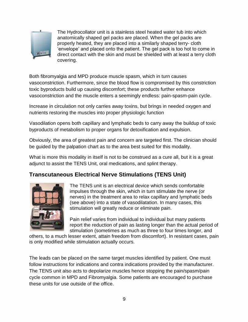
The Hydrocollator unit is a stainless steel heated water tub into which anatomically shaped gel packs are placed. When the gel packs are properly heated, they are placed into a similarly shaped terry- cloth ‘envelope’ and placed onto the patient. The gel pack is too hot to come in direct contact with the skin and must be shielded with at least a terry cloth covering.
Both fibromyalgia and MPD produce muscle spasm, which in turn causes
vasoconstriction. Furthermore, since the blood flow is compromised by this constriction
toxic byproducts build up causing discomfort; these products further enhance
vasoconstriction and the muscle enters a seemingly endless: pain-spasm-pain cycle.
Increase in circulation not only carries away toxins, but brings in needed oxygen and
nutrients restoring the muscles into proper physiologic function
Vasodilation opens both capillary and lymphatic beds to carry away the buildup of toxic
byproducts of metabolism to proper organs for detoxification and expulsion.
Obviously, the area of greatest pain and concern are targeted first. The clinician should
be guided by the palpation chart as to the area best suited for this modality.
What is more this modality in itself is not to be construed as a cure all, but it is a great
adjunct to assist the TENS Unit, oral medications, and splint therapy.
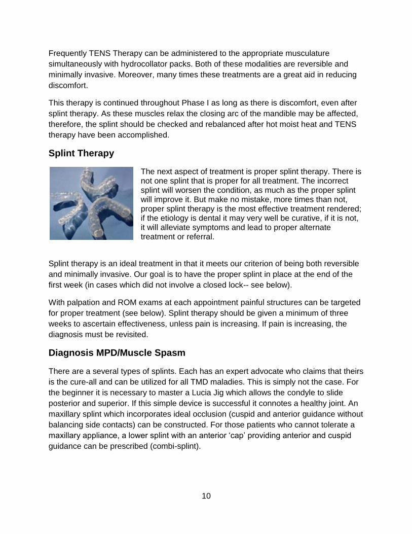
Transcutaneous Electrical Nerve Stimulations (TENS Unit)
The TENS unit is an electrical device which sends comfortable impulses through the skin, which in turn stimulate the nerve (or nerves) in the treatment area to relax capillary and lymphatic beds (see above) into a state of vasodilatation. In many cases, this stimulation will greatly reduce or eliminate pain. Pain relief varies from individual to individual but many patients report the reduction of pain as lasting longer than the actual period of stimulation (sometimes as much as three to four times longer, and
others, to a much lesser extent, attain freedom from discomfort). In resistant cases, pain is only modified while stimulation actually occurs.
The leads can be placed on the same target muscles identified by patient. One must
follow instructions for indications and contra indications provided by the manufacturer.
The TENS unit also acts to depolarize muscles hence stopping the pain/spasm/pain
cycle common in MPD and Fibromyalgia. Some patients are encouraged to purchase
these units for use outside of the office.
10
Frequently TENS Therapy can be administered to the appropriate musculature
simultaneously with hydrocollator packs. Both of these modalities are reversible and
minimally invasive. Moreover, many times these treatments are a great aid in reducing
discomfort.
This therapy is continued throughout Phase I as long as there is discomfort, even after
splint therapy. As these muscles relax the closing arc of the mandible may be affected,
therefore, the splint should be checked and rebalanced after hot moist heat and TENS
therapy have been accomplished.
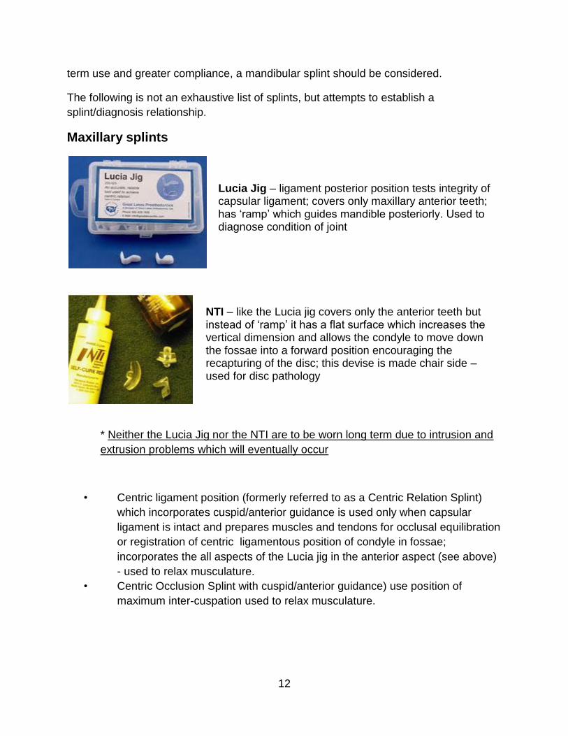
Splint Therapy
The next aspect of treatment is proper splint therapy. There is not one splint that is proper for all treatment. The incorrect splint will worsen the condition, as much as the proper splint will improve it. But make no mistake, more times than not, proper splint therapy is the most effective treatment rendered; if the etiology is dental it may very well be curative, if it is not, it will alleviate symptoms and lead to proper alternate treatment or referral.
Splint therapy is an ideal treatment in that it meets our criterion of being both reversible
and minimally invasive. Our goal is to have the proper splint in place at the end of the
first week (in cases which did not involve a closed lock-- see below).
With palpation and ROM exams at each appointment painful structures can be targeted
for proper treatment (see below). Splint therapy should be given a minimum of three
weeks to ascertain effectiveness, unless pain is increasing. If pain is increasing, the
diagnosis must be revisited.
Diagnosis MPD/Muscle Spasm
There are a several types of splints. Each has an expert advocate who claims that theirs
is the cure-all and can be utilized for all TMD maladies. This is simply not the case. For
the beginner it is necessary to master a Lucia Jig which allows the condyle to slide
posterior and superior. If this simple device is successful it connotes a healthy joint. An
maxillary splint which incorporates ideal occlusion (cuspid and anterior guidance without
balancing side contacts) can be constructed. For those patients who cannot tolerate a
maxillary appliance, a lower splint with an anterior ‘cap’ providing anterior and cuspid
guidance can be prescribed (combi-splint).

11
Combi-splint
If the Lucia Jig has caused an increase in pain then the etiology is likely the joint itself.
The Lucia Jig should be discarded and an NTI anterior splint placed.
Diagnosis Disc Displacement
This splint increases the vertical dimension while simultaneously encouraging the condyle to reposition anteriorly. If the disc injury is recent (acute) and can be recaptured, this devise will be successful. If the disc displacement is chronic a recapture splint may be attempted.
Once this diagnosis has been confirmed the treatment will likely enter Phase II and III.
Other Types of Splints
Splint therapy is often the most important aspect of long or short term dental treatment
of TMD. The question should not be if one should place an occlusal splint, but
what splint should one place. There are many types of occlusal splints available to
the clinician. Each is curative for a specific aspect of the disease. The greatest failure of
the TMD clinician is to use one splint as universal for all maladies.
In any discussion of splints one must categorize them by their major difference:
maxillary or mandibular. Maxillary splints are generally favored by the profession
because they require less attention. A maxillary splint once balanced tends to stay
balanced. This is in no small part due to the fact that although we were taught that
cranial bones are fixed joints, they are not (Upledger, J.E. & Vredevoob, J. D., 2017).
The maxillary splint tends to keep these sutures from moving, hence less adjustment.
Unfortunately along with less adjustment comes less healing and the patient cannot
achieve maximum benefit from this modality.
Moreover, not only is the mandibular splint more favored by the patient for ease of use
and esthetics, but also it allows the movement of the cranial sutures. Therefore, for long
12
term use and greater compliance, a mandibular splint should be considered.
The following is not an exhaustive list of splints, but attempts to establish a
splint/diagnosis relationship.
Maxillary splints
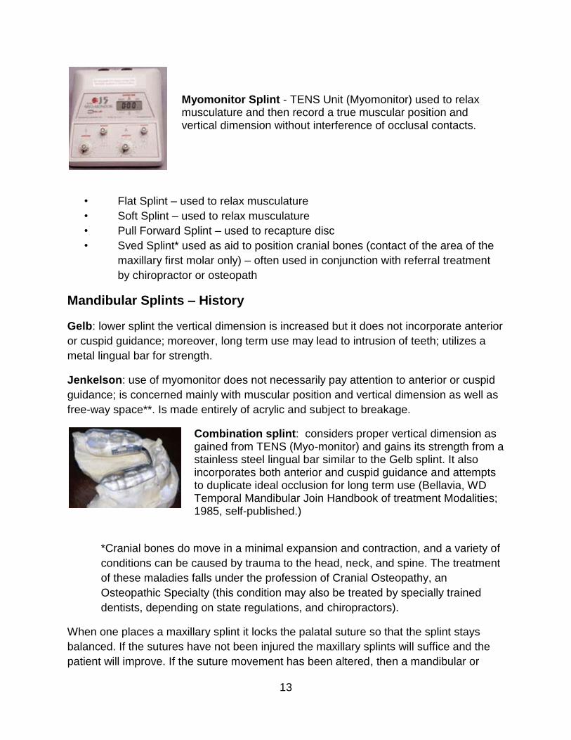
Lucia Jig – ligament posterior position tests integrity of capsular ligament; covers only maxillary anterior teeth; has ‘ramp’ which guides mandible posteriorly. Used to diagnose condition of joint
NTI – like the Lucia jig covers only the anterior teeth but instead of ‘ramp’ it has a flat surface which increases the vertical dimension and allows the condyle to move down the fossae into a forward position encouraging the recapturing of the disc; this devise is made chair side – used for disc pathology
* Neither the Lucia Jig nor the NTI are to be worn long term due to intrusion and
extrusion problems which will eventually occur
• Centric ligament position (formerly referred to as a Centric Relation Splint)
which incorporates cuspid/anterior guidance is used only when capsular
ligament is intact and prepares muscles and tendons for occlusal equilibration
or registration of centric ligamentous position of condyle in fossae;
incorporates the all aspects of the Lucia jig in the anterior aspect (see above)
- used to relax musculature.
• Centric Occlusion Splint with cuspid/anterior guidance) use position of
maximum inter-cuspation used to relax musculature.
13
Myomonitor Splint - TENS Unit (Myomonitor) used to relax musculature and then record a true muscular position and vertical dimension without interference of occlusal contacts.
• Flat Splint – used to relax musculature
• Soft Splint – used to relax musculature
• Pull Forward Splint – used to recapture disc
• Sved Splint* used as aid to position cranial bones (contact of the area of the
maxillary first molar only) – often used in conjunction with referral treatment
by chiropractor or osteopath
Mandibular Splints – History
Gelb: lower splint the vertical dimension is increased but it does not incorporate anterior
or cuspid guidance; moreover, long term use may lead to intrusion of teeth; utilizes a
metal lingual bar for strength.
Jenkelson: use of myomonitor does not necessarily pay attention to anterior or cuspid
guidance; is concerned mainly with muscular position and vertical dimension as well as
free-way space**. Is made entirely of acrylic and subject to breakage.
Combination splint: considers proper vertical dimension as gained from TENS (Myo-monitor) and gains its strength from a stainless steel lingual bar similar to the Gelb splint. It also incorporates both anterior and cuspid guidance and attempts to duplicate ideal occlusion for long term use (Bellavia, WD Temporal Mandibular Join Handbook of treatment Modalities; 1985, self-published.)
*Cranial bones do move in a minimal expansion and contraction, and a variety of
conditions can be caused by trauma to the head, neck, and spine. The treatment
of these maladies falls under the profession of Cranial Osteopathy, an
Osteopathic Specialty (this condition may also be treated by specially trained
dentists, depending on state regulations, and chiropractors).
When one places a maxillary splint it locks the palatal suture so that the splint stays
balanced. If the sutures have not been injured the maxillary splints will suffice and the
patient will improve. If the suture movement has been altered, then a mandibular or
14
maxillary Sved splint should be considered. The preferred mandibular splint, in my
opinion, is a ‘combi’ splint (designed by this author, WD Bellavia). This splint is the most
anatomic and is balanced (cuspid and anterior guidance). (See Upledger reference
above)
** Free-way space is the space between the maxillary and mandibular teeth
when the jaw is at rest.
New in Temporary Splints
According to Dental Research Journal, “The Aqualizer™, a hydrostatic oral splint, is a
newer application that automatically eliminates the distorting influence of the occlusion
on the functional position of the jaw, harmonizing muscles, bite, and body. Simple
insertion of the Aqualizer™ bite splint creates a muscle-dominant functionally generated
occlusion instantly. This occurs because the Aqualizer™ facilitates muscle-dominated
mandibular repositioning while it equalizes, axializes, balances, distributes, and makes
simultaneous all occlusal forces (Srivastava et all., 2013).
Further supporting this type of temporary splint in certain cases, is Spear Faculty and
contributing author, Gary DeWood, who states, “A posterior-contact-only appliance can
be life altering to wear when a patient is suffering from pain of an intra-articular origin.
Because they ‘unload’ the joints by minimizing the force passed through them when
teeth contact, they are often the only way to get a patient with intra-articular pain more
comfortable. When a pre-fabricated temporary posterior appliance is required, my
selection is the Aqualizer. An Aqualizer is composed of two reservoirs filled with water
that cover the occlusal surfaces of posterior teeth. The reservoirs are connected by a
channel that ‘equalizes’ the Aqualizer by moving water between the two reservoirs to
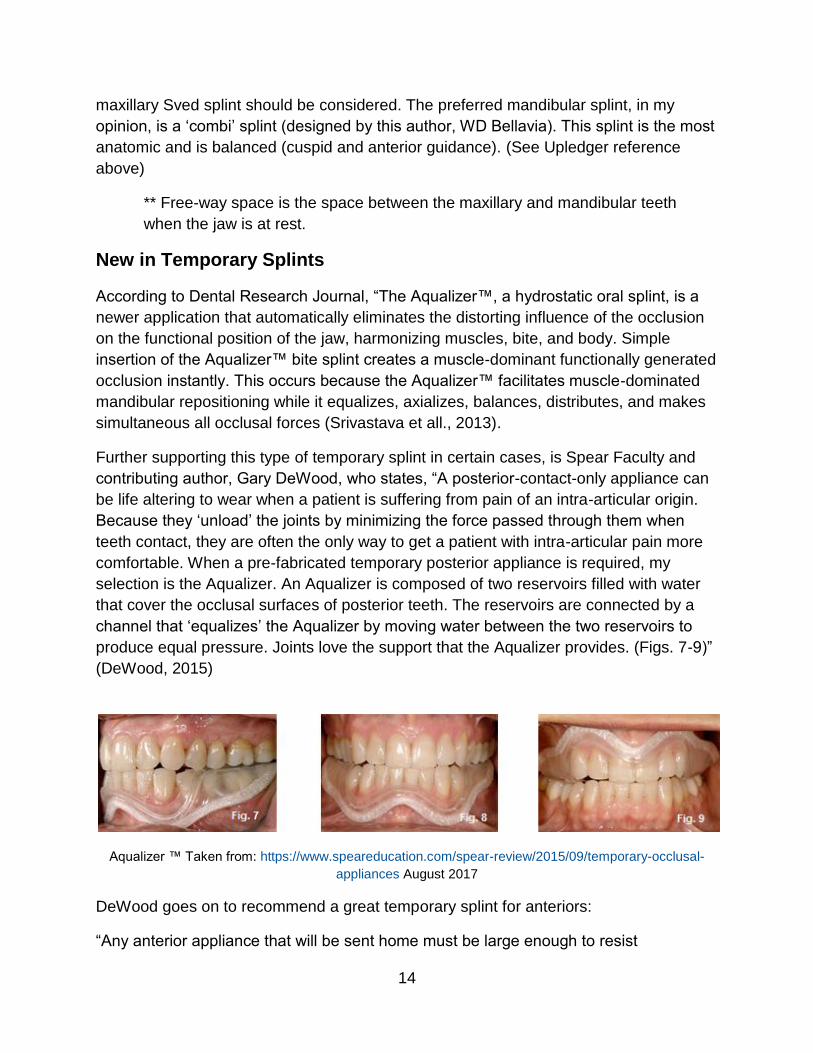
produce equal pressure. Joints love the support that the Aqualizer provides. (Figs. 7-9)”
(DeWood, 2015)
Aqualizer ™ Taken from: https://www.speareducation.com/spear-review/2015/09/temporary-occlusal-
appliances August 2017
DeWood goes on to recommend a great temporary splint for anteriors:
“Any anterior appliance that will be sent home must be large enough to resist
15
swallowing and aspiration. I want one that is very easy to fabricate, insert and adjust. It
would be beneficial if it could also provide me with some information about what a
patient does with his or her teeth by creating wear on the appliance that we can read. A
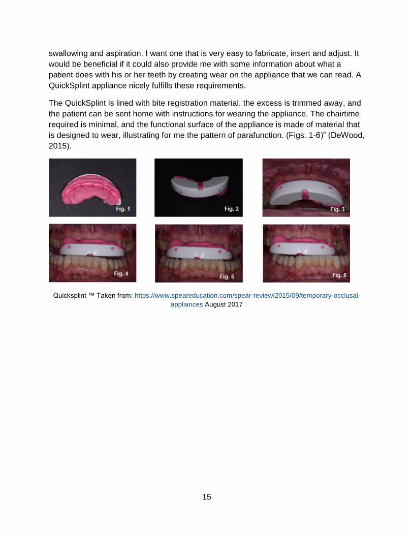
QuickSplint appliance nicely fulfills these requirements.
The QuickSplint is lined with bite registration material, the excess is trimmed away, and
the patient can be sent home with instructions for wearing the appliance. The chairtime
required is minimal, and the functional surface of the appliance is made of material that
is designed to wear, illustrating for me the pattern of parafunction. (Figs. 1-6)” (DeWood,
2015).
Quicksplint ™ Taken from: https://www.speareducation.com/spear-review/2015/09/temporary-occlusal-
appliances August 2017

16
Comparison of Splints
Diagnosis MPD - Vapo-Coolant Sprays
Treatment of spastic musculature and or trigger points has been treated successfully
with Fluormethane vapo-coolant sprays (see Travell above). Fluoromethane spray is
nontoxic and tolerated well if inhaled. The trigger area is stretched by the clinician and
the muscle is sprayed from origin to insertion (do not spray in back and forth motion).
The clinician can utilize several sweeps with an optimum speed approximately 4 inches
per second. Do not frost the skin.
This treatment should be followed by the use of hydrocollator gel pack for at least five
minutes for optimum results.
The mechanism: the spray crates an afferent barrage to the myelinated nerve fibers (hot
and cold) which in turn overwhelms the central nervous system taking up receptor sites
commonly used by unmylinated (slower) afferent neurons carrying painful stimuli.
Diagnosis MPD – Electro/Ultrasound Therapy
These are helpful modalities which can be used by a physical therapist or chiropractor.

17
Diagnosis MPD Muscle Spasm Resistant to TENS, Hydrocollator, and Splint
Therapies Consider Injections
Injection Therapy
Trigger points are common in MPD as well as Fibromyalgia. They are areas or nodules
of hyperactivity within a muscle. These areas are exquisitely painful to the touch and
readily identified via palpation http://www.triggerpoints.net/.
The areas consist of concentrations of toxic byproducts of metabolism and the ensuing
vasoconstriction does not allow adequate blood flow to bring in needed oxygen or
eliminate harmful toxins. Therefore all injections into these areas must encourage
vasodilatation. The use of epinephrine or other vasoconstrictors common in commercial
preparations of dental local anesthetics must be avoided.
The most successful treatment of these areas utilizes a technique pioneered by Janet Travell, MD called dry needling (Travell, J.B. & Simons, D. C., 2013). This technique does not utilize any drugs. The clinician stimulates the trigger area with repeated shallow penetrations of the trigger area (muscle) by the tip of the needle. This often times will produce depolarization of said muscle. The ensuing relaxation of the muscle causes vasodilatation.
If this technique is not successful then an injection with local anesthetic (w/o
vasoconstrictor) is recommended. Procaine is the drug of choice.
Successful treatment occurs when the trigger area subsides and or the relief of pain
outlives the effect of the local anesthetic. If the muscle no longer exhibits a trigger point
upon subsequent visits no further treatment is necessary. If the trigger persists, then re-
inject using the local anesthetic (w/o vasoconstrictor) should be considered.
Botox Injections
The literature is replete with clinicians expressing the virtues of Botox injections into
these muscles. Nevertheless, in a double blind study conducted by Dr. Susan Herring of
the University of Washington in Seattle, strongly indicated the effectiveness of
botulinum toxin type A for persistent myofascial TMD pain was no better than injections
of saline. Therefore, I cannot recommend the use of this pharmaceutical when less
costly and more effective drugs are readily available.

18
In a July 2015 article on the TMJ Association, Ltd website, it revealed astounding
research against botox! It says, “An article published online in the journal Bone1 by a
team of French investigators [Kun-Darbois, J.-D., Libouban, H., Chappard, D.] confirms
that injecting Botox® into jaw muscles leads to significant bone loss in adult rats. Two
jaw-closing muscles—the right masseter and right temporalis, were treated, and the rats
were compared with control animals that had salt solution (saline) injected into the same
muscles. The authors were primarily interested in the mandible and whether it would
lose bone when these muscles were injected with Botox®, which is a neurotoxin that
causes temporary muscle paralysis. (This is similar to the bone loss experienced by
astronauts in spaceflight conditions because bone is not subject to normal gravitational
forces.) The Botox® rats evidently had some trouble chewing, because they lost a little
weight, whereas the saline animals gained during the study. The mandibles were
examined in detail one month after the Botox® or saline injections. Saline injection had
no effect, with right and left sides of the mandible the same in all measurements. The
uninjected left sides of the Botox® rats remained in the normal range, but on the
injected right sides, substantial bone loss occurred. About 20% of bone was lost from a
part of the mandible that supports the teeth while 35% was lost from the head of the
mandible (the condyle). It is particularly striking that the loss was so rapid, taking place
in only 4 weeks. Another finding of interest was that on the Botox® side, a lump of bony
tissue formed where another muscle (the digastric) attaches to the mandible. The
authors speculate that the digastric had become overactive in an attempt to
compensate for the paralyzed muscles. The authors point out that if bone losses of this
scope occur in the human mandible after Botox® treatment, patients might be at serious
risk of jaw fractures. With this paper, mandibular bone loss (osteopenia) following
Botox® injection into jaw muscles has now been demonstrated in two different animal
species. A previous study on rabbits (Rafferty et al. Bone 50:651-662, 2012) also
showed extensive bone loss in both the condyle and tooth-supporting region one month
after masseter injection. That study also showed that the condyles were still osteopenic
after a 3-month recovery period, and it is not clear whether full recovery ever takes
place. This confirmation of bone loss in an unrelated mammal (rabbits are not rodents)
strongly implies that the phenomenon must affect humans as well. A preliminary study
(Raphael et al., 2014) in which members of the TMJ Association participated indicated
that humans do indeed lose condylar bone after Botox® treatment of the jaw muscles.
Patients and practitioners need to be aware that jaw muscle activity is an important
element of jaw bone health, and that removing muscle activity with paralytic agents like
Botox® has a deleterious effect.” (TMJ Association, Ltd, Kun-Darbois et all, 2015)
Continued Evaluation
If muscles, tendons, joints do not respond after four weeks (splint delivered at the end of
the first week and the patient was seen twice a week), and after a course of oral
19
analgesics and or anti-inflammatory drugs, muscle relaxants, and nutrition counseling
and if the patient claims he/she has been compliant, the following must be considered:
Is the diagnosis correct?
Patient compliance – has the patient followed the instructions for the use of the splint
and is he/she taking medications properly. What is more, have they followed nutrition
instructions?
Corticosteroid Injections
Corticosteroids - methylprednisolone acetate – brand name Depro-Medrol 10 mg/cc.
This injectable can be mixed with the local anesthetics injected into insertion sites of
affected ligaments and tendons.
Diagnosis Temporal Tendonitis/Stylomandibular Ligamentitis/TMJ
Capsilitis
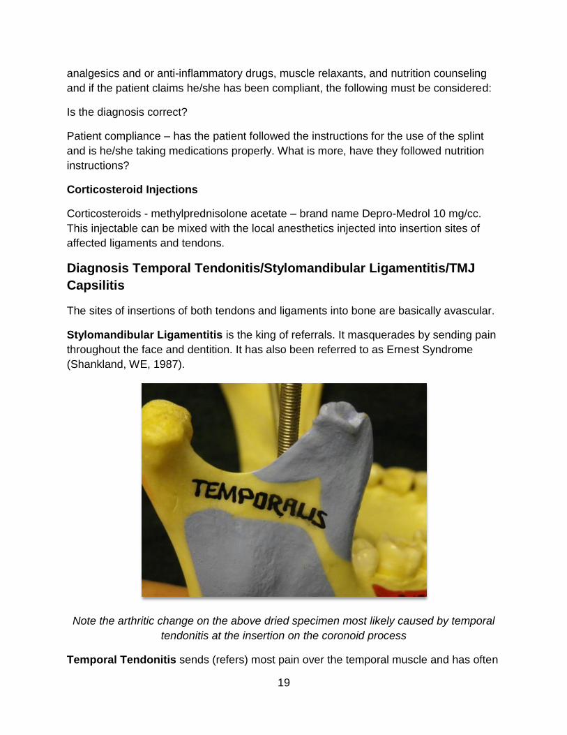
The sites of insertions of both tendons and ligaments into bone are basically avascular.
Stylomandibular Ligamentitis is the king of referrals. It masquerades by sending pain
throughout the face and dentition. It has also been referred to as Ernest Syndrome
(Shankland, WE, 1987).
Note the arthritic change on the above dried specimen most likely caused by temporal
tendonitis at the insertion on the coronoid process
Temporal Tendonitis sends (refers) most pain over the temporal muscle and has often
20
been called a “migraine-mimic”. It is associated with severe headaches (Ernest, Edwin
III, 2017).
Therefore, in order to insure long lasting pain relief at these sites, injections of local
anesthetic should contain vasoconstrictors. The drug of choice is Xylocaine (fast acting)
with vasoconstrictor. After the Xylocaine has acted and it has proven effective by
eliminating the pain at palpation and the referral sites, it should be followed by Marcaine
with epinephrine. Although Marcaine takes longer to take effect than do most other local
anesthetics, its duration is almost twice as long as others. Corticosteroids are added to
the Marcaine (see below).
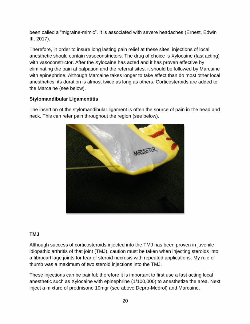
Stylomandibular Ligamentitis
The insertion of the stylomandibular ligament is often the source of pain in the head and
neck. This can refer pain throughout the region (see below).
TMJ
Although success of corticosteroids injected into the TMJ has been proven in juvenile
idiopathic arthritis of that joint (TMJ), caution must be taken when injecting steroids into
a fibrocartilage joints for fear of steroid necrosis with repeated applications. My rule of
thumb was a maximum of two steroid injections into the TMJ.
These injections can be painful; therefore it is important to first use a fast acting local
anesthetic such as Xylocaine with epinephrine (1/100,000) to anesthetize the area. Next
inject a mixture of prednisone 10mgr (see above Depro-Medrol) and Marcaine.
21
Conclusion Phase I
If Phase I treatment has been successful and pain no longer exists, the patient is
dismissed and splint is used as a night guard.
The patient is then scheduled for a follow-up ROM and muscle palpation in
approximately four weeks (new fees apply). If all is well, they then move to a six months
office visit and are dismissed back to the referring dentist or into your own re-care
system.
If the patient has not improved, referral must be considered (see below Phase II).
A third scenario is that the patient has improved, but the splint must be worn full time.
This is common in vertical dimension and/or lack of anterior guidance considerations.
Phase II should be initiated with these patients as well (see below).
Phase II
In Phase II our goal is to wean the patient from full time use of the splint and determine
their ability to live comfortably without the increase in vertical dimension and condylar
position provided by the splint.
This is a phase in which the clinician attempts to decrease the dependency of the
patient to her/him and takes a watch and wait approach to establish the success of
Phase I or necessity for more treatment. Fees are applied for treatment as it is
rendered. In this phase the dentist should consider referrals, reshaping of teeth
(occlusal equilibration), and observation.
Diagnosis MPD - Occlusal Equilibration
The only exception to the avoidance of permanent dental change in Phase II is the
process of limited occlusal equilibration. The goal of the clinician is to eliminate all
balancing side contacts and (if possible) establish cuspid and anterior guidance.
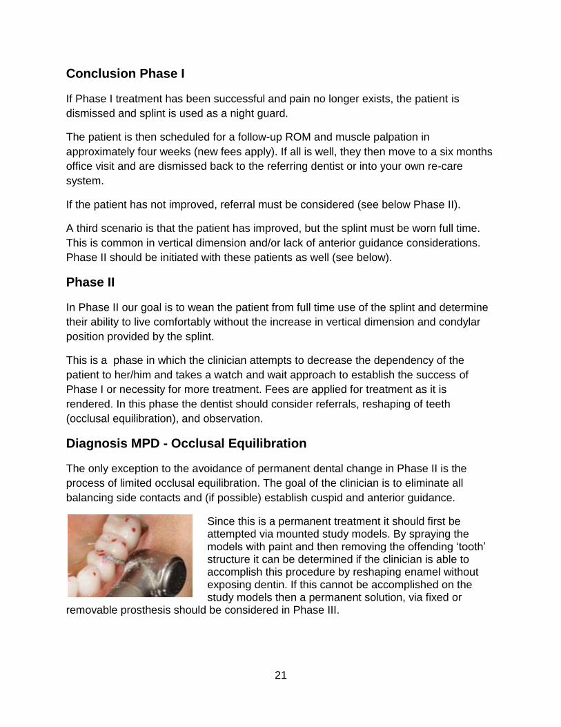
Since this is a permanent treatment it should first be attempted via mounted study models. By spraying the models with paint and then removing the offending ‘tooth’ structure it can be determined if the clinician is able to accomplish this procedure by reshaping enamel without exposing dentin. If this cannot be accomplished on the study models then a permanent solution, via fixed or
removable prosthesis should be considered in Phase III.

22
Referrals
Surgical Referral
If the joint is the definitive cause of pain; i.e. pain is relieved via auriculotemporal block
and the diagnosis of proper sophisticated radiographic surveys agree with clinical
evaluation, a surgical referral should be made. Post-surgical follow up and/or occlusal
rehabilitation will be necessary as a Phase III treatment. The surgeon will determine
whether the patient is in need of lavage or open surgery.
Referral for Counseling
When the patient presents as an emotional or depression risk, then referral to a Professional (psychologist, psychiatrist, or counselor) in a team approach must be considered.
Moreover, if the joints, musculature, tendons, and or ligaments are deemed to be the
source and have been successfully treated, but the pain persists referral for counseling
is strongly advised.
Chiropractic or Physical Therapy Referral
When Phase I treatment has had limited success, but the clinician is certain the etiology
is physical, referral to an Osteopathic Physician, Physical Therapist, or Chiropractor
should be considered.
Conclusion Phase II
Phase II treatment is concluded after approximately 8-10 months. It is mainly a referral
and ‘watch and wait’ phase to protect the patient from overzealous dental treatments
which time alone may heal. If the patient is now pain free but still dependent 24/7 (not
night guard use) on her/his splint, then phase III must be initiated.
Phase III Treatment
The goal of Phase III treatment is to render to the patient a long lasting solution for their
need of an increase in Vertical Dimension and or jaw repositioning. Therefore, other
than post-surgical rehabilitation Phase III treatment is aimed at a stable jaw position
guided and supported by the dentition.
This phase entails a permanent change in both condylar position and the dentition,
therefore should be undertaken only when the patient has been unable to function
without using her/his splint 24/7. A patient who is still in need of a ‘night guard’ does not
qualify for Phase III treatment, only those who are not able to function without the ‘splint

23
position’ and after a pain free Phase II has been accomplished.
Time Interval One Year after Phase I
If the patient has not been able to wean from the splint (and wearing full time) and every
attempt at weaning has failed (over a minimum period of 12 months), then a permanent
increase in vertical dimension should be considered. This can be accomplished by:
• Orthodontics/orthopedics
• Bonding
• Crowns and Bridges
• Overlay Partials
• Fixed and or Removable Prosthesis incorporating implants
• Full Dentures
Occlusal Position
No matter the method chosen the splint position must be the goal of treatment in Phase
III. The easiest method to capture this position is to use the splint as a guide by using a
‘wash’ material in the splint (thin or liquid material, i.e. silicone impression material, etc.)
and placing it into the splint, then over the dentition using the splint + the ‘wash’ to
mount the study models.
When fixed prosthetics is the treatment of choice it would be best to preparing one side
at a time. When one side is prepared the record may be taken (see above). The
unprepared side would act as a guide for an accurate jaw position. After preparation of
the opposite side use the impression wash/splint as a matrix for the second wash of the
newly prepared side.
In the case of orthodontics/orthopedics, removable prosthesis, and bonding mounted
study models using the splint ‘wash’ technique will act as the guide and goal for the final
orthopedic position.
Review and Detailed Treatment of Disorders Inclusive in TMD
Disc Displacement leading to Osteoarthritis
• Early click: MTI Splint or pull forward splint (both maxillary) to be worn 24/7
only removed for cleaning. Surgical soft diets are mandatory for all
attempts to ‘recapture’ disc dislocations. Attempting to recapture the disc
and place the condyle in the proper position for the healing of the capsular
ligament (6 week splint therapy).
If a ‘pull forward’ splint is used it must be slowly altered allowing the condyle
24
to slowing recede into its original position (if possible). After the flattening of
the pull forward splint or weaning off the MTI the patient must be placed on a
night guard (usually a maxillary splint reflecting their centric occlusion
incorporating anterior and cuspid guidance) for an extended period of time.
Thus, the patient will continue long term ‘night guard’ therapy. If the patient is
unable to function without full time use of the splint in the new orthopedic
position, Phase II and III treatment must be considered.
• Moderate click: treat same as early click
• Late click: treat same as early click but inform patient the later the click the
worse the prognosis for non-invasive treatment, most likely will need Phase II
and III treatment or perhaps surgical intervention
• Closed Lock: the disc is trapped anterior and medially to condyle preventing
both lateral and anterior movement on the affected side. Have patient open
as wide as possible. The clinician should place their thumb over the
mandibular molar teeth of the affected side and with a forceful and sudden
push down ward the disc should ‘pop’ back on the condyle. Then proceed as
with early click.
• Open lock (dislocation of mandible): this occurs when the disc is trapped
posterior to the condyle or the condyle has ‘over shot’ the eminence of the
fossae. In either case the joint must be reduced. Use the same method above
in the open lock to reduce the dislocation. If the condyle chronically dislocates
beyond the eminence of the glenoid fossae an eminence reduction should be
considered (referral to oral surgeon).
Myofascial Pain Dysfunction: Recommended Treatment
• Hydrocollator
• TENS
• Splint: begin with Lucia Jig to ascertain condition of joint; if successful
proceed to maxillary splint incorporating anterior and cuspid guidance; if joint
is involved see above (MTI).
• Trigger Points sprayed and injected (no epinephrine)

25
• Fibromyalgia – after diagnosis refer to primary care physician
• Nutrition
Tendonitis (Temporal)
Palpate (intra-oral) coronoid process.
If positive inject this area with Xylocaine. If the pain dissipates follow with Marcaine
(long lasting anesthetic) with epinephrine plus corticosteroid (prednisone 10mg).
Ligament tear, sprain, ligamentitis, stylomandibular ligament injury (the king of
referral pain). The key to this injury may not just be pain upon palpation at the angle of
the mandible, but the referral of pain which is often the patient’s chief complaint.
Therefore, a thorough palpation of all structures on the chart should be accomplished at
each visit in order to make a proper diagnosis and render proper ensuing treatment.
After the Xylocaine has been injected the referral pain should be resolved.
Stylomandibular Ligament Injury (tear, sprain): Palpate the angle of the mandible
(external) to ascertain the condition of the insertion of the stylomandibular ligament. If it
is inflamed the palpation will elicit pain and has several areas to which it refers pain (see
below).
If palpation is positive consider injection with Xylocaine with epinephrine. If the pain
dissipates it should be follow with Marcaine (long lasting anesthetic) with epinephrine
plus corticosteroid (prednisone 10mg.) (Shankland, WE 1987).
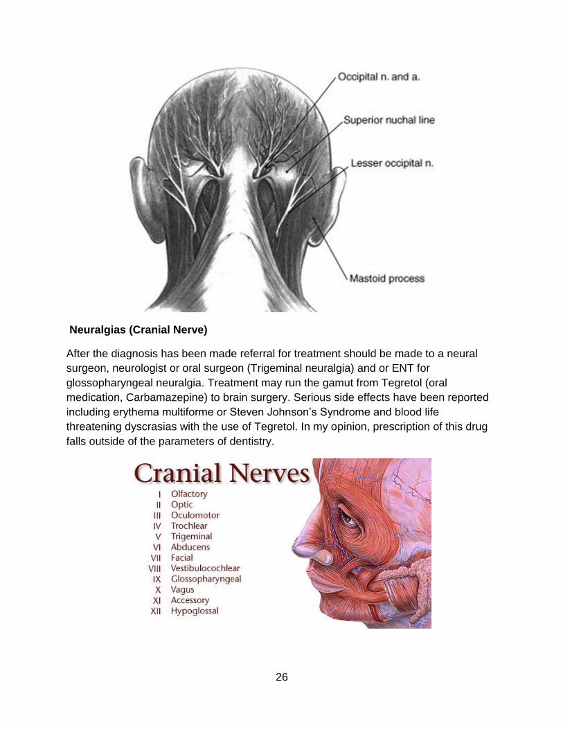
Greater and Lesser Occipital Nerve Impingement
The palpation of these structures is easily accessible. The treatment is injection at the
point of pain with Xylocaine. If the pain dissipates then the injection is followed by
Marcaine and corticosteroid. The dentist must check with her/his state dental
association to ascertain if the state practice code allows them to treat this area.
Therefore, after diagnosis referral may be necessary.
26
Neuralgias (Cranial Nerve)
After the diagnosis has been made referral for treatment should be made to a neural
surgeon, neurologist or oral surgeon (Trigeminal neuralgia) and or ENT for
glossopharyngeal neuralgia. Treatment may run the gamut from Tegretol (oral
medication, Carbamazepine) to brain surgery. Serious side effects have been reported
including erythema multiforme or Steven Johnson’s Syndrome and blood life
threatening dyscrasias with the use of Tegretol. In my opinion, prescription of this drug
falls outside of the parameters of dentistry.
27
However, that is not to say that all treatment falls outside of dental expertise. Success
has long been reported with the use of injections to obliterate peripheral nerves utilizing
absoluter alcohol. This modality is especially useful, although not a permanent cure, in
medically compromised patients or patients not wishing to undergo other forms of
treatment. Injections of Xylocaine (with epinephrine) are first used to make certain the
site is accurate via the elimination of pain. Then the clinician may proceed with the
alcohol injection (Shah, SA, Khan, MN, et. al. 2011).
These injections are frequently intraoral and likely fall within the dental practice code of
most states, but the clinician is responsible for reviewing these parameters in her/his
individual state.
What is more, I have found the routine use of occlusal splints to be of benefit to most of
these patients, and seemed to improve successful treatment (this is a clinical
observation and has not been proven via a double blind study).
Dental Disease/Differential Diagnosis
Dental Disease
Dental disease often refers pain to the TMJ, sinus, or ear, and may well cause
headaches. Although periodontal disease rarely causes referral and/or discomfort, both
pulpal and periapical pathology often do. Moreover, both frequently refer pain to other
areas in the head and neck. As stated in the diagnostic module of this course another
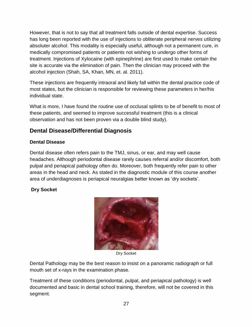
area of underdiagnoses is periapical neuralgias better known as ‘dry sockets’.
Dry Socket
Dry Socket
Dental Pathology may be the best reason to insist on a panoramic radiograph or full
mouth set of x-rays in the examination phase.
Treatment of these conditions (periodontal, pulpal, and periapical pathology) is well
documented and basic in dental school training, therefore, will not be covered in this
segment.
28
Iatrogenic TMD
It is often true that the extraction process itself, as well as long dental treatment may
precipitate, or even be the primary cause of TMD. The clinician must guard against
iatrogenic TMD by allowing the patient several breaks during treatment and placing a
balancing force opposed to the extraction force so that the TMJ will be protected from
possible dislocation.
Other areas of Differential Diagnosis include:
Sinus Pathology: sinusitis, ethmoid, sphenoid, and most often maxillary may refer
pain to many regions of the head and neck. Treatment should be referred to the
patient’s medical clinician.
CNS Diseases (i.e. TIA, stroke, Multiple Sclerosis, etc. ) can be screened via
examination of the cranial nerves (see Diagnosis), but definitive diagnosis and
treatment resides with our medical colleagues.
2017 Newer Alternative Treatments / Less Invasive
In a 2016 RDH Magazine online article titled “Treating TMD with orofacial myofunctional
therapy” by authors Timbrey Lind, RDH, and Shirley Gutkowski, RDH, BSDH they
explain an intriguing newer, less-invasive alternative for some (not all) TMD sufferers:
A study done by a TMD expert and OMT leader in São Paulo, Brazil, Claudia
Maria de Felicio, called ‘The Effects of Orofacial Myofunctional Therapy on
Temporomandibular Disorders’ reported, ‘OMT had... a significant reduction of
pain sensitivity to palpations for all muscles studied... increased measures of
mandibular range of motion... reduced frequency and severity of signs and
symptoms...’ With orofacial myofunctional therapy, a main goal is to establish
nasal breathing and correct oral posture by toning and repatterning the head and
neck muscles. By ‘repatterning,’ I literally mean re-creating strong pathways
between the brain and these muscles. This is what makes it so successful. We
do this by identifying compensations. When we focus on establishing nasal
breathing and correcting oral posture, this insinuates a lip seal and tongue to the
palate at ALL times, unless they are in use. An article written on lack of lip seal
(Harari et al., 2010) and how it leads to occlusal dysfunction goes right along with
the video I recommended. Training children to keep their lips together and
tongue to the palate encourages balanced muscles and forward growth in their
faces. Harmonious muscle pattern is important to correct a muscle-related TMD.
Myofunctional therapists are trained to identify compensations such as mouth
breathing and eliminate habits that develop because of the compensations. This
allows the muscles to habituate into a physiological normal way. Doing the least
29
invasive treatment first is always best (Lind & Gutkowski, 2016).
Conclusion
TMD is not a disease; it is a syndrome consisting of several maladies. In this author’s
opinion the disorder is best treated in phases with a global fee in Phase I. The clinician
must become adept at flowing from malady to malady as she/he peels the layers of this
debilitating condition.
All too often the patient is rushed into an extensive surgical or dental treatment without
first allowing the body a chance to heal itself. That is why I highly recommend treatment
in three phases, with only the third phase rendering the patient permanently ‘changed’.
The principal goals of treatment must remain constant:
• minimal treatment
• reversible treatment
• do the patient no harm
What is more, it should not be considered a failure if the patient is in need of long-term
or even lifelong use of a removable splint as a ‘night guard’. These devices are
minimally invasive and can prevent harmful tooth abrasion as well as muscle spasm.
The condition of these devices should be checked regularly at dental hygiene
appointments.
References
DeWood, G. (n.d.). Selecting the Best Temporary Occlusal Appliances.
https://www.speareducation.com/spear-review/2015/09/temporary-occlusal-appliances.
Accessed August 2017.
Edwin, Ernest, III, DMD, FAANaOS, Diagnosis and Treatment of Temporal Tendonitis –
a Very Common Disorder That is Often Mistaken for Migraine Practical Pain
Management http://www.practicalpainmanagement.com/pain/maxillofacial/temporal-
tendinitis-migraine-mimic Accessed August 2017
Herring, Susan Dr. University of Washington in Seattle,
http://www.painjournalonline.com/article/PIIS0304395911002491/abstract?rss=yes
Accessed August 2017.
Kun-Darbois, J.-D., Libouban, H., Chappard, D. Botulinum toxin in masticatory muscles
of the adult rat induces bone loss at the condyle and alveolar regions of the mandible
associated with a bone proliferation at a muscle enthesis. Bone 77: 75-82, 2015 Aug.
Accessed August 2017.
30
Lind, T. RDH, and Gutkowski, S. RDH, BSDH. RDH Magazine: Volume 36 Issue 8
(2016, August 23). Treating TMD with Orofacial Myofunctional Therapy.
www.rdhmag.com/articles/print/volume-36/issue-8/contents/treating-tmd-with-orofacial-
myofunctional-therapy.html. Accessed August 2017.
Shah, SA, Khan, MN, Shah SF, Ghafoor, A, Khattak A. Is Periperal Alcohol Injecito nof
Value in the Treamtent of Trigemianl Neuralgia? An Analysis of 100 Cases. Int.J Oral
Maxillofac Surg 2011, Apr 40(4) 388-92 http://www.ncbi.nlm.nih.gov/pubmed/21168309
Accessed August 2017.
Shankland, WE Ernest Syndrome as a Consequence of Stylomandibular Ligament
Injury: A Report of 68 Patients, J Prosthet Dentistry, 1987 Apr, 57 (4) 501-6
http://www.ncbi.nlm.nih.gov/pubmed/3471963 Accessed August 2017.
Srivastava R, Jyoti B, Devi P. Oral splint for temporomandibular joint disorders with
revolutionary fluid system. Dental Research Journal. 2013;10(3):307-313. Accessed
August 2017.
Timbrey Lind, RDH, and Shirley Gutkowski, RDH, BSDH: Treating TMD With Orofacial
Myofunctional Therapy, 2016 RDH Magazine
http://www.rdhmag.com/articles/print/volume-36/issue-8/contents/treating-tmd-with-
orofacial-myofunctional-therapy.html Accessed August 2017.
TMJ Association, Ltd. (n.d.). Retrieved August 21, 2017 from
http://www.tmj.org/site/page?pageId=327
The Trigger Point and Referred Pain Guide http://www.triggerpoints.net Accessed
August 2017.
Travell, J.B. & Simons, D. C., Myofunctional Pain and Dysfunction, The Trigger Point
Manual. Williams and Wilkins September 2013
Upledger, J.E. & Vredevoob, J. D., Craniosacral Therapy. Chicago Eastland Press
http://www.upledger.com/ Accessed August 2017.

31
Course Test: TMD Disease: Treatment Module Three
1. Occlusal Splint Therapy is the least important aspect of TMD treatment
a. True
b. False
2. All TMD patients must undergo permanent dental treatment in order to be cured.
a. True
b. False
3. Dentists no longer need to use study models in order to treat TMD?
a. True
b. False
4. Before occlusal equilibration is accomplished on the patient this procedure should
first be accomplished on mounted study models.
a. True
b. False
5. TMD is not considered mainly an inflammatory disease.
a. True
b. False
6. The patient exhibits trigger points upon palpation at the initial examination. What
should the clinician do next?
a. Immediately inject the site
b. Conclude the diagnosis of MPD
c. Utilize TENS and Hydrocollator Therapy
d. Continue to question the patient to ascertain if they have MPD or Fibromyalgia
32
7. After the diagnosis of MPD has been made and trigger points have been located the
following are methods of treatment:
a. Injection of trigger points with Xylocaine and epinephrine
b. Splint therapy
c. Injection of trigger points with Marcaine and corticosteroid
d. Hydrocollator and TENS Therapy
e. B & D
8. All disc displacements will need a permanent change in vertical dimension.
a. True
b. False
9. After the diagnosis of a closed lock is made, the following should be part of treatment:
a. Hydrocollator and TENS Therapy
b. Physical Reduction of the disc
c. Pull forward splint
d . All of the Above
10. After the diagnosis of a late click has been made the following can be expected:
a. The patient will respond to a pull forward splint.
b. The patient must undergo Phase III or a permanent change in vertical
dimension.
c. The joint may undergo arthritic changes.
d. The patient may respond to Phase I treatment modalities.
e. C & D only
11. The Philosophy of Treatment Changes depending on the physical condition
presented by each patient.
a. True
b. False
33
12. When trigger points are found and other modalities have been tried the drug of
choice for injection is:
a. Marcaine
b. Procaine
c. Xylocaine
13. Trigger points should always be injected without epinephrine because:
a. Local anesthetics with epinephrine cost more
b. Epinephrine causes vasoconstriction
c. The patient will not wish to be numb for a long period of time
d. Vasodilatation is caused by epinephrine
14. Spastic musculature always need:
a. Oxygen
b Vasodilatation
c. Vapo-coolant sprays
d . A & B only
15. Spastic Musculature may be helped by the following:
a. Hydrocollator, TENS, and NSAIDS
b. Splint therapy and injections of procaine w/o vasoconstrictor into trigger points
c. Occlusal equilibration
d . All of the above
16. Temporal tendonitis cause severe headaches.
a. True
b. False
34
17. Temporal tendonitis can be injected at the site of insertion on the:
a. Pterygoid process
B. Angle of the mandible
C. Coronoid Process
D. All of the above
18. Stylomandibular ligamentitis should be treated by injecting the following:
a. Xylocaine, Marcaine, and corticosteroid (anesthetics with epinephrine)
b. Xylocaine, Procaine, and corticosteroids (anesthetics without epinephrine)
c. Marcaine, Procaine and corticosteroids (anesthetics with epinephrine)
d. None of the above
19. When the clinician inquires about nutrition, his/her main goal is to encourage the
patient to lose weight.
a. True
b. False
20. Oral Medications may include the following:
a. Analgesics and muscle relaxants
b. Dose pack of corticosteroids and analgesics
c. Controlled substances and muscle relaxants
d. All of the above
21. The Goal of Phase III Treatment is to return the patient to their original condylar
position.
a. True
b. False

35
22. A patient who is still in need of a night guard after one year should enter Phase III
Treatment and have their dentition changed to that of the night guard.
a. True
b. False
23. Permanent change in the vertical dimension of the patient is undertaken only after:
a. One month
b. One week
c. One year
24. The Phase of Treatment associated with increase in vertical dimension is:
a. Phase I
b. Phase II
c. Phase III
25. The best method to record the new vertical dimension is:
a. Utilizing an accurate liquid impression material and the splint.
b. Pushing the mandible back and having the patient close gently into wax.
c. Protruding the mandible and having the patient close gently into silicone.
d. Using a Lucia Jig.