tm journal of biowiw cmm~ voi. 269, no. 42, … · its high aenity receptor* (received for...
TRANSCRIPT

T m JOURNAL OF BIOWIW C m m ~ D 1994 by The American Society for Biochemistry and Molecular Bid-, Inc
VoI. 269, No. 42, Issue of October 21, pp. 2 ~ ~ 2 6 3 7 3 , 1 9 9 4 Printed in U S A .
The Binding Site on Human Immunoglobulin E for Its High AEnity Receptor*
(Received for publication, June 1, 1994, and in revised form, August 4, 1994)
Leonard fiesta$, Robert Shields§, Lori O’Connell, Steven Lam James Porteril, Cornelia Goman**$$, and Pada JardieuO From the Departments of Protein Engineering, $Immunology, **Cell Genetics, and IlMedicinal and Analytical Chemistry, Genentech Znc., South Sun Francisco, California 94080
Immunoglobulin (Ig) E antibodies mediate allergic re- sponses by binding to specific high affinity receptors, FcdtI, on mast cells and basophils. Previous studies have shown that the principal FceRI binding site is lo- cated on the third constant domain, Fce3, of IgE. Based on a model of the IgE Fce3 (which is homologous to the second constant domain of IgG), homology scanning mu- tagenesis and replacement of individual residues were used to determine the specific amino acids of human IgE involved in binding to human FccRI. The amino acids are localized in three loops, which form a putative ridge on the most exposed side of the Fee3 domain of IgE and include Arg-408, Ser-411, Lys-415, Glu-452, Arg-465, and Met-469. The preponderance of charged residues sug- gests that IgE-FceRI binding is mediated primarily by electrostatic interaction. Furthermore, it is possible to confer FccRI binding to an IgG molecule by introducing these three IgE loops into the I& Cy2 domain.
Immunoglobulin (Ig)’ E antibodies bind to specific high af- finity receptors, FceRI, on mast cells, basophils, and Langer- hans cells (1-4) via their constant, or Fc, region in a one-to-one complex (5). Degranulation of mast cells and basophils trig- gered by binding of antigen to IgE-loaded FceRI leads to release of mediators which produce symptoms associated with allergy (6,7). IgE also binds to low affinity receptors (FceRII, CD23) on B-lymphocytes and eosinophils (8,Q). A more in-depth review of IgE and its receptors can be gleaned fmm Ref. 10. Because of the central role of cell-bound IgE in initiation of the allergic cascade, much attention has been focused on localizing the sites on IgE required for receptor binding. Over the last decade a variety of techniques have been used to determine the FcrRI binding site on IgE, including synthetic peptides corresponding to portions of the IgE Fc (11-14), anti-IgE antibodies (15-171, IgE variants (18), chimeras (19-221, and truncated forms (23). However, the exact IgE residues involved in binding have re- mained undefined. Helm et a2. (13, 14) reported mapping the FceRI binding site on human IgE to a sequence of 76 amino acids at the junction of the second (Fcr2) and third (Fce3) con- stant domains. In contrast, a recent study reported that only
* The costs of publication of this article were defrayed in part by the
“aduertisement” in accordance with 18 U.S.C. Section 1734 solely to payment of page charges. This article must therefore be hereby marked
indicate this fact. $ To whom correspondence should be addressed. fi Current address: Dept. of Biological Chemistry, University of North
Carolina, Chapel Hill, NC 27599.
San Francisco, CA 94111. $$ Current address: Megabios, ’ b o Embarcadero Center, Suite 410,
ity IgE receptor; Fc, constant domains of immunoglobulin; ELISA, en- ’ The abbreviations used are: Ig, immunoglobulin; FceRI, high aHn-
zyme-linked immunosorbent assay; CHO, Chinese hamster ovary; FITC, fluorescein isothiocyanate.
the Fce3 domain of murine IgE is involved in binding to rat FceRI (201, and this domain has also been implicated as impor- tant in binding of murine IgE to murine FceRI and FceFtII (22). In this study, we have now determined the specific amino acid residues involved in human IgE binding to human FceRI. All of the critical residues are contained in three loops, which, to- gether, form a ridge on the most exposed portion of human IgE Fce3.
The Fc portion of IgE includes three domains: Fce2, Fce3, and Fce4. The Fce3 domain has been predicted to be structurally homologous to the I& Fcy2 domain (24, 25). Hence, it com- prises seven p-strands (denoted A-G) and six connecting loops (denoted by the two p-strands they connect, e.g. loop AB), which form a p-sandwich structure representative of all immuno- globulin domains (Fig. 1). Using homology scan methodology (261, the initial variants of human IgE were designed by taking advantage of the fact that human IgGl does not bind to IgE receptors (19). These included individually replacing five Fce3 loops with their counterparts from human IgGl Fcy2 (Table I). The sixth loop (DE), which contains a conserved, structurally i m p o r t ~ t glycosylation site in both IgE and IgG, was not al- tered. Additional variants were evaluated in which solvent- exposed residues in Fce3 p-strands and selected loops in Fce2 and Fce4 were changed to alanine, as opposed to IgGl homol- ogy replacements, in order to maximize the effect of altering only 2-3 side chains per variant (Table I). Based on the results from these, a second set of variants assessed individual resi- dues within the regions found to affect the FceRI-IgE interac- tion. Residues were changed to alanine or to other amino acids in order to determine if size, charge, hydrogen-bonding capa- bility, or hydrophobi~ty were important for that particular residue.
EXPERIMENTAL PROCEDURES Phagemid Constructs-The human IgE gene was isolated from a
U266 cell line (27) library prepared at Genentech. Polymerase chain reaction (28) was used to amplify the IgE gene using primers based on previous studies (29, 30). The DNA products were isolated, digested with appropriate restriction enzymes, and purifled on acrylamide gels. The gene was then cloned into a previously described phagemid vector (311, replacing the human IgGl heavy chain with the human IgE heavy chain using the BstEII and Hind111 sites. This phagemid contains the human cytomegalovirus enhancer and promoter, a 5’ intron, and a SV40 polyadenylation signal (32). The sequence of this human IgE heavy chain matched sequences of human IgE reported previously (29, 30).
Mutagenesis was performed by the Kunkel method (33) using bders
esis kit (Bio-Rad). Sequences of the variant IgE DNAs were checked and enzymes supplied with a commercially available in vitro mutagen-
using dideox~ucleo~de sequencing. The phagemid encoding the chimeric protein IgG2/E3 was con-
structed using a previously described phagemid vector containing the human cytomegalovirus enhancer and promoter, a 5‘ intron, and SV40 polyadenylation signal (32) encoding a humanized ant i -~185~~= I&l (31). The nucleotide sequence encoding human IgGl residues Cys-239- Gly-361 was replaced with DNA encoding IgE FcB residues Cys-357-
26368

Binding Site on IgE for Its High Affinity Receptor
Fro. 1. Ribbon diagram of IgE Fcs3 and Fcr4. Fce3 p-strands A-G, connect- ing loops, and residues important in IgE binding to FceRI are labeled. Coordinates are from the Protein Data Bank (48) model described by Padlan et al. (25). The two Fee2 domains (not shown) would be at the top ofthe figure: the two F c d domains are at the bottom. p-Strands are colored blue, and a-helices are colored purple. Side chains involved in binding to Fc&I are shown with side chain carbon, nitro- gen, and oxygen atoms colored yellow, blue, and red, respectively. Carbohydrate attached at Asn-430 is shown as brown I spheres.
A
Gly-497.' The portion of the phagemid coding for IgG2E.3 was com- pletely sequenced using dideoxynucleotide sequencing. The humanized anti-p185HER2 IgGl phagemid was also used to construct the IgGEL phagemid. Using site-directed Kunkel mutagenesis (331. loop CD (IgE residues 4074201, loop EF (IgE residues 445453). loop FG (IgE resi- dues 4654691, and Fcd-Fce3 hinge residues 357-365 were substituted for their homologues in IgGl (IgG1 residues 291-305. 329-337, 349- 352, and 239-249, respectively). The portion of the phagemid coding for IgGEL was completely sequenced using dideoxynucleotide sequencing.
Protein Expression-For each IgE variant, double-stranded phage- mid DNA was isolated from Escherichia coli strain XI-1 Blue (351 with the Qiagen DNA purification kit (Qiagen Inc., Chatsworth. CAI. This DNAwas used to transfect the fetal human kidney cell line 2939 (361 by a calcium phosphate method (32). The 75% confluent cells were trans- fected with 10 pg of phagemid DNN15-mm cell culture dish and incu- bated for 15 h in serum-containing medium. The medium was removed and replaced with serum-free PS04 medium supplemented with 10 mgAiter recombinant bovine insulin. 1 mgiliter transferrin, and trace elements. The supernatant was collected at 48 and 96 h and concen- trated approximately 20-fold using a Centriprep 30 unit (Amicon). IgE variants were examined by sodium dodecyl sulfate-polyacrylamide gel electrophoresis to ascertain correct molecular weight and were quanti- fied by ELISA using purified human IgE as the reference standard. A murine anti-human IgE antibody ( M a l , Genentechl was used to cap- ture IgE in the ELISA. IgE was detected with an horseradish peroxi-
Residues are numbered according to Ref. 34.
dase goat anti-human IgE (Kirkegaard & Peny). IgE concentrations were extrapolated by non-parametric linear regression analysis. Ini- tially, IgE variants were affhty-purilied on a murine auti-human IgE antibody (MaE2, Genentech) coupled to CNBr-Sepharose (Pharmacia Biotech Inc.1. Since assays using either purified or non-purified IgE gave identical results, the purification step was eliminated for other variants.
In order to assure that. all variants were properly folded. each variant was assayed for binding to murine anti-human IgE antibody MaE1, which binds outside domain Fce3. and to three murine anti-human IgE antibodies that have been mapped to the Fcr3 domain: MaEll (371, MaE13, and MaE17 (Genentech). The binding of all four MaE antibod- ies to heat-denatured IgE (56 "C for 90 min: Ref. 15) is reduced: 10-fold for MaEl and at least 100-fold for MaE11. MaE13. and MaE17. A l l IgE variants bound as well as native IgE to M a l and to at. least one of MaE11, MaE13, or MaE17 (data not shown).
I3indirz.g Assay-Binding of human IgE variants was measured by immunofluorescence using a CHO 3D10 cell line expressing the human FceRI receptor n-chain (38). Although the FceRI recept.or comprises one e-chain, one a-chain, and two y-chains, a previous study has shown that the a-chain is sufficient for IgE binding (38 1. 5 x lo5 cells in phosphate- buffered saline containing 0.1% bovine serum albumin, 10 m M sodium azide were incubated for 30 min. at 4 "C with 0.1-10 pg/ml IgE or IgGl control (humanized anti-p185""K2 IgG1; Ref. 31). After washing, the cells were incubated for 30 min. at 4 "C with 20 pgiml affinity-purified FITC-conjugated (391 rabbit anti-human IgE (Axell, or goat FITC- F(ab)', anti-human IgG (Organon Teknica), respectively. Analyses were
26370 Binding Site on IgE for Its High Afinity Receptor
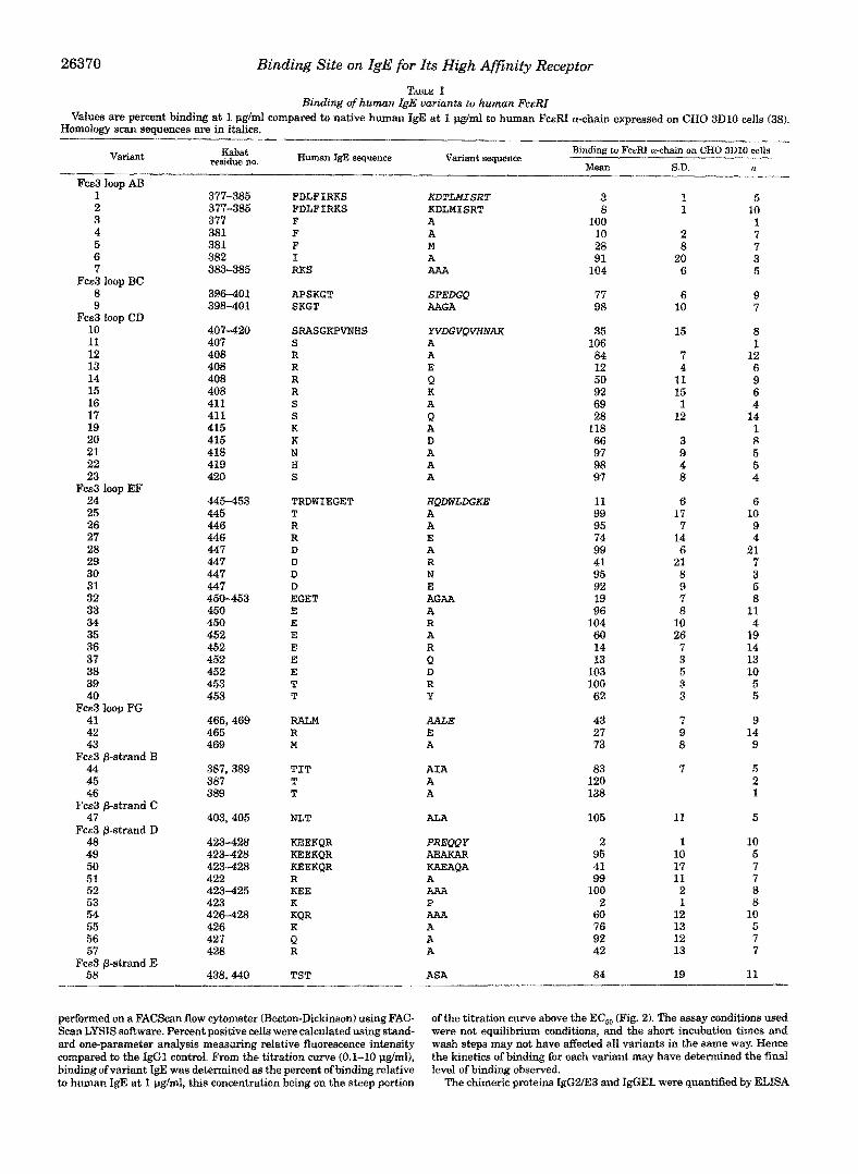
Binding of human igB variants to human F e e TABLE I
Values are percent binding at I I.lglmi compared to native human IgE at 1 pg/ml to human FcERI a-chain expressed on CHO 3D10 cells (38). Homology scan sequences are in italics.
Variant residue no. &bat Human IgE sequence Variant sequence
Binding to FcERI a-chain on CHO 3D10 cells
Mean S.D. n Fca3 loop AB
1 2 3 4 5 6 7
8 9
10 11 12 13 14 15 16 17 19 20 21 22 23
24 25 26 27 28 29 30 31 32 33 34 35 36 37 38 39 40
42 41
43
44 45 46
47
48 49 50 51 52 53 54 55 56 57
58
Fm3 loop BC
Fca3 loop CD
F C E ~ loop EF
Fcs3 loop FG
F C E ~ P-strand B
Fcs3 P-strand c Fcs3 6-strand D
FCE3 @-strand E
377-385 377-385 377 381 381 382 383-385
398-401 396-401
407 407-420
408 408 408 408 411 41 1 415 415 418 419 420
445453 445 446 446 447
447 447
447 450-453 450 450 452 452 452 452 453 453
465 465,469
469
387,389 387 389
403,405
423-428 423-428 423-428 422 423-425
426-428 423
427 426
428
438,440
FDLFIMS FDLFIRKS F F F I RKS
APSKGT SKGT
SRASGKPVNHS S R R R R S S K K N H S
TRDPJIEGET T R R D D D D EGET E E E E E E T T
R A L M R M
TIT T T
NLT
KEEXQR KEEXQR KEEKQR R KEE X KQR X Q R
TST
KDTLMISRT KDLMISRT A A M A AAA
SPEDGQ AAGA
YVDGVQVMiAK A A E Q K A Q A D A A A
H#D%LDGKE A A E A R N E AGAA A R A R Q D R Y
AALE E A
AIA A A
ALA
PREPPY AEAKAR KAEAQA A AAA P AAh
A A
A
ASA
3 8
100 10 28 92
104
77 98
35 106 84 12 50 92 69 28
118 66 97 98 97
11 99 95 74 99 41 95 92 19 96
104 60 14 13
103 100 62
43 27 73
83 120 138
105
2 95 41 99 100
2 60 76 92 42
84
I 1
2 8
20 6
6 10
15
7 4
11 15 1
12
3 9 4 8
6 17 7 14 6
21 8 9 7 8
26 10
7 3 5 3 3
7 9 8
7
11
1 10 17 11 2
12 1
13 12 13
19
5 10 1 7 7 3 5
9 7
8 1
12 6 9 6 4
14 1 8 5 5 4
6 10 9 4
21 7 3 5 8
11 4
19 14 13 10 5 5
9 14 9
5 2 1
5
10 5 7 7 8 8
10 5 7 7
11
performed on a FACScan flow cytometer (~ecton-Dic~nson) using FAC- of the titration curve above the EC,, (Fig. 2). The assay conditions used Scan LYSIS software. Percent positive cells were calculated using stand- were not equ~libnum conditions, and the short incubation times and ard one-parameter analysis measuring relative fluorescence intensity wash steps may not have affected all variants in the same way. Hence compared to the IgGl control. From the titration curve (0.1-10 pgfml), the kinetics of binding for each variant may have determined the final binding of vnu?ant IgE was determined as the percent of binding relative level of binding observed. to human IgE at 1 &I& this concentration being on the steep portion The chimeric proteins IgG2/E3 and IgSEL were quantified by ELISA

Binding Site on IgE for Its High Affinity Receptor 26371
TABLE I-continued
Variant
Fce3 P-strand F 59 60 61
Fcs3 p-strand G 62
Fce2 63 64
Fce4 65 66 67
residue no. Kaabat
455-459 460 462
471,473
329-336 361-365
498-501 594-599 595-599
Human IgE sequence
QCRVT H H
STT
QKHWLSDR DSNPR
PRAA ASPSQT S P S Q T
Variant sequence "
ACAVA D D
ATA
AAAWLAAA ASAPA
QPRE LHNHY APAAA
Binding to FcERI a-chain on CHO 3D10 cells
Mean R S.D. -
96 9 15 77 9 8 90 2 4
107 7 4
107 8 4 67 19 14
89 12 4 92 1 4 80 11 5
using a goat anti-human IgG F(ab) (Organon Teknica) to capture and an horseradish peroxidase-conjugated goat anti-human IgG F(abf to de- tect. Humanized anti-p185*RZ F(ab) (31) was used as the standard. For IgGzIE3 and IgGEL, FITC-Flab)', goat anti-human IgGl (Organon Teknica) was used in the immunofluorescence assay.
RESULTS
The homology scan suggested that IgE loops AB, CD, EF, and FG might contribute to binding {variants 1, 8, 10, 24, and 41; Table I). Among the @-strands, only changes in @-strand D significantly reduced binding (variant 48; Table I). Individual residue replacements in loop AB and @-strand D showed that in both cases only a single amino acid reduced binding. In loop A B , only substitutions at Phe-381 had an effect (variants 4 and 5; Table I). Since the crystal structure of IgE is unknown, we cannot discern whether the effect seen for Phe-381 i s confor- mational (Phe buried) or if indeed this side chain is solvent- exposed and interacts with FceRI. In @-strand D, the single amino acid change Lys-423 + Pro3 abolished binding to the same extent as did replacement of the entire loop (variants 48 and 53; Table I). However, the Lys side chain is not involved in binding since replacing the Lys-423 residue with Ala had no effect on binding (variant 52; Table I). Since Pro is accommo- dated at position 423 in IgGl (40) but not in IgE implies that @-strand D in IgE may have a different conformation than in IgG. Another single amino acid change in @-strand D, Arg-428 4 Ala, also reduced binding. Arg-428 might be distant in space from the other impo~ant residues (Fig. 1) and also precedes the conserved glycosylation site at Asn-430, suggesting that Arg- 428 may play a structural role, although direct interaction with FceRI cannot be ruled out.
In loop CD, Arg-408 is the most important binding residue. Arg-408 "-$ Ala and kg-408 "-f Lys had little effect on binding, but Arg-408 + Glu and kg-408 + Gln reduced binding by 88% and 50%, respectively. While the latter two variants show that a positive charge at this position is important, the Arg-408 +
Ala variant is difficult to interpret. While it is conceivable that the Glu and Gln substitutions might cause a local structural perturbation, the binding of the Arg-408 + Ala variant is only slightly reduced relative to IgE and this argues against any IocaI conformational perturbation when Arg-408 is substituted with the other amino acids. In addition, Glu and Gln side chains are smaller than the Arg side chain and would not be likely to prevent IgE binding t o FceRI via steric clash, Replace- ments at 2 other residues in loop CD also reduced binding. Lys-415 -+ Asp reduced binding by 34%, much less than for the charge exchange at Arg-408, and at Ser-411, Ala and Gln sub- ~-
The nomenclature for IgE variants is as follows: native amino acid, residue number, substituted amino acid, e.g. Asp-447 "-f Arg denotes substitution of Arg for Asp at residue 447.
"
100 -
80 -
o f I I I
0 1 2 3
UG I ML
FIG. 2. Binding of human IgE and selected IgE variants to hu- man FcaRI expressed on CHO 3D10 cells. Human IgE (O), variant 37 Glu-452 --i\ GIn (At, variant 38 Glu-452 4 Asp (ml, and variant 64 Asp-361 -+ ~ ~ ~ n - 3 6 3 --f AldArg-365 -+ Ala (A).
stitutions diminished binding by 31% and 72%, respectively. In murine and rat IgE residue 411 is Lys and Arg, respectively, and both rodent IgEs can bind to human FceRI (411, providing circumstantial evidence that Ser-411 --* Gln did not sterically hinder the human IgE-human FceRI interaction or perturb the conformation of loop CD.
Loop EF is the only loop for which the conformation is reli- ably discernible. The crystal structure of IgG domains Cy2 and Cy3 (40) shows that in both domains loop EF forms an a-helix
26372 Binding Site on IgE for Its High Afinity Receptor
in which a buried Trp anchors the helix and a Gly terminates the helix. In human, murine, and rat IgE, loop EF retains these characteristic amino acids and hence is also likely to be an a-helix. At Asp-447, removal of the side chain (Asp-447 + Ala), repositioning the negative charge (Asp-447 --j Glu), or neutral- ization of the charge (Asp-447 + Asn) had no effect, but charge reversal (Asp447 + Arg) reduced binding by 59%. Human IgG domain Cy3 contains an Arg at this position and its presence does not disturb the a-helix (40); hence, the Asp-447 + Arg variant is not likely to be a structural perturbation. This sug- gests that the reduced binding of the Asp-447 + Arg variant may be due to steric clash between the IgE and FceRI. In contrast to residue 447, removal of the side chain at residue 452 (Glu-452 + Ala) reduced binding and charge neutralization (Glu-452 + Gln) was as deleterious as charge reversal (Glu-452 + Arg), suggesting that Glu-452 is involved in an important salt bridge with FceRI. Repositioning the negative charge (Glu- 452 --j Asp) had no effect. One other residue in loop EF, at position 453, might be important since Thr-453 --j Arg had no effect while Thr-453 --j Tyr reduced binding. This could result from the Arg side chain being able to move out of the way during FceRI-IgE binding while the bulkier, less mobile Tyr side chain cannot, thereby sterically hindering the FceRI-IgE interaction. The importance of loop EF in rodent systems is uncertain (11, 17).
Two other regions are also involved in IgE binding. In loop FG, the charge reversal Arg-465 + Glu significantly reduced binding by 73% while Met-469 + Ala reduced binding only 27%, suggesting that Arg-465 plays the dominant role in loop FG. In the hinge region between domains FceZ and Fce3, 3 residues (at positions 361, 363, and 365) were simultaneously changed to Ala (variant 64; Table I) and binding to FceRI was reduced by 33%.
Of the variants that did not alter binding to FceRI, a few deserve comment. In addition to the glycosylation site at Asn- 430, which corresponds to that in IgG Fcy2, human IgE Fce3 contains another glycosylation site at Asn-403 (42). Variant 47 converted Asn-403 to Ala, and the loss of carbohydrate at this position did not alter binding to FceRI. One loop in domain Fce2 (variant 63; Table I) and two loops in domain F c d (variants 65-67; Table I) were evaluated. These three loops should be adjacent to domain Fce3 according to the published model of IgE (24, 25). In addition, the Fce2 loop was the N-terminal section of a peptide previously reported to bind to FceRI (13). However, none of the three variants reduced binding to FceRI, suggesting that domains FceZ and Fce4 may not be involved in IgE binding to FceRI, in agreement with previously published results on murinehuman IgE and murine IgEhuman IgG chi- meras (19-21).
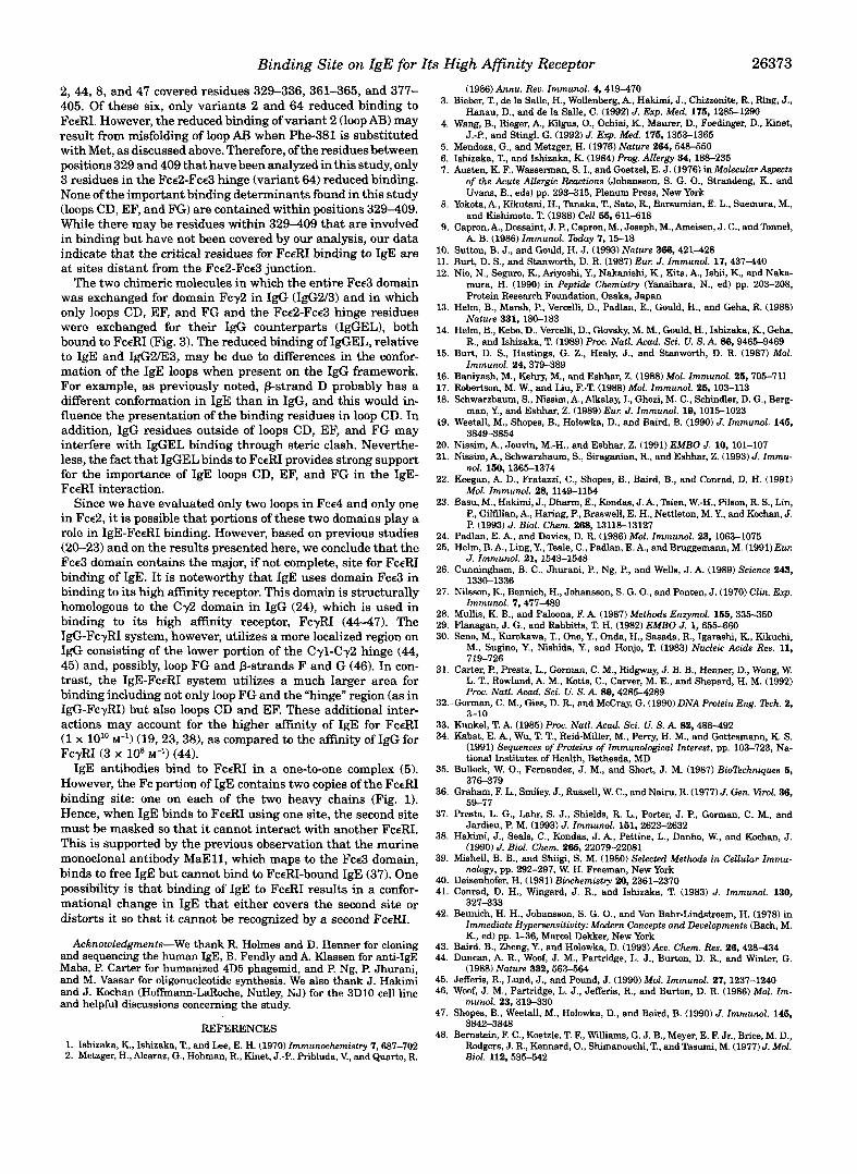
To verify that the residues determined in this study are in- deed important for binding of IgE to FceRI, we constructed two chimeric molecules. In the first, IgG2/E3, the entire IgE Fcc3 domain was substituted for the homologous Fcy2 domain in an IgG antibody and the Fce2-Fce3 hinge region (including Cys- 357, which forms the inter-heavy chain disulfide) replaced the IgG hinge. This human IgGhuman IgE chimera is similar, but not identical, to human IgG/murine IgE and murine IgE/ human IgE chimeras investigated by other groups (19, 20). In the second chimera, IgGEL, only the IgE loops critical for FceRI binding, i.e. loops CD, EF, and FG, replaced their homologous loops in an IgG antibody and again the Fce2-Fce3 hinge region replaced the IgG hinge. Although the hinge region (variant 64) contributed minimally to IgE binding, it was included so that the IgGEL chimera would have maximal likelihood of binding to FceRI. Fig. 3 shows that IgG2/E3, which contains the entire Fce3 domain, binds only about 4-fold less well (EC,, = 1.8 pd
100 1
0 5 1 0 1 5 20 25
Concentration (u!m)
IgG2/E3 and IgGEL to human FceFU expressed on CHO 3D10 FIG. 3. Binding of human IgE, IgG, and chimeric molecules
cells. 0, human IgE; 0, human IgG; 0, IgGZA33; A, IgGEL.
ml) than native human IgE (EC,, = 0.4 pglml). Furthermore, although binding of IgGEL is considerably reduced compared to native IgE, its binding to FceRI is significantly better than for the parent IgG, which does not bind at all (Fig. 3).
DISCUSSION
The binding site on human IgE for its high affinity receptor, FceRI, is localized in three loops, CD, EF, and FG, which con- nect p-strands (Fig. 1). According to the model of IgE, these loops form a ridge on the most exposed side of the Fce3 domain of IgE and include Arg-408, Ser-411, Lys-415, Glu-452,Arg-465, and Met-469. The preponderance of charged residues suggests that IgE-FceRI binding is mediated primarily by electrostatic interaction. This is supported by the fact that an anti-IgE an- tibody that blocks binding of human IgE to human FceRI uti- lizes charged residues in its binding to IgE (37).
In addition to the three loops, one other region reduced bind- ing, albeit minimally. Variant 64 consists of simultaneous al- teration to alanine of 3 residues (Asp-361, Asn-363, and Arg- 365) in the hinge connecting domains Fce2 and Fce3. This variant only reduced binding by 33% (Table I), implying that the Fce2-Fce3 hinge may play only a minor role in IgE binding. However, it has been proposed that IgE free in solution and bound to FceRI has a bent and rigid conformation (43). We cannot discern if the 3 residues in variant 64 are involved in binding directly to FceRI or if they are involved in maintaining the bent structure of IgE necessary for proper binding to FceRI.
It is also interesting to review the data from variants in the 329-409 region, which form the Fcd-Fce3 junction, in light of the report that the FceRI binding site on IgE is contained within this 76-amino acid sequence (13,141. Our variants 63,64,
Binding Site on IgE for Its High Afinity Receptor 26373
2,44, 8, and 47 covered residues 329-336, 361-365, and 377- 405. Of these six, only variants 2 and 64 reduced binding to FceRI. However, the reduced binding of variant 2 (loop AB) may result from misfolding of loop AB when Phe-381 is substituted with Met, as discussed above. Therefore, of the residues between positions 329 and 409 that have been analyzed in this study, only 3 residues in the Fce2-Fee3 hinge (variant 64) reduced binding. None of the important binding determinants found in this study (loops CD, EF, and FG) are contained within positions 329-409. While there may be residues within 329-409 that are involved in binding but have not been covered by our analysis, our data indicate that the critical residues for FceRI binding to IgE are at sites distant from the Fce2-Fce3 junction.
The two chimeric molecules in which the entire Fee3 domain was exchanged for domain Fey2 in IgG (IgG2/3) and in which only loops CD, EF, and FG and the Fcd-Fce3 hinge residues were exchanged for their IgG counterparts (IgGEL), both bound to FceRI (Fig. 3). The reduced binding of IgGEL, relative to IgE and IgG2E3, may be due to differences in the confor- mation of the IgE loops when present on the IgG framework. For example, as previously noted, p-strand D probably has a different conformation in IgE than in IgG, and this would in- fluence the presentation of the binding residues in loop CD. In addition, IgG residues outside of loops CD, EF, and FG may interfere with IgGEL binding through steric clash. Neverthe- less, the fact that IgGEL binds to FceRI provides strong support for the importance of IgE loops CD, EF, and FG in the IgE- FceRI interaction.
Since we have evaluated only two loops in Fee4 and only one in Fce2, it is possible that portions of these two domains play a role in IgE-FceRI binding. However, based on previous studies (20-23) and on the results presented here, we conclude that the Fee3 domain contains the major, if not complete, site for FceRI binding of IgE. It is noteworthy that IgE uses domain Fee3 in binding to its high affinity receptor. This domain is structurally homologous to the Cy2 domain in IgG (241, which is used in binding to its high affinity receptor, FcyRI (44-47). The IgG-FcyRI system, however, utilizes a more localized region on IgG consisting of the lower portion of the Cyl-Cy2 hinge (44, 45) and, possibly, loop FG and p-strands F and G (46). In con- trast, the IgE-FceRI system utilizes a much larger area for binding including not only loop FG and the “hinge” region (as in IgG-FcyRI) but also loops CD and EF. These additional inter- actions may account for the higher affinity of IgE for FceRI (1 x 10” M-’) (19,23,38), as compared to the affinity of IgG for FcyRI (3 x 10’ M-’) (44).
IgE antibodies bind to FceRI in a one-to-one complex (5). However, the Fc portion of IgE contains two copies of the FceRI binding site: one on each of the two heavy chains (Fig. 1). Hence, when IgE binds to FceRI using one site, the second site must be masked so that it cannot interact with another FceRI. This is supported by the previous observation that the murine monoclonal antibody MaE11, which maps to the Fee3 domain, binds to free IgE but cannot bind to FceRI-bound IgE (37). One possibility is that binding of IgE to FceRI results in a confor- mational change in IgE that either covers the second site or distorts it so that it cannot be recognized by a second FceRI.
Acknowledgments-We thank R. Holmes and D. Henner for cloning and sequencing the human IgE, B. Fendly and A. Klassen for anti-IgE Mabs, P. Carter for humanized 4D5 phagemid, and P. Ng, P. Jhurani, and M. Vassar for oligonucleotide synthesis. We also thank J. Hakimi and J. Kochan (Hofbann-LaRoche, Nutley, NJ) for the 3D10 cell line and helpful discussions concerning the study.
REFERENCES 1. Ishizaka, K., Ishizaka, T., and Lee, E. H. (1970) Immunochemistry 7,687-702 2. Metzger, H., Alcaraz, G., Hohman, R., Kinet, J . 2 , Pribluda, V., and Quarto, R.
3.
4.
5. 6. 7.
8.
9.
10. 11. 12.
13.
14.
15.
16. 17. 18.
19.
21. 20.
22.
23.
24. 25.
26.
27.
29. 28.
30.
31.
32.
33. 34.
35.
36.
37.
38.
39.
40. 41.
42.
43. 44.
Bieber, T., de la Salle, H., Wollenberg, A,, Hakimi, J., Chizzonite, R., Ring, J., (1986) Annu. Reu. Immunol. 4,419-470
Hanau, D., and de la Salle, C. (1992) J. Exp. Med. 176,1285-1290 Wang, B., Rieger, A., Kilgus, O., Oehiai, K., Maurer, D., Foedinger, D., Kinet,
Mendoza, G., and Metzger, H. (1976) Nature 264,548-550 J.-P., and Stingl, G. (1992) J. Exp. Med. 176, 1353-1366
Ishizaka, T., and Ishizaka, K. (1984) Prog. Allergy 34, 188-235 Austen, K. F., Wasserman, S. I., and Goetzel, E. J. (1976) in Molecular Aspects
of the Acute Allergic Reactions (Johansson, S. G. O., Strandeng, K., and Uvans, B., eds) pp. 293-315, Plenum Press, New York
Yokota, A., Kikutani, H., Tanaka, T., Sato, R., Barsumian, E. L., Suemura, M., and Kishimoto, T. (1988) Cell 66, 611-618
Capron, A,, Dessaint, J. P., Capron, M., Joseph, M., Ameisen, J. C., and Tonnel,
Sutton, B. J., and Gould, H. J. (1993) Nature 366,421428 A. B. (1986) Immunol. Today 7,1518
Burt, D. S., and Stanworth, D. R. (1987) Eur. J. Immunol. 17,437-440 Nio, N., Seguro, K., Ariyoshi, Y., Nakanishi, K., Kita, A,, Ishii, K, and Naka-
mura, H. (1990) in Peptide Chemistry Wanaihara, N., ed) pp. 203-208,
Helm, B., Marsh, P., Vercelli, D., Padlan, E., Gould, H., and Geha, R. (1988) Protein Research Foundation, Osaka, Japan
Helm, B., Kebo, D., Vercelli, D., Glovsky, M. M., Gould, H., Ishizaka, K, Geha, Nature 331, 180-183
Burt, D. S., Hastings, G. Z., Healy, J., and Stanworth, D. R. (1987) Mol. R., and Ishizaka, T. (1989) Proc. Natl. Acad. Sci. U. S . A. 88,9465-9469
Baniyash, M., Kehry, M., and Eshhar, Z. (1988) Mol. Immunol. 26,705-711 Immunol. 24,379-389
Robertson, M. W., and Liu, F.-T. (1988) Mol. Immunol. 26, 103-113 Schwarzbaum, S., Nissim, A,, Alkalay, I., Ghozi, M. C., Schindler, D. G., Berg-
Weetall, M., Shopes, B., Holowka, D., and Baird, B. (1990) J. Immunol. 146, man, Y., and Eshhar, Z. (1989) Eur. J. Immunol. 19, 1015-1023
3849-3854 Nissim, A,, Jouvin, M.-H., and Eshhar, Z. (1991) EMBO J. 10, 101-107 Nissim, A,, Schwarzbaum, S., Siraganian, R., and Eshhar, Z. (1993) J. Immu-
nol. 160, 1365-1374 Keegan, A. D., Fratazzi, C., Shopes, B., Baird, B., and Conrad, D. H. (1991)
Mol. Immunol. 28,1149-1154 Basu, M., Hakimi, J., Dharm, E., Kondas, J. A., Tsien, W.-H., Pilaon, R. S., Lin,
P., Gilfillan, A., Haring, P., Braswell, E. H., Nettleton, M. Y., and Kochan, J. P. (1993) J. Biol. Chem. 268,13118-13127
Padlan, E. A., and Davies, D. R. (1986) Mol. Immunol. 23, 1063-1075 Helm, B. A., Ling, Y., Teale, C., Padlan, E. A,, and Bruggemann, M. (1991) Eur.
Cunningham, B. C., Jhurani, P., Ng, P., and Wells, J. A. (1989) Science 243,
Nilsson, K, Bennich, H., Johansson, S. G. O., and Ponten, J. (1970) Clin. Ezp.
Mullis, K. B., and Faloona, F. A. (1987) Methods Enzymol. 166, 335-350 Flanagan, J. G., and Rabbitts, T. H. (1982) EMBO J. 1,655-660 Seno, M., Kurokawa, T., Ono, Y., Onda, H., Sasada, R., Igarashi, K, Kikuchi,
M., Sugino, Y., Nishida, Y., and Honjo, T. (1983) Nucleic Acids Res. 11, 719-726
Carter, P., Presta, L., Gorman, C. M., Ridgway, J. B. B., Henner, D., Wong, W. L. T., Rowland, A. M., Kotts, C., Carver, M. E., and Shepard, H. M. (1992)
Gorman, C. M., Gies, D. R., and McCray, G. (1990) DNA Protein Eng. lkch. 2, Proc. Natl. Acad. Sci. U. S. A. 89,42854289
3-10 Kunkel, T. A. (1985) Proc. Natl. Acad. Sei. U. S. A. 82, 488-492 Kabat, E. A., Wu, T. T., Reid-Miller, M., Perry, H. M., and Gottesmann, K. S. (1991) Sequences of Proteins ofImmunologica1 Interest, pp. 103-723, Na-
Bullock, W. O., Fernandez, J. M., and Short, J. M. (1987) Bwlkchniques 6, tional Institutes of Health, Bethesda, MD
376-379 Graham, F. L., Smiley, J., Russell, W. C., and Nairu, R. (1977)J. Gen. virol. 36, 59-77
Presta, L. G., Lahr, S. J., Shields, R. L., Porter, J. P., Gorman, C. M., and Jardieu, P. M. (1993) J. Immunol. 161, 2623-2632
Hakimi, J., Seals, C., Kondas, J. A,, Pettine, L., Danho, W., and Kochan, J. (1990) J. Bzol. Chem. 285,22079-22081
Mishell, B. B., and Shiigi, S. M. (1980) Selected Methods in Cellular Immu- nology, pp. 292-297, W. H. Freeman, New York
Deisenhofer, H. (1981) Biochemistry 20,2361-2370 Conrad, D. H., Wingard, J. R., and Ishizaka, T. (1983) J. Immunol. 130, 327-333
Bennich, H. H., Johansson, S. G. O., and Von Bahr-Lindstmm, H. (1978) in Immediate Hypersensitivity: Modern Concepts and Deuelopments (Bach, M. K., ed) pp. 1-36, Marcel Dekker, New York
J. Immunol. 21, 1543-1548
1330-1336
Immunol. 7,477-489
Baird, E., Zheng, Y., and Holowka, D. (1993) Ace. Chem. Res. 26,428-434 Duncan, A. R., Woof, J. M., Partridge, L. J., Burton, D. R., and Winter, G. (1988) Nature 332. 563-564
45. Jefferis, R., Lund, J., and Pound, J. (1990) Mol. Irnmunol. 27, 1237-1240 46. Woof, J. M., Partridge, L. J., Jefferis, R., and Burton, D. R. (1986) Mol. Im.
munol. 23,319330 47. Shopes, B., Weetall, M., Holowka, D., and Baird, B. (1990) J. Immunol. 146,
38423848 48. Bernstein, F. C., Koetzle, T. F., Williams, G. J. B., Meyer, E. F. Jr., Brice, M. D.,
Biol. 112,535-542 Rodgers, J. R., Kennard, O., Shimanouchi, T., and Tasumi, M. (1977) J. Mol.
I~ ~