title feiipeplomycin, and duocarmycinb2 by using capillary ......free dna was cleaved in the linker...
TRANSCRIPT
TitleInvestigating Nucleosome Accessibility for MNase,FeIIPeplomycin, and DuocarmycinB2 by Using CapillaryElectrophoresis
Author(s) Zou, Tingting; Kizaki, Seiichiro; Sugiyama, Hiroshi
Citation ChemBioChem (2018), 19(7): 664-668
Issue Date 2018-04-04
URL http://hdl.handle.net/2433/230823
Right
This is the accepted version of the following article: TingtingZou, Seiichiro Kizaki, Hiroshi Sugiyama, InvestigatingNucleosome Accessibility for MNase, FeII�Peplomycin, andDuocarmycin�B2 by Using Capillary Electrophoresis,ChemBioChem, 19(7), 664-668. which has been published infinal form at https://doi.org/10.1002/cbic.201700643. Thisarticle may be used for non-commercial purposes in accordancewith Wiley Terms and Conditions for Self-Archiving. The full-text file will be made open to the public on 06 april 2019 inaccordance with publisher's 'Terms and Conditions for Self-Archiving'. This is not the published version. Please cite onlythe published version. この論文は出版社版でありません。引用の際には出版社版をご確認ご利用ください。
Type Journal Article
Textversion author
Kyoto University
COMMUNICATION
Investigating nucleosome accessibility by MNase, Fe(II) peplomycin and duocarmycin B2 using capillary electrophoresis
Tingting Zoua, Seiichiro Kizakia, Hiroshi Sugiyamaa,b*
Abstract: Capillary electrophoresis coupled with DNA 5′ Texas Red labeling was used to investigate the ability of MNase, Fe(II) peplomycin, and duocarmycin B2 to access the nucleosome. Distinct accessibility patterns of these species to the nucleosome were observed. MNase was completely prevented from approaching the nucleosome core and exhibited a higher site specificity for targeting DNA sites located close to the core region. Intercalation of peplomycin in the nucleosomal core region was highly suppressed, but reaction sites located at the ends of the nucleosomal core remained accessible, which implied the flexibility of the core DNA end. Duocarmycin B2 was able to enter and react at the core region, although its alkylating efficiency decreased significantly.
Introduction
When eukaryotic genomic DNA is compacted into chromatin to form the DNA–protein complex, DNA-binding proteins are occluded from their target sites[1][2]. As the basic repeating unit, the nucleosome core particle has been reported to reduce the efficiency of chemical reactions by restricting the accessibility of approaching chemical reagents and proteins[3][4][5][6].
In our previous study, the nucleosomal core region underwent decreased rate of alkylation with duocarmycin B2, whereas the reaction in the linker region was enhanced, which demonstrated that histone binding in the core prevents nucleosomal DNA targeting[7]. A similar decreased reactivity in the core region was also observed with the intercalating agent N-(2,3-epoxypropyl)-1,8-naphthalimide (ENA), which mediates guanine-selective alkylation under heat treatment[8]. These results, which were based on footprinting studies with slab gel sequencing, allowed direct observation of reaction sites, but were time consuming, especially for long DNA templates.
The development of new methodologies that can respond to the above requirement and be used to investigate the accessibility of the nucleosome is in high demand. Capillary electrophoresis DNA sequencing[9][10], exhibiting improved run-to-run consistency and reliability—as well as faster turnaround time, which has been used to analyze DNA damage[11][12], and some DNA–protein interactions[13], shows the potential to overcome the limitations of the conventional slab gel technique mentioned above, and allow the reactivity of nucleosomal DNA to be investigated with higher sensitivity and efficiency.
To establish the suitability of fluorescence based capillary electrophoresis for investigating the accessibility of nucleosome, MNase digestion, which has commonly been used to determine nucleosome positioning[14], was performed to assess the technique. Peplomycin and duocarmycin B2 also were adopted as chemical probes to evaluating the reactivity of nucleosomal DNA. It was found that the nucleosome exhibited different accessibility patterns with respect to the reaction systems used.
Results and discussion
MNase-induced cleavage is enhanced at linker DNA sites close to the core region
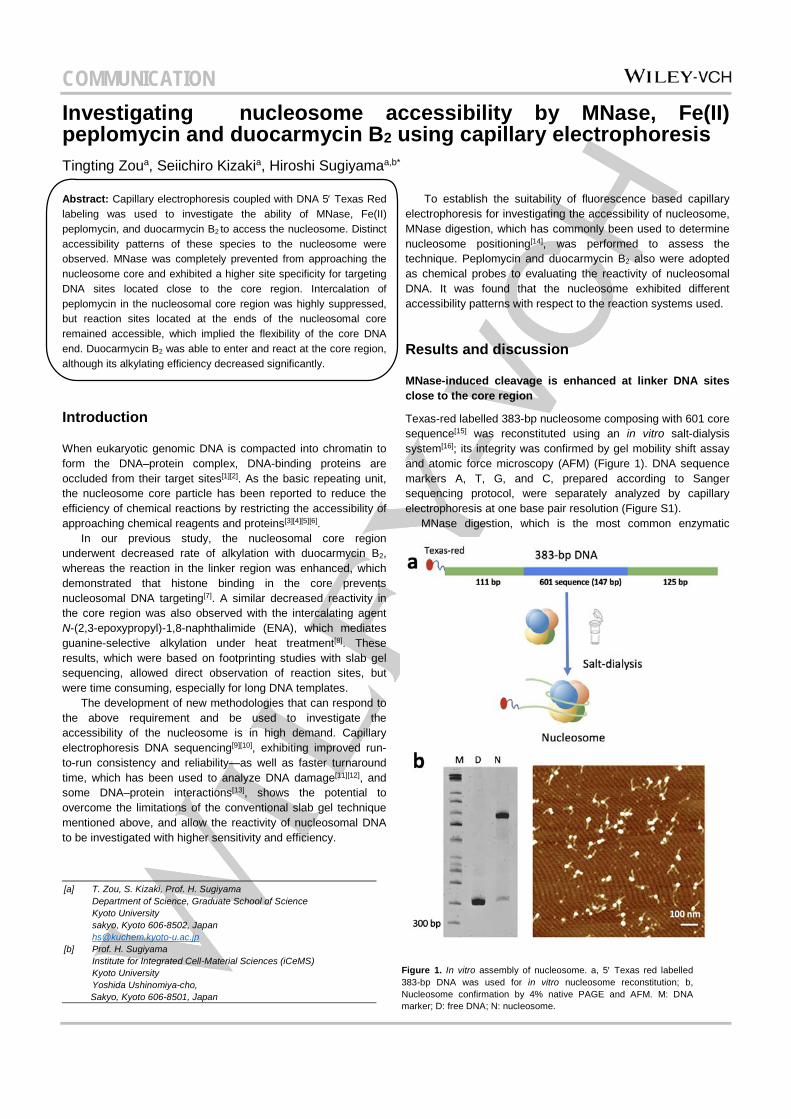
Texas-red labelled 383-bp nucleosome composing with 601 core sequence[15] was reconstituted using an in vitro salt-dialysis system[16]; its integrity was confirmed by gel mobility shift assay and atomic force microscopy (AFM) (Figure 1). DNA sequence markers A, T, G, and C, prepared according to Sanger sequencing protocol, were separately analyzed by capillary electrophoresis at one base pair resolution (Figure S1). MNase digestion, which is the most common enzymatic
[a] T. Zou, S. Kizaki, Prof. H. Sugiyama Department of Science, Graduate School of Science Kyoto University sakyo, Kyoto 606-8502, Japan [email protected]
[b] Prof. H. Sugiyama Institute for Integrated Cell-Material Sciences (iCeMS) Kyoto University Yoshida Ushinomiya-cho, Sakyo, Kyoto 606-8501, Japan
Figure 1. In vitro assembly of nucleosome. a, 5′ Texas red labelled 383-bp DNA was used for in vitro nucleosome reconstitution; b, Nucleosome confirmation by 4% native PAGE and AFM. M: DNA marker; D: free DNA; N: nucleosome.
COMMUNICATION
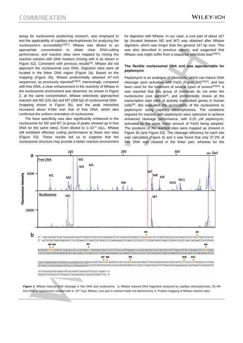
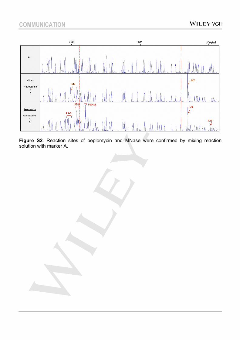
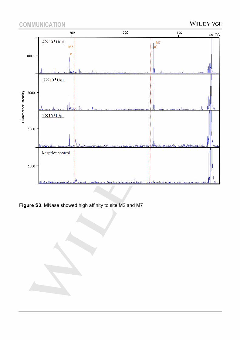
assay for nucleosome positioning research, was employed to test the applicability of capillary electrophoresis for analyzing the nucleosome’s accessibility[14][17]. MNase was diluted to an appropriate concentration to obtain clear DNA-cutting performance, and reaction sites were mapped by mixing the reaction solution with DNA markers (mixing with A as shown in Figure S2). Consistent with previous results[18], MNase did not approach the nucleosomal core DNA. Digestion sites were all located in the linker DNA region (Figure 2a). Based on the mapping (Figure 2b), MNase preferentially attacked AT-rich sequences, as previously reported[19][20]. Interestingly, compared with free DNA, a clear enhancement in the reactivity of MNase in the nucleosome environment was observed. As shown in Figure 2, at the same concentration, MNase selectively approached reaction site M2 (101 bp) and M7 (268 bp) of nucleosomal DNA (mapping shown in Figure 2b), and the peak intensities increased about 4-fold over that of free DNA, which also confirmed the uniform orientation of nucleosome. The base specificity was also significantly enhanced in the nucleosome for M2 and M7 (a group of peaks showed up in free DNA for the same sites). Even diluted to 1×10⁻⁴ U/µL, MNase still exhibited effective cutting performance at these two sites (Figure S3). These results led us to suppose that the nucleosome structure may provide a better reaction environment
for digestion with MNase. In our case, a core part of about 167 bp (located between M2 and M7) was obtained after MNase digestion, which was longer than the general 147 bp core. This was also described in previous reports, and suggested that MNase–seq might suffer from a sequence selectivity bias[20][21].
The flexible nucleosomal DNA end was approachable for peplomycin
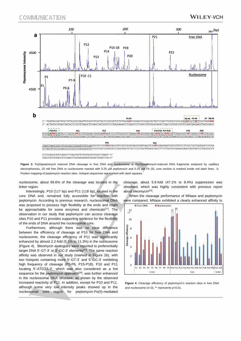
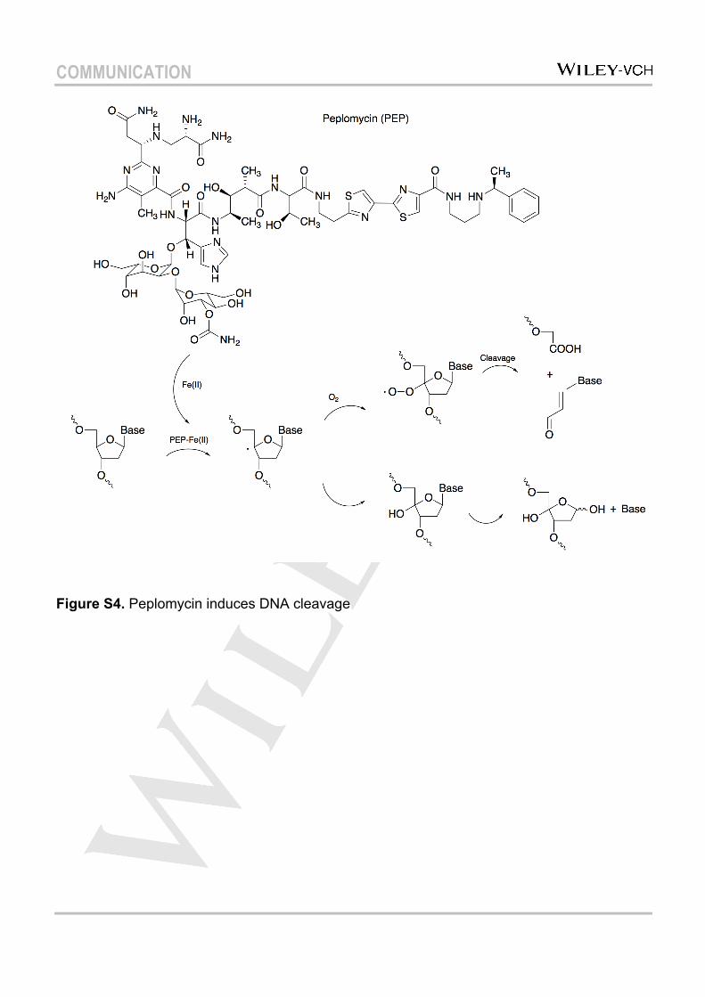
Peplomycin is an analogue of bleomycin, which can induce DNA cleavage upon activation with Fe(II) (Figure S4)[22][23], and has been used for the treatment of several types of tumors[24][25]. It was reported that this group of chemicals do not enter the nucleosome core particle[4], and preferentially cleave at the transcription start sites of actively transcribed genes in human cells[26]. We evaluated the accessibility of the nucleosome to peplomycin using capillary electrophoresis. The conditions required for reaction with peplomycin were optimized to achieve enhanced cleavage performance, with 0.25 µM peplomycin, activated by the same molar amount of Fe(II) being adopted. The positions of the reaction sites were mapped as showed in Figure 3b (and Figure S3). The cleavage efficiency for each site was calculated (Figure 4) and it was found that only 37.2% of free DNA was cleaved in the linker part, whereas for the
Figure 2. MNase induced DNA cleavage in free DNA and nucleosome. a, MNase induced DNA fragments analyzed by capillary electrophoresis, 20 nM free DNA or nucleosome reacted with 4× 10⁻⁴ U/µL MNase, core part is marked inside red dashed lines; b, Positon mapping of MNase reaction sites.
COMMUNICATION
nucleosome, about 69.6% of the cleavage was located in the linker region.
Interestingly, P10 (117 bp) and P11 (118 bp), located in the core DNA end, remained fully accessible for reaction with peplomycin. According to previous research, nucleosomal DNA was proposed to possess high flexibility at the ends and might be approachable for some enzymes and chemicals[27]. The observation in our study that peplomycin can access cleavage sites P10 and P11 provides supporting evidence for the flexibility of the ends of DNA around the nucleosomal core. Furthermore, although there was no clear difference between the efficiency of cleavage at P10 for free DNA and nucleosome, the cleavage efficiency of P11 was significantly enhanced by almost 2.2-fold (5.1% to 11.3%) in the nucleosome (Figure 4). Bleomycin analogues were reported to preferentially target DNA 5′-GT-3′ or 5′-GC-3′ elements[23]. The same reaction affinity was observed in our study (marked in Figure 2b), with two hotspots containing more 5′-GT-3′ and 5′-GC-3′ exhibiting high frequency of cleavage (P3-P6, P15-P18). P10 and P11 locating 5′-ATGTA-3′, which was also considered as a hot sequence for the peplomycin approach[28], was further enhanced in the nucleosomal DNA structure, as shown by the observed increased reactivity of P11. In addition, except for P10 and P11, although some very low intensity peaks showed up in the nucleosomal core region for peplomycin·Fe(II)-mediated
cleavage, about 5.6-fold (47.1% to 8.4%) suppression was observed, which was highly consisitent with previous report about bleomycin[29].
When the cleavage performance of MNase and peplomycin were compared, MNase exhibited a clearly enhanced affinity to
Figure 3. Fe(II)peplomycin induced DNA cleavage in free DNA and nucleosome. a, Fe(II)peplomycin-induced DNA fragments analyzed by capillary electrophoresis, 20 nM free DNA or nucleosome reacted with 0.25 µM peplomycin and 0.25 µM Fe (II), core section is marked inside red dash lines. b, Positon mapping of peplomycin reaction sites. Hotspot sequences are marked with dash squares.
Figure 4. Cleavage efficiency of peplomycin’s reaction sites in free DNA and nucleosome (n=3). ** represents p<0.01.
COMMUNICATION
the nucleosome and high preference for cleavage of DNA sites close to core region. This result confirmed the efficiency of MNase for nucleosome positioning, while might introduce longer nucleosome core particles due to the sequence selectivity. Peplomycin may produce a shorter core region in the nucleosome, and could be used to investigate the precise binding position of DNA and proteins.
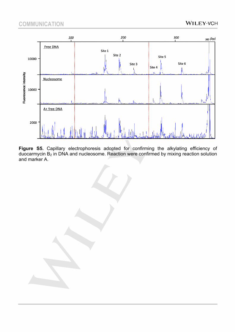
Capillary electrophoresis was applied to investigate the accessibility of duocarmycin B2 to the nucleosome For a further confirmation, the minor-groove binding agent duocarmycin B2 was applied to alkylate the nucleosomal DNA as reported previously, followed by capillary electrophoresis analysis. The fragmentations of nucleosomal DNA formed upon cleavage with duocarmycin B2 were revealed as peaks in the sequence scanner (Figure S5). The positions of the alkylation sites were confirmed by mixing the reaction solution with DNA marker A. It was found that all the DNA sites alkylated by duocarmycin B2 in free DNA were also alkylated in nucleosomal DNA, which confirmed that the minor groove of the nucleosomal core was accessible for duocarmycin B2. It has also been demonstrated that another minor-groove binding agent, pyrrole-imidazole polyamide (PIP), can access the nucleosomal core region[30]. Previously, our group has reported a group of new artificial genetic switches, conjugations of PIP with histone deacetylase inhibitor SAHA, which could specifically regulate gene expression patterns[31], and these compounds were also capable of promoting iPS cells to a neural progenitor state[32]. These results demonstrate that the nucleosome core can be a target for this group of minor-groove binding agents with consequent modification of the dynamic of the nucleosome. Similar to the previous report[7], fragments cleaved from reaction sites located in the core region, showed decreased peak intensity with about 2.3-fold suppressed alkylating efficiency (53.1% to 23.2 %). Especially site 2 (5′-TTTAA-3′), which was reported to form an extremely narrow minor groove at SHL±1.5 via a unique and conserved histone motif that ‘clamps’ the DNA[33], exhibited the clearest decrease in cleavage efficiency. In contrast, enhanced peak signals arising from cleavage of sites located on the linker region were observed. This result is consistent with previous results obtained from sequencing gel studies, thereby confirming the applicability of capillary electrophoresis for investigating the accessibility of the nucleosome.
Conclusion
In this study, capillary electrophoresis was used to investigate the accessibility of nucleosome. MNase exhibited enhanced selectivity for DNA sites close to the core region of the nucleosome. The nucleosomal core was found to be accessible for duocarmycin B2 and Fe(II) peplomycin with variable levels of
suppression, whereas the flexible core DNA end was fully approachable by peplomycin.
Expermental Section
Details list in supplemental data.
Keywords: Nucleosome • Accessibility • Capillary electrophoresis • MNase • Peplomyin
[1] W. K. M. Lai, B. F. Pugh, Nat. Rev. Mol. Cell Biol. 2017, DOI 10.1038/nrm.2017.47.
[2] C. D. Allis, T. Jenuwein, Nat. Rev. Genet. 2016, 17, 487–500. [3] J. D. Trzupek, J. M. Gottesfeld, D. L. Boger, Nat. Chem. Biol. 2006,
2, 79–82. [4] M. T. Kuo, T. C. Hsu, Nature 1978, 271, 83–84. [5] J. M. Gottesfeld, C. Melander, R. K. Suto, H. Raviol, K. Luger, P. B.
Dervan, J Mol Biol 2001, 309, 615–629. [6] S. Kizaki, T. Zou, Y. Li, Y. W. Han, Y. Suzuki, Y. Harada, H.
Sugiyama, Chem. Eur. J 2016, 22, 16598–16601. [7] T. Zou, S. Kizaki, G. N. Pandian, H. Sugiyama, Chem. Eur. J. 2016,
8502, 8756–8758. [8] G. E. Davey, B. Wu, Y. Dong, U. Surana, C. A. Davey, Nucleic
Acids Res. 2009, 38, 2081–2088. [9] K. D. Altria, Methods Mol. Biol. 1996, 52, 3–13. [10] V. Kostal, J. Katzenmeyer, E. a Arriaga, Anal. Chem. 2008, 80,
4533–4550. [11] K. Klepárník, P. Boček, Chem. Rev. 2007, 107, 5279–5317. [12] M. T. Valenzuela, M. I. Núñez, M. R. Guerrero, M. Villalobos, J. M.
Ruiz De Almodóvar, J. Chromatogr. A 2000, 871, 321–330. [13] M. Berezovski, S. N. Krylov, J. Am. Chem. Soc. 2002, 124, 13674–
13675. [14] M. Tsompana, M. J. Buck, Epigenetics Chromatin 2014, 7, 33. [15] P. . Lowary, J. Widom, J. Mol. Biol. 1998, 276, 19–42. [16] U. Muthurajan, F. Mattiroli, S. Bergeron, K. Zhou, Y. Gu, S.
Chakravarthy, P. Dyer, T. Irving, K. Luger, in Methods Enzymol., 2016, pp. 3–41.
[17] D. J. Telford, B. W. Stewart, Int. J. Biochem. 1989, 21, 127–138. [18] R. T. Simpson, Biochemistry 1978, 17, 5524–5531. [19] J. D. McGhee, G. Felsenfeld, Cell 1983, 32, 1205–1215. [20] R. V. Chereji, J. Ocampo, D. J. Clark, Mol. Cell 2017, 65, 565–
577.e3. [21] H. R. Chung, I. Dunkel, F. Heise, C. Linke, S. Krobitsch, A. E.
Ehrenhofer-Murray, S. R. Sperling, M. Vingron, PLoS One 2010, 5, DOI 10.1371/journal.pone.0015754.
[22] R. M. Burger, Chem. Rev. 1998, 98, 1153–1170. [23] K. D. Goodwin, M. A. Lewis, E. C. Long, M. M. Georgiadis, Proc.
Natl. Acad. Sci. 2008, 105, 5052–5056. [24] M. Linnert, J. Gehl, Anticancer. Drugs 2009, 20, 157–164. [25] K. Kawai, H. Akaza, Expert Opin. Drug Saf. 2003, 2, 587–596. [26] J. K. Chen, D. Yang, B. Shen, V. Murray, Int. J. Biochem. Cell Biol.
2017, 85, 56–65. [27] K. Luger, Chromosom. Res. 2006, 14, 5–16. [28] S. D. Gautam, J. K. Chen, V. Murray, J. Biol. Inorg. Chem. 2017, 22,
881–892. [29] B. L. Smith, G. B. Bauer, L. F. Povirk, J. Biol. Chem. 1994, 269,
30587–30594. [30] R. K. Suto, R. S. Edayathumangalam, C. L. White, C. Melander, J.
M. Gottesfeld, P. B. Dervan, K. Luger, J. Mol. Biol. 2003, 326, 371–380.
[31] L. Han, G. N. Pandian, S. Junetha, S. Sato, C. Anandhakumar, J. Taniguchi, A. Saha, T. Bando, H. Nagase, H. Sugiyama, Angew. Chem. Int. Ed. Engl. 2013, 52, 13410–3.
[32] Y. Wei, G. N. Pandian, T. Zou, J. Taniguchi, S. Sato, G. Kashiwazaki, T. Vaijayanthi, T. Hidaka, T. Bando, H. Sugiyama, ChemistryOpen 2016, 5, 517–521.
[33] S. Tan, C. A. Davey, Curr. Opin. Struct. Biol. 2011, 21, 128–136.
COMMUNICATION
Entry for the Table of Contents Layout 1:
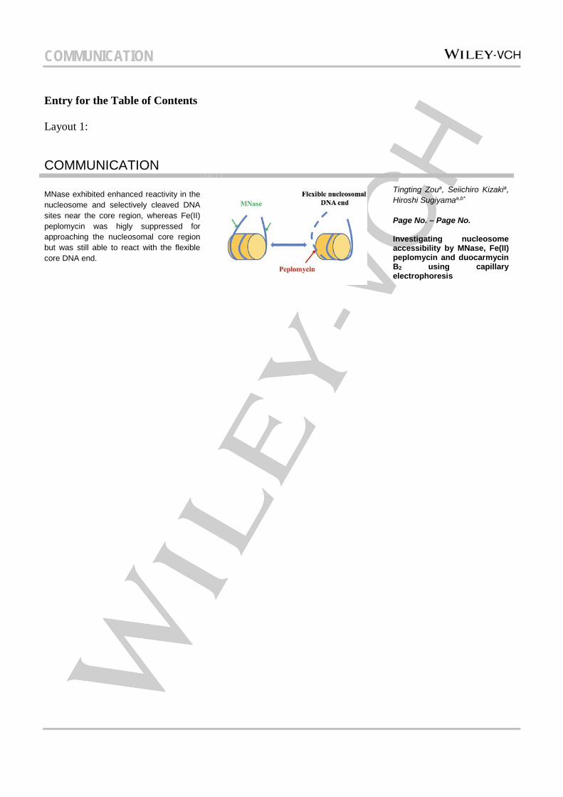
COMMUNICATION MNase exhibited enhanced reactivity in the nucleosome and selectively cleaved DNA sites near the core region, whereas Fe(II) peplomycin was higly suppressed for approaching the nucleosomal core region but was still able to react with the flexible core DNA end.
Tingting Zoua, Seiichiro Kizakia, Hiroshi Sugiyamaa,b*
Page No. – Page No. Investigating nucleosome accessibility by MNase, Fe(II) peplomycin and duocarmycin B2 using capillary electrophoresis
COMMUNICATION
Supplementary data
Experimental Section
DNA and histone preparation
383 bp DNA containing two linker DNA parts (111 bp and 125 bp) and one core 601 sequence DNA (147 bp) was constructed and amplified by PCR using primer sets: forward primer 5’-TAATACGACTCACTATAGG-3’ and reverse primer 5’-ATTTAGGTGACACTATAGAATAC-3’ from plasmid DNA pGEM3Z-601 extracted from E. coli. Forward primer 5’-texas red-TAATACGACTCACTATAGG-3’ was used for obtain texas red labelling DNA. Then amplified DNA was purified using the Wizard SV gel and PCR clean-up system (Promega, USA) and final concentration was determined by Nano drop system. Histone octamer was bought from Epicypher (NC, USA).
DNA-histone complex construction and gel confirmation
DNA-histone complex was constructed by salt-jump method previously reported for nucleosome reconstitution. The reconstituted samples were collected from dialysis tube and confirmed by 4% native PAGE gel (100 V, 90 min, 4℃) and AFM.
Duocarmycin B2 alkylating reaction
Duocarmycin B2 was dissolved in DMSO to 10 mM as a stock solution, then diluted to working solution. 20 nM of free DNA or nucleosome were mixed with duocarmycin B2 to a final 10 µL 6.25 mM sodium phosphate buffer (pH 7.0) reaction system, alkylating reaction occurred at room temperature for 18 h. After incubation, the reaction mixture was quenched by addition of calf thymus DNA.
Peplomycin reaction
Peplomycin (Kayaku, Japan) and Iron (II) Sulfate Heptahydrate were dissolved in H2O to 5 mM as stock solution, then diluted to working solution before using. 20 nM of free DNA, or nucleosome was mixed with certain amount of peplomycin and same amount of Iron (II) Sulfate Heptahydrate to a final 10 µL in a 50 mM HEPES-KOH reaction system. Reaction solution was kept in room temperature for 1 hour.
Microccocal nuclease digestion
20 nM of DNA-histone protein complex was mixed with certain amount of MNase to a final 10 µL reaction system with 1´reaction buffer (20 mM Tris-HCl, pH 8.0; 5 mM NaCl; 2.5 mM CaCl2). And reaction solution was put in 37℃ for 10 min, then add 2 µL 0.5 M EDTA (pH 8.0) to stop the reaction for 5 min on ice.
Capillary electrophoresis
After reaction completed, all the samples were mixed with denaturing buffer (1:1) and heated in 95℃ for 25 min for complete denaturation. The denatured samples were diluted with H2O to final 12 µL for capillary electrophoresis analysis. Spin column purification was performed to remove excess denaturing agents. Texas red labelled DNA fragments were determined by Applied Biosystems 3130/3130xl Genetic Analyzers (Life Technologies, USA) in standard mode. The parameter was set as: oven temperature 60℃, current stability 5.0 µAmp, pre-run voltage 15 kV, pre-run time 180 sec, injection voltage 1.6 kV, injection time 20 sec, run voltage 8.5 kV, and run time 100 min. Data was viewed and analyzed by seq-scanner software. The chromatographic images were imported to image J, peaks’ area were calculated for analyzing the cleavage efficiency.

COMMUNICATION
Figure S1. DNA markers analyzed by capillary electrophoresis.
COMMUNICATION
Figure S2. Reaction sites of peplomycin and MNase were confirmed by mixing reaction solution with marker A.
COMMUNICATION
Figure S3. MNase showed high affinity to site M2 and M7
COMMUNICATION
Figure S4. Peplomycin induces DNA cleavage
COMMUNICATION
Figure S5. Capillary electrophoresis adopted for confirming the alkylating efficiency of duocarmycin B2 in DNA and nucleosome. Reaction were confirmed by mixing reaction solution and marker A.