titanium dioxide nanostructured coatings: application … · titanium dioxide nanostructured...
TRANSCRIPT
Titanium Dioxide Nanostructured Coatings:
Application in Photocatalysis and Sensors
JA Byrne, JWJ Hamilton, TA McMurray, PSM Dunlop, V Jackson A. Donaldson,
J Rankin, G Dale, D Al Rousan
Nanotechnology and Advanced Materials Research Institute,
University of Ulster at Jordanstown, Northern Ireland, United Kingdom, BT37 0QB
Tel. +44 28 90 36 89 41 Email: [email protected]
Abstract
This paper presents some of the research work
taking place at the University of Ulster
investigating preparation, characterisation and
application of nanostructred TiO2. Four
exemplars are used to demonstrate the
potential applications of these materials i.e.
photocatalytic disinfection of water containing
chlorine resistant microorganisms,
photocatalytic ‘self-cleaning’ of surfaces
contaminated with protein, transducers for
electrochemical biosensors and finally new
opportunities presented by electrochemical
growth of TiO2 aligned nanotubes.
1.0 Introduction
Nanostructured titanium dioxide has been
widely researched for application towards the
photocatalytic treatment of purification of
water and air, “self-cleaning” and
superhydrophilic coatings for surfaces, and
dye-sensitised voltaic cells. Nanoparticle TiO2
films present a large surface area to geometric
area ratio, which is useful in water and air
purification and dye sensitised cells. In
addition, these films also give high surface
area desirable for electrochemical sensor
applications. Titanium dioxide is found in
three crystal forms, brookite, rutile and
anatase, the latter of which is the most suitable
for photocatalytic applications. Anatase TiO2
is a wide band gap semiconductor (3.2 eV) and
absorbs photons with λ < 387 nm. Band gap
excitation produces electron hole pairs, which
can take part in electrochemical reactions at
the interface and result in the production of
radical species. This photocatalytic action has
been reported to degrade organic pollutants (in
water and air) to CO2 and H2O, and kill a wide
range of microoganisms. Furthermore,
photocatalytic films can degrade protein
material (including temperature stable
proteins) adhered to their surface, and could
find application in, for example, the
sterilisation of surgical devices.
Nanostructured TiO2 thick films present a high
surface area, which is desirable for
electrochemical sensor and biosensor
applications. TiO2 is biocompatible, non-toxic,
and chemical stable under conditions found
within the body, making it a suitable material
for implantable biosensors. Furthermore, TiO2
can be used for the electrochemical detection
of hydrogen peroxide in the presence of
oxygen. This makes nano-structured TiO2
electrodes suitable for non-mediated
biosensors utilising oxidase enzymes e.g.
glucose oxidase.
Self-assembled titanium oxide nanotube arrays
with maximum packing density can be formed
by the anodic oxidation of titanium metal [1].
Such materials may prove to have enhanced
properties for photocatalytic, sensor and other
applications.
2.0 Photocatalytic disinfection of water
Cryptosporidium poses significant problems to
the drinking water industry. It is ubiquitous in
surface water, difficult to remove by
conventional drinking water treatment
processes and if ingested can cause serious
illness [2]. The cost-effective removal of
Cryptosporidium from drinking water sources
remains one of the industries greatest
challenges [3]. There have been many
outbreaks of cryptosporidiosis all over the
world associated with consumption of
contaminated drinking water [4]. The largest
outbreak occurred in Milwaukee, USA in 1993
with the death of 104 AIDS patients and an
estimated 403,000 people becoming ill [5].
The bactericidal effect of TiO2 photocatalysis
has been widely reported [6,7] however a
limited number of studies have reported the
effectiveness of photocatalysis against chlorine
resistant organisms. In this paper we present
the photocatalytic inactivation of
Cryptosporidium parvum oocysts
72 NSTI-Nanotech 2006, www.nsti.org, ISBN 0-9767985-6-5 Vol. 1, 2006
TiO2 powder (Degussa P25) was
electrophoretically immobilised onto Ti alloy
substrates [8] and placed in a quartz water-
jacketed reactor with the illumination source
focused on the coated area (125W HPR lamp
(Philips), mainline emission 365nm). An
oocyst suspension was prepared by diluting
fresh oocysts (Moredun Scientific) in saline
solution to achieve a working concentration of
2x10⁴ oocysts per cm
3. The reactor was
thermostatically controlled at 20 ± 2°C and
agitation of the 10 cm3 of oocyst suspension
was provided by a small magnetic stirrer. Air
sparging was achieved using a small aquarium
pump, flow rate of 900 cm3 min
-1. The reactor
was allowed to reach equilibrium under dark
conditions for 15 min prior to irradiation. A
100 µL sample was removed and the electrode
illuminated. Samples were removed every 60
minutes thereafter for a period of 240 minutes.
Analysis for oocyst damage was preformed
using the vital dye exclusion protocol
developed by Robertson et al [9].
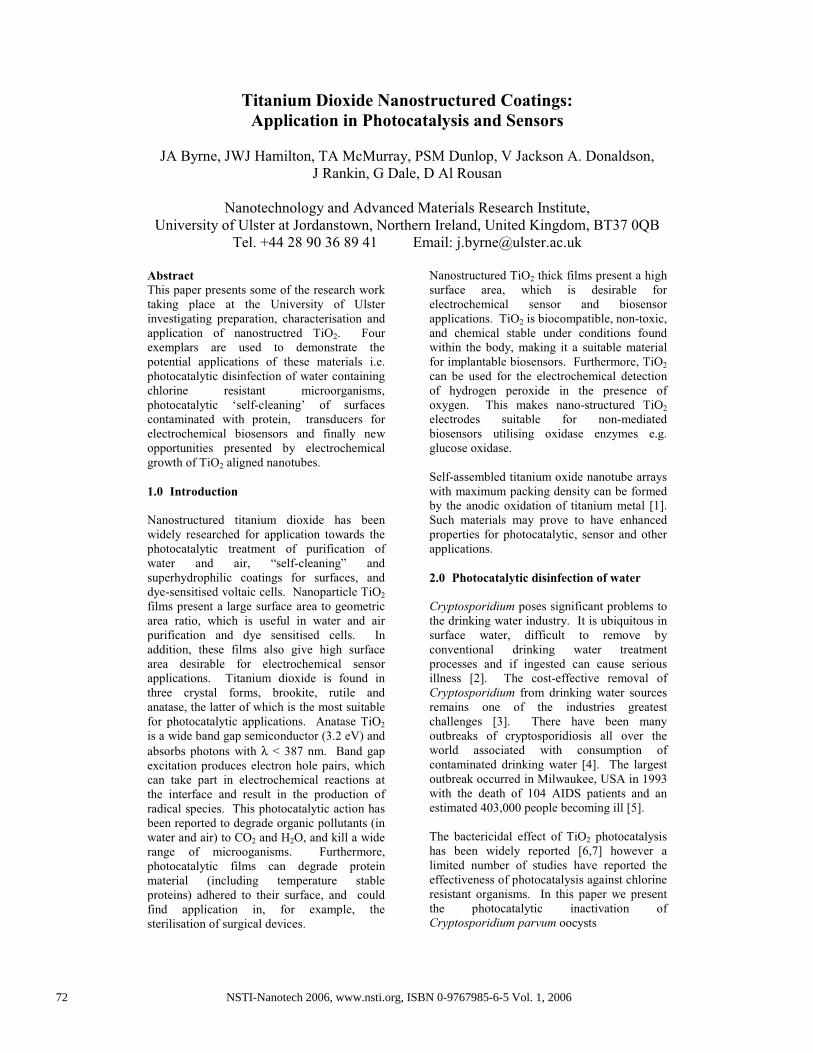
During the first hour observable changes in
oocyst shape were observed associated with a
decrease in viability (see figure 1). Following
three hours treatment viability decreased to
zero and fragmentation of the oocyst bodies
was evident along with the presence of many
ghost oocysts (oocysts missing their DNA).
Further studies are being carried out confirm
the loss of infectivity via in-vitro infectivity
and an examination to elucidate the mechanism
of disinfection will be undertaken.
3.0 Self-cleaning coatings for surface
decontamination
Decontamination of surfaces is an area of
current interest. Health care acquired
infections (HAIs) cost the Health care sector
billions of pounds every year and cause patient
discomfort, prolonged hospital stays, and even
death. Conventional approaches to the
decontamination and sterilisation of re-usable
surgical devices may not be wholly effective.
While photocatalytic coatings have been
reported to be ‘self-cleaning’ and even
commercialised for this purpose e.g. Pilkington
Activ self cleaning glass [10], there are few
published reports dealing with photocatalytic
decontamination of protein from surfaces.
In this work TiO2 thin films were prepared
using a sol gel route. Titanium IV butoxide
was hydrolysed under controlled conditions
with acetic acid as a catalyst. The resulting sol
gel was spin coated onto glass slides and then
annealed. Raman spectroscopy analysis and
glancing angle XRD confirmed the presence of
anatase crystal phase. Samples were
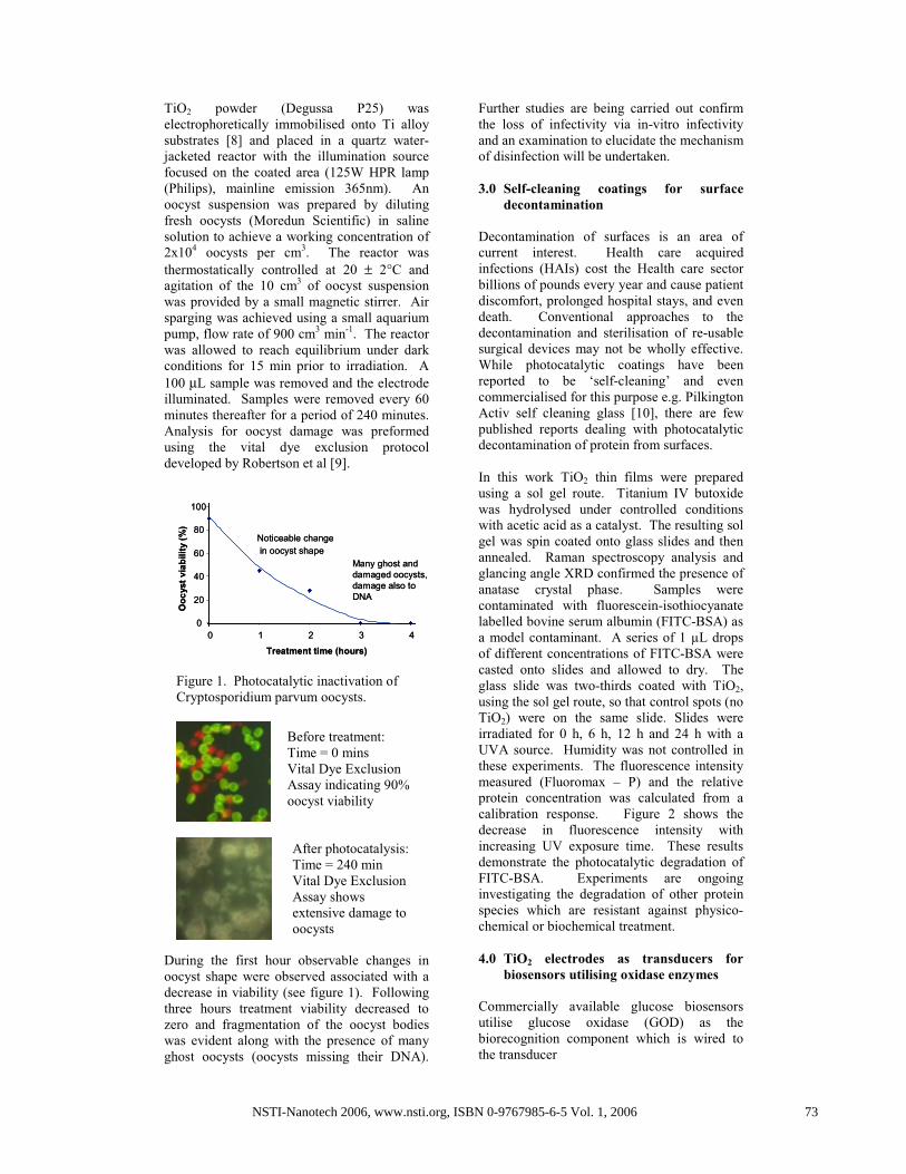
contaminated with fluorescein-isothiocyanate
labelled bovine serum albumin (FITC-BSA) as
a model contaminant. A series of 1 µL drops
of different concentrations of FITC-BSA were
casted onto slides and allowed to dry. The
glass slide was two-thirds coated with TiO2,
using the sol gel route, so that control spots (no
TiO2) were on the same slide. Slides were
irradiated for 0 h, 6 h, 12 h and 24 h with a
UVA source. Humidity was not controlled in
these experiments. The fluorescence intensity
measured (Fluoromax – P) and the relative
protein concentration was calculated from a
calibration response. Figure 2 shows the
decrease in fluorescence intensity with
increasing UV exposure time. These results
demonstrate the photocatalytic degradation of
FITC-BSA. Experiments are ongoing
investigating the degradation of other protein
species which are resistant against physico-
chemical or biochemical treatment.
4.0 TiO2 electrodes as transducers for
biosensors utilising oxidase enzymes
Commercially available glucose biosensors
utilise glucose oxidase (GOD) as the
biorecognition component which is wired to
the transducer
0
20
40
60
80
100
0 1 2 3 4
Treatment time (hours)
Oo
cyst
via
bil
ity (
%)
Noticeable change
in oocyst shape
Many ghost and
damaged oocysts,
damage also to
DNA
0
20
40
60
80
100
0 1 2 3 4
Treatment time (hours)
Oo
cyst
via
bil
ity (
%)
Noticeable change
in oocyst shape
Many ghost and
damaged oocysts,
damage also to
DNA
Figure 1. Photocatalytic inactivation of
Cryptosporidium parvum oocysts.
Before treatment:
Time = 0 mins
Vital Dye Exclusion
Assay indicating 90%
oocyst viability
After photocatalysis:
Time = 240 min
Vital Dye Exclusion
Assay shows
extensive damage to
oocysts
73NSTI-Nanotech 2006, www.nsti.org, ISBN 0-9767985-6-5 Vol. 1, 2006
using a mediator. However, mediated glucose
biosensing may not be suitable for in-vivo
sensing as mediators may be toxic or simply
lost into the blood. Non-mediated glucose
biosensing is possible using materials which
can either be directly wired to the enzyme or
which can selectively detect the H2O2 product
in the presence of oxygen. There is an
opportunity to produce a wide range of
biosensors, utilising oxidase enzymes.
The use of mesoporous titanium dioxide
electrodes has been reported previously for the
amperometric detection of glucose via electro-
reduction of released hydrogen peroxide. [11]
Electrophoretic coating may be used to
produce porous nanocrystalline TiO2
electrodes [8,12]. These electrodes have been
tested for the electrochemical reduction of
H2O2 in the presence of O2 and the response is
independent of O2 at potentials more positive
than –0.4 V vs the saturated calomel electrode
(SCE). Titanium foil samples were coated
with nanoparticle TiO2 by the electrophoretic
method. Electrical contact was made to an
area of the Ti foil not coated with TiO2 using
copper wire and conducting epoxy. The
contact and any remaining uncoated foil area
were insulated using a negative photoresist.
All electrochemical analyses were carried out
using a three-electrode electrochemical cell,
with pH 6 phosphate buffer as the supporting
electrolyte. The reference electrode was a
saturated calomel (SCE), and the counter
electrode was a platinum disc. Amperometric
detection was carried out using a BAS LC-4C
amperometric detector connected to a Lloyd
instruments PL3 x-y plotter. All potentials are
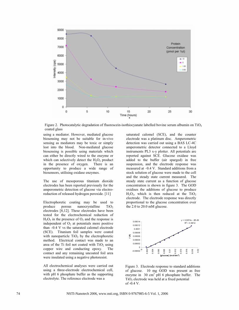
reported against SCE. Glucose oxidase was
added to the buffer (air sparged) in free
suspension, and the electrode response was
measured at –0.4 V. Standard additions from a
stock solution of glucose were made to the cell
and the steady state current measured. The
steady state current as a function of glucose
concentration is shown in figure 3. The GOD
oxidises the additions of glucose to produce
H2O2, which is then reduced at the TiO2
electrode. The electrode response was directly
proportional to the glucose concentration over
the 2.0 to 20.0 mM glucose.
y = 0.0074x - 2E-05
R2 = 0.9912
-0.00002
0
0.00002
0.00004
0.00006
0.00008
0.0001
0.00012
0.00014
0
0.0
02
0.0
04
0.0
06
0.0
08
0.0
1
0.0
12
0.0
14
0.0
16
0.0
18
0.0
2
[glucose] (m ol/dm3)
I (
A)
Figure 3. Electrode response to standard additions
of glucose. 10 mg GOD was present as free
enzyme in 30 cm3
pH 6 phosphate buffer. The
TiO2 electrode was held at a fixed potential
of -0.4 V.
Figure 2. Photocatalytic degradation of fluorescein-isothiocyanate labelled bovine serum albumin on TiO2
coated glass
74 NSTI-Nanotech 2006, www.nsti.org, ISBN 0-9767985-6-5 Vol. 1, 2006

5.0 Electrochemical growth of TiO2
nanotubes on titanium metal foil
In this work the effect of HF concentration and
anodisation potential were investigated.
Anodisation was carried out in a one
compartment cell with a titanium foil anode
platinum foil cathode. Constant potential
conditions were employed. The total cell
volume was 100 cm3 and electrode separation
was 20 mm. The salient parameters
investigated were HF concentrations 0.005%-
0.5% w/v and cell potentials in the range 5.0 to
30 V. Above concentrations of 0.15% HF the
Ti foil dissolved rapidly. The optimum HF
concentration was found to be 0.05 % w/v.
In this system, only cell potentials above 15 V
produced the nano-structuring effect and
potentials greater than 30 V destroyed the
nanotube formation. Sample morphology was
examined using an FEI quanta SEM at an
accelerating voltage of 30 kV and a beam
current of 47 pA. The mean tube diameter
was ca. 80 nm for the samples prepared at a
cell potential of 25 V in 0.005%w/v HF (see
figure 4)
Work is ongoing investigating the potential
uses of TiO2 nanotubes for photocatalysis,
biosensing and other applications
7.0 Acknowledgements
We would like to thank the following; DEL NI
for funding Rankin, Dale, Al Rousan, and
Donaldson. J Dooley and C Lowrey, UU, for
analysis of Cryptosporidium. University of
Edinburgh for FITC-BSA and analysis. Dept
of Health UK, R&D Office HPSS NI, Invest
Northern Ireland and European Commission
for funding. Degussa for samples of P25.
Philips lighting, Netherlands, for light sources.
7.0 References
1. D Gong, G.A. Grimes, O.K. Varghese, W. Hu, R.S. Singh, A. Chen, E.C. Dickey, “Titanium oxide nanotube
arrays prepared by anodic oxidation,” J.Mater.Res., 2001,
16, 3331-3334. 2. Boucher, “Cryptosporidium in Water Supplies. Third
Report of the Group of Experts” (Department of
Health/Department of the Environment, Published by HMSO, 1998).
3. Badenoch, “Cryptosporidium in Water Supplies. First Report of the Group of Experts” (Department of
Health/Department of the Environment. Published by TSO
Ltd, 1990) 4. H. V. Smith, J. B. Rose, “Waterborne cryptosporidiosis:
Current status”, Parasitology Today, 1998, 14, 14-22
5. W. R. Mackenzie, N. J. Hoxie, M. E. Proctor, M. S. Gradus, K. A. Blair, D. E. Peterson, J. J. Kazmierczak, D.
G. Addiss, K. R. Fox, J. B. Rose, J. P. Davis, “A massive
outbreak in Milwaukee of Cryptosporidium infection transmitted through the public water supply”, New
England Journal of Medicine, 1994, 331, 161-167
6. H. Zheng, P. C. Maness, D. M. Blake, E. J. Wolfrum, S. L. Smolinski, W. A. Jacoby, “Bactericidal mode of
titanium dioxide photocatalysis,” Journal of
Photochemistry and Photobiology A-Chemistry, 2000, 130,163-170.
7. D. M. Blake, P. C. Maness, Z. Huang, E. J. Wolfrum, J.
Huang, W. A. Jacoby, “Application of the photocatalytic chemistry of titanium dioxide to disinfection and the
killing of cancer cells,” Separation and Purification
Methods, 1999, 28, 1-50 8. J. A. Byrne, B. R. Eggins, N. M. D. Brown, B.
McKinney, M. Rouse, “Immobilisation of TiO2 powder for
the treatment of polluted water,” Applied Catalysis B-Environmental, 1998, 17, 25-36
9. L.J. Robertson, A.T. Campbell, H.V. Smith, “Viability
of Cryptosporidium parvum oocysts: Assessment by the dye permeability assay,” Applied and Environmental
Microbiology 1998, 64 (9) 3544
10. Ashton V., “Windows on the future.” Chemistry in Britain, 2002, 38(6): 26-28.
11. S Cosnier, C Gondran, A Senillou, M Gratzel, N
Vlachopoulos, “Mesoporous TiO2 Films: New Catalytic Electrode Materials for Fabricating Amperometric
Biosensors Based on Oxidases”, Electroanalysis, 1997,
9(18), 1387-1392. 12. J A Byrne, B R Eggins, S Linquette-Mailley and P S
M Dunlop, “The effect of hole acceptors on the
photocurrent response of particulate titanium dioxide”, Analyst, 1998, 123, 2007-2012.
Figure 4. SEM of TiO2 nantubes grown
electrochemically on Ti foil.
75NSTI-Nanotech 2006, www.nsti.org, ISBN 0-9767985-6-5 Vol. 1, 2006