this is not analogous to the blastocoel defines a...
TRANSCRIPT

Week One: November 17 November 21
Cleavage and Gastrulation – Avian embryos- Meroblastic dicoidal cleavage pattern – Cleavage is initially incomplete. The egg is a very large,
yolky egg that displaces to one side of the cytoplasm. It makes a cytoplasmic disc or blastodisc. - Initially produces a subgerminal cavity – this is not analogous to the blastocoel. - Must establish a two layered blastoderm before gastrulation – defines a blastocoel cavity. This
is distinctly different from the subgerminal cavity.- Resembles gastrulation in both the fishes and in mammals
Cleavage – discoidal- The cells gradually begin to define themselves as they produce a membrane across the yolk
layer. There is going to be a clear space that opens up – this is referred to as the area pellucida.

Formation of the blastoderm- At one edge of the egg, there is a group of cells that begin to proliferate that is referred to as the
posterior marginal zone. It will ultimately elaborate the next layer of cells – it will split the subgerminal cavity in half.
- Area over the subgerminal cavity is the area pellucida (semitransparent)- Area at marginal zones overlie yolk – area opaca- Subgerminal cavity is NOT analogous to the blastocoel- Layer of cells forming the area pellucida is the presumptive epiblast (cells from which the
embryo proper will be formed). No parts of the embryo proper will be from the hypoblast.
Formation of the blastocoel and Koller’s sickle- The posterior marginal zone will ultimately give rise to a group of cells that will be called the
Koller’s sickle. - In the formation of the blastocoel, certain cells will delaminate from the area pellucida/epiblast.
They will become embedded and form poly-invaginated islands. The islands are typically 5-20 cells. These form the primary hypoblast.
- After these poly-invaginated islands form, the group of cells sticking out from Koller’s crescent begins to project a single cell layer across the subgerminal cavity. As they do so, they will interact with the islands and push them towards the anterior end. These are the secondary hypoblast.

Formation of the Blastoderm- (A) – At the very beginning – The blue cells are going to contribute to the secondary hypoblast
cells as they project out. The Koller sickle is just a small group of cells that does not change in any situations – but seems to be acting as an inducer (not analogous to the Neiuwkoop center or the primary organizer).
- (B) – As they push out, the islands are going to connect and push forward to the anterior end. - (C) – The blue cells push out the primary hypoblast farther up. The cavity is opening up, but
there is still a subgerminal cavity underneath. The cavity that is opening will now become a true blastocoel. For gastrulation to occur, you must have a blastocoel cavity or exterior cells to move into the interior. A true blastocoel cavity had to be generated. The hypoblast only contributed to the extra-embryonic membrane.
- What does the epiblast form? Embryo proper, can also contribute to the extra-embryonic membranes (primarily endoderm)
- What does the hypoblast form? Only extra-embryonic membranes- (D) – Just behind the Koller’s crescent are cells that will form the primitive streak. The primitive
streak begins to form and is pushed forward by convergent extension. What was originally the beginning of the streak, will eventually become much thinner and narrower. Convergent extension makes the streak thinner, but longer. A lot of cells that ingress typically become presumptive mesoderm.
- (E) – Cells are ingressing to the anterior of Koller’s crescent and another group of cells projected as part of the endoderm forward. This looks like the bottle cells in the amphibian embryo – equivalent because they are going to make the foregut.

The process of Hypoblast formation- Cells extend into the subgerminal cavity from the posterior margin of the “epiblast”- Koller’s sickle – defined as thickened region of cells at the posterior margin of the epiblast –
secondary epiblast cells.- Some cells delaminate from the epiblast to drop into the subgerminal cavity and contribute to the
formation of the hypoblast – primary epiblast cells.o Function of both epiblast cells – both combine to divide the subgerminal cavity so there
is a blastocoel between the hypoblast and epiblast.- Cells posterior to Koller’s sick acts as the site for induction of the primitive streak (act as a site
for organizing/inducing) – secret Tg1 and nodalo Sounding anything like the dorsal vegetal cells in the amphibians? Nieuwkoop center-
like (not exactly like because there are no β-catenins)- The primitive streak will extend forward across the epiblast to a point 2/3 of the length of the
epiblast and for the Henson’s node.o When it reaches its most anterior position, it starts expressing two gene projects –
Chordin and Sonic Hedgehog
Chick gastrula cell movements- (A) – only looking at the projection of the primitive
streak- (B) – as it progresses forward, it extends forward by
convergent extension- (C) – the first cells to progress or migrate from the
anterior towards the streak are the cells that form the Henson’s node. Cells move from the lateral area of the epiblast and ingress to the interior through the primitive groove. The cells that pass through Henson’s node when they ingress move back through the blastocoel cavity towards the anterior – lays down a mid-dorsal line of mesoderm. This is called the chordamesoderm.
- Cells ingress through the primitive streak tend to form lateral mesoderm.
- Some contribute to the hypoblast to form extra-embryonic membranes.
- At its anterior-most extension, the primitive streak forms a pit (Henson’s node)
- The Henson’s node is analogous to… dorsal lip of the blastopore – This means that the primitive groove is analogous to the lateral lips of the blastopore.
- In true deuterostome fashion, the organizing site (opposite the Henson’s node) is going to become the anus.
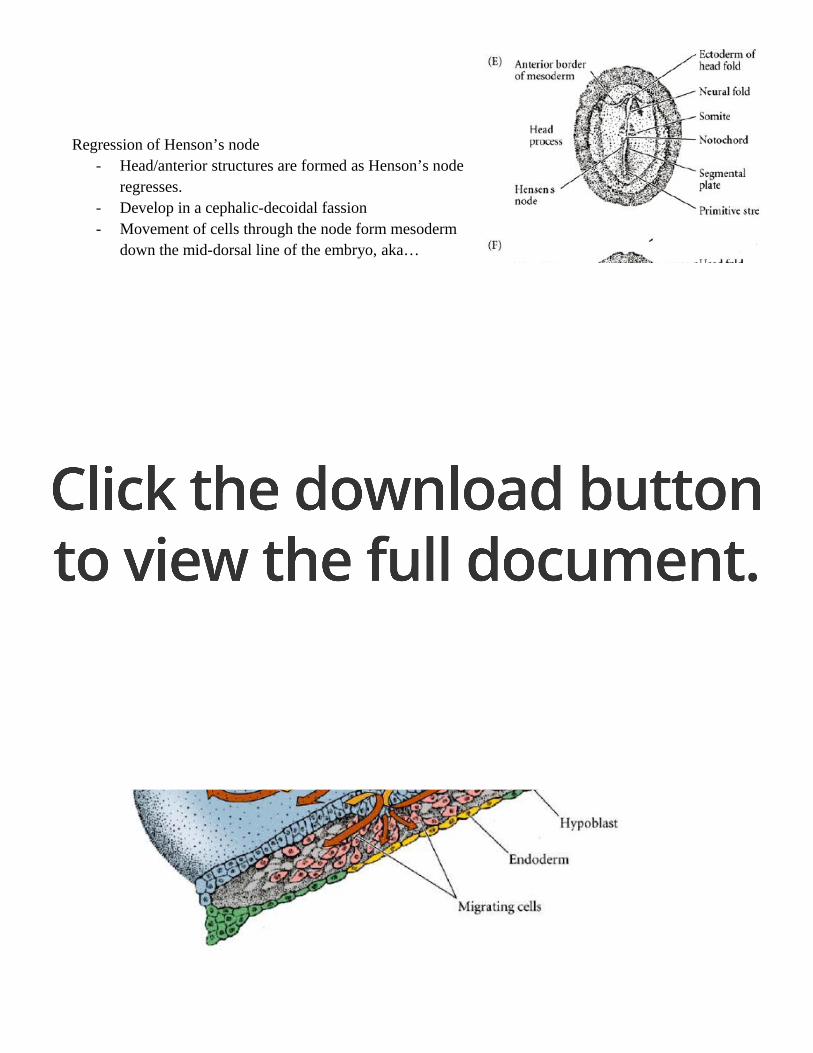
Regression of Henson’s node- Head/anterior structures are formed as Henson’s node
regresses.- Develop in a cephalic-decoidal fassion- Movement of cells through the node form mesoderm
down the mid-dorsal line of the embryo, aka… chordamesoderm. The role of chordamesoderm is to induce the overlying ectoderm to form neural structures.
Overall cell movements- As the cells move through Henson’s node, they move from the anterior and then back to the
anterior (picture is not right). Cells passing through the primitive groove migrate laterally again and become mesoderm (contributing to the embryo or extra-embryonic membranes) or will interact with the hypoblast to contribute to the endoderm (embryonic and extra-embryonic).
- Will not ask to draw this, but know what the cells are forming.

Inducing a new embryonic axis- The Henson’s node is transplanted to a chick not exactly on the primitive streak. A new
embryonic axis is produced with a new neural tube forming. This shows that Henson’s node acts exactly like the dorsal lip of the blastopore – acts as the organizer.
Formation of the extra-embryonic members- The formation of extraembryonic membranes allowed the animals to go into an environment
where others could not survive.- Amniotes – terrestrial, prolonged embryonic development, dependent on a long-term maternally
supplied nutrient, extra-embryonic membranes- Anamniotes – aquatic, rapid developers, no extra-embryonic membranes, extract nutrient from
the environment early in life history
Challenges to Amniotes- Dessication – drying out- Respiration – gas exchange across moist membranes- Nutrition source – no longer able to feed as easy, food on land is relatively scarce, - Excretion – disposal of toxic nitrogenous metabolic waste (cleidoic versus placental)
1. How? Aquatic developer can just excrete into the environment easily – it is diluted. They excrete ammonia. Land animals must have a less toxic form of waste – urea.
2. Chicken egg needs to be able to store nitrogenous wastes for a very extended period of time – need an even less nitrogenous form. This is uric acid crystals.

Cleidoic eggs of birds and reptiles- Two pairs of germ layers produce extraembryonic membranes
1. Splanchnopleure – endoderm and splanchnic mesoderm2. Somatopleure – ectoderm and mesoderm
Extraembryonic membranes – drawings- CAM – has all three germ layers, chorioallantoic membrane

Functions of extraembryonic membranes- Chorion – shock protection, later gas exchange membrane for respiration- Amnion – more important as a shock protection, fluid filled development chamber- Yolk sac – early respirator organ, digestion/nutrition- Allantois – excretion/storage (uric acid in chick); later can combine later with chorion to form
CAM (major respirator organ)
Placentation – the mammals- At some point, sperm is deposited in the female tract (vagina or uterus). The sperm are going to
swim up the fallopian tube and meet the egg in the upper 1/3 of the tube. - Notice – the ovary and the opening of the fallopian tube aren’t really directly connected. The
fimbriae will slightly connect these two in a loose association. The fimbriae and cilia need to sweep the egg into the fallopian tube to be fertilized.
- The uterine wall is beginning to build up. The blastula stage is still contained in the zona pellucida. It is very important for the zona pellucida to still exist so it does not develop in the fallopian tube – this would be an ectopic pregnancy.
- Stone babies – ectopic pregnancies that happen in the abdominal cavity. - A male could carry a baby to term if it was implanted in the abdominal cavity. - In a smoker – don’t have the cilia to push it up so it would become a stone baby
Pop Quiz #10 – structure in chick that is analogous to dorsal lip