thetrpcagemotifasascaffoldforthedisplayofa ... · opment of smaller nonimmunoglobulin protein...
TRANSCRIPT

The Trp Cage Motif as a Scaffold for the Display of aRandomized Peptide Library on Bacteriophage T7*
Received for publication, November 20, 2006, and in revised form, January 26, 2007 Published, JBC Papers in Press, January 30, 2007, DOI 10.1074/jbc.M610722200
Richard E. Herman, Douglas Badders, Mark Fuller, Ekaterina G. Makienko, Michael E. Houston, Jr., Steven C. Quay,and Paul H. Johnson1
From the Nastech Pharmaceutical Company, Inc., Bothell, Washington 98021
Phage libraries displaying linear or disulfide-constrainedpeptides often yield weak binders, upon screening against atarget, and must be optimized to improve affinity. The disad-vantages of libraries based on larger complex proteins, suchas single chain antibodies, have stimulated interest in the devel-opment of smaller nonimmunoglobulin protein scaffolds. Apromising candidate is the Trp cage motif, a 20-residue C-ter-minal sequence of exendin-4. Amino acid substitution withintheTrp cage resulted in a 20-mer peptide recognized as anultra-fast cooperative folding miniprotein, with ideal characteristicsfor the discovery of small structured nonimmunoglobulinmotifs having a stable tertiary structure. Although we wereunable to display the Trp cage onM13 phage, successful displaywas achieved using the lytic T7 phage. Interestingly, mutationswere observed at a frequency dependent on display valency. ATrp cage library designedwith randomized amino acids at sevensolvent-exposed positions was developed from 1.6 � 109 pri-mary clones in T7Select10-3b. DNA sequencing of 109 libraryclones revealed 38% mutants and 16% truncations by TAGcodons at randomized positions. Amino acid frequencies werelargely within expected bounds and DIVAA analysis revealedthat the library had an average diversity of 0.67. Utility of thelibrary was demonstrated by identification of HPQ containingTrp cage miniproteins, which bound streptavidin, and AAAD-PYAQWLQSMGPHSGRPPPR, which bound to human bron-chial epithelial cells. A high complexity library based on the Trpcage miniprotein has demonstrated potential for identifyingnovel cell and protein binding peptides that could be used forthe delivery of therapeutic molecules or as target-specific ther-apeutic agents.
Since the development of phage display technology (1), it hasbeen widely used for the screening of random combinatorialpeptide libraries in drug discovery initiatives (2). When pep-tides are displayed as unconstrained linear molecules, they canadopt numerous conformations, very few of which may repre-sent stable conformations. Panning with these unconstrainedpeptides against targets of interest usually leads to the isolationof peptides with low binding affinity. Reducing the conforma-tional freedom of the displayed peptides is necessary to
decrease entropy and increase the binding affinity (3, 4). Con-strained libraries have often achieved a reduction in conforma-tional freedom by incorporating a pair of cysteine residues atboth ends of linear peptides, resulting in disulfide bridges thatform peptide loops yielding cyclic peptide libraries reported toincrease binding affinity (5) and stability in biological fluids (6).The use of defined protein scaffolds for the generation of
random libraries has the potential to increase binding affinityfurther by providing a more rigid conformation (7). Examplesof folded protein scaffolds that have been used for peptide dis-play include the zinc finger motif, which has been used to con-struct a degenerate library for the isolation of peptides that bindto DNA and RNA (8, 9), and Kunitz domains, which have beenused as a scaffold for libraries designed for the discovery ofimproved serine protease inhibitors (10).Wehave evaluated another protein that has potential to serve
as a scaffold for the generation of a random peptide library,exendin-4, a 39-amino acid peptide isolated from the saliva ofthe Gila monster, Heloderma suspectum. NMR studies ofexendin-4 (11) showed that residues 21–38 of the C terminusform a stable tertiary structure designated a Trp cage motif.The Trp cage is a hydrophobic cluster with Trp-25 buried in acentral location, where it is shielded from solvent exposure.The residues that form the cage around Trp-25 include multi-ple proline residues (Pro-31, and prolines 36–38) where theproline rings are located on both faces of the indole ring ofTrp-25 and the Phe side chain completes the hydrophobic clus-ter. Further studies aimed at developing a miniprotein (a stableself-folding peptide with a minimum number of amino acids)showed that the C-terminal 20 amino acids of exendin-4(RLFIEWLKNGGPSSGAPPPS) are adequate for forming theTrp cage. In addition, amino acid substitution experimentsresulted in optimization ofTrp cage folding (estimated byNMRchemical shift deviations, the presence of long range nuclearOverhauser effect, the absence of cis/trans isomerizationaround proline residues, andNH exchange protection) from 40to �95% in water at physiological pH. Analysis of the confor-mation of the optimized Trp cage in the presence of 30% triflu-oroethanol, a known helix-inducing solvent, resulted in a min-imal increase in helical content. Moreover, this construct isfairly stable with a Tm of 43.5 °C (12, 13). The optimized Trpcage miniprotein TC5b (NLYIQWLKDGGPSSGRPPPS) hasbeen extensively studied, because it is an ultrafast, coopera-tively folding system (14). Within the optimized Trp cagesequence, there are several positions where the substitution ofamino acids does not compromise folding of the miniprotein.Some of those positions are solvent-exposed, making them
* The costs of publication of this article were defrayed in part by the paymentof page charges. This article must therefore be hereby marked “advertise-ment” in accordance with 18 U.S.C. Section 1734 solely to indicate this fact.
1 To whom correspondence should be addressed: Nastech PharmaceuticalCompany, 3830 Monte Villa Pkwy., Bothell, WA 98021. E-mail:[email protected].
THE JOURNAL OF BIOLOGICAL CHEMISTRY VOL. 282, NO. 13, pp. 9813–9824, March 30, 2007© 2007 by The American Society for Biochemistry and Molecular Biology, Inc. Printed in the U.S.A.
MARCH 30, 2007 • VOLUME 282 • NUMBER 13 JOURNAL OF BIOLOGICAL CHEMISTRY 9813
by guest on May 2, 2020
http://ww
w.jbc.org/
Dow
nloaded from
ideal for the display of random amino acids in a highly diverselibrary.This report describes the construction and characterization
of a peptide library of high complexity based on the 20-aminoacid Trp cage miniprotein TC5b with seven randomized posi-tions displayed on bacteriophage T7. We demonstrate thelibrary’s potential utility by the identification of a peptide con-taining the HPQmotif that binds to streptavidin and a peptidethat binds to human bronchial epithelial cells.
EXPERIMENTAL PROCEDURES
Chemicals, Media, and Reagents—General chemicals werepurchased from Sigma. Components for bacterial growthmedia were purchased fromFisher. T7Select� cloning kits, vec-tor arms, packaging extracts, S�TagTM antibody, and T7 tailfiber monoclonal antibody were purchased from Novagen(EMD Biosciences, San Diego, CA). Restriction enzymes EcoRIand HindIII and streptavidin were purchased from New Eng-land Biolabs (Beverly, MA). Mammalian cell media, Klenowfragment of DNA polymerase I, MagicMarkTM XP WesternProtein Standards, and reagents for SDS-PAGE electrophoresiswere purchased from Invitrogen. Goat anti-rabbit IgG horse-radish peroxidase and goat anti-mouse IgG horseradish perox-idase antibodies were purchased from Santa Cruz Biotechnol-ogy, Inc. (Santa Cruz, CA). SuperSignalWest FemtoMaximumSensitivity Substrate was purchased from Pierce.Bacteria, Bacteriophages, and Cell Lines—Escherichia coli
strains BL21 [F�, ompT, hsdSB (rB�mB�), gal, dcm] and
BLT5403 [F�, ompT, hsdSB (rB�mB�), gal, dcm pAR5403
(AmpR)] and bacteriophages T7Select� 415-1b, 10-3b, 1-1b andS-TagTM were obtained from Novagen (EMD Biosciences).E. coli C600 ATCC 23724 (F�, supE44, lacY1, thr-1, leuB6,mcrA, thi-1, rfbD1, fhuA21, lambda�) was obtained from theAmerican Type Culture Collection (Manassas, VA), and V517was obtained from Larry L. McKay (University of Minnesota).Human bronchial epithelial cell line 16HBE14o� was obtainedfrom D. C. Gruenert (University of California, San Francisco,CA).Growth and Purification of Bacteriophage—T7Select 415-1b
was propagated in E. coli strain BL21 using M9LB liquidmedium.T7Select 10-3b and 1-1bwere propagated in BLT5403using M9LB supplemented with carbenicillin (50 �g/ml) at37 °C. T7 phagewas enumerated using the plaque assaymethodon LB2 solid medium supplemented with carbenicillin (50�g/ml) as needed. Phage lysates were prepared following theinstructions in the T7Select System manual (Novagen, SanDiego, CA), which typically resulted in titers of 1010–1011 pfu/ml. Phage were purified and concentrated from centrifugedlysates (12,000 � g for 15 min at 4 °C) by the addition of one-sixth volumeof 20%polyethylene glycol 8000, 2.5MNaCl (PEG/NaCl) and incubating on ice for at least 60 min or overnight at4 °C. Precipitated phage were pelleted by centrifugation as
above. The phage pellet was resuspended in TBS (50 mM Tris,pH 7.5, 150 mM NaCl) and reprecipitated with one-sixth vol-ume of PEG/NaCl on ice for 15–60 min, centrifuged, resus-pended in TBS, and stored at 4 °C.Preparation of Phage DNA for Sequencing—Well-isolated
phage plaques were punched out of solid media and resus-pended in 100 �l of TE (10mMTris, pH 8.0, 1mM EDTA). Hostbacteria were grown to a density of 2� 108 cfu/ml, and 2.5�l ofthe phage suspension was added to 1ml of the bacterial culturein a 17 � 100-mm tube followed by incubation at 37 °C for 3 hwith shaking. Then 1 �l of Nuclease mix containing RNase Aand DNase I (Promega, Madison, WI) was added, and incuba-tion was continued for 15 min at 37 °C without shaking. Celldebris was removed by centrifugation in a microcentrifuge(16,000 � g, 10 min). A 900-�l volume of the supernatant wasrecovered and transferred to a new 1.5-ml tube containing 200�l of PEG/NaCl and placed at 4 °C overnight. Precipitatedphage were collected by centrifugation (16,000 � g, 10 min),resuspended in 100 �l of TE, and disrupted by extraction withan equal volume of phenol/chloroform (1:1) and then centri-fuged in a microcentrifuge (16,000 � g, 1 min). The aqueousphase was recovered, extracted with an equal volume of chlo-roform, and centrifuged for 4 min. The aqueous phase wasrecovered again, and one-tenth volume of 3 M sodium acetatewas added. Alternatively, the phage were disrupted by resus-pension in 100 �l of iodide buffer (10 mM Tris, pH 8, 1 mMEDTA, 4MNaI). PhageDNAwas precipitated by the addition of250 �l of ethanol with incubation for 10 min at room temper-ature followedby centrifugation (16,000� g, 10min). TheDNApellet was washed with 70% ethanol, dried briefly under vac-uum, and resuspended in 20–30 �l of TE.DNA Sequencing/Oligonucleotide Synthesis—T7 DNA was
submitted to Retrogen (San Diego, CA) for automated DNAsequencing by an ABI 3730 DNA analyzer using the primerT7-125 (5�-TGCGTGACTTGGCTCTGGAG-3�). A protocolmodel was constructed for sequence analysis using Teranodesoftware (Teranode Corp., Seattle, WA), which also served as asequence data base. Oligonucleotides were synthesized by Ret-rogen, using standard phosphoramidite chemistry.Insertion of DNA Sequences Encoding TC3b or TC5b into
T7Select Vectors—Oligonucleotide pairs were designed toencode the Trp cage TC3b and TC5b miniproteins (Table 1).Each oligonucleotide, 5�-phosphorylated (5�p), was resus-pended in TE to 200 pmol/�l before annealing. Oligonucleo-tide TC3b (�) (5�p-AATTTATTTATCGAATGGCTCAA-AAATGGTGGTCCTTCCAGTGGTGCTCCTCCCCCTT-CCTAA-3�) was annealed with TC3b (�) (5�p-AGCTTTA-GGAAGGGGGAGGAGCACCACTGGAAGGACCACCA-TTTTTGAGCCATTCGATAAATA-3�), and TC5b (�)(5�p-AATTCGGCAGCTGCGAATTTGTATATTCAGTG-GCTTAAGGATGGTGGTCCTTCGTCGGGGCGGCCTC-CGCCAAGTTAAA-3�) was annealed with TC5b (�) (5�p-AGCTTTTAACTTGGCGGAGGCCGCCCCGACGAAGG-ACCACCATCCTTAAGCCACTGAATATACAAATTCG-CAGCTGCCG-3�) by combining 25 �l of each of theappropriate oligonucleotides in annealing buffer (10 mM Tris,pH 7.5, 100 mM NaCl, 1 mM EDTA), the mixtures were heatedto 95 °C in a heat block for 5 min and allowed to cool at room
2 The abbreviations used are: LB, Luria-Bertani; pfu, plaque-forming units; cfu,colony-forming units; BSA, bovine serum albumin; DPBS, Dulbecco’s phos-phate-buffered saline; TBS, Tris-buffered saline; PEG, polyethylene glycol;Fmoc, N-(9-fluorenyl)methoxycarbonyl; HPLC, high pressure liquid chro-matography; ELISA, enzyme-linked immunosorbent assay.
T7 Trp Cage Library
9814 JOURNAL OF BIOLOGICAL CHEMISTRY VOLUME 282 • NUMBER 13 • MARCH 30, 2007
by guest on May 2, 2020
http://ww
w.jbc.org/
Dow
nloaded from

temperature for 2 h in the block. The annealed oligonucleotideswere ligated to the arms of T7Select 415-1b, 10-3b, or 1-1b andpackaged in vitro using the T7 Select Cloning kit according tothe manufacturer’s directions. Packaged phage were amplifiedby growth in liquid media with the appropriate host. Oligonu-cleotide insertion was confirmed by DNA sequencing.Peptide Synthesis—Peptides were synthesized by solid phase
Fmoc chemistry on Tentagel amide resin using a RaininSymphony synthesizer. Coupling steps were performedusing 5 eq of 2-(6-chloro-1H-benzotriazole-1-yl)-1,1,3,3-tetramethylaminium hexafluorophosphate and Fmoc aminoacid (NovaBiochem, San Diego, CA) with an excess ofN-meth-ylmorpholine for 40 min. Fmoc removal was accomplished bytreating the peptide resin with 20% piperidine (Aldrich) in di-methylformamide (Burdick and Jackson, Morristown, NJ) fortwo 10-min cycles. Upon completion of the entire peptide, theFmoc group was removed with piperidine and washed exten-sively with dimethylformamide. The peptides were cleavedfrom the resin by the addition of 10 ml of trifluoroacetic acid(Aldrich) containing 2.5% water and 2.5% triisopropylsilane(Aldrich) followed by gentle agitation at room temperature for2 h. The resulting crude peptide was collected by triturationwith ether followed by filtration. The crude product was dis-solved inMillipore water and lyophilized to dryness. The crudepeptide was taken up in 15 ml of water containing 0.05% triflu-oroacetic acid and 3 ml of acetic acid and loaded onto a ZorbaxRX-C8 reversed-phase column (22-mm inner diameter � 250mm, 5-�mparticle size) through a 5-ml injection loop at a flowrate of 5 ml/min. The purification was accomplished by run-ning a linear AB gradient of 0.1% B/min for 180 min, wheresolvent A is 0.05% trifluoroacetic acid in water and solvent B is0.05% trifluoroacetic acid in acetonitrile. The purified peptideswere analyzed by reversed-phase HPLC and electrospray massspectrometry. Peptideswere purified to greater than 95%purityas determined by reversed-phase HPLC.Production of Antibody AB167—The TC5b miniprotein was
synthesized with the addition of a cysteine residue at the Nterminus to allow conjugation of keyhole limpet hemocyaninand designated PN0522 (NH2-CNLYIQWLKDGGPSSGRP-PPS-amide). The keyhole limpet hemocyanin-PN0522 conju-gate was used to produce protein G-purified antibodies fromNew Zealand White rabbits by Orbigen (San Diego, CA).Phage ELISAs—For the detection of Trp cage TC5b, purified
phage were diluted in Dulbecco’s phosphate-buffered saline(DPBS), distributed 100 �l/well to a 96-well MaxisorpTM plate(NalgeNunc International, Rochester,NY), and allowed to bindfor 1 h. All incubations were done at room temperature withslow orbital shaking. After phage binding, the wells werewashed three times with 400 �l of DPBS containing 0.1%Tween 20 and 1% BSA (wash buffer) and then blocked with 200�l of wash buffer for 1 h. A 100-�l volume of primary antibody(AB167diluted 1:500 inwash buffer)was applied and allowed tobind for 1 h and then washed five times as above. Secondaryantibody (goat anti-rabbit horseradish peroxidase diluted1:1000 in wash buffer) was applied in 100 �l, incubated for 1 h,washed, and developed with OptEIA reagents (BD Biosciences)according to the manufacturer’s directions. Means were com-pared using Student’s t test.
Phage binding to 16HBE14o� cells was analyzed using con-fluent cells grown with Dulbecco’s modified Eagle’s medium/F-12 complete medium in 96-well collagen-coated plates (BDBiosciences) at 37 °C in 5% CO2. The cells were washed withDPBS, and phage diluted in DPBS containing 1% BSA (DPBS-BSA) were applied in 100 �l. Bound phage were detected usingprimary antibody (mouse IgG T7 tail fiber monoclonal anti-body diluted 1:2000 in DPBS-BSA) and secondary antibody(goat anti-mouse IgG horseradish peroxidase diluted 1:5000 inDPBS-BSA) followed by detection with OptEIA reagents asabove.Western Analysis of Phage Displaying TC5b—Purified phage
were denatured by boiling in Tris/glycine SDS sample buffer(2�) with 1% �-mercaptoethanol. Samples containing 1011pfu/ml in 10 �l were loaded into wells of a 1-mm 4–20% Tris/glycine gel and electrophoresed for 1.5 h at 150 V (constantvoltage) in Tris/glycine SDS running buffer. The separated pro-teins were transferred to a polyvinylidene difluoridemembranefor 1.5 h at room temperature and blocked with 3% nonfat drymilk in TBS containing 0.05% Tween 20 (TBS-T) for 1 h. Themembrane was probed with antibody AB167 diluted 1:2500 for2 h at room temperature, followed by three 5-min washes withTBS-T. Bound antibody was detected by incubation withgoat anti-rabbit IgG horseradish peroxidase diluted 1:5000for 1 h at room temperature followed by four 10-min washesand development with SuperSignal West Femto maximumsensitivity substrate according to the manufacturer’sinstructions. MagicMarkTM XP Western protein standardswere used for size references.Construction of the Trp Cage Library—The Trp cage library
was constructed in the T7Select 10-3b vector using oligonu-cleotide T-3 (�) (5�-CATGTTCAAGCTTGTTAMNNGGG-GGGAGGACGACCAGAMNNAGGACCMNNMNNMNN-TAACCACTGMNNGTAMNNATCTGCTGCTGCCGAAT-TCGGGGCATGTG-3�) as a template (where M representsequalmolar amounts of A andC, andN is equalmolar amountsof A, C, G, and T) and T-3 (�) (5�-CACATGCCCCGAATTC-GGCA-3�) as a primer. The template and primerwere annealedby combining 310 pmol of T-3 (�) and 820 pmol of T-3 (�) inannealing buffer (10 mM Tris, pH 7.5, 100 mM NaCl, 1 mMEDTA) to a final volume of 50 �l, the mixture was heated to95 °C in a heat block for 5min, and the blockwas allowed to coolat room temperature for 2 h. The primer was extended by com-bining 310 pmol of annealed template-primer, dNTP mixture(New England Biolabs, Beverly, MA), REact 2 buffer (Invitro-gen), andKlenow fragment (10 units) in a total volume of 250�lfor 10 min at 37 °C. The reaction was stopped by incubation at65 °C for 15min followed by phenol/chloroform extraction andethanol precipitation. The extended duplexes were doubledigested with EcoRI and HindIII, and the cleavage productswere subjected to electrophoresis in a 20% TBE (89mMTris, 89mM boric acid, 2 mM EDTA) gel. The desired band was excisedand extracted by shaking overnight in 100 mM sodium acetate,pH 4.5, 1 mM EDTA, 0.1% SDS at room temperature. Gel frag-ments were removed by centrifugation, and the DNAwas puri-fied by phenol/chloroform extraction and ethanol precipitationbefore being resuspended in TE. The extracted inserts wereligated into arms of T7Select�10-3b and packaged in vitro using
T7 Trp Cage Library
MARCH 30, 2007 • VOLUME 282 • NUMBER 13 JOURNAL OF BIOLOGICAL CHEMISTRY 9815
by guest on May 2, 2020
http://ww
w.jbc.org/
Dow
nloaded from

the T7Select� cloning kit according to the manufacturer’sdirections, and primary clones were enumerated. Ligation andpackaging was repeated until greater than 109 primary cloneswere generated and amplified. The amplified clones werepooled, treated with Nuclease mix at 37 °C for 15 min and cen-trifuged (8000 � g, 10 min) to remove cell debris. Phage wereconcentrated by adding solid PEG 8000 to 10% (w/v), incuba-tion overnight at 4 °C, centrifugation (10,400� g, 10min, 4 °C),and resuspension in 1 M NaCl, 10 mM Tris, pH 8, 1 mM EDTA.For further purification, the concentrated phage were layeredon top of a CsCl density block gradient consisting of 1.5 ml of62%, 3.0 ml of 41%, 3.0 ml of 31%, and 3.0 ml of 20% CsCl in TEand centrifuged at 28,000 rpm (150,000 � g) in a BeckmanSW28.1 rotor at 25 °C for 98 min. The phage band was recov-ered by side puncture and dialyzed against three changes of 20mM Tris, pH 8.0, 100 mM NaCl, 6 mM MgSO4.Construction of a Suppressor Host Strain for T7Select—Plas-
mid pAR5403was isolated fromBLT5403 using aQIAprep spinminiprep kit (Qiagen, Valencia, CA) following themanufactur-er’s instructions. E. coli strain C600 ATCC 23724 was madecompetent for transformation by inoculating LB broth with a1% inoculum of fresh overnight culture and incubating at 37 °Cwith shaking until the cell density reached A600 � 0.3. Cellswere pelleted by centrifugation (480 � g, 10 min), resuspendedin ice-cold 0.1 M CaCl2, recentrifuged, and resuspended in one-fiftieth volume of ice-cold 0.1 M CaCl2. Transformation wasaccomplished by combining 200�l of competent cells with 2�lof pAR5403 (200 ng/�l), incubating on ice for 30 min, heatshocking at 42 °C for 90 s, and then placing on ice for 2 min.Transformants were selected by inoculating 100 �l of heat-shocked cells into 3 ml of molten top agar, which was thenspread onto LB solid medium containing 50 �g/ml carbeni-cillin and incubated at 37 °C overnight. Plasmid DNA wasprepared from transformants by QIAprep Spin Miniprepand examined after electrophoresis (65 V for 2 h) through a10 � 15-cm 0.6% SeaKem LE (Cambrex, Rockland, ME)-agarose gel in TBE running buffer followed by staining withethidium bromide (0.5 �g/ml).Stability of Primary Clones—BLT5403 grown to A600 � 0.85
(1 � 108 cfu/ml) was split into three aliquots and infected at amultiplicity of infection of 0.02 with a total of 4 � 107 pfu ofeither mutant, nonmutant, or both mixed in a 1:1 ratio. Theinfected cultureswere incubatedwith shaking at 37 °C for 3 h oruntil lysis was complete and progeny were enumerated. Prog-eny phage plaques were randomly selected and suspended in100 �l of TE to elute phage. Lysates were prepared by infecting1-ml cultures of midlog BLT5403 with 10 �l of each eluate andconcentrated with PEG/NaCl, and phage DNA was extractedfor sequencing.One-stepGrowthExperiment—Based on the one-step growth
experiment described by Edgar (15), BLT5403 was grown in LBbroth at 37 °C to a density of 2 � 108 cfu/ml and centrifuged,and the cell pellet was resuspended in half the original volumewith LB broth and enumerated by the spread plate method todetermine input cells. 0.5 ml of resuspended cells at 4 � 108cfu/ml were placed in a 17 � 100-mm tube and incubated at30 °C for 5 min, and then phage infection was initiated by add-ing 0.5 ml of the appropriate phage stock adjusted to 4 � 109
pfu/ml to provide a multiplicity of infection of 10, and incuba-tion was continued. At 5min postinfection, 2 �g of T7 tail fibermonoclonal antibody was added to neutralize unadsorbedphage. At 10 min postinfection, the cell and phage mixture wasdiluted 104-fold into LB broth contained in a 250-ml growthflask, and incubation was continued with vigorous shaking. At15 min postinfection, 1.0 ml was removed from the growthflask, mixed with 1 ml of LB broth containing 2–3 drops ofCHCl3, and vortexed. The unadsorbed phagewere enumerated.At 20 min postinfection, 1.0 ml was removed from the growthflask for determination of infected cells (total pfu�unadsorbedpfu). At 90 min postinfection, 2–3 drops of CHCl3 were addedto the growth flask, and progeny phage were enumerated.Streptavidin Panning—A 96-well MaxisorpTM plate was
coated with 200 ng/well streptavidin (2 ng/�l) for at least 1 hand blocked with 1% BSA in DPBS with 0.1% Tween 20 (DPBS-BT) for 1 h and then rinsed three timeswith 200�l ofDPBS-BT.The crude Trp cage library (i.e. before CsCl purification) waspanned against streptavidin-coated wells by applying 1.2 � 109pfu that were allowed to bind for 1 h at room temperaturefollowed by six washes with 200 �l of DPBS-BT. Bound phagewere eluted with 1% SDS for 10 min and amplified withBLT5403. The lysate was centrifuged, and 10 �l of the phagesupernatantwas used for the next round of panning. After threerounds of panning, well isolated plaques of eluted phage wereselected for DNA sequencing.Panning against Human Bronchial Epithelial Cells—Human
bronchial epithelial cells (16HBE14o�) at low passage wereseeded into 24-well collagen-coated plates (BD Biosciences)and allowed to grow at 37 °C in 5% CO2 to confluence inDulbecco’s modified Eagle’s medium/F-12 completemedium containing 10% fetal bovine serum. To prepare thecells for panning, the medium was removed and replacedwith Dulbecco’s modified Eagle’s medium/F-12 with noserum (F12i) and incubated at 37 °C for 1.5 h. Then the F12iwas removed and replaced with 500 �l/well of 1% BSA inDPBS and incubated at 37 °C for 60 min. Just before panning,the DPBS was removed, and the cells were rinsed with 500 �lof TBS. A 50-�l aliquot of the naive T7Select Trp cage librarywas combined with 160 �l of TBS and added to the cellsfollowed by incubation at 4 °C for 60 min with gentle rock-ing. The unbound portion of the phage library was removedby 25 washes with 500 �l of TBS. Bound phage were eluted byadding 200 �l of a BLT5403 culture grown to A600 � 1.0 inM9LB followed by incubating at room temperature for 15min. Eluted phage were added to 20 ml of the BLT5403 cul-ture and amplified by incubation at 37 °C for 2–3 h until celllysis was observed. The amplified phage was concentratedwith PEG/NaCl and resuspended in 200 �l of DPBS. Subse-quent rounds of panning were performed using 20 �l of con-centrated phage from the previous round. In the secondround, unbound phage were removed by washing with TBScontaining 0.1% Tween, and in the third round TBS with0.3% Tween was used. After three rounds of panning, well-isolated plaques of eluted phage were selected for DNAsequencing.
T7 Trp Cage Library
9816 JOURNAL OF BIOLOGICAL CHEMISTRY VOLUME 282 • NUMBER 13 • MARCH 30, 2007
by guest on May 2, 2020
http://ww
w.jbc.org/
Dow
nloaded from

RESULTS
Display of Trp Cage Miniproteins on Bacteriophage T7 IsAssociated with a Valency-dependent Increase in PeptideMutation—The T7Select system, which is designed to displaypeptides with a free C terminus on the surface of bacteriophageT7 by fusion to the phage capsid protein (gp10B), was used todisplay Trp cage miniproteins (Table 1) that were based onsequences from Neidigh (13). Initial work designed to test thefeasibility of displaying the Trp cage miniprotein was per-formed with Trp cage version TC3b, and later work focused onthe further optimized version TC5b because it has better fold-ing characteristics. Three T7Select vectors were used, each onevarying by the average number of capsid fusion proteins dis-played per phage: 415-1b (average copy number � 415), 10-3b(average copy number � 5–15), and 1-1b (average copy num-ber � 0.1–1). In order to determine if Trp cage TC3b could bedisplayed on bacteriophage T7, oligonucleotides TC3b (�) andTC3b (�) were annealed and inserted into the three T7Selectvectors. After in vitropackaging and amplification, several well-isolated plaques were selected and analyzed by DNA sequenc-ing. The deduced amino acid sequences revealed a high fre-quency of mutants (7 of 8 clones) when TC3b was expressed inthe 415-1b vector and a low frequency of mutants in the 10-3band 1-1b vectors (1 of 10 and 1 of 9 clones, respectively). Acomparison of the expected TC3b peptide sequence and repre-sentative mutants (Table 2) from expression in 415-1b(mutants 415-1b A1, A2, and Ai5) revealed single base changesin the mutant sequences that resulted in nonsense or missense
mutations that prevented expression of the Trp codon TGG bytruncating the peptide upstream or changing the codon so thatan amino acid substitution occurred. Mutants that resultedfrom expression of TC3b in 10-3b and 1-1b contained deletionsthat resulted in frame shifts, which altered the amino acidsequence, including changes to the amino acids that arerequired for the Trp cage structure. Since expression of a highcopy number of Trp cage protein fusions in vector 415-1b led toa high percentage ofmutants, themedium valency vector 10-3bwas utilized for the remaining library work.TrpCageMiniproteinsAreDisplayed onT7 through Fusion to
gp10B—To show that the Trp cage was displayed as a result offusion to the capsid protein (gp10B) of T7Select10-3b, proteinsfrom phage lysates were separated by SDS-PAGE, and bindingof Trp cage-specific antibody AB167 to lysate proteins wasdetected byWestern blot analysis. As shown in Fig. 1, the anti-body bound to a protein in the lane containing the lysate fromcells infected with phage expressing the TC5b insert. This bandwas not observed in the lanes containing lysates from cellsinfected by phage with no insert or phage displaying the linear15-amino acid S�TagTM or in lysates from uninfected host bac-teria. The size of the positive band is consistent with the pre-dicted size of 39,640 daltons (376 amino acids) for theTrp cage-gp10B phage capsid protein fusion. As expected, Westernanalysis using an antibody against the S�TagTM instead of theTrp cage resulted in detection of a band only in the S�TagTMlane, which had the same relative mobility as the Trp cage-gp10B phage capsid protein fusion (data not shown).Display of Trp Cage Miniproteins Reduces the Yield of Prog-
eny Phage—One-step growth experiments were conducted inE. coli BLT5403 to determine if display of the Trp cage affectedthe production of progeny phage as measured by the averageburst size. Each experiment compared three phage:T7Select10-3b with no displayed peptide, T7Select10-3b dis-
TABLE 1Amino acid sequence of Exendin-4 and Trp cage miniproteins
Exendin-4 (residues 20–39) RLFIEWLKNGGPSSGAPPPS
TC3b NLFIEWLKNGGPSSGAPPPS
TC5b NLYIQWLKDGGPSSGRPPPS
TABLE 2Amino acid and DNA sequences from mutants resulting from insertion of TC3b into T7Select 415-1b, 10-3b, and 1-1b
T7 Trp Cage Library
MARCH 30, 2007 • VOLUME 282 • NUMBER 13 JOURNAL OF BIOLOGICAL CHEMISTRY 9817
by guest on May 2, 2020
http://ww
w.jbc.org/
Dow
nloaded from

playing the 15-amino acid S�TagTM, andT7Select10-3b display-ing Trp cage TC5b. As shown in Table 3, the results from threeindependent one-step growth experiments indicated thatalthough the average number of infected cells was similar for allphage tested, the burst size (a ratio of progeny phage to infectedcells) of phage displaying Trp cage TC5b was lower than phagewith no displayed peptide by greater than 50% (p � 0.001).Phage displaying the linear 15-amino acid peptide S�TagTMgave an intermediate value reflecting a 30% reduction (p �0.05). These results indicate that displaying the Trp cage, evenat low valency, reduces the average yield of progeny phage pro-duced per infected cell greater than a similar size linear peptide.The Diversity of the T7 Trp Cage Library Is Similar to That of
M13 Ph.D.-C7CTM—A library displaying the Trp cage TC5bminiprotein (Table 1) with seven randomized positions on bac-teriophage T7Select10-3b was produced by the primer exten-sion method. The primer T-3 (�) and template T-3 (�) oligo-nucleotides were designed to produce an insert in T7 such thatthe coding strand would include codons commonly utilized byE. coli and phage T7 for the invariable amino acids and thedegenerate codon NNK at the seven randomized positionswhere N represents G/A/T/C and K represents G/T. Utilizingthe NNK codon reduced the number of possible amino acidcodons at the randomized positions from 64 to 32 to provide amore uniform distribution of amino acids and eliminate all ofthe stop codons except TAG (amber). Expression of the insert
was designed to result in fusion of the 23-amino acid minipro-teinsAAADXYXQWLXXXGPXSGRPPPX (whereX indicates arandomized position) to the phage capsid protein. Due to theirhigh helical propensity, three alanine residues were encoded atthe N terminus to initiate and propagate the N-terminal helixand to stabilize the protein (16), followed by aspartic acid, sub-stituted for the Asm-1 of TC5b, to act as a helix-capping resi-due. The positions chosen for randomization are not requiredfor folding (13) and are located on the solvent-exposed portionof the folded protein as determined from the NMR structure(Fig. 2), where they are available to interact with potential bind-ing sites on target molecules.A total of 1.6 � 109 packaged primary phage clones were
obtained in order to provide a library complexity approachingthe number of possible combinations (207 � 1.28 � 109). Todetermine the expected proportion of the library expressing theTrp cage as designed, DNA from 100 primary phage clones wassequenced. Among the 100 clones, 64% contained the expectedsequence (of which 8% contained the stop codonTAG in one ofthe randomized positions), and 36% contained frameshiftmutations within the Trp cage sequence.Since a high percentage of the primary clones contained
mutations in the Trp cage sequence, we were concerned that
FIGURE 1. Western blot analysis of Trp cage TC5b displayed onT7Select10-3b. Proteins in lysates from phage-infected cells were separatedby SDS-PAGE as shown in the Coomassie-stained gel on the left. The lysateswere prepared from cells that were infected with T7 displaying either nopeptide (10-3b), Trp cage TC5b (TC5b), or the S�TagTM peptide (S-Tag). Thelane labeled BLT5403 contained lysate from uninfected cells. The Westerntransfer on the right shows that only the lysate from infection by phage dis-playing Trp cage TC5b contained a protein that bound the Trp cage-specificantibody AB167. MagicMarkTM XP Western protein standards (Stds) wereloaded in the first lane and are visible in the gel and the Western blot with sizesindicated in kDa.
FIGURE 2. NMR structure of Trp cage TC5b (13) (NCBI accession number1LY_A). Based on analysis of the Trp cage sequence that indicated whichamino acids must be conserved for Trp cage folding, seven numbered posi-tions (highlighted in red) that were nonconserved and solvent-exposed werechosen for randomization in the library Trp cage structure. Two correspond-ing sequences below the molecular structure represent the TC5b sequence(top) and the library sequence with variable positions, marked X (bottom), andan indication of the �-helix and a 310 helix regions.
TABLE 3Burst size analysis from one-step growth experiments (n � 3)Burst sizes were compared by Student’s t test: 10-3b � 10-3b/TC5b (p � 0.001),10-3b � 10-3b/S-Tag (p � 0.05) and 10-3b/S-Tag � 10-3b/TC5b (p � 0.05).
Phage tested 10-3b 10-3b/S-Tag
10-3b/TC5b
Average infected cells(cfu/ml)
3.56 � 108 3.88 � 108 4.20 � 108
Average burst size � S.D. 216.9 � 20.2 157.1 � 23.3 104.4 � 11.1
T7 Trp Cage Library
9818 JOURNAL OF BIOLOGICAL CHEMISTRY VOLUME 282 • NUMBER 13 • MARCH 30, 2007
by guest on May 2, 2020
http://ww
w.jbc.org/
Dow
nloaded from

further mutations during library amplification might occur.The genetic stability of nonmutant and mutant primary phagecloneswas examined by selecting one of each type and infectingBLT5403 with each phage separately or mixed in a 1:1 ratio.When progeny from the infections were examined by DNAsequencing, the mutant and nonmutant phage only producedprogeny (10 of 10 for each) with exactly the same DNAsequence as the corresponding parental phage. When 20 prog-eny plaques were picked and sequenced from the mixed infec-tion, there were nine mutant sequences and 11 nonmutantsequences. In each case, the mutant and nonmutant sequencesmatched the sequence of the corresponding parental phage.The results from the infections with a single parental phage,either mutant or nonmutant, suggest that the mutant and non-mutant genotypes are stable. Moreover, since the mixed infec-tion produced progeny in a ratio nearly equal to the ratio of theinput parental phages, we conclude that there is no apparentgrowth advantage for the mutant or nonmutant clones.The primary clones were amplified once before purification
by a CsCl step gradient. After amplification, the naive librarywas characterized by randomly selecting 109 individual clonesforDNA sequencing. Three categories of librarymembers wereobserved. There were 50 clones (46%) with deduced amino acidsequences that contained the expected Trp cage scaffold,including seven randomized positions, and these clones wereclassified as nonmutants. There were 18 clones (16%) that con-tained the designed scaffold but had a stop codon in one of therandomized positions, and they were classified as stops. Theremaining 41 clones (38%) had amino acid sequences that didnot contain the expected sequence, and these clones were clas-sified as mutants. Most of the mutations observed were singlebase deletions or insertions that resulted in missense or non-sense mutants. The most common mutation observed (Type I)was the deletion of aG, indicated by an underscore inTable 4, atthe beginning of the protein fusion where the capsid proteinends and the Trp cage starts, which changed the reading frameof the inserted sequence and resulted in a sequence encoding atruncated protein.The second most common mutant (Type II) resulted from
the insertion of a C, indicated by c in Table 4, in the areabetween the sixth and seventh randomized positions. Thesemutants usually resulted in altered reading frames that contin-ued past the designed stop codon (TAA) until an alternate stop
codon was reached downstream. The change in reading framealtered the amino acid sequence in the area of the three prolineresidues that have been shown to be important for formationand stability of the Trp cage fold.The frequency of the amino acids in the randomized posi-
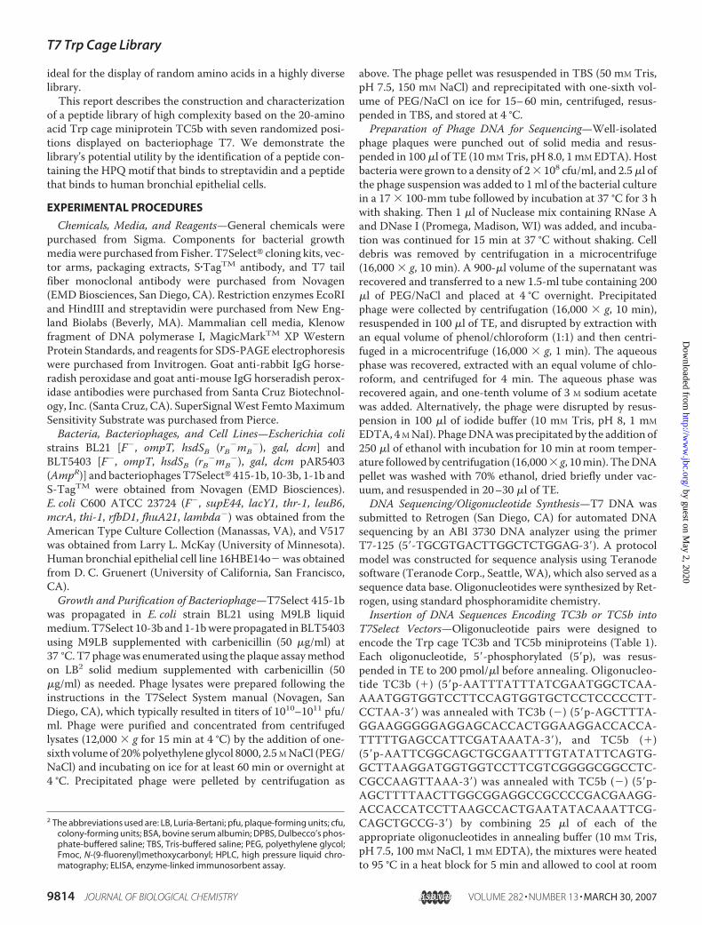
tions of 50 nonmutant clones is shown in Table 5. Confi-dence interval bounds for the frequency of amino acids ineach of the seven randomized positions were determinedusing the Clopper-Pearson method for the determination ofthe exact confidence bounds for a binomially distributed ran-dom variable (17) and was based on the use of a 32-codongenetic code. An adjustment was made to the usual 95% confi-dence level based on the simplest Bonferroni correction for theseven positions (100 � (1 � 0.05/7) � 99.3%). The resultingbounds are 0–7 for amino acids encoded by only one codon,0–10 for amino acids encoded by two codons, and 1–13 foramino acids encoded by three codons. The amino acid frequen-cies all fall within the expected bounds except the arginine atposition 5, whichwas zero but was expected to be at least 1 witha confidence of 99.3%.In addition, the amino acid sequences deduced from the 50
nonmutant clones were analyzed using the DIVAA programavailable at the RELIC Web site (relic.bio.anl.gov/) to deter-mine the diversity of the amino acids by position in the library.As shown in Fig. 3, the diversity of the randomized positionsranges from 0.55 to 0.75 with overlapping error bars. For com-parison, data available at the Web site were used to generate aplot of the diversity for theM13 Ph.D.-C7CTM library. The Trpcage library has diversity that is comparable with the con-strained M13 Ph.D.-C7CTM library at all positions except forposition 3, where the Trp cage library exhibited lower diversity.The average diversity for the Trp cage library was 0.67, com-pared with 0.70 for Ph.D.-C7CTM.The C600CR Host Cell Line Effectively Suppresses Premature
Peptide Termination—Since phage display systems typicallyencode randomized amino acids using the reduced 32-codongenetic code, which includes one stop codon, TAG, a suppres-sor host is utilized to prevent premature peptide terminationresulting from incorporation of the stop codon. Because a sup-pressor host is not available for the T7Select system, a hostcapable of suppressing the TAG stop codon while limiting thenumber of peptides displayed on the T7 phage capsid wasobtained by transforming competent cells of the carbenicillin-
TABLE 4Mutant DNA sequence and resulting peptide sequence from the two major types of mutants observed in the Trp cage T-3 libraryAn underscore indicates a deletion, and a substitution is underlined.
T7 Trp Cage Library
MARCH 30, 2007 • VOLUME 282 • NUMBER 13 JOURNAL OF BIOLOGICAL CHEMISTRY 9819
by guest on May 2, 2020
http://ww
w.jbc.org/
Dow
nloaded from
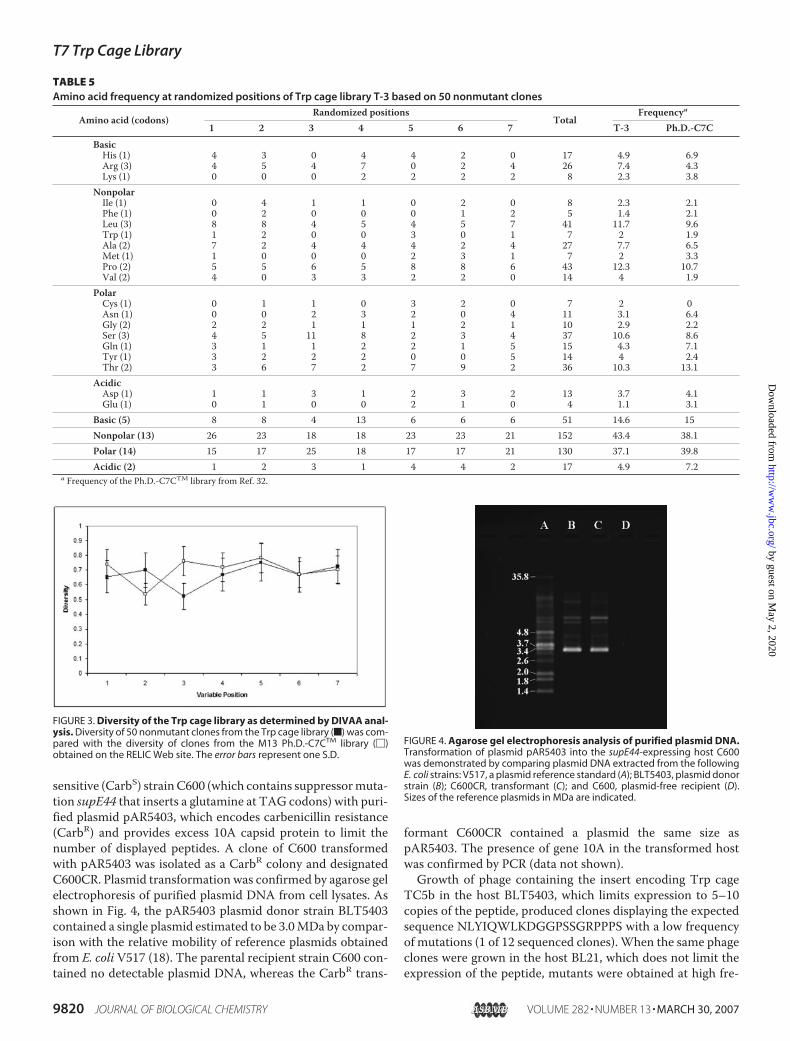
sensitive (CarbS) strainC600 (which contains suppressormuta-tion supE44 that inserts a glutamine at TAG codons) with puri-fied plasmid pAR5403, which encodes carbenicillin resistance(CarbR) and provides excess 10A capsid protein to limit thenumber of displayed peptides. A clone of C600 transformedwith pAR5403 was isolated as a CarbR colony and designatedC600CR. Plasmid transformation was confirmed by agarose gelelectrophoresis of purified plasmid DNA from cell lysates. Asshown in Fig. 4, the pAR5403 plasmid donor strain BLT5403contained a single plasmid estimated to be 3.0MDa by compar-ison with the relative mobility of reference plasmids obtainedfrom E. coli V517 (18). The parental recipient strain C600 con-tained no detectable plasmid DNA, whereas the CarbR trans-
formant C600CR contained a plasmid the same size aspAR5403. The presence of gene 10A in the transformed hostwas confirmed by PCR (data not shown).Growth of phage containing the insert encoding Trp cage
TC5b in the host BLT5403, which limits expression to 5–10copies of the peptide, produced clones displaying the expectedsequence NLYIQWLKDGGPSSGRPPPS with a low frequencyof mutations (1 of 12 sequenced clones). When the same phageclones were grown in the host BL21, which does not limit theexpression of the peptide, mutants were obtained at high fre-
FIGURE 3. Diversity of the Trp cage library as determined by DIVAA anal-ysis. Diversity of 50 nonmutant clones from the Trp cage library (f) was com-pared with the diversity of clones from the M13 Ph.D.-C7CTM library (�)obtained on the RELIC Web site. The error bars represent one S.D.
FIGURE 4. Agarose gel electrophoresis analysis of purified plasmid DNA.Transformation of plasmid pAR5403 into the supE44-expressing host C600was demonstrated by comparing plasmid DNA extracted from the followingE. coli strains: V517, a plasmid reference standard (A); BLT5403, plasmid donorstrain (B); C600CR, transformant (C); and C600, plasmid-free recipient (D).Sizes of the reference plasmids in MDa are indicated.
TABLE 5Amino acid frequency at randomized positions of Trp cage library T-3 based on 50 nonmutant clones
Amino acid (codons)Randomized positions
TotalFrequencya
1 2 3 4 5 6 7 T-3 Ph.D.-C7CBasicHis (1) 4 3 0 4 4 2 0 17 4.9 6.9Arg (3) 4 5 4 7 0 2 4 26 7.4 4.3Lys (1) 0 0 0 2 2 2 2 8 2.3 3.8
NonpolarIle (1) 0 4 1 1 0 2 0 8 2.3 2.1Phe (1) 0 2 0 0 0 1 2 5 1.4 2.1Leu (3) 8 8 4 5 4 5 7 41 11.7 9.6Trp (1) 1 2 0 0 3 0 1 7 2 1.9Ala (2) 7 2 4 4 4 2 4 27 7.7 6.5Met (1) 1 0 0 0 2 3 1 7 2 3.3Pro (2) 5 5 6 5 8 8 6 43 12.3 10.7Val (2) 4 0 3 3 2 2 0 14 4 1.9
PolarCys (1) 0 1 1 0 3 2 0 7 2 0Asn (1) 0 0 2 3 2 0 4 11 3.1 6.4Gly (2) 2 2 1 1 1 2 1 10 2.9 2.2Ser (3) 4 5 11 8 2 3 4 37 10.6 8.6Gln (1) 3 1 1 2 2 1 5 15 4.3 7.1Tyr (1) 3 2 2 2 0 0 5 14 4 2.4Thr (2) 3 6 7 2 7 9 2 36 10.3 13.1
AcidicAsp (1) 1 1 3 1 2 3 2 13 3.7 4.1Glu (1) 0 1 0 0 2 1 0 4 1.1 3.1
Basic (5) 8 8 4 13 6 6 6 51 14.6 15Nonpolar (13) 26 23 18 18 23 23 21 152 43.4 38.1Polar (14) 15 17 25 18 17 17 21 130 37.1 39.8Acidic (2) 1 2 3 1 4 4 2 17 4.9 7.2
a Frequency of the Ph.D.-C7CTM library from Ref. 32.
T7 Trp Cage Library
9820 JOURNAL OF BIOLOGICAL CHEMISTRY VOLUME 282 • NUMBER 13 • MARCH 30, 2007
by guest on May 2, 2020
http://ww
w.jbc.org/
Dow
nloaded from

quency (6 of 6 sequenced clones). Some of the mutant clonescontained the TAG stop codon within the Trp cage sequenceandwere expected to result in the display of truncated peptides.Two nonsense mutants, E7 (which contained a TAG stopcodon at Tyr-3 of the Trp cage sequence (NL*IQWLKDGGP-SSGRPPPS)) and E12 (which contained a TAG stop codon atTrp-6 of the Trp cage sequence (NLYIQ*LKDGGPSSGRPP-PS)), were chosen to test the effectiveness of the suppressorhost C600CR by ELISA. Each mutant and phage clone B1,which displays TC5b without a stop codon, was grown in thenonsuppressor strain BLT5403 and the suppressor strainC600CR. T7Select10-3b, which does not have a displayed pep-tide, was grown in BLT5403. Phage displaying Trp cage TC5bwere detected using rabbit IgG polyclonal antibody AB167,which was raised against TC5b and demonstrated to bind tophage displaying the TC5b peptide by Western analysis (seeabove). Since AB167 is a polyclonal antibody, some binding tothe truncated peptides of E7 and E12 was expected, with possi-bly greater binding to E12 because it is 3 residues longer thanE7. As shown in Fig. 5, the ELISA indicated that the antibodydetected display of the TC5b peptide when B1 was grown ineither host (B1/B and B1/C), and there was minimal binding ofthe antibody to phage T7Select10-3b (T7/B), which has no dis-played peptide. The percentage of binding to E12 after growthin BLT5403 (E12/B) was numerically lower thanwhen grown inC600CR (E12/C), but the difference was not significant (p �0.1). Antibody binding to mutant E12 was lower than the bind-ing to B1 regardless of the host. Antibody binding tomutant E7was significantly higher (p � 0.01) when grown in the suppres-sor C600CR (E7/C) compared with growth in the nonsuppres-sor BLT5403 (E7/B), and both exhibited lower binding than B1.The results from E7 indicate that C600CR was able to suppressthe codon TAG in the T7Select phage. Values lower than B1were expected, since suppression by supE44 is not 100% effi-cient (19, 20).
The T7 Trp Cage Library Allows Rapid Identification of Pep-tides Containing the HPQMotif—Streptavidin has been used asa model receptor system for analysis of linear and constrainedphage display libraries yielding the well known His-Pro-Gln(HPQ) motif (5). We used streptavidin as a target to assess thenaive Trp cage library for the display of peptides containing theHPQmotif in a limited panning experiment using 1.2� 109 pfuof the amplified library before final purification by CsCl. Afterpanning the Trp cage library against streptavidin for threerounds, a small number of clones were selected for DNAsequencing. There were three mutants among the 10 clonesanalyzed, and, as shown in Table 6, two of the seven nonmu-tant clones contained the expected HPQ motif (AAADY-YLQWLHPQGPHSGRPPPA and AAADSYRQWLHPQGP-WSGRPPPA). This result demonstrated that a motifreported to bind to streptavidin after panning with linearand constrained phage libraries can quickly be obtainedfrom the Trp cage library. Within our small sample size, theHPQ motif was only observed at the three randomized posi-tions that are contiguous, which is located at the junction ofthe �-helix and the 310 helix of the Trp cage fold.The T7 Trp Cage Library Enables Selection of Cell-binding
Peptides—One of the potential uses of the Trp cage library is torapidly discover peptides with affinity to human cell types forthe development of cell-specific targeting ligands. In order toevaluate the library’s capability to produce such peptide leads, itwas panned against the human bronchial epithelial cell line16HBE14o�. After three rounds of panning, the peptidesequences displayed by 18 randomly selected clones weredetermined fromDNA sequence analysis. As shown in Table 7,there were four full-length Trp cage sequences, including five
FIGURE 5. Phage ELISA for demonstration of TAG suppression. Binding ofantibody AB167 to T7Select10-3b phage grown in the nonsuppressor hostBLT5403 (B) was compared with binding to phage grown in the suppressorhost C600CR (C). The comparison included phage without a displayed pep-tide grown in the nonsuppressor host (T7/B) and phage displaying TC5bgrown in both hosts (B1/B and B1/C). Binding of the antibody to phagemutants E7 and E12 displaying truncated Trp cages grown in B (E7/B andE12/B, respectively) was related to antibody binding to phage clone B1 dis-playing the full-length Trp cage on a percentage basis. The phage mutantsgrown in C600CR (E7/C and E12/C) were likewise related to B1 grown inC600CR. The error bars represent one S.D.
TABLE 6Peptide sequences obtained from panning against streptavidin(variable positions are in boldface type, and the expected HPQ motifis underlined)
Clone number Peptide sequence255-104-3 AAADYYLQWLHPQGPHSGRPPPA*255-104-5 AAADSYRQWLHPQGPWSGRPPPA*255-104-2 AAADLYRQWLALCGPQSGRPPPT*255-104-4 AAADHYRQWLLGLGPGSGRPPPC*255-104-9 AAADKYTQWLSIAGPQSGRPPPQ*255-104-10 AAADLYNQWLTHSGPRSGRPPPN*255-104-11 AAADAYMQWLRYHGPFSGRPPPT*
TABLE 7Peptides obtained from panning against human bronchial epithelialcell line 16HBE14o�
Clone number Peptide sequence (variable positionsin boldface type)
Full-length Trp cage624-55-20 AAADRYPQWLNGMGPSSGRPPPN*624-55-21 AAADAYPQWLFTPGPTSGRPPPL*(5)a624-55-29 AAADPYAQWLQSMGPHSGRPPPR*624-55-36 AAADMYAQWLDNMGPHSGRPPPY*
Mutant Trp cage624-55-24 AAADAYSQWLLQTGPYSGRPSPRV624-55-32 AAADTYIQWLKINGPRLVVLPPRN624-55-33 AAADAYGQWLRTSGPLSGRSLPPR624-55-37 AAADWYQQWLPPGGPGSGRPPPHL
Truncated Trp cage624-55-22 AAADLYLQWLD*PGPSSGRPPPLL624-55-26 AAAD*YTQWLYLQGPNSGRPPPY*(5)a
a There were five instances of these peptides among the clones sequenced.
T7 Trp Cage Library
MARCH 30, 2007 • VOLUME 282 • NUMBER 13 JOURNAL OF BIOLOGICAL CHEMISTRY 9821
by guest on May 2, 2020
http://ww
w.jbc.org/
Dow
nloaded from

instances of the same sequence, which is represented in thetable by clone number 624-55-21. There were also fiveinstances of the truncated peptide represented by clone num-ber 624-55-26. Binding of the displayed peptides to16HBE14o� cells was evaluated by phage ELISA using unfixedcells. As shown in Fig. 6, binding to 16HBE14o� cells was lowerfor the negative control phage displaying the TC5b version ofthe Trp cage (NLYIQWLKDGGPSSGRPPPS) and for two ofthe phage clones selected at random after panning against the16HBE14o� cells; phage clone 624-55-29 displaying the pep-tide AAADPYAQWLQSMGPHSGRPPPR (amino acids in thevariable positions are indicated in boldface type) demonstratedsignificantly higher binding to 16HBE14o� cells, compared withthenegative control. This result indicated that theTrp cage librarycan be used to select peptides with cell binding capability.
DISCUSSION
The Trp cage miniprotein has been optimized by amino acidsubstitution to produce versions with improved folding charac-teristics, including the versions utilized in this study, TC3b(NLFIEWLKNGGPSSGAPPPS) andTC5b (NLYIQWLKDGG-PSSGRPPPS), which fold 38 and �95%, respectively, in water.Amino acid substitution analysis also revealed that some of theamino acids must be conserved to obtain a folded Trp cage, butother positions could be changed without compromising fold-ing characteristics (11, 12). The aim of this study was to con-struct a phage display library using theTrp cage as a self-foldingprotein scaffoldwith substitution of randomized amino acids insome of the nonconserved positions in order to create a libraryof novel peptides that can be used to select for target-specificbinding ligands.The stable fold adopted by peptides in the Trp cage library
should result in tighter binding to target molecules by decreas-
ing the change in conformationalentropy upon binding. Gomez andFreire (21) studied the energetics ofthe binding of pepstatin A to endo-thiapepsin and noted that the mainobstacle to binding was the reduc-tion in rotational, translational, and,most importantly, conformationalentropy of the peptide. The confor-mational entropy of a peptide can beattributed to immobilization of thepeptide backbone upon binding,burying of side chain upon binding,and a smaller term for solvent-ac-cessible side chains. Since the Trpcage library is a stable folded struc-ture, the peptide backbone isalready fixed; thus, the change inconformational entropy is minimal.Initially, we attempted to display
the Trp cage on the gene III proteinof bacteriophage M13, but thoseattempts failed to produce phagecontaining Trp cage inserts. Lack ofsuccess may have been due to
incompatibility of the highly folded Trp cage and the biologicalconstraints of the M13 system, which relies on extrusion of thephage and any displayed proteins through the cell membrane(22). Also, the tail fiber protein pIII, which is fused to displayedpeptides in theM13 system, is anchored to the innermembraneof the cell prior to phage assembly (23), and a displayed peptidewith a hydrophobic domain, such as the Trp cage, might inhibitphage assembly by altering the interaction of pIII with the innermembrane. The M13 system that we used (Ph.D. system; NewEngland Biolabs) relies on the pIII signal sequence for Sec-de-pendent protein translocation of displayed peptides (24). SinceSec translocates unfolded proteins, incorporation of signalsequences from the signal recognition pathway has recentlybeen shown to improve translocation and display of folded pro-teins on filamentous phage (25). Display of the Trp cage librarywith the M13 system might be achieved by using the signalrecognition pathway or an alternate protein translocationpathway.The T7Select system was chosen next to display the library,
because it utilizes a lytic phage maturation process and there-fore was expected to avoid potential problems with membraneinteractions and/or protein translocation. Although it is a lyticsystem, display of TC3b on T7 was not compatible with growthof the high valency vector T7Select415-1b, as evidenced by ahigh frequency ofmutations within the insert encoding the Trpcage. The other T7Select vectors 10-3b (displaying 5–10 copiesper phage) and 1-1b (displaying 0.1–1 copies per phage) bothexhibited a low mutation frequency within the insertedsequence. The one-step growth experiments showed that dis-play of Trp cage TC5b did not affect the number of infectedcells producing phage but did affect the number of phage pro-duced per infected cell greater than display of a linear peptide.This suggests that display of theTrp cage interferes in someway
FIGURE 6. Phage ELISA for detection of binding to unfixed 16HBE14o� cells. Binding of phageT7Select10-3b displaying Trp cage TC5b (Trp cage) was compared with binding of three phage clones selectedfrom the naive Trp cage library after three rounds of panning against the human bronchial epithelial cell line16HBE14o�. Results represent the average of triplicate wells, and error bars represent one S.D.
T7 Trp Cage Library
9822 JOURNAL OF BIOLOGICAL CHEMISTRY VOLUME 282 • NUMBER 13 • MARCH 30, 2007
by guest on May 2, 2020
http://ww
w.jbc.org/
Dow
nloaded from
with phage assembly.Western analysis showed that despite theaffect of the Trp cage on phage production, display on the cap-sid protein of T7Select10-3b was achieved.The design of theTrp cage librarywas based on versionTC5b
of the miniprotein with randomized amino acids at seven posi-tions, resulting in display of the sequence AAADXYX-QWLXXXGPXSGRPPPX as a C-terminal protein fusion on thephage capsid protein (gp10B). The three alanine residues(AAA) were added to the N terminus to increase helical pro-pensity (16). Secondary structure analysis of gp10B using Pep-Tool software (BioTools, Edmonton, Canada) predicted thephage protein to be highly helical, which could also contributeto the folding of the Trp cage N-terminal helix. The N-cappingresidue Asn of TC5b was replaced with Asp (D). Randomizedpositions, indicated by an X, were selected because they are notconserved and are located on the solvent-exposed surfaces ofthe Trp cage NMR structure (Fig. 2), where they are availablefor ligand binding. As a result of the fold, the randomized posi-tions are in close proximity.Characterization of the naive library by DNA sequencing
revealed that the library consisted of three groups designatednonmutants, stops, and mutants. Nonmutants comprised46% of the clones, and they contained the Trp cage scaffoldas designed with seven randomized amino acids. Stops (16%)contained the stop codon TAG in one of the randomizedpositions. In order to obtain a more uniform distribution ofamino acid frequency and reduce the occurrence of stopcodons, the oligonucleotide used to construct the Trp cagelibrary was based on a 32-codon genetic code that only con-tains one stop codon, TAG. Systems that use the bacterioph-ageM13 for phage display utilize a host that can suppress theremaining stop codon. However, the bacterial host used forthe T7 system does not contain a suppressor for the stopcodon TAG, resulting in a truncated Trp cage when thiscodon appears in one of the variable positions. Among therandom clones that were selected for sequencing, 18 con-tained the TAG stop codon in a randomized position. Thestop codon was observed in all of the randomized positionsexcept the last one. Since the frequency of TAG within the32-codon genetic code is 0.031 and 109 clones with sevenrandom positions were sequenced, the expected observa-tions of TAG occurring would be 23.6. Since we onlyobserved 18 occurrences, there does not appear to be a selec-tive pressure increasing the frequency of stop codons at therandomized positions of the library. The truncated proteinswould still be displayed but might not have a Trp cage struc-ture. To reduce the impact of the stop codon, a suppressorhost was constructed for use in any further amplifications ofthe library. Since this host contains the supE44 suppressormutation, glutamine residues would be inserted at TAGcodons raising the frequency of that amino acid in thelibrary. Mutants were observed at high frequency (38%) andcontained primarily single base insertions or deletions. It islikely that the origin of observed mutants comes from theoriginal chemical synthesis of the 103-base oligonucleotidetemplate used to construct the library (26, 27). The fre-quency of mutation indicates that the Trp cage exerts selec-
tive pressure on the phage even with the display of only 5–10copies.The inability to display the Trp cage in the M13 system, the
common occurrence of mutation resulting from display ofTC3b in the T7Select system, and mutations observed withinthe T7 Trp cage library all suggest that the Trp cage is a highlyactive structure that is selected against based on the copy num-ber and modifications resulting from randomized amino acids.Since the Trp cage affects phage growth, it is possible that someof the modified structures that are too active for compatibilitywith this systemmight be observable by ribosome display or analternate in vitro display system. After observing a high muta-tion frequency from display of TC3b, the library was designedso that the stabilizing salt bridge between Asp-9 and Arg-16 ofTC5b was sacrificed when Asp-9 was chosen as one of the ran-domized positions. We reasoned that decreasing the stabilityof the Trp cage fold by removing the salt bridge would reduceselective pressure against the Trp cage, providing the poten-tial for discovery of a greater number of active moleculesfrom the library by, in effect, reducing the stringency.Among the nonmutant clones that were sequenced from thelibrary, we observed one occurrence of a random insertionthat restored Asp-9 that was within the expected frequencyrange, indicating that the restoration of the salt bridge isneither selected for or against. The stability of any peptidesequences selected from the library by panning experimentscould potentially be enhanced by reestablishing the saltbridge as long as restoring Asp-9 does not adversely affectthe desired binding characteristics.The Trp cage library was used successfully to identify an
HPQ-containing peptide that binds to streptavidin. SuchHPQ-containing peptides were previously identified using M13phage libraries (5, 28). Crystallographic analysis has demon-strated that the HPQ sequence binds to streptavidin as a Type I�-turn (29). Selection of an HPQ sequence in the Trp cageminiprotein (Table 6) occurred at the three variable positionslocated between an �-helix and a 310 helix (Fig. 2), a regionexpected to most easily accommodate a �-turn. Detailed struc-ture-activity studies of the HPQ containing Trp cage minipro-tein are in progress and will be reported elsewhere.Despite the frequency of clones containing mutations and
stops, the library still contains enough nonmutant clones toprovide substantial complexity. One estimate of library com-plexity is the number of primary clones. Since we produced1.6 � 109 primary clones for this library, the estimated com-plexity from nonmutants is 7.4 � 108 novel peptides. An addi-tionalmeasure of library complexitywas provided by analysis of50 nonmutant clones with the DIVAA program. The Trp cagelibrary had an average diversity of 0.67, which is comparablewith the average diversity of 0.70 for the constrained Ph.D.-C7CTM library. Linear and disulfide-constrained libraries con-structed in T7 have been reported to have higher diversity thanM13 libraries after evaluation byDIVAA (30). A linear library of12 amino acids in T7 had an average diversity of 0.85 comparedwith the linear M13 library Ph.D.-12TM, which had an averageof 0.68, and a T7 library with 7 amino acids constrained by adisulfide bridge had an average diversity of 0.80. The lowerdiversity of the Trp cage library is probably due to amino acid
T7 Trp Cage Library
MARCH 30, 2007 • VOLUME 282 • NUMBER 13 JOURNAL OF BIOLOGICAL CHEMISTRY 9823
by guest on May 2, 2020
http://ww
w.jbc.org/
Dow
nloaded from

combinations that are not tolerated, as indicated by the occur-rence of mutations. The diversity of the Trp cage library waslowest at the third randomized position, which is located nearthe junction of the�-helix and the 310 helix, suggesting that thisregion is conserved.Although immunoglobulins have been relied upon for ther-
apeutic and diagnostic applications where protein scaffoldswith high binding affinity are needed, there has been recentinterest in the development of scaffolds based on nonimmuno-globulin proteins (31). Characteristics considered ideal for non-immunoglobulin binding proteins include a small single-chainprotein, high thermodynamic stability, absence of disulfidebonds, and no free cysteines. Proteins with these characteristicswould be easier to manufacture, more stable, and capable offolding in the intracellular environment, where many drug tar-gets are located, and they would be amenable to adding a singlecysteine for the coupling of effector compounds. The Trp cageminiprotein satisfies all of these characteristics, making it agood candidate nonimmunoglobulin scaffold.An interesting example of a phage display peptide containing
a disulfide bond that has some similarities to the Trp cage is the�-hairpin peptide (NLPRCTEGPWGWVCM), which binds tothe human IgE receptor (6). Both are stably folded peptides ofsmall size that have a central Gly-Pro motif structure and Trpresidues that are involved in folding. In the case of the�-hairpinpeptide, the GP motif is part of the sequence (GPWG) thatforms the hairpin, whereas in the Trp cage, the GPmotif is partof a 310 helix (GGPSSGR). The hairpin structure relies upon thedisulfide bond for conformational stability, whereas the Trpcage conformation is disulfide-independent. Unlike the Trpcage, there does not appear to be interaction between Trp andPro in the�-hairpin peptide; instead, Trp-12 contributes to foldstability by packing up against the disulfide bond. The Trp cageminiprotein contains a Trp sandwiched between prolines,whereas binding of the �-hairpin peptide to the IgE receptorappears to be dependent upon formation of a “proline sand-wich” between Pro-9 and two tryptophans in the IgE receptor.Some of the prolines of the Trp cage might also be available forsandwich formation, or peptides from the library containingproline in the randomized positions, such as the one selectedfor as a binder to human bronchial epithelial cells containing aproline at the first randomized position, could be involved insuch formations.This study demonstrated that a random amino acid library
utilizing the Trp cage miniprotein TC5b as a scaffold can bedisplayed on bacteriophage T7. A library of high complexityand diversity was obtained and was shown to support the rapididentification of peptides containing the HPQ motif whenpanned against streptavidin. This library was useful for theselection of a peptide that binds to human bronchial epithelialcells andmay therefore be valuable for the selection of other cellbinding peptides to improve drug delivery technology and forthe selection of receptor-binding ligands. Since the Trp cagehas been studied as a model for protein folding, this librarycould also be useful for the selection of novel Trp cagesequences for folding studies.
Acknowledgments—We thank Niels Andersen (University of Wash-ington) and Jianzhu Chen (Massachusetts Institute of Technology)for critical review of the manuscript; Martin Lee and Sonia Souzafor expert assistance with statistical analysis; Mohammad Ahma-dian for helpful discussions; the Teranode Corporation, Ken Coo-per, Jason Bohreer, and Jeremy Thompson for assistance in thedevelopment of Teranode software applications for processingsequence data and database construction; and Harry Wang andKen Farber for peptide synthesis and purification support.
REFERENCES1. Smith, G. P. (1985) Science 228, 1315–13172. Lowman, H. B. (1997) Annu. Rev. Biophys. Biomol. Struct. 26, 401–4243. Ladner, R. C. (1995) Trends Biotechnol. 13, 426–4304. Uchiyama, F., Tanaka, Y.,Minari, Y., and Tokui, N. (2005) J. Biosci. Bioeng.
99, 448–4565. Giebel, L. B., Cass, R. T., Milligan, D. L., Young, D. C., Arze, R., and John-
son, C. R. (1995) Biochemistry 34, 15430–154356. Nakamura, G. R., Starovasnik, M. A., Reynolds, M. E., and Lowman, H. B.
(2001) Biochemistry 40, 9828–98357. Ladner, R. C., and Ley, A. C. (2001) Curr. Opin. Biotechnol. 12, 406–4108. Rebar, E. J., and Pabo, C. O. (1994) Science 263, 671–6739. Friesen, W. J., and Darby, M. K. (1998) Nat. Struct. Biol. 5, 543–54610. Dennis, M. S., Herzka, A., and Lazarus, R. A. (1995) J. Biol. Chem. 270,
25411–2541711. Neidigh, J. W., Fesinmeyer, R. M., Prickett, K. S., and Andersen, N. H.
(2001) Biochemistry 40, 13188–1320012. Andersen, N. H., Barua, B., Fesinmeyer R. M., and Neidigh, J. (2001) in
Peptides: The Wave of the Future (Lebl, M., and Houghton, R. A., eds) pp.406–408, American Peptide Society, Springer, New York
13. Neidigh, J. W., Fesinmeyer, R. M., and Andersen, N. H. (2002)Nat. Struct.Biol. 9, 425–430
14. Qiu, L., Pabit, S. A., Roitberg, A. E., and Hagen, S. J. (2002) J. Am. Chem.Soc. 124, 12952–12953
15. Edgar, R. S. (1963) in Methodology in Basic Genetics (Burdette, W. J., ed)pp. 19–36, Holden-Day, Inc., San Francisco
16. Lin, J. C., Barua, B., and Andersen, N. H. (2004) J. Am. Chem. Soc. 126,13679–13684
17. Clopper, C. J., and Pearson, E. S. (1934) Biometrika 26, 404–41318. Macrina, F. L., Kopecko, D. J., Jones, K. R., Ayers, D. J., and McCowen,
S. M. (1978) Plasmid 1, 417–42019. Miller, J. H., and Albertini, A. M. (1983) J. Mol. Biol. 164, 59–7120. Edelmann, P., Martin, R., and Gallant, (1987) J. Mol. Gen. Genet. 207,
517–51821. Gomez, J., and Freire, E. (1995) J. Mol. Biol. 252, 337–35022. Castagnoli, L., Zucconi, A., Quondam,M., Rossi,M., Vaccaro, P., Panni, S.,
Paoluzi, S., Santonico, E., Dente, L., and Cesareni, G. (2001) Comb. Chem.High Throughput Screen. 4, 121–133
23. Marvin, D. A. (1998) Curr. Opin. Struct. Biol. 8, 150–15824. Rapoza, M. P., and Webster, R. E. (1993) J. Bacteriol. 175, 1856–185925. Steiner, D., Forrer, P., Stumpp, M. T., and Pluckthun, A. (2006) Nat. Bio-
technol. 24, 823–83126. Beaucage, S. L., and Iyer, R. P. (1992) Tetrahedron 48, 2223–231127. Krotz, A. H., Klopchin, P. G., Walker, K. L., Srivatsa, G. S., Cole, D. L., and
Ravikumar, V. T. (1997) Tetrahedron Lett. 38, 3875–387828. Devlin, J. J., Panganiban, L. C., and Devlin, P. E. (1990) Science 249, 404–40629. Katz, B. A. (1995) Biochemistry 34, 15421–1542930. Krumpe, L. R., Atkinson, A. J., Smythers, G. W., Kandel, A., Schumacher,
K. M., McMahon, J. B., Makowski, L., and Mori, T. (2006) Proteomics 6,4210–4222
31. Binz, H. K., Amstutz, P., and Pluckthun, A. (2005) Nat. Biotechnol. 23,1257–1268
32. New England Biolabs, Inc. (2002) Instruction Manual for the Ph.D.-C7CPhage Display Peptide Library Kit, p. 21, New England Biolabs, Inc.,Ipswich, MA
T7 Trp Cage Library
9824 JOURNAL OF BIOLOGICAL CHEMISTRY VOLUME 282 • NUMBER 13 • MARCH 30, 2007
by guest on May 2, 2020
http://ww
w.jbc.org/
Dow
nloaded from

Houston, Jr., Steven C. Quay and Paul H. JohnsonRichard E. Herman, Douglas Badders, Mark Fuller, Ekaterina G. Makienko, Michael E.
on Bacteriophage T7The Trp Cage Motif as a Scaffold for the Display of a Randomized Peptide Library
doi: 10.1074/jbc.M610722200 originally published online January 30, 20072007, 282:9813-9824.J. Biol. Chem.
10.1074/jbc.M610722200Access the most updated version of this article at doi:
Alerts:
When a correction for this article is posted•
When this article is cited•
to choose from all of JBC's e-mail alertsClick here
http://www.jbc.org/content/282/13/9813.full.html#ref-list-1
This article cites 26 references, 5 of which can be accessed free at
by guest on May 2, 2020
http://ww
w.jbc.org/
Dow
nloaded from