thesis full draft - university of minnesota
TRANSCRIPT

!!Pearl in the Mud: Genome Assembly and Binning of a cold seep Thiomargarita nelsonii cell and
Associated Epibionts from an Environmental Metagenome
!!!!!!!A THESIS
SUBMITTED TO THE FACULTY OF THE UNIVERSITY OF MINNESOTA
BY !!!!!Palmer Scott Fliss !!!!!!
IN PARTIAL FULFILLMENT OF THE REQUIREMENTS FOR THE DEGREE OF MASTER OF SCIENCE !!!!!!!!!!Adviser Jake V. Bailey !!!!!!!!
January 2014

!!!!!!
© Palmer Scott Fliss 2014

!!
Acknowledgements
!I would like to thank Jake Bailey for his guidance and mentorship throughout my time at the University of Minnesota. I would also like to profusely thank Beverly Flood her guidance,
support, and teachings on microbiology, geobiology, and acceptance into what was once and remains a challenging field, in addition to her extensive work on the DNA extraction and wet lab
microbiology. Dan Jones was an invaluable asset to my understanding of science of bioinformatics and geobiology, his talks and insights were key to understanding the breadth of this project. The assistance of the aforementioned scientists was critical to the success of this
project. I would like to extend my gratitude to the entire Bailey lab group for their discussions, friendship, knowledge, and aid both scholarly and emotional. Many thanks to Greg Dick and
Sunit Jain for their computational resources and their continued support and suggestions on best practices regarding metagenomic assembly and binning. Thanks to Greg Rouse, Victoria Orphan, and Lisa Levin for assistance with the collection of gastropods from Hydrate Ridge. Additional
thanks to Ying Zhang, and the MSI Bioinformatics support staff for their assistance in troubleshooting assemblies and software installation. Charles Nguyen and the rest of the Earth
Sciences IT Department were essential to the success of establishing the Bailey Geobiology Lab Server, and their assistance with installation and structuring our data was profoundly helpful.
Additional thanks to Anne-Kristin Kaster for generously donating PacBio RS sequencing wells, as well as her advice regarding assembly and data synthesis. I would like to thank D. Fox and D.
Knights for serving on my thesis defense committee. Portions of this work were supported by National Science Foundation grant EAR-1057119 and by a UMN Grant-in-Aid to Dr. Jake Bailey. I would like to thank Rachel for her moral and emotional support, grammatical advice, immense
patience, and advice that I couldn’t have done without. !!
!i

!!!
Abstract
! As the study of microbes and their impact on the environment grows, so too does the
desire to understand the genetic basis of the physiologies that make possible interactions between
microbial cells and their environment. Since it is now much more cost-effective to sequence
bacterial genomes, environmental metagenomic assembly is a very attractive option for obtaining
the genetic blueprints of bacterial physiologies. Bacteria of the genus Thiomargarita (Greek;
theio-: sulfur; margarites: pearl), pose a particularly interesting quandary. The genus includes the
world’s largest bacteria, but as uncultured organisms, their physiologies and basis for their
gigantism are not well understood. In order to investigate the genetic basis for these modes, a
single cell MDA amplification approach was used on T. nelsonii cells collected at the Hydrate
Ridge methane seep off of the coast of Oregon. These particular cells were derived from a
gastropod-attached epibiont community. Next-generation sequencing produced a metagenomic
product representing both T. nelsonii and attached bacteria (epibionts). These reads were
assembled into contigs, binned using the tetranucleotide frequency of the resultant contigs, and
finalized using a more stringent secondary assembly. The resulting draft genome shows evidence
in Thiomargarita nelsonii for a complete denitrification pathway not previously known in large,
vacuolated, sulfur-oxidizing bacteria. Additionally, the genes necessary for polyphosphate
metabolism were observed. Polyphosphate metabolism is thought to play a role in the formation
of phosphatic minerals that serve as important reservoirs in the marine phosphorous cycle.
!ii

!!
Table of Contents
!!
!List of Tables iv List of Figures v List of Abbreviations vi 1. Introduction 1 2. Methods 5 2.1. Sample Collection 5 2.2. Genomic DNA Preparation and Sequencing 5 2.3. Bioinformatic Analysis 7 2.4. Annotation and Function Mapping 9 3. Results 10 3.1. Sequence Binning 10 3.2. Phylogeny and Morphology 10 3.3. Gene Identification 11 3.4. Ribosomal Proteins and Single-Copy Genes 13 3.5. Nitrate Respiration 14 3.6. Sulfur Oxidation 14 3.7. Phosphate Accumulation 16 3.8. Oxygen Respiration 16 4. Discussion 17 4.1. Sequencing, Analysis, and Binning 17 4.2. Site and Geochemistry 19 4.3. Thiomargarita in the Environment 21 4.4. Genome and Metabolism Discussion 23 4.4.1. Nitrate 23 4.4.2. Sulfur 26 4.4.3. Phosphorous 28 4.4.4. Oxygen 29 4.4.5. Carbon Metabolism and Heterotrophy 30 4.4.6. Cell Division and Shape 32 5. Conclusion 33 6. Bibliography 35 7. Table Captions 41 8. Figure Captions 50 !
!iii
!!!
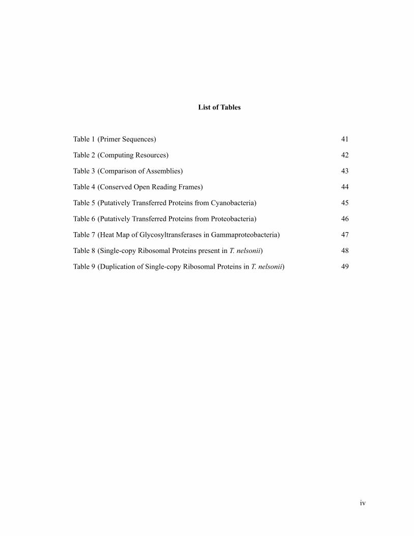
List of Tables
!Table 1 (Primer Sequences) 41
Table 2 (Computing Resources) 42
Table 3 (Comparison of Assemblies) 43
Table 4 (Conserved Open Reading Frames) 44
Table 5 (Putatively Transferred Proteins from Cyanobacteria) 45
Table 6 (Putatively Transferred Proteins from Proteobacteria) 46
Table 7 (Heat Map of Glycosyltransferases in Gammaproteobacteria) 47
Table 8 (Single-copy Ribosomal Proteins present in T. nelsonii) 48
Table 9 (Duplication of Single-copy Ribosomal Proteins in T. nelsonii) 49
!iv

!!!
List of Figures
!Figure 1 (The microbial consortia present on Provanna laevis) 50
Figure 2 (ESOM generated from MetaVelvet) 51
Figure 3 (Maximum-Likelihood in Relation to Morphology) 52
Figure 4 (Comparative Geographic Morphotype Analysis) 53
Figure 5 (Maximum-Likelihood Phylogeny of Glycosyltransferase) 54
Figure 6 (Predicted Nitrogen, Sulfur, Phosphorous, and Oxygen 55
Metabolism of T. nelsonii)
Figure 7 (Schematic of Incomplete SOX system in T. nelsonii) 56
Figure 8 (Schematic of potential energy-yielding pathways in 57
T. nelsonii in variable geochemical conditions)
!v

!List of Abbreviations
!AOM Anaerobic Oxidation of Methane
ATP Adenosine Tri-Phosphate
Contig Contiguous section of overlapping DNA that forms a consensus sequence
DMSO Dimethyl Sulfoxide
DNA DeoxyriboNucleic Acid
DNRA Dissimilatory Nitrate Reduction to Ammonia
ESOM Emergent Self-Organizing Map
GTE GlycosylTransferase-Encoding
IMG-ER Integrated Microbial Genomes Expert Review
ITS Internal Transcribed Spacer
MDA Multiple Displacement Amplification
MEGA Molecular Evolutionary Genetics Analysis
MEGAN Metagenome Analyzer
MSI Minnesota Supercomputing Institute
MUSCLE Multiple Sequence Comparison by Log-Expectation
NCBI National Center for Biotechnology Information
ORF Open Reading Frame
PCR Polymerase Chain Reaction
rDSR Reverse Dissimilatory Sulfate Reductase
ROV Remotely Operated Vehicle
rRNA Ribosomal RiboNucleic Acid
SNP Single-Nucleotide Polymorphism
(T)BLAST(N/P) (Translated) Basic Local Alignment Search Tool (for Nucleotides/Proteins)
TCA Tricarboxylic Acid
WGA Whole Genome Amplification
!!
!vi

!1. Introduction
! Deep-sea cold seeps exhibit both weak and ebullient flow of hydrocarbons such as
methane that can vary (6-105 mmol m-2 day-1) over spatial scales of only a few meters (Torres et
al., 2002). The elevated flux of hydrocarbons from the subsurface in these settings drives the
production of sulfide via the anaerobic oxidation of methane (AOM) and sulfate reduction
(Boetius et al., 2000). Microbial mats at these sites, which can be spatially extensive, are visually
dominated by large, sulfur-oxidizing bacterial ecotypes of the closely related genera Beggiatoa
and Thiomargarita that fall within the family Beggiatoaceae. These bacteria are chemolithotrophs
that obtain energy for metabolism from the oxidation of reduced sulfur species. Thiomargarita are
thought to primarily use the oxidation of electron donors available in the sediment to fuel carbon
fixation. However, the terminal electron acceptors used in these reactions can vary depending on
their location relative to the oxic/anoxic boundary. Under these highly reducing conditions,
oxygen concentrations decline rapidly upon penetration into the sediments. Where sulfide fluxes
upward into the water column, the oxic/anoxic boundary changes temporally and spatially,
forcing bacterial species to adopt alternate electron acceptors to survive the dynamic conditions.
Nitrate can be used as a terminal electron acceptor in large, vacuolated sulfur bacteria in times of
anoxia; Thiomargarita allocates up to 90% of its volume for intracellular nitrate storage (Schulz
and Jørgensen, 2001). Thiomargarita has also been shown to accumulate intracellular elemental
sulfur inclusions that serve as intermediates in the oxidation of hydrogen sulfide to sulfate, to
provide the cell with electron donor when access to sulfide is limited. The lack of motility
observed in Thiomargarita additionally illustrates the need for metabolic flexibility in the
presence of temporally and spatially variable geochemical gradients. These nitrate and sulfur
storage capacities allow them to bridge the suboxic zone, where neither sulfide nor oxygen exist,
providing Thiomargarita an advantage over sulfide-oxidizing bacteria that are incapable of
spanning this gap (Schulz et al., 1999). Beggiatoa and Thioploca have been shown to possess
!1
mechanisms for motility across the suboxic zone, with Thioploca cells spanning the entire region,
and Beggiatoa following the oxygen gradient both above and below the sediment surface (Schulz
& Jørgensen, 2001).
! Prior research has demonstrated that Thiomargarita is capable of accumulating phosphate
intracellularly as long polyphosphate (poly-p) polymers. The hydrolysis of this polyphosphate,
and concomitant release of phosphate into pore water has been linked to the formation of large
phosphorite deposits in the seafloor and the subsurface (Schulz & Schulz, 2005; Bailey et al.,
2007; Goldhammer et al., 2010). These phosphatic minerals precipitated by this release of
phosphate serve as a form of sequestration, or long-term phosphorous sink, indicating that the
role of the large sulfur bacteria in the phosphorous cycle is worth investigation.
! Thiomargarita-like bacteria have been discovered in numerous locations on the ocean
floor, including the Gulf of Mexico (Kalanetra et al., 2005; Bailey et al., 2007), mud volcanoes in
the Barents Sea (Girnth et al., 2011), the Mediterranean Sea (Grünke et al., 2011), the Costa
Rican Margin (Schulz et al., 1999), and methane seeps off the coast of Oregon. The “sulfur pearl”
Thiomargarita (Greek; theio-: sulfur; margarites: pearl) species initially discovered at seeps
along the Costa Rican Margin and later observed at Hydrate Ridge, T. nelsonii, were found
commonly attached to substrates, in particular on shells of Provanna snails. Additionally, these
attached cells appeared to undergo an apparent dimorphism (elongate vs. budding) in their life
cycle, wherein Thiomargarita elongated almost a millimeter in length, and budded into spherical,
or coccoidal, daughter cells on the free end (Bailey et al., 2011). Several of the samples collected
showed a distinct epibiont community resembling morphotypes similar to Ca. Thiomargarita
nelsonii as well as Maribeggiatoa sp. and Leucothrix sp. Due to prior failed attempts to culture
Thiomargarita (Flood & Bailey, unpublished), a bioinformatics-based de novo sequencing and
assembly approach was chosen to reveal the genetic information coding for metabolic enzymes,
cytoskeletal proteins, and the extent of lateral gene transfer in Thiomargarita. Certain
!2

Thiomargarita nelsonii morphotypes exhibit a dimorphic reproductive behavior not previously
described in other large sulfur bacteria (Bailey et al., 2011). Selection was therefore made of both
bud and stalk morphotypes for amplification and sequencing. Despite attempts at single-cell
isolation via physical cell straining, PCR screening revealed that all 4 collected samples contained
other bacterial cells in addition to the targeted T. nelsonii. As the amplification of the
Thiomargarita 16S and ITS regions produced cleaner Sanger sequencing chromatograms in the
coccoidal buds, the genome amplification, sequencing and assembly was only carried out on the
coccoid buds. It is thought that the elongate cells not sequenced here experience a larger breadth
of geochemical conditions as compared to their stationary mat-forming counterparts, as
attachment to their mobile host provides transportation.
! None of the vacuolate sulfide-oxidizing bacteria have been isolated in pure culture. Thus,
what is known about their unique morphology, their nitrate storage abilities, and their ecological
relationships is primarily based on extrapolation of isolated bacteria with seemingly similar
characteristics. Previous physiological and genetic studies have been carried out on Beggiatoa sp.
(Nelson & Jannasch, 1983; Ahmad et al., 2006; Mußman et al., 2007; MacGregor et al., 2013), a
closely related large sulfur-oxidizing bacteria. These studies showed the presence of an adaptable
suite of metabolic-enzyme-coding genes that allow Beggiatoa to persist in habitats that would be
too inhospitable for other bacterial species without said mechanisms. The primary line of
investigation into the similar ecophysiological role of Thiomargarita in its locally dynamic
environment was to sequence and investigate the genes involved in metabolism of the chemical
species present. Investigations of Thiomargarita sp. have been centered around physiology
studies, and no studies of the genome have yet been published.
! It is now standard to study genomic fragments of uncultured microbes obtained by
shotgun sequencing of bulk DNA extracted from mixed communities (Tyson et al., 2004; Dick et
al., 2009). However, the assembly of whole genome amplification (WGA) from metagenomic
!3

datasets is a technique that is still in its infancy, and has been shown to be problematic with
respect to genome coverage. Multiple displacement amplification (MDA), a type of whole
genome amplification (WGA), can amplify bacterial DNA (up to a billion-fold) from single cells
or mixed communities, can generate higher-quality sequences from uncultured environmental
microorganisms than previous WGA techniques (Lasken & Stockwell, 2005; Raghunathan et al.,
2005; Kvist et al., 2007; Podar et al., 2007). However, this technique does have disadvantages, as
it has been shown to create segments of DNA that have no corresponding representatives in the
environment, essentially rearranging sections of genetic code to produce what are known as
chimeras, or false combinations of two or more pieces of DNA. The amplification of the DNA
that is separate from the target DNA, or overrepresentation of bacterial DNA in species that are
relatively rare in the environment has also been shown to confound analysis of microbial
genomes (Binga et al., 2008). Previous methods to extract microbial genetic information from
mixed communities were unable to amplify whole genomes, as the stochastic nature of
amplifications would commonly amplify only the most abundant species. Despite background
amplification and chimera formation (Lasken & Stockwell, 2005), MDA has higher efficacy in
amplifying complex DNA than past WGA techniques (Gonzalez et al., 2005; Spits et al., 2006).
One of the ways to combat these errors in amplification is to use high-throughput Illumina
sequencing. This type of sequencing generates billions of short (100 base pair) sections of genetic
information, wherein overlapping segments of DNA can be assembled into long sections of
consensus. In addition, paired-end reads contain positional information, allowing for highly
precise alignment of reads over many times the length of the reads themselves. This positional
information, paired with the consensus sequences generated from assembly, obviates some of the
problems associated with chimera formation from amplification.
! The combination of the whole-community representation of MDA-amplified DNA with
the coverage and positional advantages of Illumina paired-end sequencing show great potential to
rapidly analyze the genomes of unculturable microbes. Using MDA, the genomic DNA of two
!4

cells of uncultured Thiomargarita from a bacterial colony were separately amplified in this study.
Both of these cells were sequenced on Illumina HiSeq lanes, and annotated using the Integrated
Microbial Genomes (IMG) Pipeline. Here I summarize the approach of constructing a draft
genome of the world’s largest bacteria from an environmental sample, highlight the metabolic
pathways inferred from genes in this genome, and discuss the potential role of those genes in
Thiomargarita nelsonii.
!!2. Methods
!2.1. Sample Collection
The Remotely Operated Vehicle (ROV) Jason was used to collect Provanna sp. snail
samples during a research expedition on board the R/V Atlantis (AT18_10) to Hydrate Ridge
North (44º 40.02687’ N 125º 5.99969’ W), Cascadian margin, off the coast of Oregon, USA on
September 3, 2011. The shells of live Provanna sp. snails collected from the methane seep
sediments hosted an attached microbial (i.e., epibiont) community that appeared to be rich in
sulfide-oxidizing bacteria (Fig. 1), including morphotypes that resembled Candidatus
Thiomargarita nelsonii, as well as Ca. Maribeggiatoa sp. and Luecothrix sp. Some snails were
preserved shipboard individually in a 1:1 mixture of sterile ethanol and Instant Ocean (Instant
Ocean, Blacksburg, VA) and then stored at -20°C for future molecular analyses. Live snails, as
well as snails fixed in 4% paraformaldehyde diluted in Instant Ocean, were also maintained and
later observed in the laboratory. An Olympus SZX16 stereo-microscope and an Olympus BX61
compound microscope equipped with an Olympus XM10 color camera were used to obtain high-
resolution images of the snails and the attached cells.
! 2.2. Genomic DNA Preparation and Sequencing
!5

Stereomicroscopy revealed coccoidal buds either partially or fully detached from the
elongated Thiomargarita nelsonii morphotypes in the ethanol-seawater mixture surrounding snail
samples. These individual Thiomargarita cells were then pipetted into a sterile 40 µm pore-
diameter cell strainer (BD Biosciences, San Diego, CA) within a well of a sterile 6-well plate.
Each well contained DNA-free 3.5% NaCl and 50% ethanol. Using aseptic techniques, the cell
strainer was then lightly gyrated to disassociate small unattached and loosely attached bacteria
from the Thiomargarita cells. Then the cell strainer was transferred to a clean well and the
process was repeated five times. The cell strainer was then placed over a 50 mL conical and ~10
mL of the saltwater-ethanol mixture was gently poured over the cell strainer. The cell strainer
was then placed into a DNA-free phosphate buffer solution. Individual Thiomargarita cells were
then transferred by pipette to 5 µL of the PBS solution and frozen separately at -20°C.
!Two coccoidal buds and two elongated cells were selected for genomic DNA
amplification. DNA extraction and amplification was performed using the Qiagen REPLI-g Midi
kit (Qiagen, Hilden, Germany) for whole genome amplification (WGA), following the
manufacturer’s protocol. Cell lysis was achieved by heating to 65ºC and using the proprietary
Qiagen lysis buffer D2. One coccoidal and one elongated cell were also subjected to an
additional step of 10 minutes of incubation on ice following the cell lysis step, to determine if
additional cooling step increased genetic yield. The DNA was eluted in Tris-EDTA and an 1:100
dilution aliquot was generated for DNA quantification via a Thermo Scientific Nanodrop 2000C
Spectrophotometer and for polymerase chain reaction (PCR) screening. The undiluted stocks
developed a white fluffy precipitate (presumably proteins and other biomass). Thus the stocks
were centrifuged at 10,000 rpm and the more pristine supernatant was removed and used as the
primary DNA stock for genomic sequencing.
! PCR utilizing the primer set 233F-ITSReub was utilized to confirm positive amplification
of Thiomargarita nelsonii. The samples were screened for contamination using the universal
!6

primers for the 16S gene for bacteria (27F-1492R) and archaea (Arc8F - 9rts) (Table 1). Agarose
gel analyses and Sanger sequencing revealed that all four samples contained bacterial DNA but
none contained archaeal DNA. The elongated cells showed less amplification of the
Thiomargarita 16S and internal transcribed spacer (ITS) region and appeared more contaminated
with environmental bacteria than the coccoid buds and thus were excluded from further analyses.
Stock DNA was submitted to University of Minnesota Genomics Center for DNA quality
screening and genomic sequencing. Sample B6 was sequenced on a full lane of a Illumina HiSeq
2000 (Illumina, San Diego, USA) with 100 bp paired-end reads with ~300 base inserts while
sample B10 was sequenced on a half a lane. Using the Lander/Waterman equation (Lander &
Waterman, 1988) for computing coverage, (L=100 bps, N=1.975x10⁸ reads, G=7.4x10⁶ bps) if the
sample was uncontaminated, we would expect ~2600x coverage, with the genome length
equaling the length of the Beggiatoa sp. PS genome (Mußmann et al., 2007). Given the fact that
the extract contained DNA from multiple organisms, this estimate served as an extreme upper
bound on sample coverage. The presence of 20+ distinct 16S rRNA gene sequences in the raw
dataset indicates that a more accurate upper coverage bound is ~130x, based on an average
microbial genome size equivalent to Beggiatoa sp. Due to the high number of genome copies, or
polyploidy, observed in large bacterial cells (Mendell et al., 2008; Viswanathan, 2012) an
overrepresentation of genetic material from Thiomargarita is likely responsible for higher
representation in the environmental sample. Dichosa et al. (2012) were able to show that inducing
artificial polyploidy in Bacillus subtilis cells markedly increased sequencing coverage, illustrating
that genome copies can improve assembly quality.
!2.3. Bioinformatic Analysis
Bioinformatic analysis was performed using supercomputing resources from the
Minnesota Supercomputing Institute (MSI), the Michigan Geomicrobiology Lab, and Symbiosys,
the Bailey Geobiology Lab server. Initial quality analysis was performed using Galaxy (Giardine
et al., 2005; Blankenberg et al., 2010; Goecks et al., 2010) through MSI, using the FASTQC
!7
module (Andrews, 2010). FASTQC-MCF was used to filter out low-quality sequences (>25
quality score) and remove adapters left over from Illumina sequencing (Aronesty, 2011).
PRINSEQ (PRINSEQ lite 0.19.3) was also used to trim low quality ends (>5 base pairs) from
otherwise high-quality sequences (Schmieder & Edwards, 2011). A custom-made Perl script was
used to remove duplicate sequences from the trimmed and filtered sequences (Jones,
unpublished). These analyses, unless otherwise noted, were performed on the MSI’s “Labs”
server, as well as on their high performance computing cluster, Itasca (Table 2).
! A separate protocol of quality analysis was carried out on the samples processed by the
Michigan Geomicrobiology Lab. A Perl script designed to remove any raw reads with 100%
identity over 100% length was first used to remove exact duplicates, reduce file size and reduce
assembly time. Trimming was performed using Sickle with default parameters, on the forward
and reverse reads separately. The trimmed and dereplicated reads were then interleaved using a
custom Perl script, and converted to a .fasta file for assembly. Assemblies were made using the
String Graph Assembler (SGA), ABySS-pe, IDBA-UD, and MetaVelvet (Table 3) (Simpson &
Durbin, 2010; Simpson et al., 2009; Peng et al., 2012; Namiki et al., 2012; respectively) with k-
mer lengths greater than 65% of raw read length (>65 bps). K-mer lengths were chosen to
maximize computational and assembly efficiency. A broad variety of assemblers were tested in
order to determine the most effective assembler for assembling genomic data from this mixed-
community sample. Outputs of assembled contigs were visualized using the Metagenome
Analyzer (MEGAN) to determine phylogenetic affiliation (Huson et al., 2011), and additionally
visualized with Hawkeye and the ABySS explorer to determine coverage (Schatz et al., 2013).
! MEGAN and the National Center for Biotechnology Information (NCBI) Basic Local
Alignment Search Tool for Nucleotides (BLASTN) were used at a minimum cutoff of 1e-10, to
determine which assembler produced the longest contigs that hit to the Thiotrichales order (to
which Thiomargarita belongs). As shown in Table 3, IDBA-UD appeared to produce the best
!8

assembly by many metrics, including number of contigs generated, number of contigs over 1000
base pairs, mean contig size, and N50 length. However, analysis of the contigs belonging to
Thiotrichales showed that a large amount of misassemblies were produced with this assembler, a
problem not observed in MetaVelvet, a more conservative assembler designed to assemble
metagenomic data. Once MetaVelvet was found to produce the longest Thiotrichales contigs,
future assemblies were carried out with MetaVelvet exclusively. Three assemblies were done at k-
mer lengths of 53, 71, and 89. The assemblies were combined using Minimus2 (Sommer et al.,
2007), an assembler in the AMOS package, with minimum overlap settings of 100 base pairs, and
minimum identity percentage of 99%. These settings were chosen in order to minimize chimeric
assemblies. A tetramer-frequency based Emergent Self-Organizing Map (tetra-ESOM) was
constructed on the Geo-omics server in the Michigan Geomicrobiology Lab following the
protocol outlined in Ultsch & Mörchen (2005) and Dick et al. (2009). The protocol that was
followed can be found at (https://github.com/tetramerFreqs/Binning). After the initial creation of
the necessary files for the SOM to run, the default parameters were selected for the ESOM run.
The network was trained with a K-Batch algorithm, 220 rows and 220 columns (~48400
neurons), and a starting radius value of 50. After binning by the tetra-ESOM method, BLASTN
was used to query the 16S and 23S rRNA sequences characteristic for Ca. Thiomargarita nelsonii
and closely related Thiomargarita sp. against the emergent bins. CAP3 (Huang & Madan, 1999)
was used to perform a final assembly with a minimum overlap of 150 base pairs on the sequences
in the bin that was identified via BLAST as having originated from Thiomargarita nelsonii.
!2.4. Annotation and Function Mapping
Annotation was carried out initially by the Integrated Microbial Genomes Expert Review
(IMG-ER) automated gene-calling pipeline (Markowitz et al. 2012). Manual curation of the
relevant metabolic and structural genes was carried out using the IMG BLAST function and
neighborhood operon analysis. In the absence of clearly annotated function from the IMG
Pipeline, individual genes were sought out using a best BLAST match from closely related
!9

organisms (Thiomicrospira, Beggiatoa sp. PS, Beggiatoa sp. Orange Guaymas). After an initial
protein-protein BLAST (BLASTP) of the gene in question, any genetic elements selected as
potentially missed by the IMG Pipeline were queried using translated nucleotide BLAST (T-
BLASTN) against the NCBI BLASTN database. A minimum e-value cutoff of 1e-100 and
minimum percent identity of 65% were used to determine annotation at the protein level.
!3. Results
!3.1. Sequence Binning
In order to identify and isolate the genetic material of Thiomargarita from sequences
originating in other bacteria from the mixed community, all contigs of our MetaVelvet-assembled
dataset were analyzed in a binning approach based on their intrinsic DNA codon usage bias. The
relative abundance of tetramers was used to statistically train an Emergent Self-Organizing Map
(ESOM) to cluster closely related sequences, as has been shown to be effective in separating even
closely related bacterial species from a metagenome (Dick et al. 2009). Fig. 2A shows the
resulting ESOM with sequences ranging from 2.5-5 kbps, with the Ca. Thiomargarita nelsonii
23S rRNA sequence used to identify the bin of interest (labeled here as Bin 1). This range was
chosen to extract the highest degree of separation between bins, as shown by the brown-colored
gaps between extracted bins. Fig. 2B shows the ESOM that was created with a range of 4-8 kbps,
with none of the bins strongly affiliated to the genome in question. The resulting bins warrant
further consideration as they are likely ecologically-relevant epibionts, with the labeled Bin 2
(Fig. 2C) identified as a Deltaproteobacterium, some of which are known to reduce sulfur.
!3.2. Phylogeny and Morphology
In both spherical (coccoidal) buds that were sequenced and analyzed, partial 16S rRNA
gene sequences were found that were identified as Ca. Thiomargarita nelsonii. Comparative
analysis of the 16S rRNA sequences and ITS regions revealed a 100% identity match between the
!10
buds along the entirety of the present 16S and ITS sequences, indicating that the separate buds are
of the same strain of Ca. Thiomargarita nelsonii. This is perhaps unsurprising given that the cells
originated from the same snail shell.
! Fig. 3 shows a maximum likelihood tree of the 16S rRNA gene of the Beggiatoaceae,
Leucotrichaceae, and Achromatiaceae in relation to the morphology of the more recently
sequenced organisms in bold (Salman et al. 2013). The sequenced buds matched with 100%
identity to the 16S fragments of Ca. Thiomargarita nelsonii HYR001 and COS (008, 015, 013,
010, 012), with 100% match of the ITS region fragment to the HYR001 phylotype. A comparative
geographic morphotype analysis as shown in Fig. 4 illustrates that the dimorphic bud and stalk
morphotypes present in Thiomargarita from Hydrate Ridge correlate to the 16S rRNA similarity
observed in this study (Bailey et al. 2011).
!3.3. Gene Identification
Beggiatoa sp. Orange Guaymas and Beggiatoa sp. PS are the closest relatives of T.
nelsonii for which whole genome sequences are available on the Integrated Microbial Genomes
(IMG) database, based on 16S rRNA similarity. A comparison of the conserved open reading
frames (ORFs) (Table 4) present in the genome against the IMG database supports this affiliation,
as best matches showed highest degrees of similarity to members of the Beggiatoaceae.
! Many genes identified as belonging to Thiomargarita showed closest phylogenetic
similarity to cyanobacterial genes from the filamentous Nostoc sp. and Cylindrospermum
stagnale, and the gliding cyanobacterium, Anabaena variabilis. Most of these ORFs encode
genes relevant to cell membrane biogenesis, transport of secondary metabolites, and conserved
hypothetical proteins (Table 5). Similar phylogenetic patters for hundreds of genes in Beggiatoa
were interpreted by Mußmann et al. (2007) and Flood et al. (2014) to result from horizontal gene
transfer. The phylogenetic clustering of genes found within the Thiomargarita genome to genes
!11

present in cyanobacteria (instead of proteobacteria) suggests that horizontal gene transfer is the
likely mode of acquisition, in contrast to descent with modification. Extensive gene exchange was
shown between filamentous cyanobacteria and Beggiatoa sp. as the putatively transferred genes
co-localized with the nitrate reductase subunit genes native to Beggiatoa. Additionally, contigs
with cyanobacteria-affiliated genes did not group in their cluster analysis, furthering the
hypothesis of horizontal transfer. In our tetra-ESOM binning method, those contigs containing
these putatively transferred genes also did not group together, which indicates an already
Thiomargarita-adapted codon usage pattern. In Fig. 5, a phylogenetic tree was constructed to
show the relationship between a putatively horizontally-transferred glycosyltransferase
(Tmarg_03686) and the top 20 closest protein BLAST (BLASTP) results. An NCBI BLASTP
search was performed using the default scoring matrix (BLOSUM62) and the default e-value of
10. The top 20 BLAST results were aligned using the Molecular Evolutionary Genetics Analysis
(MEGA 5.22) software package (Tamura et al., 2011). Sequences were aligned using the
MUSCLE (MUltiple Sequence Comparison by Log-Expectation) (Edgar, 2004) alignment
module built in to MEGA 5.22. Default alignment settings within the MUSCLE alignment
module were used. The phylogenetic tree was constructed using a maximum-likelihood tree-
building method with a 1000 replicate bootstrap analysis. Fig. 5 shows that most representatives
belong to the Cyanobacteria or Planctomycetes, with Thiomargarita nelsonii falling into the
middle of the cyanobacterial grouping.
! Additionally, genes were identified in the binned T. nelsonii that clustered most closely to
genes from Alpha-, Beta-, and Deltaproteobacteria, represented most commonly by
Rhodobacteraceae, Commonadaceae, and Desulfobacteraceae, respectively. Most of the contigs
were identified as conserved hypothetical proteins, with glycosyltransferases and other cell
membrane biogenesis proteins present in all groups, exhibiting particularly strong (>60% percent
identity) matches to the Betaproteobacteria (Table 6). Putative transfers from the
Betaproteobacteria also include transposases (Tmarg_11326, Tmarg_11829), or enzymes that
!12

serve to shuttle mobile genetic elements to another part of the genome by either a cut-and-paste
insertion mechanism or a replicative transposition.
A heat map of glycosyltransferase-encoding genes was constructed from the genome of
Thiomargarita nelsonii, the closely-related Beggiatoa sp. Orange Guaymas, Beggiatoa sp. PS, the
chemolithotrophic, sulfide-oxidizing bacteria Thiomicrospira crunogena (1-2 µm cell diameter),
and the Gammaproteobacteria Vibrio harveyi (Table 7). The large, vacuolated Beggiatoaceae and
Thiomargarita species reveal a large amount (up to 37 in Thiomargarita, 19 in Beggiatoa sp.
Orange Guaymas) of copies of the glycosyltransferase-encoding (GTE) genes. The Thiomargarita
genome contains nearly double of GTE genes in previous studies of large sulfur-oxidizing
bacteria (Mußmann et al., 2007; MacGregor et al., 2013).
!3.4. Ribosomal Proteins and Single-Copy Genes
To determine the completeness of our assembled genome, the presence and number of
duplicate genes that usually only occur once per prokaryotic genome were analyzed. Forty-one
ribosomal proteins in the T. nelsonii dataset were identified that exclusively affiliated with
Gammaproteobacteria, 7 of which identify at greater than 90% sequence similarity to the
aforementioned class (Table 8). Raes et al. (2007) proposed a novel approach for the prediction of
the number of genome equivalents in metagenomic samples, that is based on the occurrence of 35
widely conserved, single-copy marker genes present in most prokaryotic genomes. All 35 of these
genes were identified in the T. nelsonii dataset, some genes more than once (Table 9). These
duplicates could be a result of an artifact of the tetra-ESOM binning approach and the stringent
minimus2 meta-assembly parameters (OVERLAP=100, MINID=99); an indicator of single-
nucleotide polymorphisms (SNPs) among the many genome copies that Thiomargarita is known
to possess; or a product of amplification, from the polyploidy shown in Thiomargarita or from
MDA. The presence of all 35 single-copy marker genes as determined in Raes et al. (2007)
indicates the relative completeness of the T. nelsonii genome. These single-copy genes share
!13

extremely high degrees of similarity between duplicates, not only in operon makeup and function,
but high levels of nucleotide-level similarity (>99% identity).
!3.5. Nitrate Respiration
Analysis of the T. nelsonii dataset reveals two complete denitrification pathways. One
involves a nitrite reductase (nirBD, Tmarg_08782, Tmarg_08783) that catalyzes the final step in
the reduction of nitrate to ammonium (DNRA), while the other involves a nitrous oxide reductase
(nosZ, Tmarg_08173-4) that catalyzes the final step in denitrification (Fig. 6A). The nitrate
reduction pathway encompasses both membrane-bound (narGHIJ, Tmarg_01615-01618,
Tmarg_11764-11767) and periplasmic (napAGH, Tmarg_12325-12327, Tmarg_06472-06478)
nitrate reductases. The role of NapAGH in the reduction of nitrogen is unclear, but it has been
proposed that it may allow T. nelsonii and Beggiatoa sp. to support nitrate respiration at low
nitrate concentrations (Wang et al., 1999) or enable respiration of nitrate under anaerobic
conditions (Bell et al. 1990). A nitrite reductase (nirS, Tmarg_03321, Tmarg_07219,
Tmarg_12671) and two nitric oxide reductases (norBC, Tmarg_08828, Tmarg_03322) reduce
nitrite to nitric oxide and nitric oxide to nitrous oxide, respectively.
!!3.6. Sulfur Oxidation
The genomes of the coccoidal buds sequenced encode proteins of the reverse
dissimilatory sulfate reductase (rDsr) pathway (Hipp et al., 1997; Pott & Dahl, 1998) (Fig. 6B).
Gene fragments were identified that encode the cytoplasmic DsrABC (dsrABC; Bud 2) and the
membrane proteins DsrJKMOP (dsrKMJ; Tmarg_08388, Tmarg_08387, Tmarg_08389; dsrOP;
Bud 2) that serve to funnel electrons to DsrABC. Similar to the betaproteobacterium Thiobacillus
dentrificans (Beller et al., 2006) and Beggiatoa sp. (Mußmann et al., 2007), at least 3-5 homologs
of the DsrC-like subunit are present in the T. nelsonii genome. Following the formation of sulfite
by the DsrABC complex, it is oxidized and phosphorylated by an adenosin-phosphosulfate (APS)
!14

reductase to APS (aprAB; Tmarg_02567-02568). APS is then dephosphorylated with an ATP
sulfurylase (Tmarg_05810, Tmarg_00628) to produce sulfate and an ATP (Hagen & Nelson,
1997). In Beggiatoa sp. and T. nelsonii, the AprAB is linked to heterodisulfide reductases
(hdrABC; Tmarg_02860-02862) that transport electrons to AprAB, as has been shown in sulfate-
reducing bacteria (Mußmann et al., 2005, Haveman et al., 2004). Analysis revealed the presence
of a gene encoding sulfite oxioreductase (sorA; Tmarg_09133), which oxidizes sulfite directly to
sulfate.
! The oxidation of thiosulfate is catalyzed in T. nelsonii by the SoxABXYZ (soxABX, Bud
2; soxYZ, Tmarg_02753-4, Tmarg_06834-5) subunits of the SOX pathway (Friedrich et al.,
2001). As has been shown in Beggiatoa sp. and Allochromatium vinosum, some organisms that
encode the rDsr pathway form sulfur globules from the oxidation of reduced sulfur compounds
(Mußmann et al., 2007, Hensen et al., 2006), likely related to the lack of SoxCD genes. The
products of the SoxCD are responsible for the formation and release of an additional sulfate from
the SOX system, yet is not required for sulfite oxidation (Friedrich et al., 2001) Thiomargarita
nelsonii lacks these SoxCD subunits in both currently sequenced buds, and is known for its
production of elemental sulfur globules. It is therefore not expected that the SoxCD genes would
be present in any of the unsequenced portions of the genome.
! The adaptability of the respiratory pathways of T. nelsonii is demonstrated by the
presence of dimethyl sulfoxide (DMSO) reductase genes (dmsABC, Tmarg_02306-8) indicating
that DMSO can be respired in addition to nitrate or oxygen. DMSO is formed by eukaryotic
plankton burial (Besiktepe et al., 2004) and photochemical oxidation of dimethyl sulfide
(Brimblecombe & Shooter, 1986). Therefore, in anoxic, nitrate-poor waters, T. nelsonii could
access this electron acceptor at the sediment-water interface. Furthermore, the presence of
thiosulfate reductase (phsAC, Tmarg_01089, Tmarg_07692) genes in the Thiomargarita genome
!15

indicates a metabolism that could be involved in the reduction of elemental sulfur and
tetrathionate, as has been proposed in Hinsley & Berks (2002) and Mußmann et al. (2007).
!3.7. Phosphate Accumulation
Under nutrient stress, many bacterial species will accumulate intracellular phosphate
stored as polyphosphate, long chains of phosphate joined by shared oxygen atoms. Large,
vacuolated sulfur bacteria have been shown to exhibit this phosphate storage and contain large
polyphosphate granules (Schulz & Schulz, 2005). Both Thiomargarita datasets encode for
phytases (Tmarg_01186), enzymes critical to the uptake of inorganic phosphates from the
sediments known as phytates. As has been shown in related species (Mußmann et al. 2007), T.
nelsonii takes up orthophosphate and polyphosphate from the environment by way of phosphate
permeases, symporters, and phosphate-selective porins (Tmarg_02751, Tmarg_06511). Poly-p
granules are synthesized via poly-p kinase (ppk, Tmarg_11806, Tmarg_02749, Tmarg_08103)
(Fig. 6C), from phosphate taken from ATP. In addition, genes were found that encode
polyphosphate pyrophosphohydrolases/synthetases (Tmarg_08051, Tmarg_11732, Tmarg_04433,
Tmarg_07574) that serve to catalyze the release of phosphate groups from polyphosphate to aid
in purine metabolism in other bacteria. It is likely that the hydrolysis and release of this phosphate
occurs only in times of nutrient stress, when Thiomargarita is unable to uptake electron acceptor
from the surrounding pore water or sediments. The oxic-anoxic boundary may serve as a dividing
line between accumulating poly-p in aerobic sediments and degrading poly-p under anaerobic
conditions where acetate is present (Karl, 2014).
!3.8. Oxygen Respiration
The presence of both the high-affinity cytochrome c bb3 (Tmarg_00823-00825,
Tmarg_12758-12760, Tmarg_08520-08522) and the low-affinity cytochrome c aa3 (quinol
oxidase, Bud 2) (Fig. 6D) in the annotated genome demonstrates the flexibility of T. nelsonii to
respond to both microoxic and high-oxygen conditions, respectively. The differential expression
!16

of cytochrome oxidases under varying oxygen concentrations has been demonstrated for
Beggiatoa leptomitiformis and Beggiatoa sp. (Muntian et al., 2005; Mußmann et al., 2007).
Beggiatoa and their close relatives have been shown to exhibit negative chemotactic responses to
high oxygen concentrations, and preferentially oxidize sulfur compounds in microoxic
incubations (Møller et al., 1985), which could account for the observed trend of a higher number
of gene copies that encode the cytochrome c bb3 in the T. nelsonii genome.
!4. Discussion
!4.1. Sequencing, Analysis, and Binning
The assembly of raw, mixed community genetic data has been shown to be a challenging
problem in microbiology and bioinformatics. The degree of similarity observed in genes from an
environmental sample, some belonging to organisms that are very closely related, can create
misassemblies that further obfuscate downstream analysis of assembled genetic product. A
conservative protocol is necessary for the particular type of assembly and binning that must occur
to bin out genomes from a metagenome. An assembler created with the intention of cautiously
assembling the genetic material from only one phylotype is therefore imperative to later binning
success. Many types of assemblers were tested, in order to determine maximum efficiency and
effectiveness. A few types of assemblers noted for the low amount of computational resources
required were utilized (String Graph Assembler, AbYSS-PE, SPAdes, IDBA), but as these
algorithms were developed with single-cell sequencing in mind, the variable coverage found in
our dataset caused a product with many short contigs (~150 bps). A metric consisting of a
combination of the average length of assembled contigs, the number of contigs generated longer
than 1000 base pairs, and the number of contigs generated was used to evaluate assembly efficacy
(Table 3). Assemblers specifically designed to combat the uneven coverage (IDBA-UD,
MetaVelvet) were tested next, with MetaVelvet (Namiki et al., 2012) producing similarly sized
contigs, while creating the few misassemblies. IDBA-UD has a limited scaffolding capability, and
!17

has been shown to exhibit lower coverage at every taxonomic level than MetaVelvet, and lower
capability to extract coverage for genomes with a low representation in a metagenomic sample
than MetaVelvet (Namiki et al., 2012). MetaVelvet was therefore chosen as the assembler of
choice after comparison with other protocols (Table 3).
Binning of the Thiomargarita draft genome from our assembled dataset was first carried
out using principal component analysis (PCA) on a matrix of tetranucleotide frequencies inherent
to the contigs generated prior. The relative frequency of these tetramers across contigs can be
used as a fingerprinting of genetic material, a way to cluster genes with similar codon-usage bias
together. After PCA was unable to show meaningful separation among supplied contigs, we
attempted the Emergent Self-Organizing Map protocol outlined in Dick et al. (2009) to make use
of a machine-learning approach to cluster our contigs based off of their signature tetranucleotide
frequency. This technique uses a neural network algorithm to represent multidimensional data on
a two dimensional map, with map ‘elevation’ corresponding to distance in tetranucleotide
frequency between contigs. This approach was largely successful, an ESOM generated with
contigs between 2.5 and 5 kbps possessed a phylogenetic marker gene belonging to
Thiomargarita nelsonii, and showed separation from the background genetic material. Due to the
human-aided boundary markings made on our ESOM, additional analysis was necessary to
confidently assign microbial taxa to the produced bins. Contigs at the edges of the bin were
shown to belong to bacteria other than Thiomargarita that were present in the original
metagenome, and a manual curation of the annotated Thiomargarita bin was carried out to ensure
the highest quality of the draft genome. The ESOM protocol used should not stand alone as the
sole binning method, as alternate methods should always be used to corroborate any assertions
regarding assigned microbial taxa due to the subjective nature of delineating the bin boundaries.
It is possible that genes from organisms with similar codon usage biases are present in the draft
genome, yet those genes essential to metabolism have been identified as belonging to
Thiomargarita via best BLAST match analyses to closely related organisms. In addition, genes
that have been recently horizontally transferred into the Thiomargarita genome and not yet
!18

adopted its codon usage bias, or similar tetranucleotide frequency could also be missing from the
draft genome. Investigations into additional binning strategies (i.e., coverage-based binning) will
be carried out in the future, to provide a supplemental genetic clustering mechanism for analysis.
!!4.2. Site and Geochemistry
The microbial ecology of Hydrate Ridge, with its complex and variable geochemistry,
invites further exploration. Hydrate Ridge, in the Cascadia margin accretionary complex off of
the coast of Oregon, USA, is characterized by extensive deposits of methane hydrate. The ridge
itself has a northern topographic high located at (44º 40.02687’ N 125º 5.99969’ W) at about
600m water depth, which is comprised of extensive carbonate deposits, and a southern maximum
(44° 34.1’ N 125° 9.0’ W) at about 800m depth which is primarily covered in sediments. The
entirety of the range has been described as a complex hydrogeologic system wherein fluid and
methane fluxes vary by up to five orders of magnitude across the seafloor (Torres et al., 2002;
Tryon et al., 2002). There are three active fluid flow regimes present at this site, discrete sites of
methane ebullition driven by destabilization of gas hydrate deposits, large bacterial mats capping
methane hydrate crusts, and sites colonized by vesicomyid clams (which harbor sulfide oxidizing
bacteria in their gills) characterized by orders of magnitude slower gas release (Torres et al.,
2002). The bacterial mats at the northern maximum cover areas as large as 15-20 m2 and the
underlying sediment has been shown to release methane at rates ranging from 30-100 mmol m-2
day-1 (Tryon et al., 2002; Boetius & Suess, 2004), compared with 106 mmol m-2 day-1 methane
flux at discrete release points nearby.
! Sahling et al. (2002) were able to show that the distribution of benthic communities
observed at Hydrate Ridge are related chiefly to the flux of sulfide from the surface sediments. In
turn, the sulfide fluxes are regulated largely by the supply of methane from the underlying
sediments and sulfate from the water column. Methane and sulfate serve as electron donor and
!19

electron acceptors, respectively, for anaerobic oxidation of methane (AOM) mediated by an
active biological community of methanotrophic archaea and sulfate-reducing bacteria (Boetius et
al., 2000; Nauhaus et al., 2002; Treude et al., 2003; Joye et al., 2004; Bowles & Joye, 2011;
Green-Saxena et al., 2013). Production of sulfide at this interface coincides with vast mats of
large, vacuolated, sulfide-oxidizing bacteria, both Beggiatoa sp. and Thiomargarita sp. The sulfur
gradient underneath the bacterial mats measured by Boetius & Suess (2004) show sulfide
increasing from 0 mM at the sediment-water interface to 10 mM at 5 cm depth, while sulfate
decreases from 28 mM at the sediment surface to 3 mM at 5 cm depth before rising slightly. At
the sites sampled with Beggiatoa sp., the sulfide is less concentrated than in sediments not
covered with bacteria, despite significantly higher rates of sulfate reduction. This is likely due to
the higher rate of sulfide oxidation correlated to bacterial metabolism (Knittel et al., 2003;
Boetius & Suess, 2004; Torres et al., 2002).
! The ROV Jason collected snail samples, later used to collect that bacteria used in this
genome project, from the northern portion of Hydrate Ridge (44º 40.02687’ N 125º 5.99969’ W).
Provanna snails with attached microbial epibionts from the same site as those sequenced here
were collected and placed in a tank containing sulfide-rich sediments surrounding active
microbial degradation of bone, and were seen to exhibit atypical behavior, wherein they would
cross into sulfidic zones, turn onto their shells, and bury the attached sulfide-oxidizing bacteria
into the sediment (Flood & Bailey, unpublished). It is currently unknown whether the snails were
exhibiting this behavior as a protective mechanism from the toxic sulfide, or exposing their
epibionts to electron donors and subsequently feeding on these attached cells. The snails are
unable to feed on the bacteria attached to their own shells, but it is possible this is a group-
beneficial feeding strategy among snails, as they would be able to graze on the shells other snails
present in the sediments. The cross-phyla breadth of the epibiont community sequenced on the
surface of Thiomargarita nelsonii suggests a complex ecological relationship between the snails,
!20

their bacterial companions, the epibiont community on those companions, and the geochemical
surroundings.
!4.3. Thiomargarita in the Environment
The discovery of large, sulfide-oxidizing organisms in sulfidic marine sediments
distributed across the globe illustrates the potential importance of these bacteria in the global
sulfur cycle. As several major clades of Thiotrichaceae were only recently recognized, these
bacteria still lack investigation and understanding of their makeup. The role of endosymbionts
and their hosts has recently been characterized by Nakagawa et al. (2014) for a snail and its
gammaproteobacterial endosymbiont. The relationship in question is one of a snail in a harsh
geochemical environment and its bacterial symbionts that aid in its survival in such conditions.
Understanding the role of Beggiatoa sp. (a close relative of Thiomargarita), its physiology,
genetics, and how it interacts with its geochemical environment has been evaluated in previous
studies (Mußmann et al., 2007; Girnth et al., 2011; Tang et al., 2013), but the connection between
Thiomargarita and its ecological niche, morphology, and genetics remains unclear.
! Bacterial species on the micron scale typically are diffusion limited, that is, the surface
area to volume ratio of small bacterial generally dictates their maximum size if they are to
optimize the diffusion of electron donors and acceptors into the cell (Schulz & Jørgensen, 2001).
However, the disparity in surface-area-to-volume between an average bacterial cell (1 µm) to
those observed in the largest sulfur oxidizing bacteria (750 µm) varies by over 3 orders of
magnitude, indicating that Thiomargarita’s size is directly related to its survival, as it is likely its
electron-acceptor storing capabilities are selected for in its ecological niche. In contrast to
Beggiatoa and Thioploca, T. nelsonii is not motile (Schulz et al., 1999), though a limited rolling
motility has been observed in some Thiomargarita (Salman et al., 2011). Thus, the cells can
neither follow the overlapping boundary of sulfide with oxygen or nitrate as has been seen in
Beggiatoa; nor can they span the geochemical horizons of the sediment like filamentous
!21

Thioploca. The sediments inhabited by T. nelsonii are a biologically-diverse ecological
community with very high sulfide concentrations of >10 mM (Schulz & Jørgensen, 2001). This
sulfide production is powered by some of the highest AOM rates measured in marine sediments
(Treude et al., 2003). Off the coast of Namibia, the highest density cell density of Thiomargarita
namibiensis is found near the sediment surface (Schulz et al., 1999), but living cells containing
nitrate can be found down to >10 cm (Schulz & Jørgensen, 2001). Apparently, these buried
Thiomargarita cells can only come in contact with nitrate when the loose sediment becomes
suspended into the water column. This may happen as a result of turbulence by wave motion,
macrofaunal disturbance, or methane bubbling, which is known to occur regularly in areas
inhabited by Thiomargarita (Copenhagen, 1953). In the Hydrate Ridge environment, the marine
geochemical gradients can change in response to seasonal conditions, and in the case of the
Provanna-attached Thiomargarita, they can move in and out of different geochemical conditions.
The similarities of the dynamic geochemical conditions observed in these geographically-
separated environments selects for organisms share similar ecophysiologies, namely those shared
by Thiomargarita-like organisms. The substrate-attached morphotype described here is a subset
of this comparably-equipped group of organisms; a morphotype that resides in its own ecological
niche, likely possessing unique and distinctive adaptions to the environmental pressures it
experiences.
! Thiomargarita’s ability to store large amounts of intracellular nitrate allows the bacterium
to endure highly dynamic environmental conditions, provided the presence of an electron
acceptor. If one assumes a rate of nitrate reduction per volume of cytoplasm similar to that of
Thioploca as measured by Otte et al. (1999), an average-sized Thiomargarita coccoid cell (~250
µm) should be able to respire for at least 40–50 days without taking up new nitrate (Schulz et al.,
1999), though the observed survival periods of Thiomargarita have been measured to be much
longer (Schulz & Jørgensen, 2001).
!
!22

At least some Thiomargarita appear to tolerate high oxygen concentrations and can
survive exposure to oxygen-saturated waters. Thiomargarita namibiensis has also been shown to
use oxygen as an electron acceptor for the oxidation of sulfide (Schulz & de Beer, 2002). By
measuring radial oxygen microprofiles around single cells of Thiomargarita, previous studies
have indicated that cells take up oxygen and the uptake of oxygen is greatly enhanced in the
presence of sulfide and vice versa. The presence of both high- and low-affinity cytochrome c
oxidases strongly indicates a versatile oxygen-utilizing metabolism. This usage of oxygen also
suggests another adaptation to resuspension into the water column, an occurrence much more
likely in the coccoidal morphotype due to the lack of substrate attachment common in the
elongate form. If the highly sulfidic sediments are physically mixed with the oxic water column
or oxygen penetrates the sediments, Thiomargarita namibiensis has been shown to use the
available oxygen to oxidize internal sulfur or sulfide distributed into the water column without
relying on the stored intracellular nitrate (Schulz & Jørgensen, 2001). To compare, the Provanna-
attached Thiomargarita nelsonii experiences similarly dynamic geochemical conditions, as
attachment to a mobile snail subjects the bacterial species to differing conditions depending on
the placement of the gastropod both spatially and temporally, in relation to the oxic-anoxic
boundary. While both the sediment-inhabiting Thiomargarita namibiensis and the host-attached
Thiomargarita nelsonii compensate for their lack of active motility with metabolic flexibility, the
mobility imparted to T. nelsonii by its host affords a greater opportunity for suspension in the
water column, in addition to a more constant exposure to electron acceptors.
!!4.4. Genome and Metabolism Discussion
4.4.1. Nitrate
!23

Under anoxic conditions, Thiomargarita has been shown to respire vacuolar nitrate
(McHatton et al., 1996; Sayama et al., 2005; Kamp et al., 2006) with concentrations up to 800
mM NO3- measured in Thiomargarita namibiensis (Schulz, 2006). The annotated draft genome
for Bud 1 encodes both membrane-bound nitrate reductases (narGHIJ; Tmarg_01615-01618,
Tmarg_11764-11767) and periplasmic nitrate reductases (napAGH; Tmarg_12325-12327,
Tmarg_06472-06478) (Fig. 6A). Mobile genetic elements annotated as group II catalytic introns
were found to be spliced in between the napA gene and the napGH genes (Tmarg_06472-06478),
in contrast to the other full, uninterrupted operon (Tmarg_12325-12327). Nitrate reductases can
also operate in the reverse direction in nitrite-oxidizing bacteria, where they are considered nitrite
oxidoreductases (Starkenburg et al., 2006). As there is physiological evidence for nitrite oxidation
in anaerobic ammonium oxidizing (anammox) bacteria with narG as candidate enzyme (Schulz &
Jørgensen, 2001), the use of environmental and intracellular nitrite is as an electron donor is
possible in Thiomargarita, under nutrient stress. In Beutler et al. (2012), it was shown in a similar
sulfide-oxidizing bacterium, Ca. Allobeggiatoa halophila, that the reduction of oxidized nitrogen
species takes place both in the cytoplasm and in the vacuolar membrane. According to their
model, nitrate is reduced in the cytoplasm by a nitrate reductase, resulting in a proton flux
through the vacuolar membrane, thereby creating a pH gradient over the membrane. This pH
gradient is responsible for a proton motive force that not only allows for the synthesis of ATP and
pyrophosphates, but also uptake of additional nitrate from the environment. Nitrite reductases are
responsible for the further reduction of the nitrite to nitric oxide (NO) inside the vacuole.
Additionally, Beutler et al. propose that NO reductases mediating NO consumption are present in
the vacuolar membrane, and reduce NO to the intermediate N2O.
! The preferred pathway and regulation of nitrate respiration in Thiomargarita remains
incompletely understood (Jørgensen & Nelson, 2004). It is assumed that the resulting nitrogen
species from dissimilatory nitrate reduction through the pathways linked to sulfide oxidation in T.
nelsonii is ammonia or an ammonium ion (dissimilatory nitrate reduction to ammonia; DNRA)
!24

(Teske & Nelson, 2004). However, Thiomargarita namibiensis cells that were removed from the
sediment and incubated with 15N-labeled nitrate revealed a build up of 15N-labeled molecular
nitrogen (Schulz, 2006), providing evidence for the presence of a full denitrification pathway.
Brunet & Garcia-Gil (1996) showed that only in sulfide-enriched slurries was nitrate appreciably
reduced to ammonia, while at low concentrations of sulfide, denitrification was preferentially
utilized. A correlation of dissimilatory nitrate reduction to ammonia (DNRA) to oxidation of
sulfide to elemental sulfur was also observed, a process potentially responsible for the production
of the elemental sulfur globules seen in Thiomargarita sp. Sulfide inhibition of NO- and N2O-
reductases is proposed as being responsible for the preferential reduction of nitrate to ammonia.
Analysis of the Thiomargarita genome revealed the presence of an NAD(P)H-dependent nitrite
reductase (nirBD, the large and small subunits respectively) which is responsible for the direct
reduction of nitrite to ammonia. The genes encoding both subunits (Tmarg_08782,
Tmarg_08783) were found on the same operon, and were shown to have a best BLASTP match to
the Gammaproteobacterium Thioalkavibrio, a chemolithoautotrophic sulfur-oxidizing bacterium
commonly isolated from soda lake sediments. The intact nature of the two subunits and its close
affiliation to a closely-related bacterium indicates the likelihood of a correct annotation and
assembly. Further in the denitrification pathway, 3 nitrite reductases (nirS; Tmarg_03321,
Tmarg_07219, Tmarg_12671) and four nitric oxide reductases (norB; Tmarg_03323,
Tmarg_08827 and norC; Tmarg_08828 and Tmarg_03322) reduce nitrite and nitric oxide,
respectively, to nitrous oxide (Fig. 6A). In two of the genes encoding for the nirS enzyme, the
norBC genes are immediately downstream, suggesting linkage of the subsequent transcription.
Lastly, the presence of a nitrous oxide reductase (nosZ, Tmarg_08173-4) is responsible for the
final step in the denitrification pathway, a reduction of N2O to dinitrogen. This enzyme was
predicted in Beggiatoa sp. PS (Mußmann et al., 2007), but was not observed. In summary, the
genomic data presented here provide the first unambiguous genomic evidence of the significant
denitrification potential of large marine sulfur bacteria.
!
!25

4.4.2. Sulfur
Recent studies on nitrate-respiring Beggiatoa have pointed to a two-step oxidation of
sulfide (Sayama et al., 2005; Kamp et al., 2006). In the absence of oxygen, sulfide is oxidized to
elemental sulfur and sulfate by reduction of intracellular nitrate. The further oxidation of the
internally stored sulfur globules likely occurs in the absence of sulfide, using environmental
oxygen as the terminal electron acceptor. The initial oxidation of hydrogen sulfide to elemental
sulfur is likely catalyzed via one of two pathways: (1) a sulfide quinone oxidoreductase (sqr; Bud
2) or (2) a flavocytochrome c/sulfide dehydrogenase (fccAB; Bud 2) (Fig. 6B). The two genes
seem to have differing expression at varying concentrations of sulfide, with Sqr preferentially
oxidizing under high sulfide, low oxygen conditions and FccAB more prevalent at low sulfide,
high oxygen concentrations in the upper sediment (Reinartz et al., 1998; Eddie & Hanson, 2013).
! In T. nelsonii and related sulfide-oxidizing bacteria, the reverse dissimilatory sulfite
reduction (rDSR) pathway is likely responsible for the continued oxidation of intracellular
elemental sulfur to sulfite (Dahl et al., 2005). The genes encoding the rDSR pathway include the
common dissimilatory sulfite reductase genes (dsrABC; Bud 2) and the complete transmembrane
electron-transporting complex (dsrKMJ; Tmarg_08388, Tmarg_08387, Tmarg_08389; dsrOP;
Bud 2). The secondary DsrKMJOP complex is present in three separate fragments throughout the
T. nelsonii genome, with the former three genes encoding a protein with high similarity to
heterosulfide reductase from methanogenic archaea, a triheme cytochrome c mediating transport
across the cytoplasm, and a membrane-bound b-type cytochrome, respectively. Their closest
phylogenetically-affiliated homologs, as determined using the IMG database, are a conserved Fe-
S oxioreductase, a respiratory nitrate reductase, and an electron transport protein. The dsrOP
encoding genes are absent from the Bud 1 sample, but have multiple strong BLASTP matches in
the Bud 2 dataset (dsrO, 4e-117; dsrP, 0.0), and respectively encode a periplasmic iron-sulfur
protein and integral membrane protein (Dahl et al., 2005; Ulrich et al., 2013). The rDSR pathway
!26

as described here is likely essential for mobilizing intracellular elemental sulfur and binding the
produced sulfite for the final oxidation step to sulfate.
! The oxidation of sulfur species to sulfate is the final step of the sulfur oxidation pathway,
and can be reached in three ways. First, the sulfite produced from the rDSR pathway can be
oxidized and phosphorylated via an adenosin-phosphosulfate (APS) reductase (aprAB;
Tmarg_02567-02568) to produce APS. The APS is further dephosphorylated to produce an ATP
and a sulfate ion via an ATP sulfurylase (Tmarg_05810, Tmarg_00628). It has been proposed by
Mußmann et al. (2007) and Haveman et al. (2004) that heterodisulfide reductases (hdrABC;
Tmarg_02860-02862) transport electrons to the AprAB enzymes, a role previously described in
sulfate-reducing bacteria. The second potential method of producing sulfate utilizes sulfite
dehydrogenase (sorA; Tmarg_09133) to further oxidize the sulfite produced from the rDSR
pathway by employing ferricytochromes as electron acceptors. The flexibility of the sulfite-
sulfate oxidation pathway further increases options available for Thiomargarita to metabolize the
available environmental and intracellular sulfur species (Mußmann et al., 2007). The final
metabolic enzymes found in T. nelsonii capable of producing sulfate complete the sulfur
oxidation pathway (soxABX, Bud 2; soxYZ, Tmarg_02753-4, Tmarg_06834-5), which is capable
of both oxidizing sulfite and thiosulfate to ultimately produce sulfate.
! A complete SOX system, as found in Paracoccus pantotrophus (Friedrich et al., 2001), is
capable of fully oxidizing sulfite and thiosulfate to produce sulfate without producing
intermediates like elemental sulfur. The incomplete SOX system present in Thiomargarita is
thought to work in a co-dependent fashion, with SoxXA initiating the cycle by aiding in the
bonding of thiosulfate with the SoxYZ group to form a SoxY-thiocysteine-S-sulfate complex
(Fig. 7A). SoxB would hydrolyze sulfate from the thiocysteine-S-sulfate residue to give
thiocysteine-S (Fig. 7B). The method of hydrolyzing the final sulfur from the thiocysteine to
produce elemental sulfur is not wholly understood (Fig. 7C), though the presence of a SoxCD
!27
group (not found) has been shown to form an additional sulfate in place of the final sulfur
(Haveman et al., 2004). Therefore, it is likely that an unknown process is responsible for the
production of the intracellular sulfur noted in Thiomargarita, or that the SoxCD group is not
represented in our incomplete genome. It is also possible that the cytochrome c oxidase just
upstream of the soxYZ genes plays some role (Tmarg_02752). Fig. 7D illustrates the similar
mechanisms by which the SOX system would carry out oxidation to sulfate via oxidation of
sulfite, as opposed to thiosulfate, with the absence of a step to create elemental sulfur.
!4.4.3. Phosphorous
Polyphosphate metabolism occurs in most organisms, with prokaryotes and some fungi
able to store large amounts intracellularly (Kornberg, 1995). It has been shown that
Thiomargarita and related large sulfur bacteria are capable of creating large polyphosphate
inclusions to utilize them under anoxic or sulfidic conditions for sulfide oxidation (Schulz &
Schulz, 2005; Goldhammer et al., 2010; Brock & Schulz, 2011). In oxic conditions, it is thought
that Thiomargarita accumulates environmental nitrate and phosphate in the cytoplasm, in order to
readily adapt to changing geochemical conditions. While this chemical uptake and release has
been experimentally verified, a mechanism for synthesis and hydrolysis of polyphosphate has yet
to be shown in Thiomargarita. Analysis of the T. nelsonii datasets reveal the presence of genes
that encode for polyphosphate kinases (Tmarg_11806, Tmarg_02749, Tmarg_08103), the latter
two directly downstream from a putative transposase (Tmarg_08101, Tmarg_02747). These
kinases are likely responsible for the formation of polyphosphate chains from ATP as shown in
Fig. 6C. The hydrolysis of these molecules is likely carried out by polyphosphate
pyrophosphohydrolases, which are encoded for multiple times in the T. nelsonii genome
(Tmarg_08051, Tmarg_11732, Tmarg_04433, Tmarg_07574). These genes are also annotated as
intermediates in purine metabolic pathways, responsible for the release of phosphate from
Guanosine-5’-triphosphate (GTP). The release of phosphate into the environment from large,
vacuolated sulfur bacteria has been shown in prior experiments (Schulz & Schulz, 2005; Brock &
!28

Schulz, 2011), and it was likely through this mechanism that the phosphate was taken into the
cell, and subsequently hydrolyzed. The polyphosphate stored intracellularly has been implicated
in numerous studies for geologic deposition in phosphatic minerals and phosphorites (Williams &
Reimers., 1983; Reimers et al., 1990; Nathan et al., 1993; Krajewski et al., 1994; Goldhammer et
al. 2010; Crosby & Bailey, 2012), and has potential as a large phosphorous sink for
environmental phosphorous. Diagenetic precipitation of phosphatic minerals has been reported
from the Santa Barbara Basin, wherein elevated pore water nitrate concentrations were correlated
to the metabolic activity of large sulfur bacteria (Reimers et al., 1996). Schulz & Schulz (2005)
were also able to show that under anoxic conditions, Thiomargarita namibiensis released enough
phosphate to account for the formation of hydroxyapatite observed to be precipitating in modern
sediments off the coast of Namibia. Additionally, Goldhammer et al. (2010) incubated
Thiomargarita- and Beggiatoa-rich sediments with a 33P radiotracer to determine the ultimate fate
of pore-water phosphorous. Under both oxic and anoxic conditions, the bacteria accumulated 33P
intracellularly, and catalyzed the formation of phosphate to apatite, with the largest amount of
apatite formation under anoxia. The removal of this phosphorous from the water column via
apatite sequestration (and ultimately phosphorite deposition) is likely a long-term geologic sink
for phosphorous. The occurrence of modern or recent phosphorites and the high Beggiatoaceae
biomass are roughly correlated geographically, though phosphorites are not known from methane
seep or hydrothermal vent settings. As phosphorite precipitation through the hydrolysis and
release of phosphate could be a relatively common phenomenon in marine sediment surface, it
could have also preserved potential microbial presence in past phosphatic minerals (e.g. Bailey et
al., 2013).
!4.4.4. Oxygen
In order to survive changing geochemical conditions, it is necessary for Thiomargarita to
be able to make use of multiple electron acceptors, including oxygen. Intracellular nitrate is
thought to serve as an energetically favorable acceptor when the bacteria reside in anoxic
!29

sediments, but the respiration of oxygen is also important, given the likely transportation of the
attached cells between the sulfidic, anoxic sediments (Fig. 8A) and the oxic water column via
movement from the host snails (Fig. 8B). It has been shown by Girnth et al. (2011) that highly
dynamic geochemical conditions favor Thiomargarita species over other sulfur-oxidizing
bacteria, illustrating a potential survival strategy for bacteria both associated with mobile hosts
and highly variable environmental chemistries. In the organic-rich sediments, oxygen penetrates
only the upper few millimeters beneath the sediment-water interface. The chemotactic response
away from high oxygen concentrations that has been observed in other large, vacuolated sulfur
bacteria (Mußmann et al., 2007) has not been observed in Thiomargarita, but it is likely that the
oxidation of sulfur compounds is most energetically favorable in microoxic conditions. The
presence of the low-affinity terminal oxidase (cytochrome c bb3) was noted in multiple locations
in the genome (Tmarg_00823-00825, Tmarg_12758-12760, Tmarg_08520-08522), and is
assumed to be used under microoxic concentrations, and is likely the chief terminal oxidase
utilized. The high-affinity terminal oxidase (cytochrome c aa3) was not present in the Bud 1
dataset, but a putative cytochrome c aa3-oxidase is present in the Bud 2 dataset, and is likely
responsible for oxygen utilization under high oxygen concentrations. Differential expression of
cytochrome c oxidases has been shown in freshwater Beggiatoa species (Muntian et al., 2005),
and is likely occurring in Thiomargarita under highly variable oxygen concentrations.
!!4.4.5. Carbon Metabolism and Heterotrophy
The flux of hydrocarbons from subsurface methane clathrates is an important source of
carbon to microbial communities at Hydrate Ridge and other cold seep sites. The subsequent
bacterial oxidation of this abundant organic carbon in turn has an important impact on the seep
environment. Bacterial oxidation of hydrocarbons lowers the concentration of oxygen in the
sediment and makes conditions favorable for the bacterial reduction of sulfate (Aharon & Fu,
2000). The sediment at methane seeps would therefore contain an ample supply of H2S, in
!30

addition to organic compounds (hydrocarbons and metabolic intermediates derived from
heterotrophic consumption of those hydrocarbons). These conditions would support microbial
heterotrophy as well as autotrophy. Chemoautotrophic growth in the closely-related bacteria
Beggiatoa sp. has been described by Nelson et al. (1986) in oxygen-sulfide microgradients.
Analyses done both on metabolic enzymes and carbon isotope analysis indicate that Beggiatoa sp.
preferentially use CO2 generated by the bacterial oxidation of hydrocarbons as their carbon
source, and oxidize H2S for energy (Nelson et al., 1989; Sassen et al., 1993).
In T. nelsonii, fixation of CO2 for autotrophic growth is facilitated by a form-I ribulose-
bisphosphate carboxylase oxygenase (RuBisCO, Tmarg_03576, Tmarg_05222). Additionally, as
has been shown in Beggiatoa sp., phosphoribulokinase (Tmarg_08893) and carbonic anhydrase
(Tmarg_07234, Tmarg_09509) genes were found. However, closely related Beggiatoa species (B.
alba, B. leptomitiformis, Beggiatoa sp.) have been shown to grow heterotrophically on acetate
and other organic carbon sources (Faust & Wolfe, 1961; Strohl et al., 1981; Grabovich et al.,
1998). The Thiomargarita genome possesses acetate-cation symporters (Tmarg_03415,
Tmarg_06685) to shuttle acetate into the cell. Thiomargarita has additionally been shown to
stabilize sulfur oxidation gradients in the presence of low concentrations of acetate in the
environment (Schulz & de Beer, 2002). As has been observed in Beggiatoa, during growth on
acetate or organic carbon, the glyoxylate cycle is likely used for gluconeogenesis (Strohl et al.,
1981; Muntian et al., 2005). As has been shown in the Beggiatoa sp. genome (Mußmann et al.,
2007), key enzymes in the tricarboxylic acid (TCA) cycle—malate synthase and isocitrate lyase
—were not identified in the sequenced genome. However, due to the presence of several key
enzymes (isocitrate, succinate, malate, and 2-oxoglutarate dehydrogenase, fumarate dehydratase,
and succinyl-coenzyme A synthase; Tmarg_04553, Tmarg_08994-6, Tmarg_09218,
Tmarg_05369, Tmarg_13445, Tmarg_06689, Tmarg_10119-20) and the consistency with
previous findings, it is possible that Thiomargarita possesses an incomplete TCA cycle, though
the absence of these key enzymes can also be attributed to the incomplete genome. Additionally,
!31
the presence of glycolate oxidase subunits (glcDEF, Tmarg_04834-6, Tmarg_10672-4) suggests
glycolate utilization by Thiomargarita, as has been seen in Beggiatoa sp. (Mußmann et al., 2007).
Photosynthetic organisms and some photoautotrophic chemolithotrophs create glycolate, and
while the former organisms are thought to co-occur with the order Thiotrichales, it is possible that
these glycolate utilization genes are remnants of horizontal transfer similar to that identified
elsewhere in the genome.
The synthesis of glycogen or polyglucoses in Thiomargarita has been previously
recorded (Schulz & Schulz, 2005), and both genome datasets reveal the capability to synthesize
glycogen preferentially under oxic conditions, as shown by genes encoding glycogen synthase
(Tmarg_04661, Tmarg_06659, Tmarg_06941) and glycogen-debranching enzymes
(glucosyltransferase and beta-glucosidase; Tmarg_07642-3. Tmarg_06849; Tmarg_00898). T.
nelsonii could also produce ATP via phosphorylation from pyruvate by way of a potential
fermentative lactate dehydrogenase (ldh, Tmarg_00514). This fermentation of storage compounds
may enable Thiomargarita to survive during periods of oxygen, and nitrate depletion, for
example, when the oxic–anoxic interface is located above the sediment-water interface (Fig. 8A).
!4.4.6. Cell Division and Shape
Cell wall biogenesis proteins, crucial for the formation of the laterally-extensive cell wall
observed in large, vacuolated sulfur bacteria, were putatively transferred in some amount from
cyanobacteria and/or Alpha-, Beta-, and Deltaproteobacteria to Thiomargarita based on best
match comparisons to previously sequenced organisms. It is also possible that the dimorphic
morphology observed in Thiomargarita nelsonii is the result of a horizontal gene transfer event,
as similar elongate and budding morphologies have been observed in cyanobacterial species
(Chamaesiphon) and Alphaproteobacteria (Caulobacter crescentus) (Brun & Janakiraman, 2000).
The budding of the coccoidal Thiomargarita cells is likely a response to nutrient stress, as
elongation increases the surface area to volume ratio from the original spherical cell (i.e. a rod has
!32
a more surface area than a sphere of the same volume) (Young, 2006). Reductive division has
been seen in the Costa Rica Margin Thiomargarita sp. inhabiting sediments near the budding
Thiomargarita morphotype (Kalentra et al., 2005; Bailey et al., 2007). It is feasible that the
Thiomargarita sp. observed belong to the same species, yet exist in different stages of a complex
life cycle. One possibility is that elongation increases surface area in attached cells that have
access to nitrate and other metabolites can readily enlarge or divide; whereas reductive division
parcels stored nitrate into smaller, more easily dispersible cells with higher surface area to volume
ratios when nitrate is limited. The greater the radius of a spherical Thiomargarita cell, the larger
its vacuole, and thus its potential store of nitrate, but at the cost of decreasing its surface area-to-
volume ratio. Growth of a cell into an elongate filament, as opposed to simply expanding the
sphere volume, minimizes the decrease in surface area-to-volume ratio that accompanies growth
(Bailey et al., 2011).
!5. Conclusion
This study uses a combination of whole genome amplification, Illumina paired-end
sequencing, and binning via ESOM to sequence the genome of Thiomargarita nelsonii, an
organism that has thus far proven unculturable. Thiomargarita possesses the genes encoding the
denitrification pathway that reduces nitrate to dinitrogen, the dissimilatory nitrate reduction to
ammonia (DNRA) pathway to reduce nitrate to ammonia, and the genes for the synthesis and
hydrolysis of polyphosphates. In the absence of an isolated culture, these genes are our only lines
of evidence that these transformations are catalyzed by Thiomargarita nelsonii. It is also likely
that Thiomargarita has also received some of its genetic material from other prokaryotic species
via horizontal gene transfer, rather than from vertical descent with modification. The role that
Thiomargarita and other similarly-equipped large sulfur-oxidizing bacteria play in the global
phosphorous, sulfur, and nitrogen cycles is increasing in importance with further study. The
sequestration of pore water phosphorous into phosphatic minerals and phosphorites is mediated
by the polyphosphate accumulation and release by Thiomargarita sp. and related organisms. The
!33

cycling of toxic, reduced sulfur species is crucial to metabolism of this bacterial species and the
long-term viability of the ecological niche it inhabits. The denitrification potential and the DNRA
exhibited by Thiomargarita has been shown in a laboratory environment, but prior to this study,
had never been shown in sequence data from this clade. The metabolic activity and availability of
electron donors and acceptors seems to also inform the morphotype of the bacteria. In times of
nutrient abundance, an elongate form can be taken to increase surface area, and thusly uptake of
the oxygen, nitrate, and phosphorous to be stored intracellularly. The still poorly understood
adaptation of the non-motile Thiomargarita to environments with dynamic geochemical
conditions and redox gradients illustrates the need to fully explore all lines of investigation
available in the absence of pure culturing. Indeed, the sequencing and analysis of this genome and
others yet to come, may help improve our chances of growing and studying these organisms in
the laboratory.
!!
!34

!6. Bibliography !Aharon P, Fu B (2000) Microbial sulfate reduction rates and sulfur and oxygen isotope
fractionations at oil and gas seeps in deepwater Gulf of Mexico. Geochimica et Cosmochimica Acta 64, 233-246.
Andrews S (2010) FASTQC A quality control tool for high throughput sequence data URL http://wwwbioinformaticsbabrahamacuk/projects/fastqc
Aronesty E (2011) Command-line tools for processing biological sequencing data Expression analysis.
Bailey JV, Joye SB, Kalanetra KM, Flood BE, Corsetti FA (2006) Evidence of giant sulphur bacteria in Neoproterozoic phosphorites. Nature 445, 198-201.
Bailey JV, Salman V, Rouse GW, Schulz-Vogt HN, Levin LA, & Orphan VJ (2011) Dimorphism in methane seep-dwelling ecotypes of the largest known bacteria. The ISME journal 5, 1926-1935.
Bell LC, Richardson DJ, Ferguson SJ (1990) Periplasmic and membrane-bound respiratory nitrate reductases in Thiosphaera pantotropha: The periplasmic enzyme catalyzes the first step in aerobic denitrification. FEBS letters 265, 85-87.
Beller HR, Chain PS, Letain TE, Chakicherla A, Larimer FW, Richardson PM, Kelly DP (2006) The genome sequence of the obligately chemolithoautotrophic, facultatively anaerobic bacterium Thiobacillus denitrificans. Journal of bacteriology 188,1473-1488.
Besiktepe S, Tang K, Vila M, Simó R (2004) Dimethylated sulfur compounds in seawater, seston and mesozooplankton in the seas around Turkey. Deep Sea Research Part I: Oceanographic Research Papers 51, 1179-1197.
Beutler M, Milucka J, Hinck S, Schreiber F, Brock J, Mußmann M, Schulz-Vogt H N, de Beer D (2012) Vacuolar respiration of nitrate coupled to energy conservation in filamentous Beggiatoaceae. Environmental Microbiology 14, 2911–2919.
Binga EK, Lasken RS, Neufeld JD (2008) Something from (almost) nothing: the impact of multiple displacement amplification on microbial ecology. The ISME Journal 2, 233-241.
Blankenberg D, Kuster GV, Coraor N, Ananda G, Lazarus R, Mangan M, Taylor J (2010) Galaxy: A Web Based Genome Analysis Tool for Experimentalists. Current protocols in molecular biology 19, 1-21.
Boetius A, Ravenschlag K, Schubert CJ, Rickert D, Widdel F, Gieseke A, Pfannkuche O (2000) A marine microbial consortium apparently mediating anaerobic oxidation of methane Nature 407, 623-626.
Boetius A, Suess E (2004) Hydrate Ridge: a natural laboratory for the study of microbial life fueled by methane from near-surface gas hydrates. Chemical Geology 205, 291-310.
Bowles M, Joye S (2010). High rates of denitrification and nitrate removal in cold seep sediments. The ISME journal 5, 565–567
Brimblecombe P, Shooter D (1986) Photo-oxidation of dimethylsulphide in aqueous solution. Marine Chemistry 19, 343-353.
Brock J, Schulz-Vogt H (2011) Sulfide induces phosphate release from polyphosphate in cultures of a marine Beggiatoa strain. The ISME journal 5, 497-506.
Brun YV, Janakiraman R (2000) The dimorphic life cycle of Caulobacter and stalked bacteria. In: Prokaryotic Development (Brun YV and Shimkets LJ (eds)). American Society for Microbiology, 297–317.
Brunet RC, Garcia-Gil LJ (1996) Sulfide-induced dissimilatory nitrate reduction to ammonia in anaerobic freshwater sediments. FEMS Microbiology Ecology 21, 131-138.
Copenhagen W (1953) The periodic mortality of fish in the Walvis region: A phenomenon within the Benguela Current. Division of Sea Fisheries.
Crosby CH, Bailey JV (2012) The role of microbes in the formation of modern and ancient phosphatic mineral deposits. Front. Microbio. 3, 241.
!35

Dahl C, Engels S, Pott-Sperling AS, Schulte A, Sander J, Lübbe Y, Brune DC (2005) Novel genes of the dsr gene cluster and evidence for close interaction of Dsr proteins during sulfur oxidation in the phototrophic sulfur bacterium Allochromatium vinosum. Journal of Bacteriology 187, 1392-1404.
Dichosa AEK, Fitzsimons MS, Lo CC, Weston LL, Preteska LG, et al (2012) Artificial Polyploidy Improves Bacterial Single Cell Genome Recovery. PLoS ONE 7.
Dick GJ, Andersson AF, Baker BJ, Simmons SL, Thomas BC, Yelton AP, Banfield JF (2009) Community-wide analysis of microbial genome sequence signatures. Genome Biology 10, R85.
Eddie BJ, Hanson TE (2013) Chrolobaculum tepidum displays a complex transcriptional response to sulfide addition. Journal of Bacteriology 195, 399-408
Edgar RC (2004) MUSCLE: multiple sequence alignment with high accuracy and high throughput Nucleic Acids Research 32, 1792-1797
Faust L, Wolfe R (1961) Enrichment and cultivation of Beggiatoa alba. Journal of Bacteriology 81, 99.
Flood BE, Bailey JV, Biddle JF (2014) Horizontal gene transfer and the rock record: comparative genomics of phylogenetically distant bacteria that induce wrinkle structure formation in modern sediments. Geobiology doi: 10.1111/gbi.12072
Friedrich CG, Rother D, Bardischewsky F, Quentmeier A, Fischer J (2001) Oxidation of reduced inorganic sulfur compounds by bacteria: emergence of a common mechanism? Applied and Environmental Microbiology 67, 2873-2882.
Giardine B, Riemer C, Hardison RC, Burhans R, Elnitski L, Shah P, Taylor J (2005) Galaxy: a platform for interactive large-scale genome analysis. Genome Research15, 1451-1455.
Girnth AC, Grünke S, Lichtschlag A, Felden J, Knittel K, Wenzhöfel,F, Boetius A (2011) A novel, mat-forming Thiomargarita population associated with a sulfidic fluid flow from a deep-sea mud volcano. Environmental Microbiology 13, 495-505.
Goecks J, Nekrutenko A, Taylor J, Team TG (2010) Galaxy: a comprehensive approach for supporting accessible, reproducible, and transparent computational research in the life sciences. Genome Biology 11, R86.
Goldhammer T, Brüchert V, Ferdelman TG, Zabel M (2010) Microbial sequestration of phosphorus in anoxic upwelling sediments. Nature Geoscience 3, 557-561.
Gonzalez JM, Portillo MC, Saiz-Jimenez C (2005) Multiple displacement amplification as a pre-polymerase chain reaction (pre-PCR) to process difficult to amplify samples and low copy number sequences from natural environments. Environmental Microbiology 7, 1024-1028.
Grabovich MY, Dubinina G, Lebedeva VY, Churikova V (1998) Mixotrophic and lithoheterotrophic growth of the freshwater filamentous sulfur bacterium Beggiatoa leptomitiformis D-402. Microbiology 67, 383-388.
Green-Saxena A, Dekas AE, Dalleska VF, Orphan VJ (2013) Nitrate-based niche differentiation by distinct sulfate-reducing bacteria involved in the anaerobic oxidation of methane. The ISME journal 8, 150-163.
Grünke S, Felden J, Lichtschlag A, Girnth AC, De Beer D, Wenzhöfer F, Boetius A (2011) Niche differentiation among mat-forming, sulfide-oxidizing bacteria at cold seeps of the Nile Deep Sea Fan (Eastern Mediterranean Sea). Geobiology 9, 330-348.
Hagen KD, Nelson DC (1997) Use of reduced sulfur compounds by Beggiatoa spp: Enzymology and physiology of marine and freshwater strains in homogeneous and gradient cultures. Applied and Environmental Microbiology 63, 3957-3964.
Haveman SA, Greene EA, Stilwell CP, Voordouw JK, Voordouw G (2004) Physiological and gene expression analysis of inhibition of Desulfovibrio vulgaris Hildenborough by nitrite. Journal of Bacteriology 186, 7944-7950.
Hensen D, Sperling D, Trüper HG, Brune DC, Dahl C (2006) Thiosulphate oxidation in the phototrophic sulphur bacterium Allochromatium vinosum. Molecular Microbiology 62, 794-810.
!36

Hinsley AP, Berks BC (2002) Specificity of respiratory pathways involved in the reduction of sulfur compounds by Salmonella enterica. Microbiology 148, 3631-3638.
Hipp WM, Pott AS, Thum-Schmitz N, Faath I, Dahl C, Trüper HG (1997) Towards the phylogeny of APS reductases and sirohaem sulfite reductases in sulfate-reducing and sulfur-oxidizing prokaryotes. Microbiology 143, 2891-2902.
Huang X, Madan A (1999) CAP3: A DNA sequence assembly program. Genome Research 9, 868-877.
Huson DH, Mitra S, Ruscheweyh HJ, Weber N, Schuster SC (2011) Integrative analysis of environmental sequences using MEGAN4. Genome Research 21, 1552-1560.
Jørgensen BB, Nelson DC (2004) Sulfide oxidation in marine sediments: Geochemistry meets microbiology In: Sulfur biogeochemistry—Past and present Boulder (Amend JP, Edwards KJ, Lyons TW) Geological Society of America, 63–81.
Joye SB, Boetius A, Orcutt BN, Montoya JP, Schulz HN, Erickson MJ, Lugo SK (2004) The anaerobic oxidation of methane and sulfate reduction in sediments from Gulf of Mexico cold seeps. Chemical Geology 205, 219-238.
Kalanetra K, Joye S, Sunseri N, Nelson D (2005) Novel vacuolate sulfur bacteria from the Gulf of Mexico reproduce by reductive division in three dimensions. Environmental Microbiology 7, 1451-1460.
Kamp A, Stief P, Schulz-Vogt HN (2006) Anaerobic sulfide oxidation with nitrate by a freshwater Beggiatoa enrichment culture. Applied and Environmental Microbiology 72, 4755-4760.
Karl DM (2014) Microbially mediated transformations of phosphorous in the sea: new views of an old cycle. The Annual Review of Marine Science 6, 279-337.
Knittel K, Boetius A, Lemke A, Eilers H, Lochte K, Pfannkuche O et al. (2003) Activity, distribution, and diversity of sulfate reducers and other bacteria in sediments above gas hydrate (Cascadia Margin, Oregon). Geomicrobiol J 20, 269–294.
Kornberg A (1995) Inorganic polyphosphate: toward making a forgotten polymer unforgettable. Journal of Bacteriology 177, 491-496.
Krajewski K, Vancappellen P, Trichet J, Kuhn O, Lucas J, Martinalgarra A, Knight R (1994) Biological processes and apatite formation in sedimentary environments. Eclogae Geologicae Helvetiae 87, 701-745.
Kvist T, Ahring BK, Lasken RS, Westermann P (2007) Specific single-cell isolation and genomic amplification of uncultured microorganisms. Applied Microbiology and Biotechnology 74, 926-935.
Lander ES, Waterman MS (1988) Genomic mapping by fingerprinting random clones: a mathematical analysis Genomics 2, 231-239.
Lasken RS, Stockwell TB (2005) Multiple displacement amplification from single bacterial cells in: Whole genome amplification (Hughes S, Lasken RS, editors) Scion Publishing, 117– 148.
MacGregor B, Biddle J, Harbort C, Matthysse A, Teske A (2013) Sulfide oxidation, nitrate respiration, carbon acquisition, and electron transport pathways suggested by the draft genome of a single orange Guaymas Basin Beggiatoa (Cand Maribeggiatoa) sp filament. Marine Genomics 11, 53-65.
Markowitz VM, Chen I-MA, Palaniappan K, Chu K, Szeto E, Grechkin Y, Williams P (2012) IMG: the integrated microbial genomes database and comparative analysis system. Nucleic Acids Research 40, D115-D122.
McHatton SC, Barry JP, Jannasch HW, Nelson DC (1996) High nitrate concentrations in vacuolate, autotrophic marine Beggiatoa spp Applied and Environmental Microbiology 62, 954-958.
Mendell JE, Clements KD, Choat JH, Angert ER (2008) Extreme polyploidy in a large bacterium. Proceedings of the National Academy of Sciences 105, 6730-6734,.
Møller, MM, Nielsen, LP, Jørgensen BB (1985) Oxygen responses and mat formation by Beggiatoa spp Applied and Environmental Microbiology 50, 373-382.
!37
Muntian, M, Grabovich, M, Patritskaia, V, & Dubinina, G (2005) Regulation of metabolic and electron transport pathways in the freshwater bacterium Beggiatoa leptomitiformis D-402 Mikrobiologiia 74, 452.
Mußmann M, Richter M, Lombardot T, Meyerdierks A, Kuever J, Kube M, Amann R (2005) Clustered genes related to sulfate respiration in uncultured prokaryotes support the theory of their concomitant horizontal transfer. Journal of bacteriology 187, 7126-7137.
Mußmann, M, Hu, F Z, Richter, M, de Beer, D, Preisler, A, Jørgensen, BB, Koopman, WJ (2007) Insights into the genome of large sulfur bacteria revealed by analysis of single filaments PLoS biology 5, 230.
Nakagawa S, Shimamura S, Takaki Y, Suzuki Y, Murakami S, Watanabe T, Fujiyoshi S, Mino S, Sawabe T, Maeda T, Makita H, Nemoto S, Nishimura S, Watanabe H, Watsuji T, Takai K (2014) Allying with armored snails: the complete genome of gammaproteobacterial endosymbiont. The ISME Journal 8, 40-51.
Namiki T, Hachiya T, Tanaka H, Sakakibara Y (2012) MetaVelvet: an extension of Velvet assembler to de novo metagenome assembly from short sequence reads. Nucleic Acids Research 40, 155.
Nathan Y, Bremner JM, Loewenthal RE, Monteiro P (1993) Role of bacteria in phosphorite genesis. Geomicrobiology Journal 11, 69-76.
Nauhaus K, Boetius A, Krüger M, Widdel F (2002) In vitro demonstration of anaerobic oxidation of methane coupled to sulphate reduction in sediment from a marine gas hydrate area. Environmental Microbiology 4, 296-305.
Nelson DC, Jannasch HW (1983) Chemoautotrophic growth of a marine Beggiatoa in sulfide-gradient cultures. Archives of Microbiology 136, 262-269.
Nelson DC, Revsbech NP, Jorgenson BB (1986) Microoxic- anoxic niche of Beggiatoa spp.: microelectrode survey of marine and freshwater strains. Applied and Environmental Microbiology 52, 161-168.
Nelson DC, Williams CA, Farah BA, Shively JM (1989) Occurrence and regulation of calvin cycle enzymes in non-autotrophic Beggiatoa strains. Archives of Microbiology 151, 15-19.
Otte S, Kuenen JG, Nielsen LP, Paerl HW, Zopfi J, Schulz HN, Jørgensen BB (1999) Nitrogen, Carbon, and Sulfur Metabolism in Natural Thioploca Samples. Applied and Environmental Microbiology, 65, 3148-3157.
Peng Y, Leung HC, Yiu S-M, Chin FY (2012) IDBA-UD: a de novo assembler for single-cell and metagenomic sequencing data with highly uneven depth. Bioinformatics 28, 1420-1428.
Podar M, Abulencia CB, Walcher M, Hutchison D, Zengler K, et al (2007) Targeted access to the genomes of low abundance organisms in complex microbial communities. Appl. Environ. Microbiol. 73, 3205–3214.
Pott AS, Dahl C (1998) Sirohaem sulfite reductase and other proteins encoded by genes at the dsr locus of Chromatium vinosum are involved in the oxidation of intracellular sulfur. Microbiology 144, 1881-1894.
Raes J, Korbel JO, Lercher MJ, von Mering C, Bork P (2007) Prediction of effective genome size in metagenomic samples. Genome biology 8, R10.
Raghunathan A, Ferguson HR, Bornarth CJ, Song WM, Driscoll M, et al (2005) Genomic DNA amplification from a single bacterium. Appl Environ Microbiol 71, 3342–3347.
Reimers C, Kastner M, Garrison R (1990) The role of bacterial mats in phosphate mineralization with particular reference to the Monterey Formation. Phosphate Deposits of the World 3, 300-311.
Reimers C, Ruttenberg KC, Canfield DE, Christiansen MB, Martin JB (1996) Porewater pH and authigenic phases formed in the uppermost sediments of the Santa Barbara Basin. Geochimica et Cosmochimica Acta 60, 4037-4057.
Reinartz M, Tschäpe J, Brüser T, Trüper HG, Dahl C (1998) Sulfide oxidation in the phototrophic sulfur bacterium Chromatium vinosum. Archives of Microbiology 170, 59-68.
!38

Sahling H, Rickert D, Lee RW, Linke P, Suess E (2002) Macrofaunal community structure and sulfide flux at gas hydrate deposits from the Cascadia convergent margin, NE Pacific. Marine Ecology Progress Series 231, 121-138.
Salman V, Amann R, Girnth A-C, Polerecky L, Bailey JV, Høgslund S, Schulz-Vogt H N (2011) A single-cell sequencing approach to the classification of large, vacuolated sulfur bacteria. Systematic and Applied Microbiology 34, 243-259.
Salman V, Bailey JV, Teske A (2013) Phylogenetic and morphologic complexity of giant sulphur bacteria. Antonie van Leeuwenhoek 104, 169-186.
Sassen R, Roberts HH, Aharon P, Larkin J, Chinn EW, Carney R (1993) Chemosynthetic bacterial mats at cold hydro- carbon seeps, Gulf of Mexico continental slope. Organic Geochemistry 20,77-89.
Sayama M, Risgaard-Petersen N, Nielsen LP, Fossing H, Christensen PB (2005) Impact of bacterial NO3− transport on sediment biogeochemistry. Applied and Environmental Microbiology 71, 7575-7577.
Schatz MC, Phillippy AM, Sommer DD, Delcher AL, Puiu D, Narzisi G, Pop M (2013) Hawkeye and AMOS: visualizing and assessing the quality of genome assemblies. Briefings in Bioinformatics 14, 213-224.
Schmieder R, Edwards R (2011) Quality control and preprocessing of metagenomic datasets. Bioinformatics 27, 863-864.
Schulz HN, Brinkhoff T, Ferdelman T, Mariné M, Teske A, Jorgensen B (1999) Dense populations of a giant sulfur bacterium in Namibian shelf sediments. Science 284, 493-495.
Schulz HN, Jørgensen BB (2001) Big bacteria. Annual Reviews in Microbiology 55, 105-137. Schulz HN, de Beer D (2002) Uptake rates of oxygen and sulfide measured with individual
Thiomargarita namibiensis cells by using microelectrodes. Applied and Environmental Microbiology 68, 5746-5749.
Schulz HN, Schulz HD (2005) Large sulfur bacteria and the formation of phosphorite. Science 307, 416-418.
Schulz, HN (2006) The genus Thiomargarita. Prokaryotes, 1156-1163. Simpson JT, Wong K, Jackman SD, Schein JE, Jones SJ, Birol İ (2009) ABySS: a parallel
assembler for short read sequence data. Genome Research 19, 1117-1123. Simpson JT, Durbin R (2010) Efficient construction of an assembly string graph using the FM-
index Bioinformatics 26, i367-i373. Spits C, Le Caignec C, De Rycke M, Van Haute L, Van Steirteghem A, Liebaers I, Sermon K
(2006) Whole-genome multiple displacement amplification from single cells. Nature Protocols 1, 1965-1970.
Sommer D, Delcher A, Salzberg S, Pop M (2007) Minimus: a fast, lightweight genome assembler. BMC Bioinformatics 8, 64.
Starkenburg SR, Chain PS, Sayavedra-Soto LA, Hauser L, Land ML, Larimer FW, Arp D J (2006) Genome sequence of the chemolithoautotrophic nitrite-oxidizing bacterium Nitrobacter winogradskyi Nb-255. Applied and Environmental Microbiology 72, 2050-2063.
Strohl WR, Cannon GC, Shively JM, Güde H, Hook LA, Lane CM, Larkin JM (1981) Heterotrophic carbon metabolism by Beggiatoa alba. Journal of Bacteriology 148, 572-583.
Tamura K, Peterson D, Peterson N, Stecher G, Nei M, and Kumar S (2011) MEGA5: Molecular Evolutionary Genetics Analysis using Maximum Likelihood, Evolutionary Distance, and Maximum Parsimony Methods. Molecular Biology and Evolution 28, 2731-2739.
Tang K, Liu K, Jiao N, Zhang Y, Chen C-T A (2013) Functional metagenomic investigations of microbial communities in a shallow-sea hydrothermal system. PloS one.
Teske A, Nelson D C (2004) The genera Beggiatoa and Thioploca. In:The prokaryotes: An evolving electronic resource for the microbial community (Dworkin M, Falkow S, Rosenberg E, Schleifer KH, Stackebrandt E, editors) Fischer Verlag.
!39

Teske A, Sørensen KB (2007) Uncultured archaea in deep marine subsurface sediments: have we caught them all? The ISME journal 2, 3-18.
Torres M, McManus J, Hammond D, De Angelis M, Heeschen K, Colbert S, Suess E (2002) Fluid and chemical fluxes in and out of sediments hosting methane hydrate deposits on Hydrate Ridge, OR, I: Hydrological provinces. Earth and Planetary Science Letters 201, 525-540.
Treude T, Boetius A, Knittel K, Wallmann K, Jørgensen BB (2003) Anaerobic oxidation of methane above gas hydrates at Hydrate Ridge, NE Pacific Ocean. Marine Ecology Progress Series 264, 1-14.
Tryon M, Brown K, Torres M (2002) Fluid and chemical flux in and out of sediments hosting methane hydrate deposits on Hydrate Ridge, OR, II: Hydrological processes. Earth and Planetary Science Letters 201, 541-557.
Tyson GW, Chapman J, Hugenholtz P, Allen EE, Ram RJ, et al (2004) Community structure and metabolism through reconstruction of microbial genomes from the environment. Nature 428, 37–43.
Ulrich T, Lanzén A, Stokke R, Pedersen RB, Bayer C, Thorset IH, Schleper C, Steen IH, Øvreas L (2013) Microbial community structure and functioning in marine sediments associated with diffuse hydrothermal venting assessed by integrated meta-omics. Environmental Microbiology doi:10.1111/1462-2920.12283
Ultsch A, Mörchen F (2005) ESOM-Maps: tools for clustering, visualization, and classification with Emergent SOM.
Viswanathan V K (2012) Sizing up microbes. Gut microbes 3, 483-484. Waldron F (1900) On the appearance and disappearance of a mud island at Walfish Bay.
Transactions of the South African Philosophical Society 11, 185-188. Wang H, Tseng CP, Gunsalus RP (1999) The napF and narG nitrate reductase operons in
Escherichia coli are differentially expressed in response to submicromolar concentrations of nitrate but not nitrite. Journal of Bacteriology 181, 5303-5308.
Weisburg WG, Barns SM, Pelletier DA, Lane DJ (1991) 16S ribosomal DNA amplification for phylogenetic study Journal of Bacteriology 173, 697-703.
Williams LA, Reimers C (1983) Role of bacterial mats in oxygen-deficient marine basins and coastal upwelling regimes: preliminary report. Geology 11, 267-269.
Young KD (2006) The selective value of bacterial shape. Microbiol. Mol. Biol. Rev. 70, 660–703. !!!!!!!!!!!!!!!!!!!!!!!40
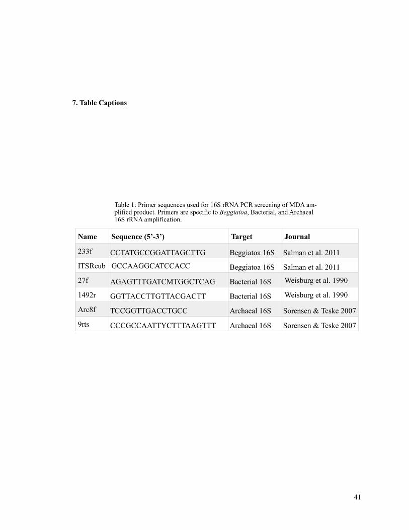
!!!! !7. Table Captions
!41

!
!42

!
!43

!!!!!!
!!!!
!44

!
!45
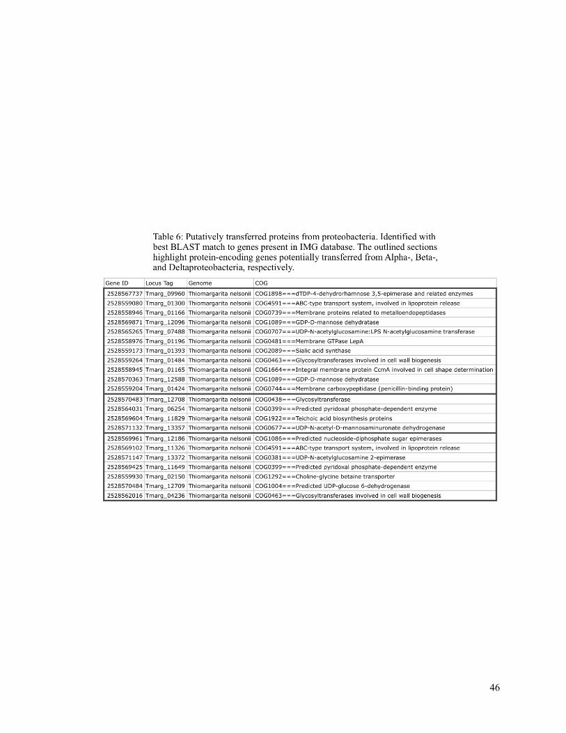
!
!46

!
!47

!
!48

!
!49

!!! 8. Figure Captions
!50

!
!51

!
!52
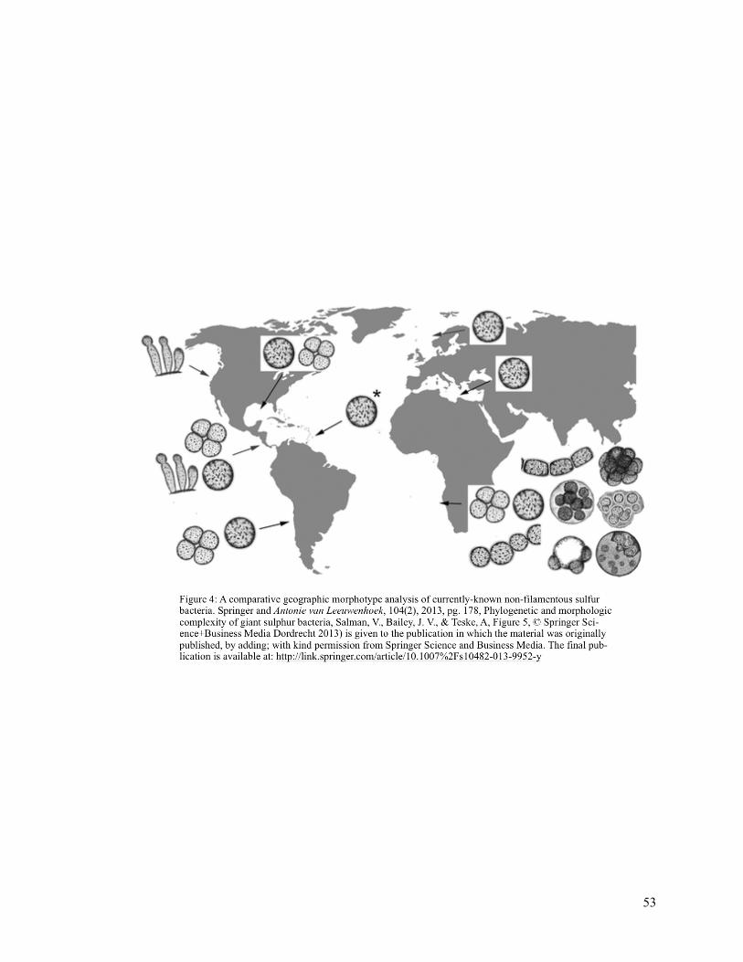
!!!! !!
!53

!
!54

!
!55

!
!56

!57