therosmall identification of the rornas · proc. nati. acad. sci. usa vol. 81, pp. 1996-2000, april...
TRANSCRIPT

Proc. Nati. Acad. Sci. USAVol. 81, pp. 1996-2000, April 1984Biochemistry
The Ro small cytoplasmic ribonucleoproteins: Identification of theantigenic protein and its binding site on the Ro RNAs
(small cytoplasmic RNAs/rheumatic disease/autoantibodies/RNA-protein interactions)
SANDRA L. WOLIN AND JOAN A. STEITZDepartment of Molecular Biophysics and Biochemistry, Yale University, P.O. Box 3333, New Haven, CT 06510
Contributed by Joan A. Steitz, December 19, 1983
ABSTRACT Patients with systemic lupus erythematosusand other rheumatic diseases often possess autoantibodies di-rected against discrete classes of small ribonucleoprotein parti-cles (RNPs). The class of particles recognized by anti-Ro anti-bodies contains from two to four small cytoplasmic RNAs, de-pending on the mammalian species examined. We find that anantigenic polypeptide of 60 kDa is the major protein residingin Ro RNPs from human HeLa cells. To determine what com-mon feature of Ro RNA sequence or structure is recognized bythe Ro protein, we carried out ribonuclease protection experi-ments on isolated Ro RNPs from HeLa cells. For each of thethree human Ro RNAs whose sequence is known, the mosthighly protected portion found in immunoprecipitates corre-sponded to the lower section of a stem formed by base-pairingthe 5' and 3' ends of the RNA. Within this protected helix is ahighly conserved region composed of seven identical base pairswith a single bulged cytidine. We discuss possible functions forthe Ro RNPs.
Anti-Ro antibodies from patients with systemic lupus erythe-matosus precipitate several small cytoplasmic ribonucleo-proteins (scRNPs) from mammalian cells (1). The RNA com-ponents of these particles, designated hYl-hY5 in humancells and mY1 and mY2 in mouse cells, range in size from 83to 112 nucleotides (2-4). Sequences have been reported forthree of the four unique human Ro RNAs (hY2 is a process-ing or degradation product of hYl). They exhibit many se-quence and secondary structure homologies (3, 4). Each RoRNA is present in about 105 copies per cell, or about 1% thenumber of ribosomes. Because of their relatively low abun-dance, Ro scRNPs were not detected before the use of pa-tient antibodies as probes.Ro RNAs are also precipitated by another lupus antibody,
anti-La, which recognizes a 50-kDa protein that binds manynascent RNA polymerase III transcripts, including adenovi-rus VA RNAs, Epstein-Barr virus EBER RNAs, and pre-cursors to tRNA and SS rRNA (1, 2, 5-8). The Ro RNAs arealso synthesized by RNA polymerase III (4); reassembly ex-periments have demonstrated that a proportion of the RoscRNPs actually contain the La protein in addition to pro-tein(s) carrying the Ro determinant (2), although anti-Ro andanti-La antibodies frequently are present simultaneously inpatient sera (9).The exact number ot proteins associated with Ro scRNPs
has not been well defined. Proteins are required for antigeni-city as the isolated RNAs are not precipitable by anti-Roantibodies (1). In the mouse, Ro RNAs are found exclusivelyin anti-Ro-precipitable scRNPs, indicating that the majorityof Ro RNAs are bound by the Ro protein(s) (4).We show here that the major protein component of the Ro
scRNPs is a single antigenic polypeptide of 60 kDa. Ribonu-
clease protection experiments indicate that this protein bindsto a highly conserved feature of all Ro RNAs, a stem formedby pairing the 5' and 3' ends of the molecules.
MATERIALS AND METHODSCells, Extracts, and Sera. Cells were maintained as de-
scribed (4). Cell sonicates (5) were prepared for RNA analy-sis by labeling 2 x 10⁷ cells with 32P04 (50 ACi/ml; 1 Ci = 37GBq) in phosphate-free minimal essential medium (GIBCO)for 14-16 hr, or for protein analysis by labeling 1 x 10⁷ cellswith [35S]methionine (10 ,uCi/ml) in methionine-free minimalessential medium for 20 hr.
Sera from patients with systemic lupus erythematosus orrelated autoimmune disorders were provided by J. Hardin(Yale University), S. Malawista (Yale University), M.Reichlin (Oklahoma Medical Research Foundation, Oklaho-ma), M. Akizuki (Keio University, Tokyo), and G. McCarty(Georgetown University).Immunoprecipitation of Proteins and RNA. A procedure
based on that of Matter et al. (10) was used. Protein A Seph-arose CL-4B (Pharmacia) preswollen in NET-2 (150 mM so-dium chloride/ 10 mnM Tris HCl, pH 7.5/0.05% Nonidet P-40) was incubated with 2 Al of crude serum for 1 hr at roomtemperature, and then washed 3 times with NET-2. The anti-body-bound beads were incubated for 15 min at 40C with analiquot of labeled cell sonicate corresponding to 5 ml of cells.After three washes with NET-2, the bound material was ex-tracted either with NaDodSO4 gel sample buffer (for pro-teins) (11) or treatment with phenol/NaDodSO4 (phenol/chloroform/isoamyl alcohol, 50:50:1/0.1% NaDodSO4) (forRNA) as described by Lerner and Steitz (12).RNAs were fractionated on 15% polyacrylamide (acrylam-
ide/bisacrylamide, 27:1) gels in 7M urea/45 mM Tris borate,pH 8.3/1.25 mM EDTA. Bands were extracted by the crushand soak method (13). T1 and pancreatic ribonuclease fin-gerprints of eluted RNAs were prepared (14) using thin-layerhomochromatography on PEI 300 (Brinkmann) for the sec-ond dimension (12), and the resulting oligonucleotides weresubjected to secondary analysis (14).Proteins were analyzed by electrophoresis on 10% poly-
acrylamide/NaDodSO4 gels (11). Gels were soaked for 30min in 0.5 M sodium salicylate, dried, and autoradiographed.
Ribonuclease Protection Experiments. 32P-labeled immunecomplexes bound on protein A-Sepharose beads (see above)were resuspended in 500 ,u of NET-2 containing 5 mMMgCl2 and 40 ,ug of carrier yeast RNA. This mixture wasdigested for 15 min at 25°C with pancreatic ribonuclease atconcentrations ranging from 10 ,ug/ml to 1 mg/ml. The beadswere then washed 4 times with NET-2 and extracted withphenol/NaDodSO4. The nuclease-resistant RNA fragmentswere precipitated with ethanol in the presence of 20 ,g ofcarrier RNA, and electrophoresed as described above. As
Abbreviations: RNP, ribonucleoprotein; scRNP, small cytoplasmicribonucleoprotein.
1996
The publication costs of this article were defrayed in part by page chargepayment. This article must therefore be hereby marked "advertisement"in accordance with 18 U.S.C. §1734 solely to indicate this fact.

Biochemistry: Wolin and Steitz
controls, immunoprecipitates were extracted with phe-nol/NaDodSO4, precipitated with ethanol, resuspended inthe buffer described above and mixed with antibody boundon protein A-Sepharose beads prior to digestion with pancre-atic ribonuclease. The digested mixture was extracted withphenol/NaDodSO4 and processed as described above.
Protein Blots. Cytoplasmic extracts of HeLa cells (15)(provided by E. Gottlieb, Yale University) were separatedby electrophoresis on 10% polyacrylamide/NaDodSO4 gelsand the protein was transferred to nitrocellulose sheets (16,17). Nitrocellulose sheets were blocked overnight in 1% gel-atin in phosphate-buffered saline (50 mM KH2PO4, pH7.4/130 mM NaCl). The sheets were washed 3 tinips (5 mineach) in phosphate-buffered saline containing 0.05% TritonX-100 (buffer T), then incubated with antisera in buffer T for2 hr at room temperature. After 3 washes in buffer T, thesheets were probed with 125I-labeled protein A in buffer T,washed as described above, and autoradiographed.
Proc. Nati. Acad. Sci. USA 81 (1984) 1997
Ro scRNPs, immunoblots were prepared using a cytoplas-mic extract ofHeLa cells (15). Fig. 1B compares the proteinsrecognized by a serum containing anti-La antibodies (lane 6),a serum containing mostly anti-Ro antibodies but also low-titer anti-La antibodies (lane 4), and a normal (nonimmune)serum (lane 8). The serum containing high-titer anti-Ro anti-bodies reacts with a 60-kDa protein as well as the 50-kDa Laprotein (lane 4) (8, 18); normal serum (lane 8) and the serumcontaining only anti-La antibodies do not detect the 60-kDaprotein. These results indicate that the 60-kDa protein thatdominates anti-Ro immunoprecipitates is antigenic, and thatthe presence of bound RNA is not absolutely required for Roantigenicity. All seven anti-Ro sera blot a 60-kDa protein,although in certain cases it is necessary to use an anti-Roimmunoprecipitate as a concentrated source of antigenicprotein. All seven sera also show anti-La specificity (as seenin Lane 4) in immunoblots, although these secondary auto-antibodies are not usually evident from examining immuno-precipitated RNAs.
RESULTSAnti-Ro Sera Precipitate a Single Major Antigenic Protein
of 60 kDa from Mammalian Cell Extracts. To examine theprotein components of Ro scRNPs we labeled human HeLacells with [35S]methionine and incubated samples of a whole-cell extract with anti-Ro sera. The gel in Fig. 1A shows thatautoantibodies from four different Ro patients (lanes 3-6)and a Ro,La patient (lane 7) precipitate a common majorpolypeptide of -60 kDa, although additional bands unique toindividual sera are sometimes seen. Anti-Ro sera from sevendifferent patients all immunoprecipitate this protein. When amixture of 3H amino acids was used to label cells, no addi-tional polypeptides were seen (data not shown). The 60-kDaprotein appears conserved across mammalian species: a pro-tein that comigrates with the HeLa cell protein is precipitat-ed from mouse Friend erythroleukemia cells (data notshown) by anti-Ro antibodies.To determine whether the 60-kDa polypeptide seen in
immunoprecipitates corresponds to the antigenic moiety of
Ac _
'p
12 34 5678
B Ppt Immunoblots
0 I- 040 IC0
-x i
.ctr * X x0XrCaa-1
* 69
* -46
.3
-930
9 - 1
Amg/ml RNose
w0
B
5S
tRNA
xc
f-
.P
94- 967- _ I43- O
30- _
20- -
2 3 4 5 6 7 8
FIG. 1. Proteins reactive with anti-Ro autoantibodies. (A) 35S-labeled HeLa cell proteins contained in immunoprecipitates wereanalyzed as described in Materials and Methods. Lanes: 1, a lightexposure of total cell proteins; 2 and 9, '4C-methylated molecularsize marker proteins (in kDa); 3-6, proteins immunoprecipitated byanti-Ro sera from four different patients; 7, proteins immunoprecipi-tated by a patient serum characterized as containing both anti-Roand anti-La antibodies; 8, proteins immunoprecipitated by normal(nonimmune) human serum. (B) Immunoblot comparison of proteinsidentified by serum from a patient with anti-Ro antibodies (lane 4),serum from a patient with anti-La antibodies (lane 6), and a normal(nonimmune) human serum (lane 8). Lanes 1, 3, 5, and 7 contain125I-labeled molecular size marker proteins (in kDa). Lane 2, a 35S-labeled anti-Ro immunoprecipitate from a HeLa whole-cell soni-cate. The anti-Ro serum used in lane 4 is the same as that used in A,lane 6.
2 3 5.7.....
2 3 4 5 6 7 8
FIG. 2. RNA fragments protected by Ro protein from pancreaticribonuclease digestion. (A) Anti-Ro immunoprecipitates isolatedfrom equal portions of 32P-labeled HeLa whole-cell extracts weretreated with increasing amounts of pancreatic ribonuclease (0.01,0.06, 0.30, or 1.0 mg/ml), and the nuclease-resistant bound frag-ments were analyzed (lanes 3-6). A profile of total cellular RNAs(lane 1) and undigested Ro RNAs (lane 2) representing 1/5th of theamount of sample loaded in lanes 3-6 are also shown. Bands num-bered 1-10 were eluted and analyzed. XC and BP indicate the posi-tions of the xylene cyanole FF and bromophenol blue marker dyes.In a separate experiment, we compared nuclease-resistant frag-ments obtained from immunoprecipitates (lane 7) with fragments ob-tained from immunoprecipitates that were extracted with phenol(lane 8) prior to digestion with pancreatic ribonuclease (1.0 mg/ml).(B) Pancreatic ribonuclease fingerprints of nuclease-resistant bands4 and 7 were prepared, and the resulting oligonucleotides were ana-lyzed (12, 14). Electrophoresis was from right to left and homochro-matography was from bottom to top.
_O_
1998 Biochemistry: Wolin and Steitz
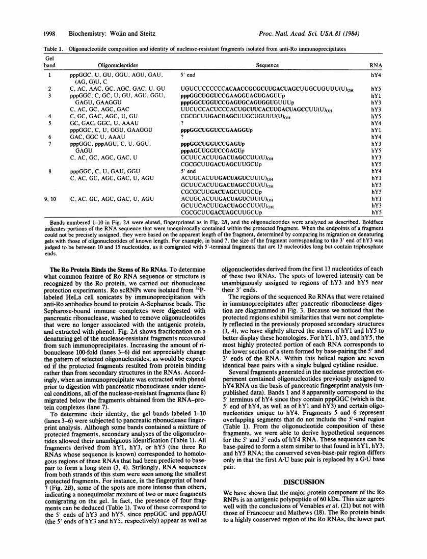
Table 1. Oligonucleotide composition and identity of nuclease-resistant fragments isolated from anti-Ro immunoprecipitates
Gelband Oligonucleotides Sequence RNA
1 pppGGC, U, GU, GGU, AGU, GAU, 5'end hY4(AG, G)U, C
2 C, AC, AAC, GC, AGC, GAC, U, GU UGUCUCCCCCCACAACCGCGCUUGACUAGCUUGCUGUUU(U)OH hY53 pppGGC, C, GC, U, GU, AGU, GGU, pppGGCUGGUCCGAAGGUAGUGAGUUp hYl
GAGU, GAAGGU pppGGCUGGUCCGAGUGCAGUGGUGUUUp hY3C, AC, GC, AGC, GAC UUCUCCACUCCCACUGCUUCACUUGACUAGCCUU(U)oH hY3
4 C, GC, GAC, AGC, U, GU CGCGCUUGACUAGCUUGCUGUUU(U)oH hY55 GC,GAC,GGC,UAAAU ? hY4
pppGGC, C, U, GGU, GAAGGU pppGGCUGGUCCGAAGGUp hY16 GAC, GGC U, AAAU ? hY47 pppGGC, pppAGU, C, U, GGU, pppGGCUGGUCCGAGUp hY3
GAGU pppAGUUGGUCCGAGUp hYSC, AC, GC, AGC, GAC, U GCUUCACUUGACUAGCCUU(U)oH hY3
CGCGCUUGACUAGCUUGCUp hY58 pppGGC, C, U, GAU, GGU 5' end hY4
C, AC, GC, AGC, GAC, U, AGU ACUGCACUUGACUAGUCUU(U)oH hY1GCUUCACUUGACUAGCCUU(U)oH hY3CGCGCUUGACUAGCUUGCUp hYS
9, 10 C, AC, GC, AGC, GAC, U, AGU ACUGCACUUGACUAGUCUU(U)oH hY1GCUUCACUUGACUAGCCUU(U)oH hY3CGCGCUUGACUAGCUUGCUp hY5
Bands numbered 1-10 in Fig. 2A were eluted, fingerprinted as in Fig. 2B, and the oligonucleotides were analyzed as described. Boldfaceindicates portions of the RNA sequence that were unequivocally contained within the protected fragment. When the endpoints of a fragmentcould not be precisely assigned, they were based on the apparent length of the fragment, determined by comparing its migration on denaturinggels with those of oligonucleotides of known length. For example, in band 7, the size of the fragment corresponding to the 3' end of hY3 wasjudged to be between 10 and 15 nucleotides, as it comigrated with 5'-terminal fragments that are 13 nucleotides long but contain triphosphateends.
The Ro Protein Binds the Stems of Ro RNAs. To determinewhat common feature of Ro RNA sequence or structure isrecognized by the Ro protein, we carried out ribonucleaseprotection experiments. Ro scRNPs were isolated from 32p_labeled HeLa cell sonicates by immunoprecipitation withanti-Ro antibodies bound to protein A-Sepharose beads. TheSepharose-bound immune complexes were digested withpancreatic ribonuclease, washed to remove oligonucleotidesthat were no longer associated with the antigenic protein,and extracted with phenol. Fig. 2A shows fractionation on a
denaturing gel of the nuclease-resistant fragments recoveredfrom such immunoprecipitates. Increasing the amount of ri-bonuclease 100-fold (lanes 3-6) did not appreciably changethe pattern of selected oligonucleotides, as would be expect-ed if the protected fragments resulted from protein bindingrather than from secondary structures in the RNAs. Accord-ingly, when an immunoprecipitate was extracted with phenolprior to digestion with pancreatic ribonuclease under identi-cal conditions, all of the nuclease-resistant fragments (lane 8)migrated below the fragments obtained from the RNA-pro-tein complexes (lane 7).To determine their identity, the gel bands labeled 1-10
(lanes 3-6) were subjected to pancreatic ribonuclease finger-print analysis. Although some bands contained a mixture ofprotected fragments, secondary analyses of the oligonucleo-tides allowed their unambiguous identification (Table 1). Allfragments derived from hY1, hY3, or hY5 (the three RoRNAs whose sequence is known) corresponded to homolo-gous regions of these RNAs that had been. predicted to base-pair to form a long stem (3, 4). Strikingly, RNA sequencesfrom both strands of this stem were seen among the smallestprotected fragments. For instance, in the fingerprint of band7 (Fig. 2B), some of the spots are more intense than others,indicating a nonequimolar mixture of two or more fragmentscomigrating on the gel. In fact, the presence of four frag-ments can be deduced (Table 1). Two of these correspond tothe 5' ends of hY3 and hY5, since pppGGC and pppAGU(the 5' ends of hY3 and hY5, respectively) appear as well as
oligonucleotides derived from the first 13 nucleotides of eachof these two RNAs. The spots of lowered intensity can beunambiguously assigned to regions of hY3 and hY5 neartheir 3' ends.The regions of the sequenced Ro RNAs that were retained
in immunoprecipitates after pancreatic ribonuclease diges-tion are diagrammed in Fig. 3. Because we noticed that theprotected regions exhibit similarities that were not complete-ly reflected in the previously proposed secondary structures(3, 4), we have slightly altered the stems of hY1 and hY5 tobetter display these homologies. For hY1, hY3, and hY5, themost highly protected portion of each RNA corresponds tothe lower section of a stem formed by base-pairing the 5' and3' ends of the RNA. Within this helical region are sevenidentical base pairs with a single bulged cytidine residue.
Several fragments generated in the nuclease protection ex-periment contained oligonucleotides previously assigned tohY4 RNA on the basis of pancreatic fingerprint analysis (un-published data). Bands 1 and 8 apparently correspond to the5' terminus of hY4 since they contain pppGGC (which is the5' end of hY4, as well as of hY1 and hY3) and certain oligo-nucleotides unique to hY4. Fragments 5 and 6 representoverlapping segments that do not include the 5'-end region(Table 1). From the oligonucleotide composition of thesefragments, we were able to derive hypothetical sequencesfor the 5' and 3' ends of hY4 RNA. These sequences can bebase-paired to form a stem similar to that found in hY1, hY3,and hY5 RNA; the conserved seven-base-pair region differsonly in that the first AU base pair is replaced by a G&U basepair.
DISCUSSIONWe have shown that the major protein component of the RoRNPs is an antigenic polypeptide of 60 kDa. This size agreeswell with the conclusions of Venables et al. (21) but not withthose of Francoeur and Mathews (18). The Ro protein bindsto a highly conserved region of the Ro RNAs, the lower part
Proc. NatL Acad Sci. USA 81 (1984)

Biochemistry: Wolin and Steitz
of a stem formed by base-pairing the 5' and 3' ends of themolecule.
Antigenic Proteins Contained in Ro scRNPs. Although the60-kDa protein is clearly the major protein component of RoscRNPs, other proteins may also be associated with theseparticles. A small percentage of the Ro RNPs contain the 50-kDa La protein, a polypeptide that binds at least initially tovirtually every known RNA polymerase III transcript. [Aprotein that comigrates with the La protein is visible as afaint band in 35S-labeled immunoprecipitates on long expo-sures (Fig. 1A, lanes 3-6).] Similarly, if other proteins wereassociated either transiently or permanently with only one ofthe less abundant Ro scRNPs, such as that containing hY1(4), they might not be detectable by immunoprecipitation oftotal cellular Ro particles.Our results suggest, but do not rigorously prove, that each
Ro RNA is contained in a separate antigenic complex. Theidentification of a binding site for the Ro protein on each ofthe four unique human Ro RNAs argues that each RNA mol-ecule is bound by at least one molecule of the antigenic pro-tein. Ro scRNPs sediment at =7 S in sucrose gradients (un-published data), consistent with a total molecular size ofabout 93 kDa for each Ro RNP (60 kDa for one protein mole-cule and 33 kDa for one RNA). This is somewhat lower thanthe 100- to 150-kDa size determined by gel filtration (22).The reaction of anti-Ro antibodies with the 60-kDa Ro pro-
tein in immunoblots is quite weak, in comparison to thatseen with other classes of autoimmune antisera (8, 17, 18, 21,23-25). This might be due to the low abundance of this anti-gen in cell extracts relative to other autoantigens, or to anintrinsic lability of the Ro antigenic determinant on exposureto NaDodSO4 and transfer to nitrocellulose. Alternatively,bound Ro RNA, which is eliminated by NaDodSO4 gel elec-trophoresis, might enhance Ro antigenicity.One curious observation made in our immunoblot studies
is that most anti-Ro sera contain detectable levels of anti-La
Proc. Natl. Acad. Sci. USA 81 (1984) 1999
antibodies. This is true even of sera that have been designat-ed "monospecific" anti-Ro based on immunodiffusion (T.Mimori, personal communication) as well as on immuno-precipitation of 32p- or 35S-labeled cell extracts. The frequentpresence of anti-Ro antibodies in patients with anti-La anti-bodies, but not the converse, has been noted (9). The factthat even our most "monospecific" anti-Ro sera recognize a50-kDa protein on immunoblots suggests that these sera docontain low levels of anti-La antibodies. Certain other speci-ficities have been noted to co-occur in patients with systemiclupus erythematosus, including anti-(Ul)RNP and anti-Sm(26), and antibodies to histones H1 and H2b (27). As in theRo-La case, the two autoantigens coexist in specific pro-tein-nucleic acid complexes (i.e., small RNPs or nucleo-somes), suggesting that the immune system may target theparticle as a whole. Alternatively, the two antigenic proteinsmight be related at the sequence or structural level.RNA Binding Site of the Ro Protein. The assignment of the
Ro protein binding site to the base of a stem formed by the 5'and 3' ends of Ro RNAs agrees well with data on the inclu-sion of shortened forms of these RNAs in anti-Ro precipita-ble particles. Earlier, we observed that hY2 and hY3*RNAs, which are slightly truncated forms of hY1 and hY3RNAs, respectively, are immunoprecipitable by anti-Roantibodies (4). (hY2 terminates between nucleotides 103 and107 of the hY1 sequence, while hY3* terminates betweennucleotides 92 and 96 of the hY3 sequence.) Conversely, aseverely shortened form of hY3 called hY3**, which termi-nates with a UOH between nucleotides 59 and 61, is not im-munoprecipitable (4) presumably because it totally lacks the3' portion of the stem to which Ro protein binds.The Ro protein binding site contains a single bulged nucle-
otide within a helix. This observation adds weight to the ideathat bulged helices may be a general structural feature of ri-bonucleic acid-protein binding sites (28). Uhlenbeck and co-workers (29, 30), studying the interaction of R17 coat protein
CC UU UC G
so-A UA UG C-70A C U C
A 50 AC C U
40-C C A G U C A G U U A G A UU G U U A G U U A A U C U A
U 30 C UU U
G C C C C20- A U
G CU A-90G CA UU AIGC U
UGoC4-IAA $)I-100
Cu
C GG U
pppG C U U(U)110
hYl
uc
C-so
u uu cU C
A U
G U-60A U
40 C GA
cc 50-A U c cC C AA A GUU U UUU G UU U C AA C
A AA 30 C UAu c
C
J-70
UC C
G C U
U A-so20- G C
G UU GIG C I
A U ,
G C @-U AG CA U
10- C U -90
~CC:
AiU
C GG C
pppG C U U(U)OH100
hY3
U GU AG UA U
30-A UU A-40U AG C
U U U
A
u c c c uG C
20-G C
G CU AG CU AU A 60oGC
GG C GU GIG C
uA
GO - 70
a uU A
U G_ G CpppA U U G C U G U U U(U)oH
so
hY5FIG. 3. Diagrammatic representation of regions of Ro RNAs protected from nuclease digestion in immunoprecipitates. Secondary struc-
tures of hY1 and hY5 (modified slightly from refs. 3 and 4) have calculated stabilization energies (-zAG) (19, 20) that are similar to the previouslyproposed structures (3, 4). The secondary structure of hY3 is from ref. 4. Solid lines indicate oligonucleotides that were unambiguouslyidentified in fingerprints of protected fragments. In some cases the endpoints of a fragment could not be precisely determined and are indicatedby broken lines. The stippled area identifies a region exactly conserved between hY1, hY3, and hY5 RNAs.
m

2000 Biochemistry: Wolin and Steitz PS
with its RNA, have constructed synthetic binding sites inwhich the single bulged adenine was deleted or replaced bycytidine. Both of these alterations greatly decreased coatprotein binding. More extensive studies of the Ro proteinbinding site, such as using in vitro mutagenesis to alter basesin this region, will be required to determine what features ofthe site are absolutely essential for protein recognition.
Finally, it must be noted that the ribonuclease protectionexperiments presented here can only identify those parts ofthe RNAs that remain tightly bound after digestion. Theremay be additional sites that contact the protein but are re-leased by nuclease treatment.
Cytoplasmic Roles for Ro scRNPs. Although the function ofthe Ro scRNPs has not yet been determined, it seems likelythat they participate in translation-related events. Three oth-er scRNPs have been recently assigned roles as positive ornegative effectors of protein synthesis in mammalian cells(31-33). Using cloned human Ro RNA genes (4) to probeRNA extracted from mouse and rat tissues, we have ob-served that Ro RNPs are about 10-fold more abundant, rela-tive to total cytoplasmic RNA, in brain and heart tissue thanthey are in liver (unpublished data). One intriguing possibili-ty is that Ro scRNPs function in the translation of a subset ofmRNAs abundant in brain and heart. It is perhaps relevantthat maternal anti-Ro antibodies are strongly associated withthe occurrence of atrioventricular conduction defects in neo-nates (34, 35). One prediction would be that the cells of theatrioventricular node, at least at some stage in development,have high concentrations of Ro scRNPs.
We thank H. LaBranche-Chabot for help with fingerprinting; Drs.S. Mount, 0. Uhlenbeck, H. Robertson, and J. Stefano for adviceand criticism; and E. Gottlieb for HeLa cell extracts. We are grate-ful to Drs. J. Hardin, S. Malawista, M. Reichlin, M. Akizuki, T.Mimori, and G. McCarty for providing patient sera; and to Drs. J.Carey and 0. Uhlenbeck for communication of results prior to publi-cation. This work was supported by a grant from the National Insti-tutes of Health (GM 26154) to J.A.S. and a U.S. Public Health Ser-vice training grant to S.L.W.
1. Lerner, M. R., Boyle, J. A., Hardin, J. A. & Steitz, J. A.(1981) Science 211, 400-402.
2. Hendrick, J. P., Wolin, S. L., Rinke, J., Lerner, M. R. &Steitz, J. A. (1981) Mol. Cell. Biol. 1, 1138-1149.
3. Kato, N., Hoshino, H. & Harada, F. (1982) Biochem. Biophys.Res. Commun. 108, 363-370.
4. Wolin, S. L. & Steitz, J. A. (1983) Cell 32, 735-744.5. Lerner, M. R., Andrews, N. C., Miller, G. & Steitz, J. A.
(1981) Proc. Natl. Acad. Sci. USA 78, 805-809.6. Rosa, M. D., Gottlieb, E., Lerner, M. R. & Steitz, J. A. (1981)
Mol. Cell. Biol. 1, 785-786.7. Rinke, J. & Steitz, J. A. (1982) Cell 29, 149-159.8. Steitz, J. A., Wolin, S. L., Rinke, J., Pettersson, I., Mount,
S. M., Lerner, E. A., Hinterberger, M. & Gottlieb, E. (1983)Cold Spring Harbor Symp. Quant. Biol. 47, 893-899.
9. Mattioli & Reichlin (1974) Arth. Rheum. 17, 421-429.10. Matter, L., Schopfer, K., Wilhelm, J. A., Nyffenegger, T.,
Parisot, R. F. & De Robertis, E. M. (1982) Arth. Rheum. 25,1278-1283.
11. Laemmli, U. K. (1970)-Nature (London) 227, 680-685.12. Lerner, M. R. & Steitz, J. A. (1979) Proc. Natl. Acad. Sci.
USA 76, 5495-5499.13. Maxam, A. M. & Gilbert, W. (1980) Methods Enzymol. 65,
499-560.14. Barrell, B. G. (1971) in Procedures in Nucleic Acid Research,
eds. Cantoni, G. L. & Davies, D. R. (Harper, New York),Vol. 2, pp. 751-779.
15. Weil, P. A., Segall, J. Harris, B., Ng, S.-Y. & Roeder, R. G.(1979) J. Biol. Chem. 254, 6163-6173.
16. Towbin, H., Staehelin, T. & Gordon, J. (1979) Proc. Nail.Acad. Sci. USA 76, 4350-4354.
17. Mimori, T., Hinterberger, M., Pettersson, I. & Steitz, J. A.(1984) J. Biol. Chem. 259, 560-565.
18. Francoeur, A. M. & Mathews, M. B. (1982) Proc. Natl. Acad.Sci. USA 79, 6772-6776.
19. Tinoco, I., Borer, P. N., Dengler, B., Levine, M. D., Uhlen-beck, 0. C., Crothers, D. M. & Gralla, J. (1973) Nature (Lon-don) New Biol. 246, 40-41.
20. Salser, W. (1977) Cold Spring Harbor Symp. Quant. Biol. 42,985-1002.
21. Venables, P. J. W., Smith, P. R., Maini, R. N. (1983) Clin.Exp. Immunol. 54, 731-738.
22. Clark, G., Reichlin, M. & Tomasi, T. B. (1976) J. Immunol.102, 117-122.
23. White, P. J. & Hoch, S. 0. (1981) Biochem. Biophys. Res.Commun. 102, 365-371.
24. Takano, M., Golden, S. S., Sharp, G. C. & Agris, P. F. (1981)Biochemistry 20, 5929-5936.
25. Billings, P. B., Allen, R. W., Jensen, F. C. & Hoch, S. 0.(1982) J. Immunol. 128, 1176-1180.
26. Mattioli, M. & Reichlin, M. (1973) J. Immunol. 110, 1318-1324.
27. Hardin, J. A. & Thomas, J. 0. (1983) Proc. Natl. Acad. Sci.USA 80, 7410-7414.
28. Peattie, D. A., Douthwaite, S., Garrett, R. A. & Noller, H. F.(1981) Proc. Nail. Acad. Sci. USA 78, 7331-7335.
29. Carey, J., Lowary, P. T. & Uhlenbeck, 0. C. (1983) Biochem-istry 22, 4723-4730.
30. Uhlenbeck, 0. C., Carey, J., Romaniuk, P., Lowary, P. &Beckett, D. (1983) J. Biomol. Struct. Dyn. 1, 539-552.
31. Thimmappaya, B., Weinberger, C., Schneider, R. J. & Shenk,T. (1983) Cell 31, 543-551.
32. Bhat, R. A. & Thimmappaya, B. (1983) Proc. Natl. Acad. Sci.USA 80, 4789-4793.
33. Walter, P. & Blobel, G. (1982) Nature (London) 299, 691-698.34. Scott, J. S., Maddison, P. J., Taylor, P. V., Esscher, E.,
Scott, 0. & Skinner, R. P. (1983) N. Engl. J. Med. 309, 209-212.
35. Weston, W. L., Harmon, C., Peebles, C., Manchester, D.,Franco, H. L., Huff, J. C. & Norris, D. A. (1982) Br. J. Der-matol. 107, 377-382.
Proc. NatL Acad Sci. USA 81 (1984)