thec-terminal50aminoacidresiduesofdenguens3 ... · by competitive ns3-ns5 interaction elisa that...
TRANSCRIPT
The C-terminal 50 Amino Acid Residues of Dengue NS3Protein Are Important for NS3-NS5 Interaction and ViralReplication*
Received for publication, September 1, 2014, and in revised form, December 5, 2014 Published, JBC Papers in Press, December 8, 2014, DOI 10.1074/jbc.M114.607341
Moon Y. F. Tay‡, Wuan Geok Saw§, Yongqian Zhao‡¶, Kitti W. K. Chan‡, Daljit Singh‡, Yuwen Chong‡,Jade K. Forwood�, Eng Eong Ooi‡, Gerhard Gruber§, Julien Lescar**, Dahai Luo‡‡, and Subhash G. Vasudevan‡¶1
From the ‡Program in Emerging Infectious Diseases, Duke-National University of Singapore Graduate Medical School, 8 CollegeRoad, Singapore 169857, Singapore, the §School of Biological Sciences, Nanyang Technological University, 60 Nanyang Drive,Singapore 637551, Singapore, the ¶NUS Graduate School for Integrative Sciences and Engineering, National University ofSingapore, 28 Medical Drive, Singapore 117456, Singapore, the �School of Biomedical Sciences, Charles Sturt University, WaggaWagga, New South Wales 2650, Australia, the **Division of Structural Biology and Biochemistry, School of Biological Sciences,Nanyang Technological University, Singapore 138673, Singapore, and the ‡‡Lee Kong Chian School of Medicine, NanyangTechnological University, 61 Biopolis Drive, Proteos Building, 07-03, Singapore 138673, Singapore
Background: NS3-NS5 interaction is important for the dengue virus life cycle.Results: NS3 residue Asn-570 is essential for its interaction with NS5; mutation in an infectious cDNA abolished virus produc-tion and reduced positive-strand RNA synthesis.Conclusion: NS3-NS5 interaction may be required for coordinated positive- and negative-strand RNA synthesis.Significance: NS3-NS5 interaction may be a target for rational design of antiviral drugs.
Dengue virus multifunctional proteins NS3 protease/helicaseand NS5 methyltransferase/RNA-dependent RNA polymeraseform part of the viral replication complex and are involved inviral RNA genome synthesis, methylation of the 5�-cap of viralgenome, and polyprotein processing among other activities.Previous studies have shown that NS5 residue Lys-330 isrequired for interaction between NS3 and NS5. Here, we showby competitive NS3-NS5 interaction ELISA that the NS3 pep-tide spanning residues 566 –585 disrupts NS3-NS5 interactionbut not the null-peptide bearing the N570A mutation. Smallangle x-ray scattering study on NS3(172– 618) helicase andcovalently linked NS3(172– 618)-NS5(320 –341) reveals a rigidand compact formation of the latter, indicating that peptideNS5(320 –341) engages in specific and discrete interaction withNS3. Significantly, NS3:Asn-570 to alanine mutation intro-duced into an infectious DENV2 cDNA clone did not yielddetectable virus by plaque assay even though intracellular dou-ble-stranded RNA was detected by immunofluorescence. Detec-tion of increased negative-strand RNA synthesis by real timeRT-PCR for the NS3:N570A mutant suggests that NS3-NS5interaction plays an important role in the balanced synthesis ofpositive- and negative-strand RNA for robust viral replication.Dengue virus infection has become a global concern, and thelack of safe vaccines or antiviral treatments urgently needs to beaddressed. NS3 and NS5 are highly conserved among the fourserotypes, and the protein sequence around the pinpointedamino acids from the NS3 and NS5 regions are also conserved.
The identification of the functionally essential interactionbetween the two proteins by biochemical and reverse geneticsmethods paves the way for rational drug design efforts to inhibitviral RNA synthesis.
Dengue virus (DENV),2 of which there are four distinct sero-types (DENV1– 4), is an important re-emerging mosquito-borne flavivirus that is endemic in more than 100 countries,causing �390 million human infections that result in �100million dengue fever (DF) cases (1). Infections with DENV areeither asymptomatic or can result in self-limiting febrile illness(DF) that leads to a broad spectrum of pathologies, includingsevere DF without hemorrhagic symptoms, dengue hemor-rhagic fever, or dengue shock syndrome. The current treatmentfor dengue infection is mainly supportive, and there is no pre-ventative vaccine or effective antiviral agents to treat DF or themore severe disease manifestations.
The �11-kb positive-sense single-stranded RNA genome ofDENV serotypes share around 70% sequence identity and con-tain a 5�-type-1 cap as well as 5�- and 3�-untranslated regions(UTR) that flank a single open reading frame (ORF) (2). An�3300-amino acid polyprotein precursor is translated from theORF and is processed by both host and viral proteases to yieldthree structural proteins (capsid, premembrane protein, andenvelope protein (E)) and seven nonstructural proteins (NS)(NS1, NS2A, NS2B, NS3, NS4A, NS4B, and NS5) that are
* This work was supported by the Duke-National University of Singapore Sig-nature Research Program (funded by the Ministry of Health, Singapore),National Medical Research Council, Singapore, Grants NMRC/1315/2011and NMRC/TCR/005/2008, and the Ministry of Education MOE Tier 3 GrantMOE2012-T3-1-008, Singapore.
1 To whom correspondence should be addressed. Tel.: 65-6516-6718; Fax:65-6221-2529; E-mail: [email protected].
2 The abbreviations used are: DENV, Dengue virus; SAXS, small-angle x-rayscattering; RdRP, RNA-dependent RNA polymerase; P/S, penicillin/strepto-mycin; bNLS, importin b-mediated nuclear localization sequence; NSD,normalized spatial discrepancy; NSD, normalized spatial discrepancy; IFA,immunofluorescence assay; EOM, ensemble optimization method; E,envelope protein; DF, dengue fever; RC, replication complex; Y2H, yeasttwo-hybrid.
THE JOURNAL OF BIOLOGICAL CHEMISTRY VOL. 290, NO. 4, pp. 2379 –2394, January 23, 2015© 2015 by The American Society for Biochemistry and Molecular Biology, Inc. Published in the U.S.A.
JANUARY 23, 2015 • VOLUME 290 • NUMBER 4 JOURNAL OF BIOLOGICAL CHEMISTRY 2379
by guest on April 7, 2020
http://ww
w.jbc.org/
Dow
nloaded from
involved in formation of mature virion and viral RNA replica-tion, respectively. Among the NS proteins, NS3 and NS5 con-tain the enzymatic activities that are essential for DENV repli-cation (3).
Both NS3 (residues 1– 618) and NS5 (residues 1–900) arelarge multifunctional proteins with sequence identity of around60 and 70% among the four serotypes, respectively (3). NS3contains an N-terminal serine protease domain (residues1–170) that requires NS2B to be an active protease (4 – 8). ItsC-terminal domain contains ATPase/helicase activity forunwinding of the double-stranded RNA (dsRNA) intermediate(9 –14) and RNA 5�-triphosphatase activity for viral RNA5�-capping reaction that is carried out together with the N-ter-minal domain of NS5, which has methyltransferase activity (3,9, 13, 15–22). The C-terminal domain of NS5 has RNA-depen-dent RNA polymerase (RdRP) activity, which is crucial for RNAreplication (19, 23–26). NS3 and NS5 have been shown to inter-act and colocalize in infected cells, and NS3 RNA 5�-triphos-phatase activity has been reported to be stimulated by NS5 invitro (9, 27–29). These observations are consistent with thefunctional roles of both NS3 and NS5 in the replication com-plex (RC) (30, 31).
During viral RNA replication within the RC, many criticalRNA-RNA, RNA-protein, and protein-protein interactionsoccur to synthesize both positive- and negative-strand viralRNA (31, 32). There have been several reports of NS3-NS5interactions that include biochemical pulldown assays frominfected cell extracts (28, 32–34) and two-hybrid (Y2H) studiesthat mapped the interaction to the C-terminal region of NS3helicase (residues 303– 618) and the N-terminal region of NS5RdRP (residues 320 –368; known as bNLS (nuclear localizationsequence)) (35, 36). The NS5-binding site appears to be cen-tered at residue Lys-330 because the mutation to alanine dis-rupted its interaction with NS3 and abolished RNA replication,although the in vitro RdRP activity was unaffected (37). Basedon available crystal structures of the RdRP domain of NS5, it hasbeen proposed that the cavity occupied by Lys-330 may be apotential target for antiviral drug design by blocking NS3-NS5interaction (37). However, the details of the interaction from
the NS3 perspective is missing to fully exploit structure-guideddrug design.
In this study, through the use of both NS3 WT and mutantpeptides in competitive NS3-NS5 interaction ELISA, we iden-tified a conserved amino acid in subdomain III of DENV NS3protein, Asn-570, as being critical for its interaction with NS5.Mutation of NS3:Asn-570 to alanine in the DENV2 cDNAclone abolished infectious virus production and reduced viralprotein production and RNA replication. This mutation alsosuggests that the NS3-NS5 interaction is essential for viral RNAreplication by possibly coordinating positive- and negative-strand synthesis. Small angle x-ray scattering (SAXS) dataof NS3 helicase (residues 172– 618) covalently linked toNS5(320 –341) supports the observation that physical interac-tion occurs in the region of interaction between NS3 and NS5.
EXPERIMENTAL PROCEDURES
Cell Lines and Virus—Baby hamster kidney cells (BHK-21)were cultured in RPMI 1640 medium (Invitrogen) supple-mented with 10% fetal bovine serum (FBS), 100 units/ml peni-cillin, and 100 �g/ml streptomycin (1% P/S) in 5% CO2 at 37 °C.
Human hepatoma (Huh-7) cells were cultured in DMEM(Invitrogen) supplemented with 10% FBS and 1% P/S at 37 °C,with 5% CO2. Aedes albopictus mosquito (C3/36) cells werecultured in RPMI 1640 medium supplemented with 25 mM
HEPES, 10% FBS, and 1% P/S at 28 °C, in the absence of CO2.DENV2 of cosmopolitan genotype (GenBankTM number
EU081177.1) that was used in this study was grown in C3/36cells and titered in BHK-21 cells before storage at �80 °C. Thisvirus was isolated during a local dengue outbreak that occurredin 2005 as part of Early Dengue Infection and Outcome (EDEN)Study in Singapore (38).
Plasmid Construction—Plasmids for the expression ofDENV3 NS5 RdRP (residues 273–900), NS3 full-length protein(NS2B18NS3; residues 49 – 66 of NS2B and 1– 618 of NS3 con-nected by a flexible linker G4SG4), and DENV4 NS3 helicasedomain (NS3(172– 618)) have been described previously (Fig.1A and Table 1) (11, 25, 39).
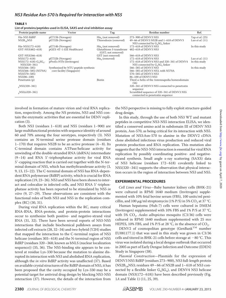
TABLE 1List of proteins/peptides used in ELISA, SAXS and viral inhibition assay
Protein/peptide name Vector Tag Residue number Ref.
His-NS5 RdRP pET15b (Novagen) His6 (not removed) 273–900 of DENV3 NS5 Yap et al. (25)NS2B18NS3 pET32b (Novagen) Thioredoxin (removed) 49–66 of DENV3 NS2B and 1–618 of DENV3
NS3 connected by G4SG4 linkerLuo et al. (11)
His-NS3(172–618) pET14b (Novagen) His6 (not removed) 172–618 of DENV3 NS3 In this studyGST-NS3(482–618) pGEX-4T-1 (GE Healthcare) Glutathione S-transferase
(GST; not removed)482–618 of DENV3 NS3
GST-NS3(566–618) GST (not removed) 566–618 of DENV3 NS3NS3(172–618) pET32b (Novagen) His6 (removed) 172–618 of DENV4 NS3 Luo et al. (11)NS3(172–618)-G4SG4-
NS5(320–341)pProEx HTb (Invitrogen) 172–618 of DENV4 NS3 and 320–341 of DENV4
NS5 connected by G4SG4 linkerIn this study
NS3(566–585) Synthesized by NTU peptide synthesiscore facility (Singapore)
566–585 of DENV3 NS3 In this studyNS3(566–585) (N570A) 566–585 of DENV3 NS3, with N570ANS3(570–585) 570–585 of DENV3 NS3NS3(86–100) 86–100 of DENV3 NS3Penetratin (p) Biotin Third �-helix of the Antennapedia homeodomain
(56)pNS5(320–341) 320–341 of DENV3 NS5 connected to penetratin
sequencepNS5(s320–341) Scrambled sequence of 320–341 of DENV3 NS5
connected to penetratin sequence
NS3 Residue Asn-570 Is Required for Interaction with NS5
2380 JOURNAL OF BIOLOGICAL CHEMISTRY VOLUME 290 • NUMBER 4 • JANUARY 23, 2015
by guest on April 7, 2020
http://ww
w.jbc.org/
Dow
nloaded from
DENV3 NS3(172– 618), NS3(482– 618), and NS3(566 – 618)fragments were amplified from DENV3 NS2B18NS3 construct(as mentioned above). NS3(172– 618) fragment was amplifiedwith forward primer 5�-CAAACAAATGCACATATGGATG-GACCGACACC-3� and the reverse primer 5�-GTGCTCGAG-TGCGGATCCAAGCTTTTACTTTC-3. The underlined sequencecorresponds to NdeI and BamHI sites, respectively. The PCRfragment was digested with NdeI and BamHI and cloned intopET14b (Novagen, Germany). The NS3(482– 618) fragmentwas amplified with the forward primer 5�-CCAGCCTCTCG-GATCCGATGAAGACCATG-3� and the reverse primer5�-CACAAGATCAAGCTCGAGTCACTTTCTGCC-3�. TheNS3(566–618) fragment was amplified with the forward primer5�-GAAAATGGTGCGGATCCGGGCAACGCAATAATC-3�and the same reverse primer as above. The underlined sequencecorresponds to BamHI and XhoI sites, respectively. Both PCRproducts were digested with BamHI and XhoI and cloned intopGEX-4T-1 (GE Healthcare) (Fig. 1A and Table 1).
DENV4 NS3(172– 618)-NS5(320 –341) fragment (con-nected by a flexible linker G4SG4, Fig. 3A and Table 1) wasamplified from DENV4 NS2B18NS3 construct (as mentionedabove). The full-length PCR product was generated by first ampli-fying the helicase region with the forward primer 5�-GAAAAC-CTGTATTTTCAGGGCGCCGGCGGTGAGCCAGATTAT-GAAGTGG-3� and the reverse primer 5�-CTCCATTCACCA-TTCCTCCTCCTCCTGATCCTCCTCCTCCCTTTCTTCC-ACTGGC-3�, followed by two separate PCRs that added theflexible linker sequence and NS5(320 –341) sequence to thehelicase via two reverse primers, 5�-GGAATCACAT-CCCAGGGTTTTGTCAGCAGTTTCACCACTCCATTCA-CCATTCCTC-3� and 5�-GCGGCCGCGACTAGTGAGCTC-GTCGACTTACTGGGTCACCATTGGAATCACATCCCA-GGG-3�. The underlined sequence corresponds to NaeI and SalIsites, respectively. The PCR product was digested with NaeI andSalI and cloned into pProEx HTb vector.
Mutation of NS3 Asn-570 to alanine in DENV3 NS2B18NS3was done using QuikChange II XL site-directed mutagenesis kit(Stratagene), according to the manufacturer’s protocol. The fol-lowing primers were used: NS3 N570A forward (5�-GATGGGC-AACGCAATGCTCAAATTTTAGAGGAG-3�) and reverse (5�-CTCCTCTAAAATTTGAGCATTGCGTTGCCCATC-3�). Theunderlined nucleotide corresponds to the mutation that was beingmade. Mutations were confirmed by automated DNA sequencing.
Protein Expression and Purification—For ELISA and ATPaseactivity assay, NS3 and NS5 constructs (Table 1) were trans-formed into Escherichia coli BL21 CodonPlus (DE3)-RIL cells(Stratagene) for protein expression and purified as previouslypublished (11, 25, 39).
For SAXS, NS3(172– 618) and NS3(172– 618)-NS5(320 –341) constructs were transformed into E. coli BL21 CodonPlus(DE3)-RIL cells (Stratagene) for protein expression and puri-fied as described below. The cells were disrupted on ice by son-ication three times for 1 min in buffer A (20 mM sodium phos-phate, pH 7.4, 500 mM NaCl, 0.8 mM DTT, and 2 mM PefablocSC
(Biomol)). Precipitated material was separated by centrifuga-tion at 12,500 � g for 25 min, and the supernatant was filtered(0.45 �m; Millipore). The filtered supernatant was incubatedwith 2 ml of nickel-nitrilotriacetic acid-agarose (Qiagen) for
1.5 h at 4 °C, and the His6-tagged protein was eluted with animidazole gradient from 20 to 200 mM in buffer A. Fractionscontaining the protein of interest were pooled and subjected toovernight cleavage with thrombin for NS3(172– 618) ortobacco etch virus protease for NS3(172– 618)-NS5(320 –341)and dialyzed overnight in buffer B (20 mM sodium phosphate,pH 7.4, 200 mM NaCl) at 4 °C. Following incubation, the dia-lyzed sample was incubated with 1 ml of nickel-nitrilotriaceticacid-agarose for 1 h at 4 °C, and the flow-through containingcleaved NS3(172– 618) or NS3(172– 618)-NS5(320 –341) wascollected and applied to a gel filtration column (SuperdexTM
200 HR 10/300 column, GE Healthcare) in buffer C (20 mM
Tris/HCl, pH 7.4, 200 mM NaCl, 5% glycerol, 1 mM DTT). Frac-tions containing NS3(172– 618) or NS3(172– 618)-NS5(320 –341) were pooled and concentrated using Amicon Ultra-4 cen-trifugal unit (10-kDa molecular mass cutoff; Millipore).
Peptide Synthesis—Peptides were synthesized at the Nan-yang Technological University peptide synthesis core facility(Singapore).
Competitive NS3-NS5 Interaction ELISA—The ELISA (Fig.1B) was performed as published previously (33). The concen-tration of NS2B18NS3 was fixed at 60 or 80 nM and mixed withincreasing concentrations of truncated NS3 proteins (DENV3NS3(172– 618), GST-NS3(482– 618), GST-NS3(566 – 618), orGST (negative control)) or NS3 peptides (NS3(566 –585) andNS(566 –585) (N570A), NS(571–585), or NS(86 –100) (nega-tive control); listed in Table 1). The concentration of NS3 proteinat which 50% inhibition of NS2B18NS3 protein binding occursrepresents the apparent KD value for the DENV3 NS5 RdRP-NS3protein interaction. Data were fitted to the sigmoidal dose-re-sponse equation (variable slope) by nonlinear regression usingGraphPad Prism 5 from triplicate measurements.
Viral Inhibition Assay—2 � 105 Huh-7 cells were seeded intoa 12-well plate and incubated overnight at 37 °C with 5% CO2.Cells were infected with DENV2 at a multiplicity of infection of1 for 1 h, after which the virus inocula were then removed andreplaced with 5% FBS/DMEM maintenance media. At 6 h post-infection, the infected cells were treated with 7.5 �M NS3 pep-tides complexed with 22.5 �M penetratin in a molar ratio of 1:3or 7.5 �M NS5-penetratin fusion peptides (Table 1). At 24 hpost-infection, cells were washed once with PBS prior to lysis byTRIzol for cellular viral RNA quantification by real time RT-PCR analysis with primers that binds to the NS1 gene (forward5�-CCGCTGACATGAGTTTTGAGTC-3� and reverse 5�-CATGACAGGAGACATCAAAGGA-3�) (40).
ATPase Activity Assay—The assay was carried out as described(13), with slight modifications. Purified NS2B(320 –368)NS3WT or N570A protein of 2.5 nM was preincubated at 37 °C withpoly(U) (10 �g/ml) in 40 �l of reaction buffer (50 mM Tris/HCl,pH 7.5, 2 mM MgCl2, 1.5 mM dithiothreitol, 0.05% Tween 20,0.25 �g/ml bovine serum albumin (Sigma)) for 5 min. The reac-tion was initiated by the addition of 10 �l of varying ATP con-centrations (2-fold serial dilution, starting from 2000 �M) andcarried out for 10 min at 37 °C. 10 �l of malachite green reagent(BioAssay Systems) was added to stop the reaction. Absorbancewas read at 635 nm after 30 min at room temperature. The Kmof the protein was determined with GraphPad Prism 5, withMichaelis-Menten Equation 1,
NS3 Residue Asn-570 Is Required for Interaction with NS5
JANUARY 23, 2015 • VOLUME 290 • NUMBER 4 JOURNAL OF BIOLOGICAL CHEMISTRY 2381
by guest on April 7, 2020
http://ww
w.jbc.org/
Dow
nloaded from
Vo � �Vmax�S/�Km � �S (Eq. 1)
SAXS—SAXS data of the NS3(172– 618) and NS3(172– 618)-NS5(320 –341) were measured by the NanoStarTM instrument(Bruker), equipped with a METALJETTM x-ray source andVantec 2000-detector system. The METALJETTM source usesthe liquid gallium source to deliver a high intensity x-ray beamat the wavelength of � � 1.34 Å. The SAXS measurements werecarried out with the source to sample distance of 145 cm, atwo-pinhole collimation system, and the sample to detectordistance of 67 cm (41). SAXS experiments of both proteins werecarried out at 1.2, 2.2, and 4 mg/ml in a sample volume of 40 �lat 15 °C. For each sample, a total of nine measurements at5-min intervals were recorded. The data were flood-field andspatially corrected, and processed using the built-in SAXS soft-ware. We tested the possible radiation damage by comparing alldata sets, and no changes were detected. The scattering inten-sity of the buffer was subtracted, and the difference curves werescaled for the concentration. All the data processing steps wereperformed automatically using the program package PRIMUS(42). The forward scattering I(0) and the radius of gyration Rgwere evaluated using the Guinier approximation (43). Theseparameters were also computed from the entire scattering pat-terns using the indirect transform package GNOM (44), whichalso provides the distance distribution function p(r). Tenlow resolution models of NS3(172– 618) or NS3(172– 618)-NS5(320 –341) were independently built by the programGASBOR (45). The spatial discrepancy (NSD), which is a mea-sure of similarity between sets of three-dimensional points,was computed between all 10 reconstructions using theDAMAVER program (46). The reconstruction with the leastNSD was selected for NS3(172– 618) or the fusion proteinNS3(172– 618)-NS5(320 –341). The ensemble optimizationmethod (EOM) suite was used to select an ensemble of confor-mations that best fit the experimental data, and the dimensionsof selected conformations were compared with the randompool to evaluate the flexibility and compactness of NS3(172–618)-NS5(320 –341) (47, 48).
DENV2 Full-length cDNA Clone Construction and Site-di-rected Mutagenesis—To construct a full-length DENV2 cDNAclone (GenBankTM accession EU081177.1, cosmopolitan geno-type; Fig. 4A), low passage virus stock was subjected to viralRNA extraction by RNeasy kit (Qiagen), and three cDNA frag-ments (fragment boundaries indicated by nucleotide numbers;Fig. 4A) covering the complete genome were amplified fromviral RNA by RT-PCR using SuperScript III one-step RT-PCRkits (Invitrogen). Fragment 1 contained the SphI restrictionsite, a T7 promoter sequence, and DENV2 cDNA nucleotides1– 4498, which also contained the KpnI restriction site. Frag-ment 2 spanned from the KpnI site (nucleotide position 4493)to the XbaI site (nucleotide position 6008). Fragment 3 spannedfrom XbaI site (nucleotide position 6003) to the 3� end of thegenome (nucleotide position 10,723), containing the SacI site atthe end of the genome. The PCR product of each cDNA frag-ment was digested and cloned into a pre-digested and modifiedlow copy number plasmid pWSK29 (49). The plasmid wasmodified by the replacement of the BssHII site that was locatedbefore the T7 promoter with the SphI site by site-directed
mutagenesis of the plasmid with the following primer: forward5�-GGCCAGTGAGCATGCGTAATACGAC-3� and reverse5�-GTCGTATTACGCATGCTCACTGGCC-3�. The under-lined nucleotide corresponds to the mutation that was beingmade. The subclone that maintained each fragment was namedaccordingly as follows: pWSK29 D2 fragment 1, fragment 2,and fragment 3, respectively, and each subclone was validatedby DNA sequencing by 1st BASE DNA Sequencing Services(Singapore) before proceeding for assembly. Subsequently,fragment 2 was inserted into the subclone pWSK29 D2 frag-ment 1 at the KpnI and XbaI site to generate subclone pWSK29D2 fragment 1 � 2. Finally, fragment 3 was inserted to generatesubclone pWSK29 D2 fragment 1 � 2 at XbaI and SacI sites togenerate the full-length cDNA clone, pWSK29 D2 full length.The E. coli XL-1 Blue chemically competent cell (Stratagene)was used for construction and propagation of the cDNA clones.Standard cloning procedures were performed with the excep-tion that the cDNA clones were propagated at 30 °C for at least20 h. All restriction enzymes were purchased from New Eng-land Biolabs.
The genome-length cDNA clones with NS3:N570A and NS5:K330A mutations were constructed using the subclonepWSK29 D2 fragment 3. The mutations were generated usingQuikChange II XL site-directed mutagenesis kit (Stratagene)and performed according to the manufacturer’s protocol. Thefollowing primers were used for the generation of bothmutants: NS3:N570A forward (5�-CTTTGATGGAGTCAAG-AACGCCCAAATCTTGGAAGAAAATG-3�) and reverse (5�-CATTTTCTTCCAAGATTTGGGCGTTCTTGACTCCAT-CAAAG-3�); NS5:K330A forward (5�-GTGGTTAGGCTGCT-AACAGCACCTTGGGATGTCATCCCC-3�) and reverse (5�-GGGGATGACATCCCAAGGTGCTGTTAGCAGCCTAAC-CAC-3�). The underlined nucleotides correspond to themutation that was being made. Mutations were confirmed byautomated DNA sequencing, and fragment 3 bearing the muta-tion was excised from the plasmid by XbaI and SacI andinserted into subclone pWSK29 D2 fragments 1 � 2 that weresimilarly cut with XbaI and SacI.
In Vitro Transcription, RNA Electroporation, Plaque Assay,Real Time RT-PCR (Reverse Transcription-PCR), Immunofluo-rescence Assay (IFA), and Western Blot—BHK-21 cells weretrypsinized, washed twice with cold PBS, and resuspended inOpti-MEM (Invitrogen) at a cell density of 1 � 107 cells/ml. 10�g of in vitro transcribed RNA with T7 mMESSAGEmMACHINE kit (Ambion) of DENV2 WT and mutants weremixed with 800 �l of cell suspension in a pre-chilled 0.4-cmcuvette and electroporated at settings of 850 V and 25 micro-farads, 2 pulses with an interval of 3 s. Electroporated cells wereallowed to recover at room temperature for 10 min before resus-pending in complete RPMI 1640 medium for cell recovery. Cells(3 � 105) were then seeded into a 12-well plate and incubated at37 °C in the presence of 5% CO2. Media were changed to 2% FBSmaintenance media after 6 h post-transfection. Samples were har-vested every 24 h post-transfection until 120 h. Supernatants werecollected and clarified for titering of the infectious virus particle bystandard plaque assay and extracellular viral RNA quantificationby real time RT-PCR analysis (40). Cells were then washed oncewith PBS prior to lysis by TRIzol reagent (Invitrogen) or 1� SDS-
NS3 Residue Asn-570 Is Required for Interaction with NS5
2382 JOURNAL OF BIOLOGICAL CHEMISTRY VOLUME 290 • NUMBER 4 • JANUARY 23, 2015
by guest on April 7, 2020
http://ww
w.jbc.org/
Dow
nloaded from

PAGE reducing loading dye for cellular viral RNA quantificationand Western blot, respectively.
For cellular viral RNA quantification, total RNA was isolatedusing the TRIzol extraction method from the cell lysate. 500 ngof total RNA was used for cDNA synthesis using the Improm IIreverse transcription system (Promega) with random primer inaccordance to manufacturer’s instructions. 40 ng of cDNA wasused for real time RT-PCR analysis of viral RNA in Bio-Rad iQ-5real time thermal cycler with the use of SYBR Green supermix(Bio-Rad) as described previously with primers that bind to theNS1 gene (forward 5�-CCGCTGACATGAGTTTTGAGTC-3�and reverse 5�-CATGACAGGAGACATCAAAGGA-3�) (40).Absolute numbers of intracellular viral RNA genome copy werequantitated using the DENV standard curve, normalized to actinlevels, and reported as absolute number of viral RNA genome copyper �g of RNA used for real time RT-PCR.
For intracellular viral RNA genome copy of both positive andnegative strands, 5�-tagged primers that bind to the E gene(Eden2_forward_RT, 5�-GGCCGTCATGGTGGCGAATAACA-GGCTATGGCACTGTCACGAT-3�, and Eden2_reverse_RT,5�-GGCCGTCATGGTGGCGAATAACCATTTGCAGCAA-CACCATCTC-3�) were used for transcribing cDNAs of bothpolarities (50, 51). The forward primer was used to transcribecDNA from the negative-strand RNA, whereas the reverse primerwas used to transcribe cDNA from positive-strand RNA by usingImprom II reverse transcription system (Promega). 40 ng of cDNAwas used for real time RT-PCR analysis of viral RNA in Bio-RadiQ-5 real time thermal cycler with the use of SYBR Green super-mix (Bio-Rad) with the appropriate primer pair for either negative-strand (forward 5�-GGCCGTCATGGTGGCGAATAA-3� andreverse 5�-CCATTTGCAGCAACACCATCTC-3�) or positive-strand (forward 5�-GGCCGTCATGGTGGCGAATAA-3� andreverse 5�-CAGGCTATGGCACTGTCACGAT-3�) detection.The underlined sequence corresponds to the 5�-tagged sequence(51). Absolute positive- and negative-strand copy numbers werequantitated and reported as described above.
For extracellular viral RNA quantification, viral RNA fromthe supernatant was extracted by Qiagen viral RNA extractionkit according to the manufacturer’s instructions, and for itsquantification, a SYBR Green one-step real time RT-PCR (Bio-Rad) was conducted using the same PCR conditions as cellularRNA quantification, with primers that bind to the NS1 gene (asmentioned above). Absolute viral RNA genome copy was cal-culated based on the DENV standard curve generated andreported as absolute number of viral RNA genome copy per mlof supernatant. The detection limit of each real time RT-PCRassay is indicated as a gray line in the graph.
IFA against E protein by anti-E mouse antibody 4G2, NS3protein by anti-NS3 human antibody 3F8, and dsRNA by anti-dsRNA mouse antibody J2 (Scicons), and Western blot againstNS3 protein by anti-NS3 3F8 were performed as described pre-viously (33). IFA images were captured on an inverted fluores-cence microscope (Olympus IX71, Center Valley) at �20 mag-nification, and image analysis was performed with ImageJsoftware (52).
Statistical Analysis—Student’s t test was used to determinestatistical significance. p values of �0.05 were considered assignificant.
RESULTS
NS3(566 – 618) Interacts with NS5 RdRP—Even thoughNS3(303– 618) of the helicase domain (residues 172– 618, seeFig. 1A) was previously shown by Y2H study to interact withNS5(320 –368) (bNLS) (36) of the RdRP domain (residues 273–900) (35, 36), the amino acid residues of NS3 helicase that areresponsible for binding to NS5 RdRP have not been pinpointed.However, it was hypothesized that helicase subdomain III,NS3(482– 618), contains the interaction site (13, 22). To testthis, we first established that the binding of NS2B18NS3 tocoated NS5 RdRP increased in a dose-dependent manner asdescribed previously (33) and carried out the NS3-NS5 interac-tion assay in a competitive ELISA format, with an increasingamount of the following competing proteins, namely NS3(172–618), GST-NS3(482– 618) (subdomain III, or GST-NS3(566 –618) (Fig. 1, A and B). From these data, all three NS3 truncatedproteins were found to be able to compete with NS2B18NS3protein binding to coated NS5 RdRP (Fig. 1C). The apparent Kdvalues of the NS3-NS5 interaction for NS3(172– 618), GST-NS3(482– 618), and -(566 – 618) were found to be comparable(5.33 0.35, 10.18 0.91, and 10.25 1.14 �M (mean S.D.,n � 3) respectively) and also similar to the Kd values forNS3(172– 618) as in previous reports (33, 37). Next, we alsodetermined the IC50 value for NS3(172– 618), which was 6.07 2.60 �M. The IC50 values for GST-NS3(482– 618) and GST-NS3(566 – 618) were not determined as we were unable toreach 100% inhibition due to limited availability of truncatedprotein. Protein expression constructs spanning other NS3subdomain regions (NS3(172– 482), -(307– 619), and (172–566)) were also generated in this study, but due to protein stick-iness, protein instability, or low protein yield, these proteinscould not be purified to sufficient quantity for the competitionassay.
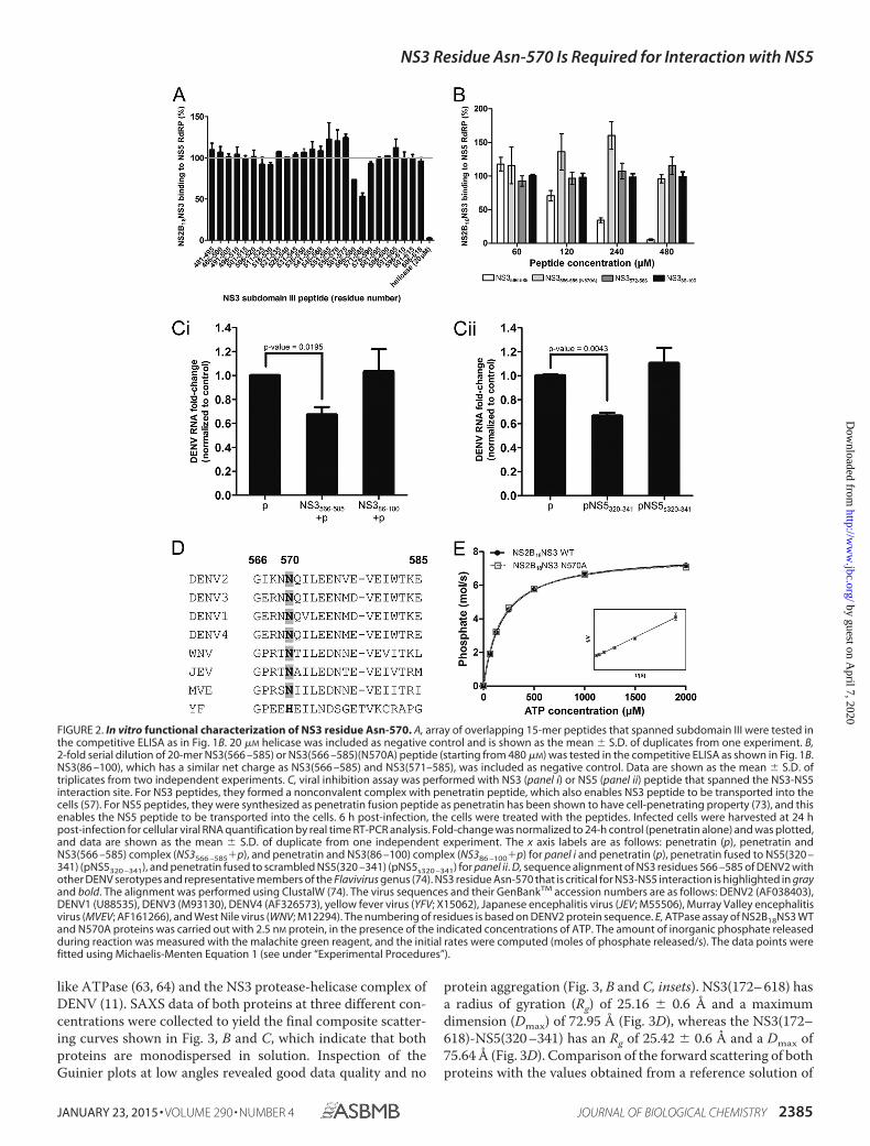
NS3 Asn-570 Is Critical for the Interaction—To map the NS3sequence that interacts with NS5 more precisely, we proceededto screen an array of overlapping 15-mer peptides (Mimotopes)(53, 54) that spanned subdomain III in the same competitiveELISA format (Fig. 2A), and we identified two peptides(NS3(566 –580) and -(571–585)) that moderately (p value �0.06 and 0.009, respectively) blocked NS3-NS5 interaction,thus narrowing down the interaction region to residues 566 –585 of NS3. We next tested the synthetic peptide NS3(566 –585) in ELISA and showed that it could also disrupt NS3-NS5interaction in a dose-dependent manner with an IC50 value of128.8 2.57 �M (Fig. 2B). At the same time, another peptide,NS3(86 –100) with a similar charge as NS3(566 –585), did notcompete. This indicates NS3-NS5 interaction involvessequence-specific residues on the NS3 protein. Additionally,the same peptide also inhibited NS3-NS5 interaction in theAlphaScreen assay format where the interacting partners weresynthesized in vitro using the wheat germ expression system(data not shown) (55).
Next, we also used the peptide in the viral inhibition assay todetermine whether blocking the interaction site could reduceviral replication (Fig. 2C). Uptake of NS3(566 –585) peptideinto infected cells was facilitated by a well characterized cell-penetrating peptide, penetratin (56), which forms a nonconva-
NS3 Residue Asn-570 Is Required for Interaction with NS5
JANUARY 23, 2015 • VOLUME 290 • NUMBER 4 JOURNAL OF BIOLOGICAL CHEMISTRY 2383
by guest on April 7, 2020
http://ww
w.jbc.org/
Dow
nloaded from

lent complex with the peptide (57). As shown in Fig. 2C,NS3(566 –585) peptide could reduce viral replication by �33%(p value � 0.0195, Fig. 2C, panel i). As a positive control, we alsotested the NS5(320 –341) peptide that was covalently linked topenetratin, which can also facilitate the uptake of the peptideinto infected cells (58). NS5(320 –341) peptide could alsoreduce viral replication by �33% (p value � 0.0043, Fig. 2C,panel ii). This suggests that blocking interaction between NS3and NS5 could be a potential therapeutic target.
Sequence alignment of NS3(566 –585) of DENV1– 4 andother flaviviruses (Fig. 2D) suggested that Asn-570 (highlightedin gray) is highly conserved within this region and may be crit-ical for the NS3-NS5 interaction. To test this, we synthesizedNS3(566 –585)(N570A) peptide and carried out the same com-petitive ELISA. We found that the replacement of asparagine byalanine at position 570 of NS3 resulted in a null-peptide withrespect to its ability to block the NS3-NS5 interaction (Fig. 2B).Next, we expressed and purified the NS3:N570A full-lengthprotein to measure its ATPase activity (59), and we found that itwas comparable with the WT NS3 protein (Fig. 2E). The affinity
of ATP for NS3 WT and NS3 N570A was similar (Km � 185.8 10.86 and 176.1 8.73 �M, respectively). Both proteins also hadsimilar turnover numbers (kcat � 3.15 and 3.11 s�1, respec-tively). From these results, we surmised that Asn-570 of NS3helicase subdomain III appears to be critical for the NS3-NS5interaction without affecting the in vitro ATPase activity.
NS5(320 –341) of RdRP bNLS Binds to NS3 Helicase inSolution—To gain some insights on the NS3-NS5 interaction atthe structural level, we constructed a synthetic fusion ofNS3(172– 618) with NS5(320 –341) (within bNLS) connectedby a flexible linker (G4SG4, NS3(172– 618)-NS5(320 –341);Fig. 3A) for SAXS study. NS5(320 –341) was selected basedon surface plasma resonance data for peptide binding andNS5 peptide-phage ELISA that supported the interaction ofNS5(320 –341) to NS3(172– 618) (data not shown). Throughmodifications of the purification strategy, NS3(172– 618) andNS3(172– 618)-NS5(320 –341) were purified to high purity andused in the SAXS experiments, which provide three-dimen-sional low resolution structures in solution (60) as described forsingle proteins (41, 61, 62), as well as multidomain complexes
FIGURE 1. Competitive NS3-NS5 interaction ELISA with NS3-truncated proteins. A, schematic representation and diagram of the recombinant NS3 and NS5proteins that were expressed in E. coli and used in B for competitive NS3-NS5 interaction ELISA. C, during incubation of NS2B18NS3 protein with coated NS5RdRP, 2-fold serial dilution of either NS3 or GST protein (starting from 20 �M) was added in triplicate competition experiments. GST protein was included asnegative control. Data are shown as the mean S.D. of triplicates from two independent experiments.
NS3 Residue Asn-570 Is Required for Interaction with NS5
2384 JOURNAL OF BIOLOGICAL CHEMISTRY VOLUME 290 • NUMBER 4 • JANUARY 23, 2015
by guest on April 7, 2020
http://ww
w.jbc.org/
Dow
nloaded from
like ATPase (63, 64) and the NS3 protease-helicase complex ofDENV (11). SAXS data of both proteins at three different con-centrations were collected to yield the final composite scatter-ing curves shown in Fig. 3, B and C, which indicate that bothproteins are monodispersed in solution. Inspection of theGuinier plots at low angles revealed good data quality and no
protein aggregation (Fig. 3, B and C, insets). NS3(172– 618) hasa radius of gyration (Rg) of 25.16 0.6 Å and a maximumdimension (Dmax) of 72.95 Å (Fig. 3D), whereas the NS3(172–618)-NS5(320 –341) has an Rg of 25.42 0.6 Å and a Dmax of75.64 Å (Fig. 3D). Comparison of the forward scattering of bothproteins with the values obtained from a reference solution of
FIGURE 2. In vitro functional characterization of NS3 residue Asn-570. A, array of overlapping 15-mer peptides that spanned subdomain III were tested inthe competitive ELISA as in Fig. 1B. 20 �M helicase was included as negative control and is shown as the mean S.D. of duplicates from one experiment. B,2-fold serial dilution of 20-mer NS3(566 –585) or NS3(566 –585)(N570A) peptide (starting from 480 �M) was tested in the competitive ELISA as shown in Fig. 1B.NS3(86 –100), which has a similar net charge as NS3(566 –585) and NS3(571–585), was included as negative control. Data are shown as the mean S.D. oftriplicates from two independent experiments. C, viral inhibition assay was performed with NS3 (panel i) or NS5 (panel ii) peptide that spanned the NS3-NS5interaction site. For NS3 peptides, they formed a nonconvalent complex with penetratin peptide, which also enables NS3 peptide to be transported into thecells (57). For NS5 peptides, they were synthesized as penetratin fusion peptide as penetratin has been shown to have cell-penetrating property (73), and thisenables the NS5 peptide to be transported into the cells. 6 h post-infection, the cells were treated with the peptides. Infected cells were harvested at 24 hpost-infection for cellular viral RNA quantification by real time RT-PCR analysis. Fold-change was normalized to 24-h control (penetratin alone) and was plotted,and data are shown as the mean S.D. of duplicate from one independent experiment. The x axis labels are as follows: penetratin (p), penetratin andNS3(566 –585) complex (NS3566 –585�p), and penetratin and NS3(86 –100) complex (NS386 –100�p) for panel i and penetratin (p), penetratin fused to NS5(320 –341) (pNS5320 –341), and penetratin fused to scrambled NS5(320 –341) (pNS5s320 –341) for panel ii. D, sequence alignment of NS3 residues 566 –585 of DENV2 withother DENV serotypes and representative members of the Flavivirus genus (74). NS3 residue Asn-570 that is critical for NS3-NS5 interaction is highlighted in grayand bold. The alignment was performed using ClustalW (74). The virus sequences and their GenBankTM accession numbers are as follows: DENV2 (AF038403),DENV1 (U88535), DENV3 (M93130), DENV4 (AF326573), yellow fever virus (YFV; X15062), Japanese encephalitis virus (JEV; M55506), Murray Valley encephalitisvirus (MVEV; AF161266), and West Nile virus (WNV; M12294). The numbering of residues is based on DENV2 protein sequence. E, ATPase assay of NS2B18NS3 WTand N570A proteins was carried out with 2.5 nM protein, in the presence of the indicated concentrations of ATP. The amount of inorganic phosphate releasedduring reaction was measured with the malachite green reagent, and the initial rates were computed (moles of phosphate released/s). The data points werefitted using Michaelis-Menten Equation 1 (see under “Experimental Procedures”).
NS3 Residue Asn-570 Is Required for Interaction with NS5
JANUARY 23, 2015 • VOLUME 290 • NUMBER 4 JOURNAL OF BIOLOGICAL CHEMISTRY 2385
by guest on April 7, 2020
http://ww
w.jbc.org/
Dow
nloaded from
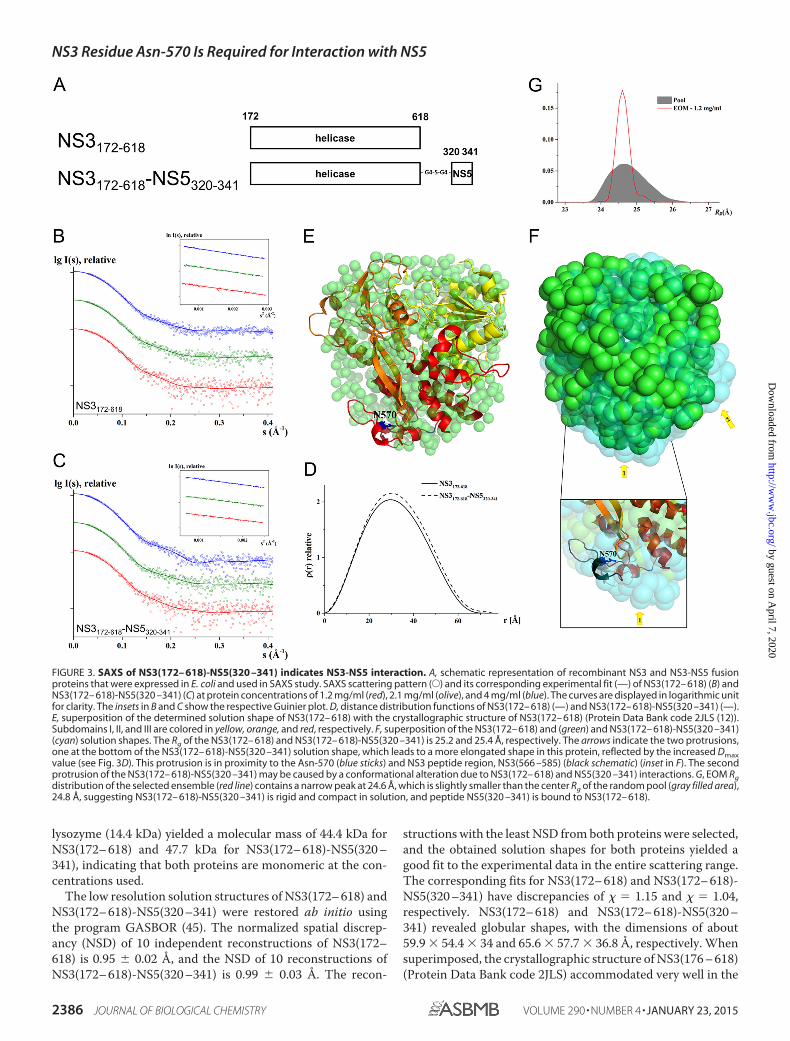
lysozyme (14.4 kDa) yielded a molecular mass of 44.4 kDa forNS3(172– 618) and 47.7 kDa for NS3(172– 618)-NS5(320 –341), indicating that both proteins are monomeric at the con-centrations used.
The low resolution solution structures of NS3(172– 618) andNS3(172– 618)-NS5(320 –341) were restored ab initio usingthe program GASBOR (45). The normalized spatial discrep-ancy (NSD) of 10 independent reconstructions of NS3(172–618) is 0.95 0.02 Å, and the NSD of 10 reconstructions ofNS3(172– 618)-NS5(320 –341) is 0.99 0.03 Å. The recon-
structions with the least NSD from both proteins were selected,and the obtained solution shapes for both proteins yielded agood fit to the experimental data in the entire scattering range.The corresponding fits for NS3(172– 618) and NS3(172– 618)-NS5(320 –341) have discrepancies of � � 1.15 and � � 1.04,respectively. NS3(172– 618) and NS3(172– 618)-NS5(320 –341) revealed globular shapes, with the dimensions of about59.9 � 54.4 � 34 and 65.6 � 57.7 � 36.8 Å, respectively. Whensuperimposed, the crystallographic structure of NS3(176 – 618)(Protein Data Bank code 2JLS) accommodated very well in the
FIGURE 3. SAXS of NS3(172– 618)-NS5(320 –341) indicates NS3-NS5 interaction. A, schematic representation of recombinant NS3 and NS3-NS5 fusionproteins that were expressed in E. coli and used in SAXS study. SAXS scattering pattern (E) and its corresponding experimental fit (—) of NS3(172– 618) (B) andNS3(172– 618)-NS5(320 –341) (C) at protein concentrations of 1.2 mg/ml (red), 2.1 mg/ml (olive), and 4 mg/ml (blue). The curves are displayed in logarithmic unitfor clarity. The insets in B and C show the respective Guinier plot. D, distance distribution functions of NS3(172– 618) (—) and NS3(172– 618)-NS5(320 –341) (—).E, superposition of the determined solution shape of NS3(172– 618) with the crystallographic structure of NS3(172– 618) (Protein Data Bank code 2JLS (12)).Subdomains I, II, and III are colored in yellow, orange, and red, respectively. F, superposition of the NS3(172– 618) and (green) and NS3(172– 618)-NS5(320 –341)(cyan) solution shapes. The Rg of the NS3(172– 618) and NS3(172– 618)-NS5(320 –341) is 25.2 and 25.4 Å, respectively. The arrows indicate the two protrusions,one at the bottom of the NS3(172– 618)-NS5(320 –341) solution shape, which leads to a more elongated shape in this protein, reflected by the increased Dmaxvalue (see Fig. 3D). This protrusion is in proximity to the Asn-570 (blue sticks) and NS3 peptide region, NS3(566 –585) (black schematic) (inset in F). The secondprotrusion of the NS3(172– 618)-NS5(320 –341) may be caused by a conformational alteration due to NS3(172– 618) and NS5(320 –341) interactions. G, EOM Rgdistribution of the selected ensemble (red line) contains a narrow peak at 24.6 Å, which is slightly smaller than the center Rg of the random pool (gray filled area),24.8 Å, suggesting NS3(172– 618)-NS5(320 –341) is rigid and compact in solution, and peptide NS5(320 –341) is bound to NS3(172– 618).
NS3 Residue Asn-570 Is Required for Interaction with NS5
2386 JOURNAL OF BIOLOGICAL CHEMISTRY VOLUME 290 • NUMBER 4 • JANUARY 23, 2015
by guest on April 7, 2020
http://ww
w.jbc.org/
Dow
nloaded from
solution form of the protein (Fig. 3E), indicating the high qual-ity of the solution data. When the solution form of NS3(172–618) (green) and NS3(172– 618)-NS5(320 –341) (cyan) weresuperimposed (Fig. 3F), two protrusions were observed and aredenoted by arrows in Fig. 3F. Protrusion 1 that is at the bottomof the NS3(172– 618)-NS5(320 –341) solution shape leads to amore elongated conformation in NS3(172– 618)-NS5(320 –341) than NS3(172– 618). This is also reflected by the increasedDmax in NS3(172– 618)-NS5(320 –341) (75.64 Å) when com-pared with the Dmax of NS3(172– 618) (72.95 Å) (Fig. 3D). Theprotrusion is in close proximity to residue Asn-570 (shown asblue sticks in the inset of Fig. 3F) and to the NS3 peptide region,NS3(566 –585) that used in ELISA (shown as black schematic inthe inset of Fig. 3F). The arrangement of NS3(566 –585) enablesan interaction with NS5(320 –341) to occur.
Comparison of the NS3(172– 618) and NS3(172– 618)-NS5(320 –341) low resolution structures also revealed a secondprotrusion of the NS3(172– 618)-NS5(320 –341) that mayreflect a conformational alteration due to the NS3(172– 618)and NS5(320 –341) interaction. Taken together, these resultsappear to be consistent with the ELISA data and suggest a phys-ical interaction between NS3(172– 618) and NS5(320 –341) insolution.
To eliminate the possibility that the peptide may be flexiblein solution, the x-ray scattering data set of NS3(172– 618)-NS5(320 –341) had been further analyzed using the EnsembleOptimization Method (EOM) (47, 48) to assess the compact-ness and flexibility of NS3(172– 618)-NS5(320 –341) in solu-tion. Based on the width and position of the selected ensemblepeak relative to random pool in Rg distribution, the flexibilityand compactness of the protein in solution can be deter-mined. In the case of NS3(172– 618)-NS5(320 –341), the Rgdistribution of the ensemble contains a narrow peak, indi-cating that NS3(172– 618)-NS5(320 –341) is rigid in solu-tion. This peak is centered at 24.6 Å, which is slightly smallerthan the center Rg of a random pool, 24.8 Å, suggesting thatNS3(172– 618)-NS5(320 –341) is compact in solution. TheEOM data demonstrate that peptide NS5(320 –341) is boundto NS3(172– 618).
NS3:N570A Mutant Has Reduced Infectious Virus Produc-tion and Viral Protein Synthesis—To study the impact of NS3:N570A mutation on NS3-NS5 interaction during the virus lifecycle, we first generated a DENV2 WT cDNA clone by standardmolecular cloning techniques. DENV2 of strain D2/SG/05K3295DK1/2005 of the cosmopolitan genotype (GenBankTM
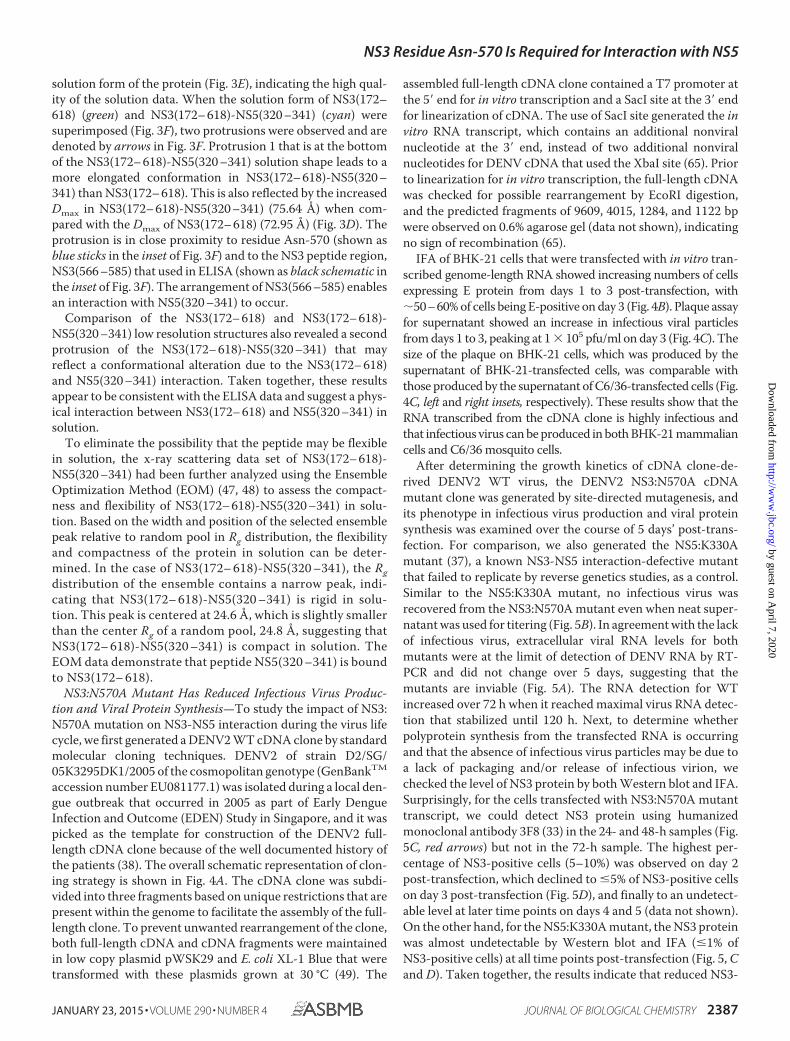
accession number EU081177.1) was isolated during a local den-gue outbreak that occurred in 2005 as part of Early DengueInfection and Outcome (EDEN) Study in Singapore, and it waspicked as the template for construction of the DENV2 full-length cDNA clone because of the well documented history ofthe patients (38). The overall schematic representation of clon-ing strategy is shown in Fig. 4A. The cDNA clone was subdi-vided into three fragments based on unique restrictions that arepresent within the genome to facilitate the assembly of the full-length clone. To prevent unwanted rearrangement of the clone,both full-length cDNA and cDNA fragments were maintainedin low copy plasmid pWSK29 and E. coli XL-1 Blue that weretransformed with these plasmids grown at 30 °C (49). The
assembled full-length cDNA clone contained a T7 promoter atthe 5� end for in vitro transcription and a SacI site at the 3� endfor linearization of cDNA. The use of SacI site generated the invitro RNA transcript, which contains an additional nonviralnucleotide at the 3� end, instead of two additional nonviralnucleotides for DENV cDNA that used the XbaI site (65). Priorto linearization for in vitro transcription, the full-length cDNAwas checked for possible rearrangement by EcoRI digestion,and the predicted fragments of 9609, 4015, 1284, and 1122 bpwere observed on 0.6% agarose gel (data not shown), indicatingno sign of recombination (65).
IFA of BHK-21 cells that were transfected with in vitro tran-scribed genome-length RNA showed increasing numbers of cellsexpressing E protein from days 1 to 3 post-transfection, with�50–60% of cells being E-positive on day 3 (Fig. 4B). Plaque assayfor supernatant showed an increase in infectious viral particlesfrom days 1 to 3, peaking at 1 � 105 pfu/ml on day 3 (Fig. 4C). Thesize of the plaque on BHK-21 cells, which was produced by thesupernatant of BHK-21-transfected cells, was comparable withthose produced by the supernatant of C6/36-transfected cells (Fig.4C, left and right insets, respectively). These results show that theRNA transcribed from the cDNA clone is highly infectious andthat infectious virus can be produced in both BHK-21 mammaliancells and C6/36 mosquito cells.
After determining the growth kinetics of cDNA clone-de-rived DENV2 WT virus, the DENV2 NS3:N570A cDNAmutant clone was generated by site-directed mutagenesis, andits phenotype in infectious virus production and viral proteinsynthesis was examined over the course of 5 days’ post-trans-fection. For comparison, we also generated the NS5:K330Amutant (37), a known NS3-NS5 interaction-defective mutantthat failed to replicate by reverse genetics studies, as a control.Similar to the NS5:K330A mutant, no infectious virus wasrecovered from the NS3:N570A mutant even when neat super-natant was used for titering (Fig. 5B). In agreement with the lackof infectious virus, extracellular viral RNA levels for bothmutants were at the limit of detection of DENV RNA by RT-PCR and did not change over 5 days, suggesting that themutants are inviable (Fig. 5A). The RNA detection for WTincreased over 72 h when it reached maximal virus RNA detec-tion that stabilized until 120 h. Next, to determine whetherpolyprotein synthesis from the transfected RNA is occurringand that the absence of infectious virus particles may be due toa lack of packaging and/or release of infectious virion, wechecked the level of NS3 protein by both Western blot and IFA.Surprisingly, for the cells transfected with NS3:N570A mutanttranscript, we could detect NS3 protein using humanizedmonoclonal antibody 3F8 (33) in the 24- and 48-h samples (Fig.5C, red arrows) but not in the 72-h sample. The highest per-centage of NS3-positive cells (5–10%) was observed on day 2post-transfection, which declined to �5% of NS3-positive cellson day 3 post-transfection (Fig. 5D), and finally to an undetect-able level at later time points on days 4 and 5 (data not shown).On the other hand, for the NS5:K330A mutant, the NS3 proteinwas almost undetectable by Western blot and IFA (�1% ofNS3-positive cells) at all time points post-transfection (Fig. 5, Cand D). Taken together, the results indicate that reduced NS3-
NS3 Residue Asn-570 Is Required for Interaction with NS5
JANUARY 23, 2015 • VOLUME 290 • NUMBER 4 JOURNAL OF BIOLOGICAL CHEMISTRY 2387
by guest on April 7, 2020
http://ww
w.jbc.org/
Dow
nloaded from
NS5 interaction in NS3:N570A mutant impairs infectious virusproduction and viral protein synthesis.
NS3:N570A Mutant Showed Accumulation of Negative-strand RNA—The reduction in viral protein synthesis for theNS3:N570A mutant and the lack of detectable infectiousplaques suggest that attenuated RNA replication could occur atan early time point for this mutant. Therefore, we examined theRNA replication kinetics by real time RT-PCR and IFA fordetection of dsRNA. The results showed that intracellular RNAcan be detected for NS3:N570A and WT but, intriguingly, notfor NS5:K330A-transfected cells (Fig. 6A and B). The RNA copyfor WT increased from around 106 copies/�g at 6 h post-trans-
fection to 109 copies/�g of RNA at 120 h, as detected by real-time RT-PCR. Although less than WT, a 10-fold increase inintracellular viral RNA copy was observed from day 1 to 2 (pvalue � 0.008) for NS3:N570A mutant and thereafter, the leveldeclined till day 5. The RNA copy for the NS5:K330A mutantdeclined from 6 to 48 h post-transfection and remained rela-tively stable from 48 to 120 h, suggesting that there was no viralRNA replication, and only the input RNA that was transfectedby electroporation was being detected. Interestingly, the detec-tion of the peak intracellular RNA level for NS3:N570A mutanton day 2 correlated with the highest level of NS3 proteindetected by Western blotting (Fig. 5C).
FIGURE 4. Construction of DENV2 cDNA infectious clone. A, schematic representation of full-length DENV2 cDNA clone generation. The DENV genome(represented approximately to scale) contains a single open reading frame that encodes three structural proteins (c, premembrane protein, and E) and sevenNS proteins (NS1, NS2A, NS2B, NS3, NS4A, NS4B, and NS5). Three fragments (fragment boundaries indicated by nucleotide numbers) covering the entire viralgenome were made and digested using unique restriction sites to facilitate the assembly of full-length cDNA clone (refer to “Experimental Procedures” fordetails). The complete DENV2 clone has a T7 promoter at 5� end for in vitro transcription and SacI site at 3� end for linearization of plasmid. B and C, BHK-21 cellswere electroporated with 10 �g of genomic length RNA, and samples were harvested daily for 3 days. B, infected cells were analyzed for the presence of Eprotein by IFA with mouse anti-E 4G2 antibody on 48 and 72 h post-transfection, and percentage infection was determined. C, culture supernatants were usedto determine viral titer by plaque assay on BHK-21 cells. The size of plaque on BHK-21 cells that was derived from the supernatant of either BHK-21 (left inset)or C6/36 (right inset) cells, which was transfected with DENV2 WT RNA, is shown.
NS3 Residue Asn-570 Is Required for Interaction with NS5
2388 JOURNAL OF BIOLOGICAL CHEMISTRY VOLUME 290 • NUMBER 4 • JANUARY 23, 2015
by guest on April 7, 2020
http://ww
w.jbc.org/
Dow
nloaded from
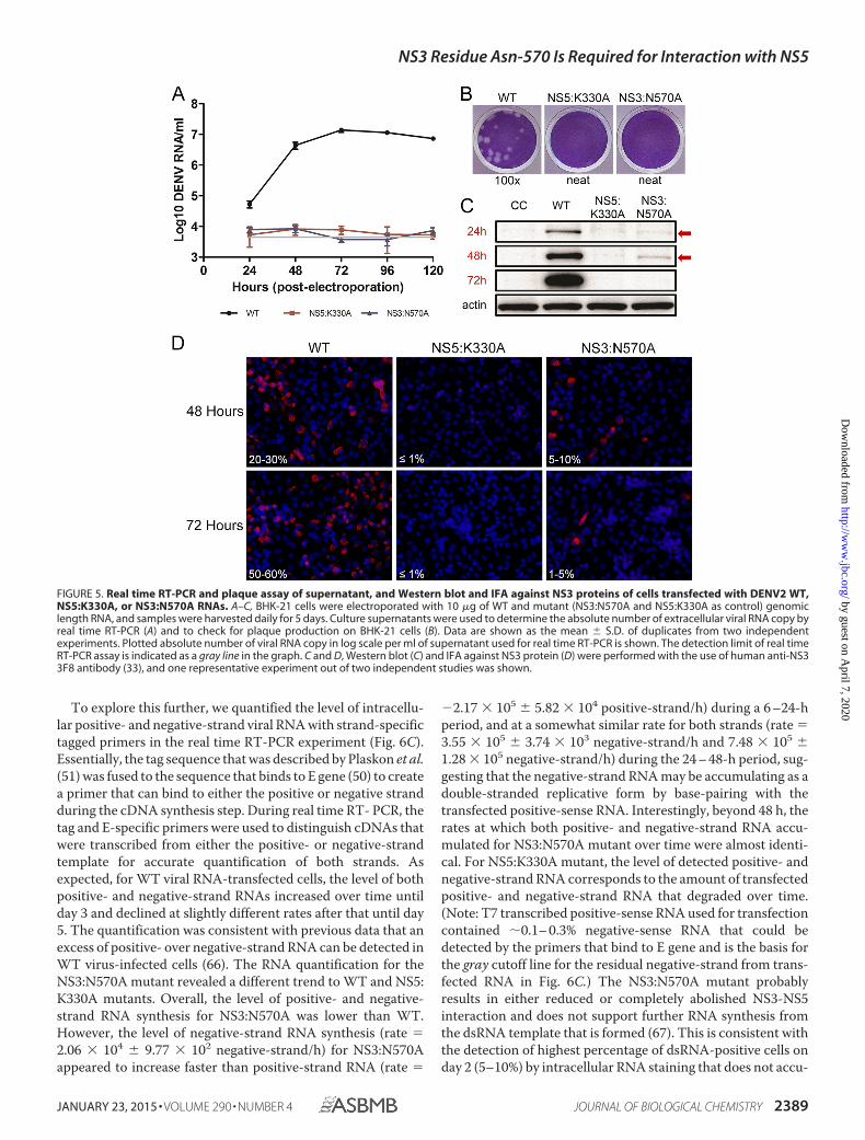
To explore this further, we quantified the level of intracellu-lar positive- and negative-strand viral RNA with strand-specifictagged primers in the real time RT-PCR experiment (Fig. 6C).Essentially, the tag sequence that was described by Plaskon et al.(51) was fused to the sequence that binds to E gene (50) to createa primer that can bind to either the positive or negative strandduring the cDNA synthesis step. During real time RT- PCR, thetag and E-specific primers were used to distinguish cDNAs thatwere transcribed from either the positive- or negative-strandtemplate for accurate quantification of both strands. Asexpected, for WT viral RNA-transfected cells, the level of bothpositive- and negative-strand RNAs increased over time untilday 3 and declined at slightly different rates after that until day5. The quantification was consistent with previous data that anexcess of positive- over negative-strand RNA can be detected inWT virus-infected cells (66). The RNA quantification for theNS3:N570A mutant revealed a different trend to WT and NS5:K330A mutants. Overall, the level of positive- and negative-strand RNA synthesis for NS3:N570A was lower than WT.However, the level of negative-strand RNA synthesis (rate �2.06 � 104 9.77 � 102 negative-strand/h) for NS3:N570Aappeared to increase faster than positive-strand RNA (rate �
�2.17 � 105 5.82 � 104 positive-strand/h) during a 6 –24-hperiod, and at a somewhat similar rate for both strands (rate �3.55 � 105 3.74 � 103 negative-strand/h and 7.48 � 105 1.28 � 105 negative-strand/h) during the 24 – 48-h period, sug-gesting that the negative-strand RNA may be accumulating as adouble-stranded replicative form by base-pairing with thetransfected positive-sense RNA. Interestingly, beyond 48 h, therates at which both positive- and negative-strand RNA accu-mulated for NS3:N570A mutant over time were almost identi-cal. For NS5:K330A mutant, the level of detected positive- andnegative-strand RNA corresponds to the amount of transfectedpositive- and negative-strand RNA that degraded over time.(Note: T7 transcribed positive-sense RNA used for transfectioncontained �0.1– 0.3% negative-sense RNA that could bedetected by the primers that bind to E gene and is the basis forthe gray cutoff line for the residual negative-strand from trans-fected RNA in Fig. 6C.) The NS3:N570A mutant probablyresults in either reduced or completely abolished NS3-NS5interaction and does not support further RNA synthesis fromthe dsRNA template that is formed (67). This is consistent withthe detection of highest percentage of dsRNA-positive cells onday 2 (5–10%) by intracellular RNA staining that does not accu-
FIGURE 5. Real time RT-PCR and plaque assay of supernatant, and Western blot and IFA against NS3 proteins of cells transfected with DENV2 WT,NS5:K330A, or NS3:N570A RNAs. A–C, BHK-21 cells were electroporated with 10 �g of WT and mutant (NS3:N570A and NS5:K330A as control) genomiclength RNA, and samples were harvested daily for 5 days. Culture supernatants were used to determine the absolute number of extracellular viral RNA copy byreal time RT-PCR (A) and to check for plaque production on BHK-21 cells (B). Data are shown as the mean S.D. of duplicates from two independentexperiments. Plotted absolute number of viral RNA copy in log scale per ml of supernatant used for real time RT-PCR is shown. The detection limit of real timeRT-PCR assay is indicated as a gray line in the graph. C and D, Western blot (C) and IFA against NS3 protein (D) were performed with the use of human anti-NS33F8 antibody (33), and one representative experiment out of two independent studies was shown.
NS3 Residue Asn-570 Is Required for Interaction with NS5
JANUARY 23, 2015 • VOLUME 290 • NUMBER 4 JOURNAL OF BIOLOGICAL CHEMISTRY 2389
by guest on April 7, 2020
http://ww
w.jbc.org/
Dow
nloaded from
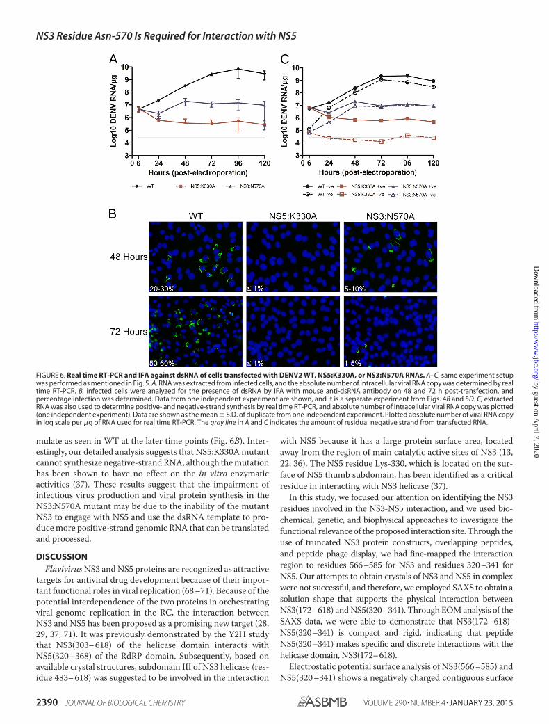
mulate as seen in WT at the later time points (Fig. 6B). Inter-estingly, our detailed analysis suggests that NS5:K330A mutantcannot synthesize negative-strand RNA, although the mutationhas been shown to have no effect on the in vitro enzymaticactivities (37). These results suggest that the impairment ofinfectious virus production and viral protein synthesis in theNS3:N570A mutant may be due to the inability of the mutantNS3 to engage with NS5 and use the dsRNA template to pro-duce more positive-strand genomic RNA that can be translatedand processed.
DISCUSSIONFlavivirus NS3 and NS5 proteins are recognized as attractive
targets for antiviral drug development because of their impor-tant functional roles in viral replication (68 –71). Because of thepotential interdependence of the two proteins in orchestratingviral genome replication in the RC, the interaction betweenNS3 and NS5 has been proposed as a promising new target (28,29, 37, 71). It was previously demonstrated by the Y2H studythat NS3(303– 618) of the helicase domain interacts withNS5(320 –368) of the RdRP domain. Subsequently, based onavailable crystal structures, subdomain III of NS3 helicase (res-idue 483– 618) was suggested to be involved in the interaction
with NS5 because it has a large protein surface area, locatedaway from the region of main catalytic active sites of NS3 (13,22, 36). The NS5 residue Lys-330, which is located on the sur-face of NS5 thumb subdomain, has been identified as a criticalresidue in interacting with NS3 helicase (37).
In this study, we focused our attention on identifying the NS3residues involved in the NS3-NS5 interaction, and we used bio-chemical, genetic, and biophysical approaches to investigate thefunctional relevance of the proposed interaction site. Through theuse of truncated NS3 protein constructs, overlapping peptides,and peptide phage display, we had fine-mapped the interactionregion to residues 566–585 for NS3 and residues 320–341 forNS5. Our attempts to obtain crystals of NS3 and NS5 in complexwere not successful, and therefore, we employed SAXS to obtain asolution shape that supports the physical interaction betweenNS3(172–618) and NS5(320–341). Through EOM analysis of theSAXS data, we were able to demonstrate that NS3(172–618)-NS5(320–341) is compact and rigid, indicating that peptideNS5(320–341) makes specific and discrete interactions with thehelicase domain, NS3(172–618).
Electrostatic potential surface analysis of NS3(566 –585) andNS5(320 –341) shows a negatively charged contiguous surface
FIGURE 6. Real time RT-PCR and IFA against dsRNA of cells transfected with DENV2 WT, NS5:K330A, or NS3:N570A RNAs. A–C, same experiment setupwas performed as mentioned in Fig. 5. A, RNA was extracted from infected cells, and the absolute number of intracellular viral RNA copy was determined by realtime RT-PCR. B, infected cells were analyzed for the presence of dsRNA by IFA with mouse anti-dsRNA antibody on 48 and 72 h post-transfection, andpercentage infection was determined. Data from one independent experiment are shown, and it is a separate experiment from Figs. 4B and 5D. C, extractedRNA was also used to determine positive- and negative-strand synthesis by real time RT-PCR, and absolute number of intracellular viral RNA copy was plotted(one independent experiment). Data are shown as the mean S.D. of duplicate from one independent experiment. Plotted absolute number of viral RNA copyin log scale per �g of RNA used for real time RT-PCR. The gray line in A and C indicates the amount of residual negative strand from transfected RNA.
NS3 Residue Asn-570 Is Required for Interaction with NS5
2390 JOURNAL OF BIOLOGICAL CHEMISTRY VOLUME 290 • NUMBER 4 • JANUARY 23, 2015
by guest on April 7, 2020
http://ww
w.jbc.org/
Dow
nloaded from
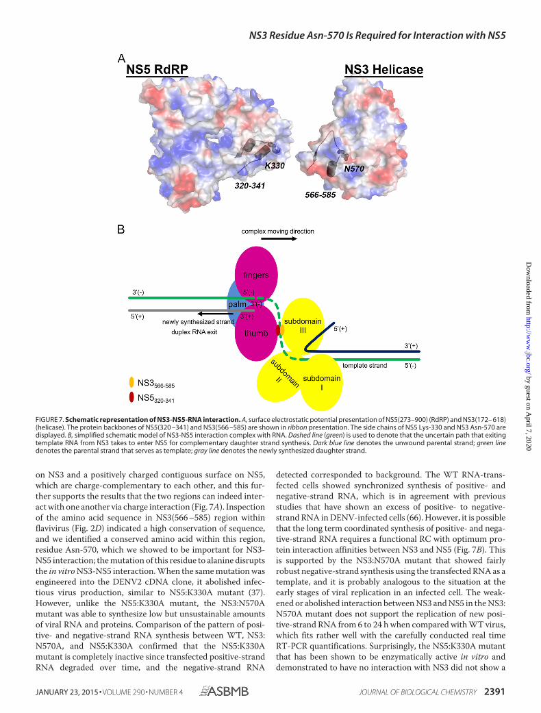
on NS3 and a positively charged contiguous surface on NS5,which are charge-complementary to each other, and this fur-ther supports the results that the two regions can indeed inter-act with one another via charge interaction (Fig. 7A). Inspectionof the amino acid sequence in NS3(566 –585) region withinflavivirus (Fig. 2D) indicated a high conservation of sequence,and we identified a conserved amino acid within this region,residue Asn-570, which we showed to be important for NS3-NS5 interaction; the mutation of this residue to alanine disruptsthe in vitro NS3-NS5 interaction. When the same mutation wasengineered into the DENV2 cDNA clone, it abolished infec-tious virus production, similar to NS5:K330A mutant (37).However, unlike the NS5:K330A mutant, the NS3:N570Amutant was able to synthesize low but unsustainable amountsof viral RNA and proteins. Comparison of the pattern of posi-tive- and negative-strand RNA synthesis between WT, NS3:N570A, and NS5:K330A confirmed that the NS5:K330Amutant is completely inactive since transfected positive-strandRNA degraded over time, and the negative-strand RNA
detected corresponded to background. The WT RNA-trans-fected cells showed synchronized synthesis of positive- andnegative-strand RNA, which is in agreement with previousstudies that have shown an excess of positive- to negative-strand RNA in DENV-infected cells (66). However, it is possiblethat the long term coordinated synthesis of positive- and nega-tive-strand RNA requires a functional RC with optimum pro-tein interaction affinities between NS3 and NS5 (Fig. 7B). Thisis supported by the NS3:N570A mutant that showed fairlyrobust negative-strand synthesis using the transfected RNA as atemplate, and it is probably analogous to the situation at theearly stages of viral replication in an infected cell. The weak-ened or abolished interaction between NS3 and NS5 in the NS3:N570A mutant does not support the replication of new posi-tive-strand RNA from 6 to 24 h when compared with WT virus,which fits rather well with the carefully conducted real timeRT-PCR quantifications. Surprisingly, the NS5:K330A mutantthat has been shown to be enzymatically active in vitro anddemonstrated to have no interaction with NS3 did not show a
FIGURE 7. Schematic representation of NS3-NS5-RNA interaction. A, surface electrostatic potential presentation of NS5(273–900) (RdRP) and NS3(172– 618)(helicase). The protein backbones of NS5(320 –341) and NS3(566 –585) are shown in ribbon presentation. The side chains of NS5 Lys-330 and NS3 Asn-570 aredisplayed. B, simplified schematic model of NS3-NS5 interaction complex with RNA. Dashed line (green) is used to denote that the uncertain path that exitingtemplate RNA from NS3 takes to enter NS5 for complementary daughter strand synthesis. Dark blue line denotes the unwound parental strand; green linedenotes the parental strand that serves as template; gray line denotes the newly synthesized daughter strand.
NS3 Residue Asn-570 Is Required for Interaction with NS5
JANUARY 23, 2015 • VOLUME 290 • NUMBER 4 JOURNAL OF BIOLOGICAL CHEMISTRY 2391
by guest on April 7, 2020
http://ww
w.jbc.org/
Dow
nloaded from
similar increase in negative-strand RNA as compared with NS3:N570A mutant. There may be two possible reasons for the inac-tivity of the NS5:K330A mutant in making negative-strandRNA. First, it may be due to impaired intramolecular signalingbetween the two NS5 domains that probably coordinate thetwo functional activities of NS5 (72) required for in vivo poly-merase activity. Second, its weak/abolished interaction withNS3 may affect the unwinding of secondary structures in thetransfected positive-sense RNA that is needed for negative-strand synthesis. Taken together, our study indicates that dif-ferent NS3-NS5 interaction-defective mutants can impairinfectious virus production, viral protein synthesis, and RNAreplication to varying degrees, which is likely to be dependenton the importance of the amino acids that are involved in NS3-NS5 interaction, and also possibly the intramolecular interac-tions in NS5. It is interesting to note that the coordinated syn-thesis of positive- and negative-strand RNA at the early stagesof replication can be further explored by studying NS3-NS5interaction mutants displaying varying strengths/degrees ofbinding and also the contribution of intramolecular cross-talkbetween the domains of NS5 in strand-specific RNA synthesisin the RC (Fig. 7B). Importantly, we also note that although theNS3 residue Asn-570 is conserved among the four DENV sero-types and several members of flavivirus genus, it is not con-served in yellow fever virus NS3, which has a histidine in placeof asparagine at this position in NS3 and tyrosine instead oflysine at position 330 of NS5 (for sequence alignment, refer toFigs. 1B and 5 in Ref. 36).
Overall, this study has identified a potentially new druggabletarget for the development of antiviral drugs to block NS3-NS5interaction that is essential for viral replication. The availablecrystal structures of NS3 and NS5 together with in vitro assaysfor interaction can be used for in silico and high throughputscreening campaigns to find lead molecules for antiviral drugdevelopment. Alternatively, because the NS3:N570A mutantgenome can be translated to a low level and is able to synthesizenegative-strand RNA, it may serve as a potential RNA-medi-ated vaccine, although the basis for this requires developmentof new technology platforms.
Acknowledgments—We thank Drs. Mallur Madhusudhan, Chan-drakala Basavannacharya, Yoichi Suzuki, Yin Hoe Yau, and SusanaGeifman Shochat for valuable suggestions, discussion, and experi-mental assistance.
REFERENCES1. Bhatt, S., Gething, P. W., Brady, O. J., Messina, J. P., Farlow, A. W., Moyes,
C. L., Drake, J. M., Brownstein, J. S., Hoen, A. G., Sankoh, O., Myers, M. F.,George, D. B., Jaenisch, T., Wint, G. R., Simmons, C. P., Scott, T. W.,Farrar, J. J., and Hay, S. I. (2013) The global distribution and burden ofdengue. Nature 496, 504 –507
2. Lindenbach, B. D., Thiel, H., and Rice, C. M. (2007) in Fields Virology(Knipe, D. M. and Howley, P. M., eds) 5th Ed., pp. 1101–1152, WoltersKluwer/Lippincott Williams & Wilkins, Philadelphia
3. Bartholomeusz, A. I., and Wright, P. J. (1993) Synthesis of dengue virusRNA in vitro: initiation and the involvement of proteins NS3 and NS5.Arch. Virol. 128, 111–121
4. Falgout, B., Pethel, M., Zhang, Y. M., and Lai, C. J. (1991) Both nonstruc-tural proteins NS2B and NS3 are required for the proteolytic processing of
dengue virus nonstructural proteins. J. Virol. 65, 2467–24755. Preugschat, F., Yao, C. W., and Strauss, J. H. (1990) In vitro processing of
dengue virus type 2 nonstructural proteins NS2A, NS2B, and NS3. J. Virol.64, 4364 – 4374
6. Wu, C. F., Wang, S. H., Sun, C. M., Hu, S. T., and Syu, W. J. (2003)Activation of dengue protease autocleavage at the NS2B-NS3 junction byrecombinant NS3 and GST-NS2B fusion proteins. J. Virol. Methods 114,45–54
7. Zhang, L., Mohan, P. M., and Padmanabhan, R. (1992) Processing andlocalization of Dengue virus type 2 polyprotein precursor NS3-NS4A-NS4B-NS5. J. Virol. 66, 7549 –7554
8. Chambers, T. J., Nestorowicz, A., Amberg, S. M., and Rice, C. M. (1993)Mutagenesis of the yellow fever virus NS2B protein: effects on proteolyticprocessing, NS2B-NS3 complex formation, and viral replication. J. Virol.67, 6797– 6807
9. Yon, C., Teramoto, T., Mueller, N., Phelan, J., Ganesh, V. K., Murthy,K. H., and Padmanabhan, R. (2005) Modulation of the nucleoside triphos-phatase/RNA helicase and 5�-RNA triphosphatase activities of Denguevirus type 2 nonstructural protein 3 (NS3) by interaction with NS5, theRNA-dependent RNA polymerase. J. Biol. Chem. 280, 27412–27419
10. Sampath, A., Xu, T., Chao, A., Luo, D., Lescar, J., and Vasudevan, S. G.(2006) Structure-based mutational analysis of the NS3 helicase from den-gue virus. J. Virol. 80, 6686 – 6690
11. Luo, D., Xu, T., Hunke, C., Gruber, G., Vasudevan, S. G., and Lescar, J.(2008) Crystal structure of the NS3 protease-helicase from dengue virus.J. Virol. 82, 173–183
12. Luo, D., Xu, T., Watson, R. P., Scherer-Becker, D., Sampath, A., Jahnke,W., Yeong, S. S., Wang, C. H., Lim, S. P., Strongin, A., Vasudevan, S. G.,and Lescar, J. (2008) Insights into RNA unwinding and ATP hydrolysis bythe flavivirus NS3 protein. EMBO J. 27, 3209 –3219
13. Xu, T., Sampath, A., Chao, A., Wen, D., Nanao, M., Chene, P., Vasudevan,S. G., and Lescar, J. (2005) Structure of the Dengue virus helicase/nucleo-side triphosphatase catalytic domain at a resolution of 2.4 A. J. Virol. 79,10278 –10288
14. Matusan, A. E., Pryor, M. J., Davidson, A. D., and Wright, P. J. (2001)Mutagenesis of the Dengue virus type 2 NS3 protein within and outsidehelicase motifs: effects on enzyme activity and virus replication. J. Virol.75, 9633–9643
15. Bartelma, G., and Padmanabhan, R. (2002) Expression, purification, andcharacterization of the RNA 5�-triphosphatase activity of dengue virustype 2 nonstructural protein 3. Virology 299, 122–132
16. Benarroch, D., Selisko, B., Locatelli, G. A., Maga, G., Romette, J. L., andCanard, B. (2004) The RNA helicase, nucleotide 5�-triphosphatase, andRNA 5�-triphosphatase activities of Dengue virus protein NS3 are Mg2�-dependent and require a functional Walker B motif in the helicase cata-lytic core. Virology 328, 208 –218
17. Egloff, M. P., Benarroch, D., Selisko, B., Romette, J. L., and Canard, B.(2002) An RNA cap (nucleoside-2�-O)-methyltransferase in the flavivirusRNA polymerase NS5: crystal structure and functional characterization.EMBO J. 21, 2757–2768
18. Egloff, M. P., Decroly, E., Malet, H., Selisko, B., Benarroch, D., Ferron, F.,and Canard, B. (2007) Structural and functional analysis of methylationand 5�-RNA sequence requirements of short capped RNAs by the meth-yltransferase domain of dengue virus NS5. J. Mol. Biol. 372, 723–736
19. Lu, G., and Gong, P. (2013) Crystal structure of the full-length Japaneseencephalitis virus NS5 reveals a conserved methyltransferase-polymeraseinterface. PLoS Pathog. 9, e1003549
20. Malet, H., Egloff, M. P., Selisko, B., Butcher, R. E., Wright, P. J., Roberts, M.,Gruez, A., Sulzenbacher, G., Vonrhein, C., Bricogne, G., Mackenzie, J. M.,Khromykh, A. A., Davidson, A. D., and Canard, B. (2007) Crystal structureof the RNA polymerase domain of the West Nile virus non-structuralprotein 5. J. Biol. Chem. 282, 10678 –10689
21. Ray, D., Shah, A., Tilgner, M., Guo, Y., Zhao, Y., Dong, H., Deas, T. S.,Zhou, Y., Li, H., and Shi, P. Y. (2006) West Nile virus 5�-cap structure isformed by sequential guanine N-7 and ribose 2�-O-methylations by non-structural protein 5. J. Virol. 80, 8362– 8370
22. Wu, J., Bera, A. K., Kuhn, R. J., and Smith, J. L. (2005) Structure of theflavivirus helicase: Implications for catalytic activity, protein interactions,
NS3 Residue Asn-570 Is Required for Interaction with NS5
2392 JOURNAL OF BIOLOGICAL CHEMISTRY VOLUME 290 • NUMBER 4 • JANUARY 23, 2015
by guest on April 7, 2020
http://ww
w.jbc.org/
Dow
nloaded from
and proteolytic processing. J. Virol. 79, 10268 –1027723. Bartholomeusz, A., and Thompson, P. (1999) Flaviviridae polymerase and
RNA replication. J. Viral Hepat. 6, 261–27024. Nomaguchi, M., Ackermann, M., Yon, C., You, S., and Padmanabhan, R.
(2003) De novo synthesis of negative-strand RNA by Dengue virus RNA-dependent RNA polymerase in vitro: nucleotide, primer, and templateparameters. J. Virol. 77, 8831– 8842
25. Yap, T. L., Xu, T., Chen, Y. L., Malet, H., Egloff, M. P., Canard, B., Vasude-van, S. G., and Lescar, J. (2007) Crystal structure of the dengue virusRNA-dependent RNA polymerase catalytic domain at 1.85-angstrom res-olution. J. Virol. 81, 4753– 4765
26. You, S., Falgout, B., Markoff, L., and Padmanabhan, R. (2001) In vitro RNAsynthesis from exogenous dengue viral RNA templates requires longrange interactions between 5�- and 3�-terminal regions that influenceRNA structure. J. Biol. Chem. 276, 15581–15591
27. Gualano, R. C., Pryor, M. J., Cauchi, M. R., Wright, P. J., and Davidson,A. D. (1998) Identification of a major determinant of mouse neuroviru-lence of dengue virus type 2 using stably cloned genomic-length cDNA.J. Gen. Virol. 79, 437– 446
28. Kapoor, M., Zhang, L., Ramachandra, M., Kusukawa, J., Ebner, K. E., andPadmanabhan, R. (1995) Association between NS3 and NS5 proteins ofdengue virus type 2 in the putative RNA replicase is linked to differentialphosphorylation of NS5. J. Biol. Chem. 270, 19100 –19106
29. Welsch, S., Miller, S., Romero-Brey, I., Merz, A., Bleck, C. K., Walther, P.,Fuller, S. D., Antony, C., Krijnse-Locker, J., and Bartenschlager, R. (2009)Composition and three-dimensional architecture of the dengue virus rep-lication and assembly sites. Cell Host Microbe 5, 365–375
30. Mackenzie, J. M., and Westaway, E. G. (2001) Assembly and maturation ofthe flavivirus Kunjin virus appear to occur in the rough endoplasmic re-ticulum and along the secretory pathway, respectively. J. Virol. 75,10787–10799
31. Uchil, P. D., and Satchidanandam, V. (2003) Architecture of the flaviviralreplication complex. Protease, nuclease, and detergents reveal encase-ment within double-layered membrane compartments. J. Biol. Chem. 278,24388 –24398
32. Chen, C. J., Kuo, M. D., Chien, L. J., Hsu, S. L., Wang, Y. M., and Lin, J. H.(1997) RNA-protein interactions: Involvement of NS3, NS5, and 3� non-coding regions of Japanese encephalitis virus genomic RNA. J. Virol. 71,3466 –3473
33. Moreland, N. J., Tay, M. Y., Lim, E., Rathore, A. P., Lim, A. P., Hanson, B. J.,and Vasudevan, S. G. (2012) Monoclonal antibodies against dengue NS2Band NS3 proteins for the study of protein interactions in the flaviviralreplication complex. J. Virol. Methods 179, 97–103
34. Yu, L., Takeda, K., and Markoff, L. (2013) Protein-protein interactionsamong West Nile non-structural proteins and transmembrane complexformation in mammalian cells. Virology 446, 365–377
35. Johansson, M., Brooks, A. J., Jans, D. A., and Vasudevan, S. G. (2001) Asmall region of the dengue virus-encoded RNA-dependent RNA poly-merase, NS5, confers interaction with both the nuclear transport receptorimportin-ss and the viral helicase, NS3. J. Gen. Virol. 82, 735–745
36. Brooks, A. J., Johansson, M., John, A. V., Xu, Y., Jans, D. A., and Vasudevan,S. G. (2002) The interdomain region of dengue NS5 protein that binds tothe viral helicase NS3 contains independently functional importin beta 1and importin �/-recognized nuclear localization signals. J. Biol. Chem.277, 36399 –36407
37. Zou, G., Chen, Y. L., Dong, H., Lim, C. C., Yap, L. J., Yau, Y. H., Shochat,S. G., Lescar, J., and Shi, P. Y. (2011) Functional analysis of two cavities inflavivirus NS5 polymerase. J. Biol. Chem. 286, 14362–14372
38. Schreiber, M. J., Holmes, E. C., Ong, S. H., Soh, H. S., Liu, W., Tanner, L.,Aw, P. P., Tan, H. C., Ng, L. C., Leo, Y. S., Low, J. G., Ong, A., Ooi, E. E.,Vasudevan, S. G., and Hibberd, M. L. (2009) Genomic epidemiology of adengue virus epidemic in urban Singapore. J. Virol. 83, 4163– 4173
39. Li, J., Lim, S. P., Beer, D., Patel, V., Wen, D., Tumanut, C., Tully, D. C.,Williams, J. A., Jiricek, J., Priestle, J. P., Harris, J. L., and Vasudevan, S. G.(2005) Functional profiling of recombinant NS3 proteases from all fourserotypes of dengue virus using tetrapeptide and octapeptide substratelibraries. J. Biol. Chem. 280, 28766 –28774
40. Paradkar, P. N., Ooi, E. E., Hanson, B. J., Gubler, D. J., and Vasudevan, S. G.
(2011) Unfolded protein response (UPR) gene expression during anti-body-dependent enhanced infection of cultured monocytes correlateswith dengue disease severity. Biosci. Rep. 31, 221–230
41. Dip, P. V., Kamariah, N., Manimekalai, M. S., Nartey, W., Balakrishna,A. M., Eisenhaber, F., Eisenhaber, B., and Gruber, G. (2014) The ups anddowns in AhpF: structure, mechanism and ensemble formation of thealkylhydroperoxide reductase subunits AhpC and AhpF from Escherichiacoli. Acta Crystallogr. D Biol. Crystallogr. 70, 2848 –2862
42. Svergun, D. (1993) A direct indirect method of small-angle scattering datatreatment. J. Appl. Crystallogr. 26, 258 –267
43. Lipson, H. (1956) Small-angle scattering of x-rays by Guinier, A., andFournet, G. Acta Crystallogr. 9, 839
44. Svergun, D. (1992) Determination of the regularization parameter in in-direct-transform methods using perceptual criteria. J. Appl. Crystallogr.25, 495–503
45. Svergun, D. I., Petoukhov, M. V., and Koch, M. H. (2001) Determination ofdomain structure of proteins from x-ray solution scattering. Biophys. J. 80,2946 –2953
46. Kozin, M. B., and Svergun, D. I. (2002) Automated matching of high- andlow-resolution structural models. J. Appl. Crystallogr. 34, 33– 41
47. Bernado, P., Mylonas, E., Petoukhov, M. V., Blackledge, M., and Svergun,D. I. (2007) Structural characterization of flexible proteins using small-angle x-ray scattering. J. Am. Chem. Soc. 129, 5656 –5664
48. Petoukhov, M. V., Franke, D., Shkumatov, A. V., Tria, G., Kikhney, A. G.,Gajda, M., Gorba, C., Mertens, H. D., Konarev, P. V., and Svergun, D. I.(2012) New developments in the ATSAS program package for small-anglescattering data analysis. J. Appl. Crystallogr. 45, 342–350
49. Wang, R. F., and Kushner, S. R. (1991) Construction of versatile low-copy-number vectors for cloning, sequencing and gene expression in Esche-richia coli. Gene 100, 195–199
50. Johnson, B. W., Russell, B. J., and Lanciotti, R. S. (2005) Serotype-specificdetection of dengue viruses in a fourplex real time reverse transcriptasePCR assay. J. Clin. Microbiol. 43, 4977– 4983
51. Plaskon, N. E., Adelman, Z. N., and Myles, K. M. (2009) Accurate strand-specific quantification of viral RNA. PLoS One 4, e7468
52. Collins, T. J. (2007) ImageJ for microscopy. BioTechniques 43, 25–3053. Moreland, N. J., Tay, M. Y., Lim, E., Paradkar, P. N., Doan, D. N., Yau, Y. H.,
Geifman Shochat, S., and Vasudevan, S. G. (2010) High affinity humanantibody fragments to dengue virus non-structural protein 3. PLoS Negl.Trop. Dis. 4, e881
54. Rivino, L., Kumaran, E. A., Jovanovic, V., Nadua, K., Teo, E. W., Pang,S. W., Teo, G. H., Gan, V. C., Lye, D. C., Leo, Y. S., Hanson, B. J., Smith,K. G., Bertoletti, A., Kemeny, D. M., and MacAry, P. A. (2013) Differentialtargeting of viral components by CD4� versus CD8� T lymphocytes indengue virus infection. J. Virol. 87, 2693–2706
55. Takahashi, H., Takahashi, C., Moreland, N. J., Chang, Y. T., Sawasaki, T.,Ryo, A., Vasudevan, S. G., Suzuki, Y., and Yamamoto, N. (2012) Establish-ment of a robust dengue virus NS3-NS5 binding assay for identification ofprotein-protein interaction inhibitors. Antiviral Res. 96, 305–314
56. Derossi, D., Calvet, S., Trembleau, A., Brunissen, A., Chassaing, G., andProchiantz, A. (1996) Cell internalization of the third helix of the Anten-napedia homeodomain is receptor-independent. J. Biol. Chem. 271,18188 –18193
57. Keller, A. A., Mussbach, F., Breitling, R., Hemmerich, P., Schaefer, B.,Lorkowski, S., and Reissmann, S. (2013) Relationships between cargo, cellpenetrating peptides and cell type for uptake of non-covalent complexesinto live cells. Pharmaceuticals 6, 184 –203
58. Zorko, M., and Langel, U. (2005) Cell-penetrating peptides: mechanismand kinetics of cargo delivery. Adv. Drug Deliv. Rev. 57, 529 –545
59. Luo, D., Wei, N., Doan, D. N., Paradkar, P. N., Chong, Y., Davidson, A. D.,Kotaka, M., Lescar, J., and Vasudevan, S. G. (2010) Flexibility between theprotease and helicase domains of the dengue virus NS3 protein conferredby the linker region and its functional implications. J. Biol. Chem. 285,18817–18827
60. Svergun, D. I., Koch, M. H., Timmins, P. A., and May, R. P. (2013) SmallAngle X-Ray and Neutron Scattering from Solutions of Biological Macro-molecules, Oxford University Press, Oxford, UK
61. Gruber, A., Gunalan, K., Ramalingam, J. K., Manimekalai, M. S., Gruber,
NS3 Residue Asn-570 Is Required for Interaction with NS5
JANUARY 23, 2015 • VOLUME 290 • NUMBER 4 JOURNAL OF BIOLOGICAL CHEMISTRY 2393
by guest on April 7, 2020
http://ww
w.jbc.org/
Dow
nloaded from
G., and Preiser, P. R. (2011) Structural characterization of the erythrocytebinding domain of the reticulocyte binding protein homologue family ofPlasmodium yoelii. Infect. Immun. 79, 2880 –2888
62. Ho, C. S., Rydstrom, A., Manimekalai, M. S., Svanborg, C., and Gruber, G.(2012) Low resolution solution structure of HAMLET and the importanceof its �-domains in tumoricidal activity. PLoS ONE 7, e53051
63. Balakrishna, A. M., Manimekalai, M. S., Hunke, C., Gayen, S., Rossle, M.,Jeyakanthan, J., and Gruber, G. (2010) Crystal and solution structure of theC-terminal part of the Methanocaldococcus jannaschii A1AO ATP syn-thase subunit E revealed by X-ray diffraction and small-angle X-ray scat-tering. J. Bioenerg. Biomembr. 42, 311–320
64. Svergun, D. I., Konrad, S., Huss, M., Koch, M. H., Wieczorek, H., Alten-dorf, K., Volkov, V. V., and Gruber, G. (1998) Quaternary structure of V1and F1 ATPase: significance of structural homologies and diversities. Bio-chemistry 37, 17659 –17663
65. Davidson, A. D. (2014) Development and application of dengue virus re-verse genetic systems. Methods Mol. Biol. 1138, 113–130
66. Tomassini, J. E., Boots, E., Gan, L., Graham, P., Munshi, V., Wolanski, B.,Fay, J. F., Getty, K., and LaFemina, R. (2003) An in vitro Flaviviridae rep-licase system capable of authentic RNA replication. Virology 313,274 –285
67. Chu, P. W., and Westaway, E. G. (1985) Replication strategy of Kunjinvirus: evidence for recycling role of replicative form RNA as template insemiconservative and asymmetric replication. Virology 140, 68 –79
68. Lescar, J., Luo, D., Xu, T., Sampath, A., Lim, S. P., Canard, B., and Vasude-van, S. G. (2008) Towards the design of antiviral inhibitors against flavivi-ruses: the case for the multifunctional NS3 protein from Dengue virus as a
target. Antiviral Res. 80, 94 –10169. Rawlinson, S. M., Pryor, M. J., Wright, P. J., and Jans, D. A. (2006) Dengue
virus RNA polymerase NS5: a potential therapeutic target? Curr. DrugTargets 7, 1623–1638
70. Bollati, M., Alvarez, K., Assenberg, R., Baronti, C., Canard, B., Cook, S.,Coutard, B., Decroly, E., de Lamballerie, X., Gould, E. A., Grard, G.,Grimes, J. M., Hilgenfeld, R., Jansson, A. M., Malet, H., Mancini, E. J.,Mastrangelo, E., Mattevi, A., Milani, M., Moureau, G., Neyts, J., Owens,R. J., Ren, J., Selisko, B., Speroni, S., Steuber, H., Stuart, D. I., Unge, T., andBolognesi, M. (2010) Structure and functionality in flavivirus NS-proteins:perspectives for drug design. Antiviral Res. 87, 125–148
71. Geiss, B. J., Stahla, H., Hannah, A. M., Gari, A. M., and Keenan, S. M.(2009) Focus on flaviviruses: current and future drug targets. Future Med.Chem. 1, 327–344
72. Li, X. D., Shan, C., Deng, C. L., Ye, H. Q., Shi, P. Y., Yuan, Z. M., Gong, P.,and Zhang, B. (2014) The interface between methyltransferase and poly-merase of NS5 is essential for flavivirus replication. PLoS Negl. Trop. Dis. 8,e2891
73. Brugidou, J., Legrand, C., Mery, J., and Rabie, A. (1995) The retro-inversoform of a homeobox-derived short peptide is rapidly internalised by cul-tured neurones: a new basis for an efficient intracellular delivery system.Biochem. Biophys. Res. Commun. 214, 685– 693
74. Thompson, J. D., Higgins, D. G., and Gibson, T. J. (1994) CLUSTAL W:improving the sensitivity of progressive multiple sequence alignmentthrough sequence weighting, position-specific gap penalties and weightmatrix choice. Nucleic Acids Res. 22, 4673– 4680
NS3 Residue Asn-570 Is Required for Interaction with NS5
2394 JOURNAL OF BIOLOGICAL CHEMISTRY VOLUME 290 • NUMBER 4 • JANUARY 23, 2015
by guest on April 7, 2020
http://ww
w.jbc.org/
Dow
nloaded from
Luo and Subhash G. VasudevanYuwen Chong, Jade K. Forwood, Eng Eong Ooi, Gerhard Grüber, Julien Lescar, Dahai
Moon Y. F. Tay, Wuan Geok Saw, Yongqian Zhao, Kitti W. K. Chan, Daljit Singh,NS3-NS5 Interaction and Viral Replication
The C-terminal 50 Amino Acid Residues of Dengue NS3 Protein Are Important for
doi: 10.1074/jbc.M114.607341 originally published online December 8, 20142015, 290:2379-2394.J. Biol. Chem.
10.1074/jbc.M114.607341Access the most updated version of this article at doi:
Alerts:
When a correction for this article is posted•
When this article is cited•
to choose from all of JBC's e-mail alertsClick here
http://www.jbc.org/content/290/4/2379.full.html#ref-list-1
This article cites 72 references, 31 of which can be accessed free at
by guest on April 7, 2020
http://ww
w.jbc.org/
Dow
nloaded from