the use of mechanical circulatory support and passive...
TRANSCRIPT

Linköping University Medical Dissertations No. 1178
The use of mechanical circulatory support and passive ventricular constraint in patients with
acute and chronic heart failure
Hans Granfeldt
Division of Cardiothoracic Surgery Department of Medicine and Care
Faculty of Health Sciences Linköping University, Sweden
Linköping 2010
1

©Hans Granfeldt, 2010 Illustration: “Kroppsvärme”, Siri Granfeldt Published articles have been reprinted with the permission of the copyright holder. Printed in Sweden by LiU‐Tryck, Linköping, Sweden, 2010 ISBN 978‐91‐7393‐414‐5 ISSN 0345‐0082
2

To Eva, Axel & Erik
3

4
Abstract Many patients are diagnosed as having chronic heart failure (CHF) and apart from the fact that daily activities are impaired, they are great consumers of health care, and the prognosis is poor. The distinction between acute heart failure (AHF) and CHF may be difficult and is more a question of time rather than severity. The “gold standard” treatment for end‐stage heart failure is heart transplantation. Due to organ shortage this is reserved for selected patients only. Since the introduction of mechanical circulatory support (MCS) more and more patients with progressive CHF have been bridged‐to‐heart‐transplantation. There are MCS systems available for both short‐ and long‐term support. Newer concepts such as ventricular constraint to prevent ventricular remodelling are on the way. We have investigated short‐ (ImpellaTM) and long‐term (HeartMateTM I and II) MCS and ventricular constraint (CorCapTM CSD) as treatment concepts for all forms of heart failure, the aims being: bridge‐to‐decision, bridge‐to‐transplant and extended therapy, called “destination therapy” (DT).
Methods and results In Paper I, the use of HM‐ITM pulsatile MCS in bridge‐to‐transplantation patients in Sweden was retrospectively investigated regarding outcome and risk factors for mortality and morbidity. Fifty‐nine patients were treated between 1993 and 2002. The dominating diagnosis was dilated cardiomyopathy in 61%. Median support time was 99.5 days. 18.6% died before transplantation. Four patients needed RV assist due to right ventricular failure. Haemorrhage was an issue. Six patients (10%) suffered a cerebrovascular thromboembolic lesion. 15% developed driveline infection. 45% of the MCS patients were discharged home while on pump treatment. Massive blood transfusion was a predictor for mortality and morbidity, p<0.001. In Paper II the second generation long‐term MCS, the continuous axial flow pump HM‐IITM, was prospectively evaluated for mortality and morbidity. Eleven patients, from 2005 until 2008, were consecutively included at our institution. One patient received the pump for DT. The median pump time was 155 days. Survival to transplantation was 81.8%. Ten patients could be discharged home before transplantation after a median time of 65 days. Paper III investigated the Swedish experience and outcome of short‐term axial flow MCS, the ImpellaTM, in patients with AHF. Fifty patients were collected between 2003 and 2007 and divided into two groups: 1. Surgical group (n=33) with cardiogenic shock after cardiac surgery; and 2. Non‐surgical group (n=17), patients with AHF due to acute coronary syndromes with cardiogenic shock (53%) and myocarditis (29%). The 1‐year survival was 36% and 70%, respectively. 52% were reoperated because of bleeding. Predictors for survival at 30 days were preoperatively placed IABP (p=0.01), postoperatively cardiac output at 12 hours and Cardiac Power Output at 6 and 12 hours. In Paper IV we evaluated the use and long term outcome of ventricular constraint CorCapTM CSD. Since 2003, 26 consecutive patients with chronic progressive heart failure were operated with CSD via sternotomy (n=25) or left mini‐thoracotomy (n=1). Seven patients were operated with CorCapTM only. Nineteen patients had concomitant cardiac surgery. There were three early and three late deaths. The remaining cohort (n=18) was investigated in a cross‐sectional study regarding QoL with SF‐36. There was no difference in QoL measured with SF‐36 after a mean 3‐years follow up period, when compared to an age‐ and sex‐matched control group from the general population. The one‐year survival was 86%, and after three years 76%. Echocardiographic dimensions had improved significantly after three years.
Conclusion In our unit, a non‐transplanting medium‐sized cardiothoracic department, short‐ and long‐term MCS (ImpellaTM resp. HMTM) in patients with acute or chronic HF have been used with good results. The use of ventricular constraint early in the course of the disease is a good adjunct to other treatment options in progressive chronic HF patients.
5

6
List of original papers
This thesis is based on the following papers, referred to in the text by their
Roman numbers
I. Risk factor analysis of Swedish left ventricular assist device (LVAD)
patients.
Granfeldt H, Koul B, Wiklund L, Peterzén B, Lönn U, Babic A, Ahn H.
Ann Thor Surg 2003;76:1993‐99
II. A single center experience with the HeartMate‐IITM left ventricular assist
device (LVAD)
Granfeldt H, Peterzén B, Hübbert L, Jansson K, Ahn H.
Scand Cardiovasc J. 2009;43(6):360‐365.
III. The experience with the ImpellaTM recovery axial‐flow system for acute
heart failure at three cardiothoracic centers in Sweden.
Granfeldt H, Hellgren L, Dellgren G, Myrdal G, Wassberg E, Kjellman
U, Ahn H. Scand Cardiovasc J. 2009;43(4):233‐9
IV. Long‐term Quality of Life (QoL) in patients with progressive chronic
heart failure after surgical ventricular restoration with passive
ventricular constraint (CorCap CSDTM). Comparison with a patient‐
matched reference group from the general population.
Granfeldt H, Holmberg E, Träff S, Jansson K, Ahn H. Manuscript.
7

8
Table of contents
Abbreviations…………………………………………………………….. 10
Introduction………………………………………………………………..13
Heart failure………………………………………………………………. 15
Devices…………………………………………………………………….. 27
Statistics…………………………………………………………………… 32
Aims..……………………………………………………………………… 33
Methods…………………………………………………………………… 35
Results……………………………………………………………………... 41
Discussion………………………………………………………………… 49
Appendix………………………………………………………………….. 58
Conclusions……………………………………………………………….. 59
Acknowledgements……………………………………………………… 60
References…………………………………………………………………. 61
Papers I‐ IV………………………………………………………………... 71
9

10
Abbreviations
ACE Angiotensin Converting Enzyme
ARB Angiotensin Receptor Blocker
ARF Acute Renal Failure
ASA Acetylsalicylic Acid
BiVAD Biventricular Ventricular Assist Device
BNP B‐type Natriuretic Peptide
BSA Body Surface Area
CABG Coronary Artery Bypass Grafting
CAD Coronary Artery Disease
CHF Chronic Heart Failure
CO Cardiac Output
CPB Cardiopulmonary Bypass
CPO Cardiac Power Output
CPR Cardio‐Pulmonary Resuscitation
CRP C‐reactive Proteine
CRT Cardiac Resynchronization Therapy
CSD Cardiac Support Device
CT Computed Tomography
DCM Dilated Cardiomyopathy
DT Destination Therapy
ECMO Extra‐Corporeal Membrane Oxygenation
EF Ejection Fraction
ESC European Society of Cardiology
FDA Food and Drug Administration
HF Heart Failure
HM HeartMate
HTx Heart Transplantation
IABP Intra‐Aortic Balloon Pump
ICD Implantable Cardiac Defibrillator
ICU Intensive Care Unit
IHD Ischemic Heart Disease
INR International Normalised Ratio
ISHLT International Society for Heart and Lung Transplantation
KNS Coagulase Negative Staphylococcus
LD Left Direct
LMWH Low Molecular Weight Heparin
LOS Low Output Syndrome
LP Left Peripheral
LV Left Ventricular
LVAD Left Ventricular Assist Device
LVEDD Left Ventricular End‐Diastolic Dimension
LVEDDi Left Ventricular End‐Diastolic Dimension index
MCS Mechanical Circulatory Support
MCS(SF‐36) Mental Composite Summary
MLHF Minnesota Living with Heart Failure
MRSA Methicillin Resistant Staphylococcus Aureus
NYHA New York Heart Association Class
PCI Percutaneous Coronary Intervention

PCS(SF‐36) Physical Composite Summary
QoL Quality of Life
RD Right Direct
RVAD Right Ventricular Assist Device
RVF Right Ventricular Failure
6‐MWT Six Minute Walk Test
SD Standard Deviation
SF‐36 Medical Outcomes Study Short Form General Health Survey
SvO2 Mixed Venous Oxygen Saturation
SVR Systemic Vascular Resistance
TAH Total Artificial Heart
TEE Trans‐Esophageal Echocardiography
TEG Thrombelastogram
VAD Ventricular Assist Device
X‐clamp Aortic Cross Clamp
11

12

Introduction
Heart failure (HF) is a complex clinical syndrome characterised by
haemodynamic abnormalities, neurohumoral and cytokine activation, fluid
retention and reduced exercise capacity. Many patients are diagnosed with the
disease and apart from impaired daily activitiy, they are great consumers of
health‐care, and the prognosis is poor. The distinction between acute and
chronic HF may be difficult and is more an indicator of time rather than
severity. Pharmacological treatment options have expanded, targeting on
different pathways in the vicious circle of heart failure progression. Surgically,
there are several treatment options for this category of patient. The “gold
standard” for end‐stage heart failure is still heart transplantation. Due to organ
shortage this is available for selected patients only and the long‐term
morbidity and mortality remains high. Valve plasty or replacement and
coronary artery revascularization are the most common surgical procedures
performed to prevent further progression of the disease. Various techniques
for ventricular restoration have been used for many years and new concepts
are on the way. The development and use of mechanical circulatory support
(MCS) devices have increased dramatically over the last decade as a form of
therapy for both acute and chronic heart failure. The idea and the dream of a
total artificial heart arose almost 50 years ago [1].
Our increased knowledge of HF pathophysiology plus technical advances in
the field has resulted in the indications for MCS becoming wider and
treatment duration longer. Different pump systems are available for short‐
(hours to days), intermediate‐ (days to weeks) and long‐term use (months to
years). Treatment concepts have been developed, bridge‐to‐recovery, bridge‐
to‐bridge, bridge‐to‐transplant and bridge‐to‐destination. There is also the
13

possibility of a bridge‐to‐decision period during which cases may be further
evaluated and ethical considerations made, so that correct treatment for the
individual patient is provided. Destination therapy (DT) has gradually
developed parallel to improvement in long‐time reliability of the assist
devices. Furthermore donor shortage now makes this a realistic option for an
increasing number of patients. All kinds of severe HF have a treatment option
regardless of cause. Cardiogenic shock, post‐cardiotomy heart failure, and
decompensated chronic heart failure can be treated by techniques ranging
from bridge‐to‐decision to DT. As an adjunct in the management of chronic
heart failure, ventricular constraint, in particular, has been introduced. There
are results indicating that reverse remodelling can be achieved with such a
device. This thesis describes the use and the strategy of MCS and ventricular
constraint treatment in a non‐transplanting University Hospital.
14
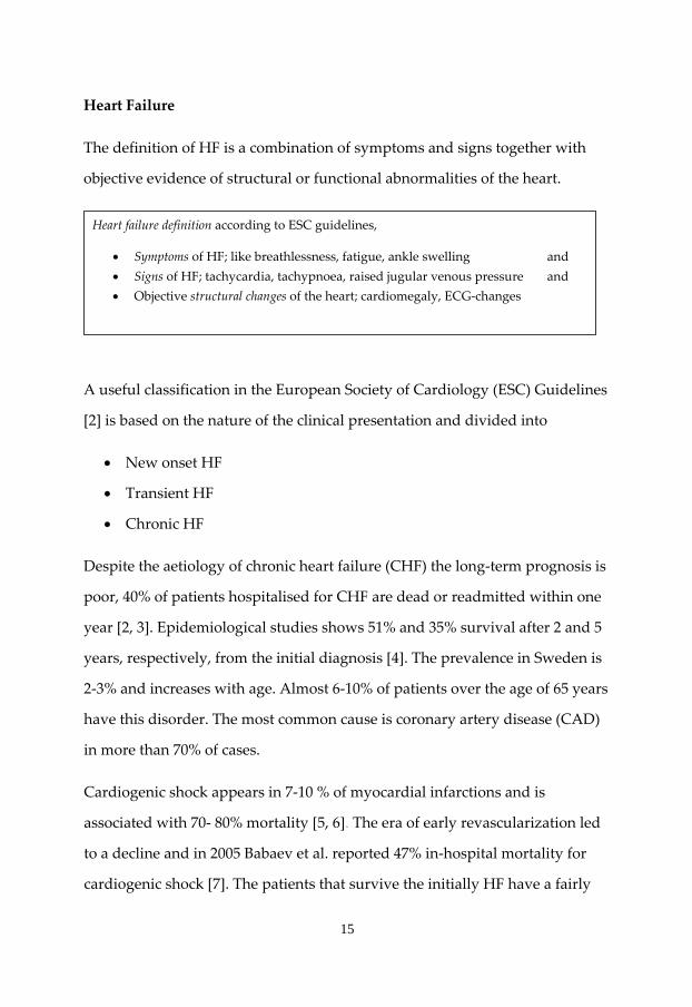
Heart Failure
The definition of HF is a combination of symptoms and signs together with
objective evidence of structural or functional abnormalities of the heart.
Heart failure definition according to ESC guidelines,
• Symptoms of HF; like breathlessness, fatigue, ankle swelling and • Signs of HF; tachycardia, tachypnoea, raised jugular venous pressure and • Objective structural changes of the heart; cardiomegaly, ECG‐changes
A useful classification in the European Society of Cardiology (ESC) Guidelines
[2] is based on the nature of the clinical presentation and divided into
• New onset HF
• Transient HF
• Chronic HF
Despite the aetiology of chronic heart failure (CHF) the long‐term prognosis is
poor, 40% of patients hospitalised for CHF are dead or readmitted within one
year [2, 3]. Epidemiological studies shows 51% and 35% survival after 2 and 5
years, respectively, from the initial diagnosis [4]. The prevalence in Sweden is
2‐3% and increases with age. Almost 6‐10% of patients over the age of 65 years
have this disorder. The most common cause is coronary artery disease (CAD)
in more than 70% of cases.
Cardiogenic shock appears in 7‐10 % of myocardial infarctions and is
associated with 70‐ 80% mortality [5, 6]. The era of early revascularization led
to a decline and in 2005 Babaev et al. reported 47% in‐hospital mortality for
cardiogenic shock [7]. The patients that survive the initially HF have a fairly
15
good two‐years survival of 80% [8]. Survival after cardiac arrest in hospital is
25% and with increasing cardiac support systems such as IABP and
extracorporeal membrane oxygenation (ECMO) survival rates have increased
to 40% [9]. Acute heart failure (AHF) can resolve or progress to CHF
depending of the initial cause. Postcardiotomy HF occurs in 2‐5% of all cardiac
operations [10]. Early mortality is high, but has declined with the use of MCS
[11].
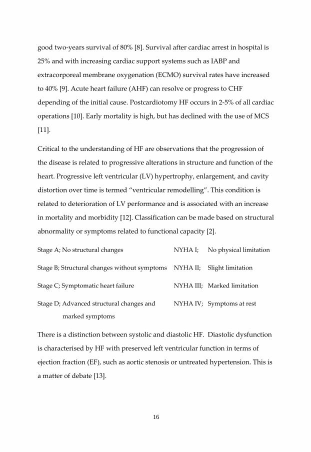
Critical to the understanding of HF are observations that the progression of
the disease is related to progressive alterations in structure and function of the
heart. Progressive left ventricular (LV) hypertrophy, enlargement, and cavity
distortion over time is termed “ventricular remodelling”. This condition is
related to deterioration of LV performance and is associated with an increase
in mortality and morbidity [12]. Classification can be made based on structural
abnormality or symptoms related to functional capacity [2].
Stage A; No structural changes NYHA I; No physical limitation
Stage B; Structural changes without symptoms NYHA II; Slight limitation
Stage C; Symptomatic heart failure NYHA III; Marked limitation
Stage D; Advanced structural changes and NYHA IV; Symptoms at rest
marked symptoms
There is a distinction between systolic and diastolic HF. Diastolic dysfunction
is characterised by HF with preserved left ventricular function in terms of
ejection fraction (EF), such as aortic stenosis or untreated hypertension. This is
a matter of debate [13].
16
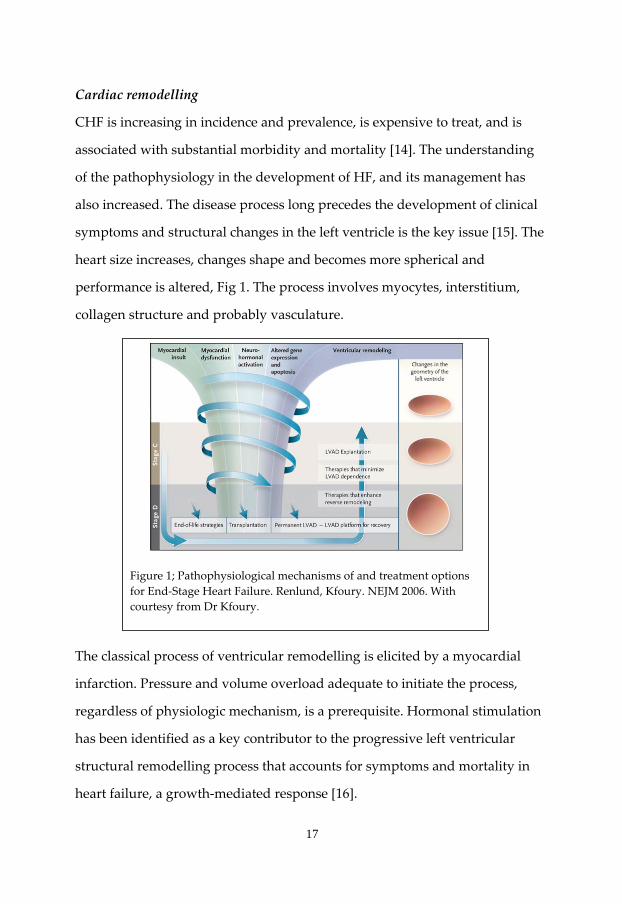
Cardiac remodelling
CHF is increasing in incidence and prevalence, is expensive to treat, and is
associated with substantial morbidity and mortality [14]. The understanding
of the pathophysiology in the development of HF, and its management has
also increased. The disease process long precedes the development of clinical
symptoms and structural changes in the left ventricle is the key issue [15]. The
heart size increases, changes shape and becomes more spherical and
performance is altered, Fig 1. The process involves myocytes, interstitium,
collagen structure and probably vasculature.
Figure 1; Pathophysiological mechanisms of and treatment options for End‐Stage Heart Failure. Renlund, Kfoury. NEJM 2006. With courtesy from Dr Kfoury.
The classical process of ventricular remodelling is elicited by a myocardial
infarction. Pressure and volume overload adequate to initiate the process,
regardless of physiologic mechanism, is a prerequisite. Hormonal stimulation
has been identified as a key contributor to the progressive left ventricular
structural remodelling process that accounts for symptoms and mortality in
heart failure, a growth‐mediated response [16].
17

The role of the neurohumoral systems (sympathetic nervous system, renin‐
angiotensin‐aldosteronesystem, endothelin and others) has led to the
development of several pharmacological inhibitors with favourable effect on
the clinical syndrome. Angiotensin‐converting enzyme (ACE) inhibitors,
angiotensin‐receptor blockers (ARB), ß‐adrenoreceptor inhibitors and
aldosterone inhibitors have all been shown to exert a favourable effect of the
disease and probably on the course of structural remodelling of the left
ventricle.
The impaired function of the ventricle is the fundamental cause of the
symptoms, whether mediated directly by left ventricular haemodynamics or
indirectly by mechanical changes on ventilation, renal sodium retention, or
neurohormonal activation. Diuretics used to reduce filling pressure are
effective in reversing symptoms, as are vasodilators and inotropes to improve
left ventricular ejection fraction and reduce filling pressure. Symptom relief is
not necessarily effective in reversing or slowing the progressive structural
remodelling process. Reversion of the heart toward more normal shape and
function is called reverse remodelling [17] and is the goal of treatment with
ventricular constraint devices such as the CorCap CSD.
Remodelling may be an adaptive process like ventricular dilatation as
compensatory response to volume overload in valve insufficiency with
regurgitation in order to maintain a sufficient cardiac stroke volume. In
conditions like myocardial infarction, non‐ischaemic forms of myocarditis, and
cardiomyopathy, structural changes are maladaptive from the beginning.
There is a relationship between impaired left ventricular function (ejection
fraction (EF), left ventricular end‐diastolic dimension (LVEDD)) and poor
prognosis [18]. Natriuretic peptide levels, especially B‐type natriuretic peptide
18

(BNP) strongly correlates with left ventricular remodelling and prognosis [19].
They can be used as a complement to clinical assessment in the management
of heart failure [20, 21].
Pharmacological treatment;
Objectives in the treatment are to relieve symptoms and signs, improve
Quality of Life (QoL) and prevent the occurrence and/or progression of
myocardial damage in order to slow the process of ventricular remodelling
and reduce mortality. New pharmacological treatment have been developed
during the last two decades including ACE‐inhibitors, ARBs, beta‐blockers
(Class I, Level A) and spironolactone (Class I, Level B), which all interact in the
process of ventricular remodelling [15, 22].
Angiotensin‐Converting‐Enzyme (ACE)‐inhibitors (Class I, Level A) are strongly
recommended in patients with CHF, regardless of symptoms and an EF<40%.
It improves the ventricular function, patients QoL, reduces hospital
readmissions and improve survival [23].
ß‐Blockers (Class I, Level A) are also strongly recommended with the same
indications and treatment results as for the ACE‐inhibitors [24].
Aldosterone antagonists (Class I, Level B) are recommended for severe HF with
EF<35%. Hospital readmissions are reduced and survival improved when
added to existing therapy [25].
Angiotensin receptor blockers (ARB) (Class I, Level A) are used when patients
still are symptomatic despite ACE‐inhibitors and ß‐blocker treatment [26].
19
There are several other pharmacological medications used in the HF
treatment, but the levels of evidence are lower. Complementary drugs include
Hydralazine (Class IIa, Level B), Digoxin (Class I, Level C) and diuretics (Class I,
Level B). These have a more symptomatic profile and do not affect survival.
Experimental studies suggest that gene transfer may be effective in the process
of ventricular remodelling, but the clinical implication still remains to be seen.
The use of embryonic and adult stem cells in the treatment of ventricular
remodelling is interesting. The process called plasticity or transdifferentiation
shows great potential, though there is a long way left to clinical application.
The development of stem cell therapy, after animal testing, stands in front of
clinical studies in randomized trials [27].
Surgical treatment;
There are several treatment options for this category of patients. The presence
of surgically correctable conditions causing HF constitutes an indication for
surgical correction.
Coronary artery disease (CAD) is the most common cause of HF [28]. In
myocardial infarction early reduction of wall stress and restoration of blood
flow to the infarcted area can minimize myocyte damage, limit infarct size and
remodelling [29], and improve function [30]. Two revascularisation modalities
are available, percutaneous angioplasty/stenting (PCI) and coronary artery
bypass grafting (CABG). The choice is depending on time frame, availability,
indications, co‐morbidity and the extent of coronary atherosclerosis.
Hibernating myocardium represents dysfunctional tissue distal to a severe
stenosis where the metabolic function is markedly down‐regulated.
20

Improvement in function and reduced mortality can be achieved with
revascularisation [30‐32]. Detection of viability and the potential for regained
function in hibernating myocardium is important before revascularisation is
carried out [33] (Class IIa, Level C). Recently a score system, the SYNTAX‐
score [34], based on the angiographic pattern of coronary stenosis, have been
suggested as a help in deciding whether to perform PCI or CABG.
Valvular heart disease, i.e significant aortic stenosis (Class I, Level C)/
regurgitation (Class I, Level B), mitral valve regurgitation (Class I, Level C)
and tricuspid insufficiency (Class III, Level C) should be handled according to
the ESC Guidelines for Valvular Heart Disease [35]. Preoperative optimisation
is important to reduce the perioperative risk for morbidity and mortality and
acute surgery should be avoided.
Cardiac Resynchronization Therapy (CRT). Patients with CHF frequently
develop electrical abnormalities leading to mechanical abnormalities. CRT is
recommended to reduce mortality and morbidity [36‐38] in patients with low
EF and QRS width >120 ms on the electrocardiogram, and with symptoms
despite optimal medical treatment (Class I, Level A) [39]. A broad QRS
complex is associated with poor long‐term survival [40]. There is also reason
to combine the CRT with an internal defibrillator (CRT‐D) due to the risk of
sudden cardiac death (Class I, Level A) [41]. CRT is an adjunct to
pharmacological medication in many patients with CHF to reduce mortality.
Therapy development is heading at preventive implantation to reduce heart
failure events [42].
21

Heart transplantation; The gold standard in patients with end‐stage CHF is
heart transplantation (Class I, Level C) [43]. Due to organ shortage, this option
is only for a limited number of patients and the waiting time for an adequate
organ can be long. World‐wide about 3.300 transplants are performed
annually. Ten‐year survival is approximately 50% [44] but is continually
improving. There is a significant risk of morbidity due to long‐term
immunosupression therapy with hypertension, aggressive CAD and
development of malignant disease. Non‐CMV infections, graft failure and
rejection are the dominant cause of mortality in the first year [45]. Newer
pharmacological treatments reduce the risk for rejection. The dominant
primary indication for transplantation has shifted towards non‐coronary
cardiomyopathy. In a recent audit of the ISHLT register 29% of transplant
candidates were on some form of mechanical support preoperatively, an
increase from previous estimates [45]. There has been an improvement in early
post‐transplant survival in MCS patients, but no significant difference long‐
term mortality. In recent years fewer recipients have been hospitalized prior to
surgery due to outpatient treatment with MCS. The disappointing long‐term
survival is explained by the fact that recent recipients have more risk factors
than before.
Mechanical assist devices (Class IIa, Level C) have been used since the end of
the Eighties and they are mostly used for bridging treatment in critically ill
patients waiting for transplantation [46, 47]. Reversing multi‐organ failure,
allows patients to rehabilitate and gain strength, increasing their survival
while awaiting transplantation. Improved tissue microcirculation [48] allows
end‐organ function to recover. Achieving a time frame, > 30 days, prior to
transplantation also improves post‐transplant survival [49], at least with the
22

first generation of MCS devices. The use of left ventricular assist devices
(LVAD) has shown signs of reverse remodelling [50‐52] including regression
in myocardial fibrosis, reduction in apoptosis and myocytolysis, and
improved myocyte function [53]. With time there is a decrease in the
neurohumoral activation [54].
A milestone in the development and progress of the use of MCS was the
REMATCH‐study [55], reporting better survival after one year for LVAD
patients compared to a control group of patients receiving optimal
pharmacological therapy for advanced HF and not eligible for heart
transplantation. Newer pump generations are smaller and have improved
long‐term reliability. This has made DT a more feasible alternative [56], where
transplantation can be postponed or even avoided altogether [57, 58]. Survival
at one year has improved with the second generation LVAD [46]. Some
patients can be bridged to recovery [59], but the rate is low.
There are several pump systems, both for acute and chronic HF, giving new
possibilities for MCS to work as bridge‐to‐bridge, bridge‐to‐destination and
also bridge‐to‐recovery. The systems can be used for unloading the left
ventricle (LVAD), right ventricle (RVAD) or both ventricles (BiVAD) of the
heart. There is also the totally artificial heart device (TAH), where the entire
heart is replaced. Patient selection is demanding and based on a multi‐
professional team‐work [60]. There are difficulties in conducting randomised
studies in this group of very sick patients where pharmacological treatment
has failed. Morbidity such as bleeding [61], infections, thrombo‐embolic events
and right ventricular failure, are demanding issues during MCS support [62].
Deplacement pumps were the first generation of flow pumps, the axial flow
pumps were the second generation, and the third generation includes small
23

centrifugal assist devices. There are several other available pumps systems on
the market. First generation pump systems; HM‐I, Berlin Heart, CardioWest
(TAH), second generation; HM‐II, DeBakey, Jarvik 2000, Incor, and the third
generation; DuraHeart, HeartWare. The TAH is under constant development
and has so far been used as bridge‐to‐transplant.
Surgical restoration of the left ventricle in order to reshape the heart to
improve pump function has been done [63, 64]. There are several methods of
ventricular surgery to reduce wall stress and decrease ventricular size
according to the law of LaPlace. The Dor‐procedure [65], and ventricular
constraint are examples [66]. There has been an ongoing debate since the
STICH‐trial was published 2009 [67].
A novel option for CHF patients is the concept of passive ventricular
constraint, the Acorn Cardiac Support Device (CSD). The theory behind this is
to slow down the process of ventricular remodelling by wrapping a net
around the ventricles of the heart, named reverse remodelling. According to
the law of LaPlace, wall stress decreases when there is a mechanical restriction
outside the wall. This stimulates reverse remodelling leading to improved
function of the heart [68]. Animal studies shows decreased echocardiographic
dimensions [69] and signs of reverse remodelling at the cellular level [70].
Initial clinical evaluation indicates smaller dimensions and better performance
of the heart with improved QoL [71]. It is even more convincing when
combining reverse remodelling with mitral valve surgery [72]. Long‐term
follow‐up shows sustained improvement regarding echocardiographic
dimensions and cardiac function after five years [73, 74].
24

Acute heart failure (New onset heart failure)
This can be defined as an acute onset of HF symptoms necessitating rapid
treatment measures [2]. The clinical presentation can be divided into acute
decompensated HF and acute vascular failure (hypertensive, pulmonary
oedema, cardiogenic shock, high output failure) [75]. The underlying
mechanism can be described like an afterload mismatch with elevated
systemic vascular resistance (SVR) in combination with impaired systolic
performance [76], a combination of a cardiac and a vascular pathway. Cotter et
al suggests that fluid accumulation, ischaemia, and arrhythmias play a minor
role for the initiation of AHF, but other mechanisms such as neurohormonal
activation, decrease in vascular plasticity, and fluid redistribution are more
important. There are several aetiologies like IHD including acute coronary
syndrome, acute myocardial infarctions, and also decompensation of CHF,
valvular disease, myopathy, myocarditis and also other non‐cardiac conditions
such as septicaemia. Acute coronary syndrome is the cause in 42% of patients
admitted for AHF for the first time [77]. In a French study, of patients
admitted to the ICU because of AHF, 61% were diagnosed with IHD and 29%
presented with cardiogenic shock. Early mortality (30 days) was 43% and after
one year 62% [78]. The authors also report that patients presenting with
cardiogenic shock had a 58% early mortality, but during the time period from
30 days to one year there was no difference in mortality between shock and
non‐shock patients. The immediate goal with these patients is to stabilise the
haemodynamic situation for optimal tissue oxygenation and relief of
symptoms.
Various forms of inotropic support are used (Class IIb, Level B) in
deteriorating patients with cardiogenic shock. In patients with acute coronary
25

syndromes early revascularisation is mandatory [32]. The use of IABP or MCS
(ECMO, LVAD) as adjuvant therapy may be necessary. The IABP is the most
widespread cardiac support device in use. It reduces afterload and increase
coronary perfusion by increasing mean arterial pressure during diastole. The
cardiac work‐load is decreased and the oxygen demand lower [79]. It is mostly
used in patients with cardiogenic shock [80], but also in coronary syndromes
and after cardiac surgery. Evidence for the prevention of remodelling is
lacking. The IABP has [81] been in clinical use for many years, but its use has
been less than expected [82]. Only 20‐ 30% of patients with cardiogenic shock
world‐wide are treated with IABP, possibly due to lack of large randomised
trials, even though its use is recommended in the European Society of
Cardiology guidelines (Class I, Level C) for myocardial infarction [83]. Since it
is relatively cheap and easy to use it is widely spread around the world. The
idea behind the ImpellaTM LVAD axial flow pump originates from the
HemopumpTM [84], used in the 1990ties as short‐term MCS [85].
A clinical programme, involving cardiologists, anesthesiologists and
cardiovascular surgeons, for new onset heart failure is important, because time
matters [86]. It is also important to have a referral network based on the use of
implantable MCS [87]. This should also include patients after cardiac surgery
with postoperative cardiogenic shock. The subject of cardiac metabolism in
heart failure is interesting. Impaired metabolic flexibility in the heart may
reduce the contractile function [88]. Substrate selection, glucose control and
improving mitochondrial function are targets for improving contractility. A
metabolic strategy has been shown to reduce mortality and the use of
inotropic agents in ischaemic patients with reduced left ventricular function
and new onset heart failure after coronary artery bypass grafting [89].
26
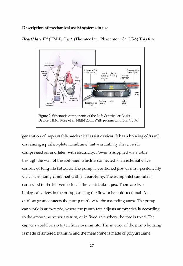
Description of mechanical assist systems in use
HeartMate ITM (HM‐I); Fig 2. (Thoratec Inc., Pleasanton, Ca, USA) This first
Figure 2; Schematic components of the Left Ventricular Assist Device, HM‐I. Rose et al. NEJM 2001. With permission from NEJM.
generation of implantable mechanical assist devices. It has a housing of 83 mL,
containing a pusher‐plate membrane that was initially driven with
compressed air and later, with electricity. Power is supplied via a cable
through the wall of the abdomen which is connected to an external drive
console or long‐life batteries. The pump is positioned pre‐ or intra‐peritoneally
via a sternotomy combined with a laparotomy. The pump inlet cannula is
connected to the left ventricle via the ventricular apex. There are two
biological valves in the pump, causing the flow to be unidirectional. An
outflow graft connects the pump outflow to the ascending aorta. The pump
can work in auto‐mode, where the pump rate adjusts automatically according
to the amount of venous return, or in fixed‐rate where the rate is fixed. The
capacity could be up to ten litres per minute. The interior of the pump housing
is made of sintered titanium and the membrane is made of polyurethane.
27
These raw surfaces reduce the need for anticoagulation due to the
development of a pseudointima. It is intended for long‐term use.
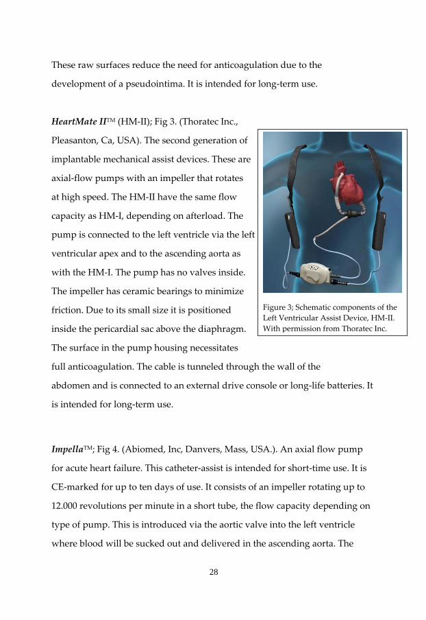
HeartMate IITM (HM‐II); Fig 3. (Thoratec Inc.,
Pleasanton, Ca, USA). The second generation of
implantable mechanical assist devices. These are
axial‐flow pumps with an impeller that rotates
at high speed. The HM‐II have the same flow
capacity as HM‐I, depending on afterload. The
pump is connected to the left ventricle via the left
ventricular apex and to the ascending aorta as
with the HM‐I. The pump has no valves inside.
The impeller has ceramic bearings to minimize
Figure 3; Schematic components of the friction. Due to its small size it is positioned Left Ventricular Assist Device, HM‐II.
inside the pericardial sac above the diaphragm. With permission from Thoratec Inc.
The surface in the pump housing necessitates
full anticoagulation. The cable is tunneled through the wall of the
abdomen and is connected to an external drive console or long‐life batteries. It
is intended for long‐term use.
ImpellaTM; Fig 4. (Abiomed, Inc, Danvers, Mass, USA.). An axial flow pump
for acute heart failure. This catheter‐assist is intended for short‐time use. It is
CE‐marked for up to ten days of use. It consists of an impeller rotating up to
12.000 revolutions per minute in a short tube, the flow capacity depending on
type of pump. This is introduced via the aortic valve into the left ventricle
where blood will be sucked out and delivered in the ascending aorta. The
28

electromagnetic motor is placed in the pump, near the impeller. A power cable
is connected to a drive console bedside. There is a monitor on the pump
housing that can measure the pressure gradient between the ventricle and the
Figure 4, Different ImpellaTM pumps. LP; left peripheral, LD; left direct, RD; right direct With permission from Abiomed Inc.
ascending aorta, indicating correct placement. There are three different types,
Left Direct (LD) 5.0 placed via the ascending aorta through a synthetic graft. It
provides flows up to 5 litres per minute. Sternotomy is required for
implantation and removal. Left Peripheral (LP) 5.0 and 2.5, the same principle
as the LD but access via the femoral artery. It is positioned in the left ventricle
with the aid of fluoroscopy or echocardiography. There is one pump for right
ventricular failure, RV, which unloads the right ventricle when connected
29

between the right atrium and the pulmonary artery. This pump, however, has
been withdrawn by the manufacturer and is no longer commercially available.
CorCap Cardiac Support DeviceTM (CSD); Fig 5. (Acorn Cardiovascular, Inc, St.
Paul, MN). A synthetic net, to surgically be wrapped
around both ventricles of the heart, providing diastolic
support and reducing wall stress. It is made of
polyurethane weave with a bidirectional stretch to
promote the ellipsoid shape of the heart. The first
generation necessitates sternotomy and is sewn with
interrupted sutures to the AV‐groove and adjusted to
fit the size of the ventricles. The sternotomy access is
easy when in combination with valve‐ or bypass‐surgery.
Figure 5; CorCap CSD. With permission from Acorn Inc.
The second generation has a delivery tool to be used
with a left mini‐thoracotomy, where the net in a parachute manner can be
placed around the ventricles. The size of the net is
determined from preoperative CT‐scans.
Intra‐aortic ballon pump (IABP); A catheter‐based
balloon, placed in the descending aorta via the femoral
artery, Fig 6. During diastole, the balloon inflates and
thereby displacing blood from the descending aorta
and then deflates immediately before systole, creating
a void in the aorta, and thereby producing its haemo‐ Figure 6; Intra‐aortic balloon Pump, IABP. With courtesy Texas Heart Institute.
dynamic effect. Inflation causes a rise in the aortic
30

pressure causing an increase in the coronary pressure gradient and therefore
increases coronary flow. Aortic counter‐pulsation also causes a drop in systolic
blood pressure due to balloon deflation just prior to systole. The increase in
diastolic pressure is typically greater than the decrease in systolic pressure,
resulting in an increase in mean arterial pressure. Helium is used in the
balloon.
31
Statistics;
In Paper I all continuous variables had normal distribution. Statistical analysis
was performed with analysis of variance (parametric tests) and discriminant
analysis using SPSS software (v 10.1.0; SPSS Inc, Chicago, IL). Descriptive data
are expressed as median and range. In the variance analyses mean ±standard
deviation was used. For the non‐parametric ordinal data a χ2‐test was used. A
p value < 0.05 was considered statistically significant. In Papers II, III and IV
the samples were analysed using the software STATISTICA (StatSoft, Inc.
2004, version 7, Tulsa, Ok, USA). The data was analysed using the non‐
parametric Mann‐Whitney U‐test for group comparison and Wilcoxon
matched pair tests for longitudinal comparison. p < 0.05 was considered
significant.
32
33
Aims
‐ to investigate the outcome and risk factors for mortality and morbidity in
patients treated with mechanical circulatory support for long‐term use in
patients accepted for heart transplantation. (Paper I)
‐ to study the morbidity and mortality in patients treated with the axial flow
pump, HeartMate‐IITM as bridge‐to‐transplantation and destination therapy.
(Paper II)
‐ to investigate the use and outcome in patients with acute heart failure treated
with ImpellaTM axial flow pump as short‐term assist. (Paper III)
‐ to study the use and long‐term outcome, especially regarding Quality of Life
in patients operated with ventricular constraint CorCap‐CSDTM, in patients
with chronic heart failure, compared to a reference group from the general
population. (Paper IV)

34
Methods
Paper I
All Swedish patients (n=59) on the waiting list for heart transplantation, since
1993 until May 2002, and treated with long‐term assist device as bridge‐to‐
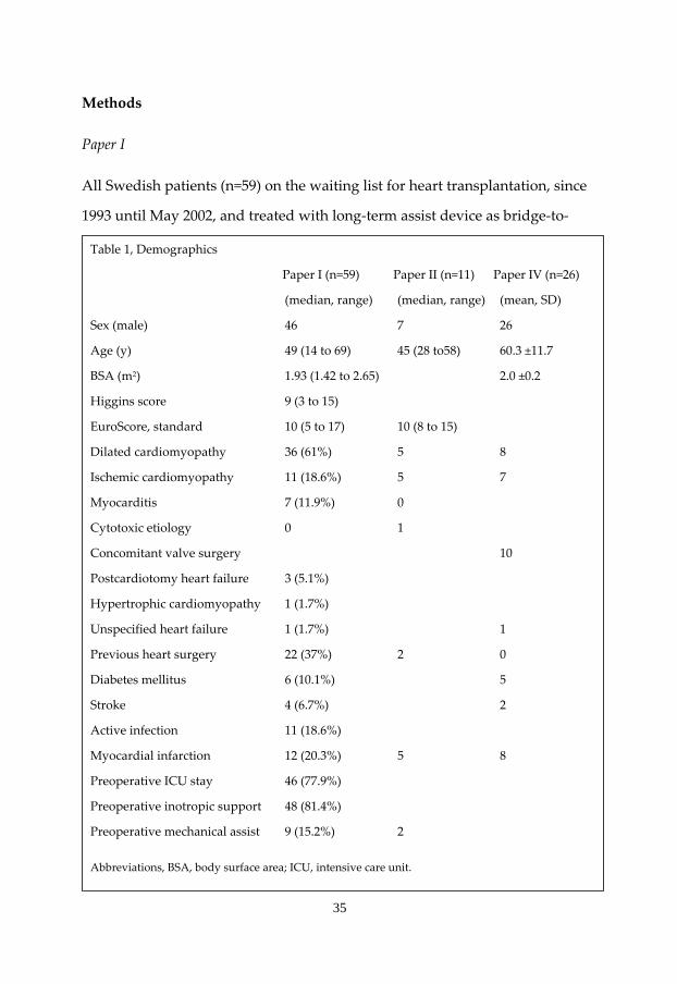
Table 1, Demographics
Paper I (n=59) Paper II (n=11) Paper IV (n=26)
(median, range) (median, range) (mean, SD)
Sex (male) 46 7 26
Age (y) 49 (14 to 69) 45 (28 to58) 60.3 ±11.7
BSA (m2) 1.93 (1.42 to 2.65) 2.0 ±0.2
Higgins score 9 (3 to 15)
EuroScore, standard 10 (5 to 17) 10 (8 to 15)
Dilated cardiomyopathy 36 (61%) 5 8
Ischemic cardiomyopathy 11 (18.6%) 5 7
Myocarditis 7 (11.9%) 0
Cytotoxic etiology 0 1
Concomitant valve surgery 10
Postcardiotomy heart failure 3 (5.1%)
Hypertrophic cardiomyopathy 1 (1.7%)
Unspecified heart failure 1 (1.7%) 1
Previous heart surgery 22 (37%) 2 0
Diabetes mellitus 6 (10.1%) 5
Stroke 4 (6.7%) 2
Active infection 11 (18.6%)
Myocardial infarction 12 (20.3%) 5 8
Preoperative ICU stay 46 (77.9%)
Preoperative inotropic support 48 (81.4%)
Preoperative mechanical assist 9 (15.2%) 2
Abbreviations, BSA, body surface area; ICU, intensive care unit.
35

transplantation due to deteriorating heart failure, were retrospectively
investigated. Demographics are depicted in Table 1. Dominating diagnoses
were dilated cardiomyopathy (61%) and ischaemic heart disease (18.6%). Nine
(15.2%) patients had a mechanical assist device before the LVAD implant. Pre‐,
per‐ and postoperative variables were recorded from the LVAD implant to
heart transplantation. The variables were evaluated regarding mortality and
morbidity prior to transplantation and risk factors for mortality, right
ventricular failure and infection.
Paper II
Patients receiving the second generation of long‐term mechanical assist
devices with axial flow at our department were consecutively included. Eleven
patients from October 2005 until May 2008 with ischaemic cardiomyopathy
(n=5), dilated cardiomyopathy (n=5) and cytotoxic aethiology (n=1) were
treated with HeartMate‐II due to deteriorating heart failure. They were
prospectively studied using a protocol. Demographics are depicted in Table 1.
In ten patients the HM‐II was implanted electively and in one patient acutely
due to rapid irreversible deterioration of the disease despite maximal
pharmacologic treatment. Two patients had a temporary left ventricular (LV)
assist (ImpellaTM) before implantation of the HM‐II, one due to cardiogenic
shock in association with myocardial infarction and one with deteriorating
dilated cardiomyopathy (CMP). In ten of the patients the indication for
implantation was bridge‐to‐heart transplantation. One patient with recent
malignancy and cytotoxic CMP received the pump as bridge‐to‐recovery or
‘‘destination’’ therapy.
36

Paper III
Data on all Swedish patients treated with the Impella axial flow pump for
acute heart failure between 2003 and 2007 were retrospectively collected from
those Swedish cardiothoracic centres using the device. Fifty patients were
divided into two groups, the Surgical group (n=33) which involved patients
with acute heart failure after cardiac surgery, and the Non‐surgical group
(n=17) where patients suffered from acute heart failure due to cardiological
conditions. Demographics are shown in Table 2. The main treatment intension
was bridge‐to‐recovery. In the surgical group 55% of the patients had a
severely reduced (EF<20%) left ventricular function preoperatively. Prior to
surgery, eight patients (25%) were on intra‐aortic balloon counter pulsation
(IABP), one (3%) was supported with a LVAD, 11 patients (33%) were on
inotropic support and seven patients (21%) were mechanically ventilated.
Fifteen of 19 failure‐to‐wean patients (79%) received the Impella prior to
weaning from the CPB. The remaining 14 patients developed their
postoperative cardiac failure in the ICU. Transoesophageal echocardiography
(TEE) was used to verify correct positioning of the pump in the LV. The
patients were considered to be responding to the therapy when the
haemodynamics were stable, with improved cardiac output (CO) and
lowering of the filling pressures and/or demonstration of an increased mixed
venous oxygen saturation (SvO2). Our definition of therapy response also
required a combination of improved echocardiographic movement, small to
moderate doses of inotropic support and an acceptable diuresis. The weaning
procedure started when all parameters were stable for at least 24 hours.
37

Table 2, Demographics (Paper III)
Surgical group (n=33) Non‐surgical group(n=17)
(n) (n)
Age, years (mean, range) 58.1 (27 to 84) 47.5 (36 to 63)
Male 24 (73%) 11 (79%)
BSA, m2 (mean, range) 1.9 (1.5 to 2.7) 1.9 (1.1 to 2.1)
Previous cardiac surgery 7 (21%) 0 (0%)
Myocardial infarction 14 (42%) 6 (43%)
Stroke 1 (3%) 0 (0%)
Diabetes Mellitus 7 (21%) 2 (14%)
Atrial fibrillation 11 (33%) 1 (7%)
Impaired kidney function 4 (12%) 2 (14%)
CPB‐time, min (mean, range) 249 (57 to 452)
X‐clamp time, min (mean, range) 100 (0 to 236)
NYHA (mean) 3.6 3.8
EuroScore standard (mean, range) 9.1 (2 to 18) 10.1 (3 to 17)
Indication for treatment
Postcardiotomy LOS 19 (58%)
Cardiogenic shock 10 (30%) 9 (53%)
Myocarditis 1 (3%) 5 (29%)
Prophylactic use 3 (18%)
Other 3 (9%)
Treatment aim
Bridge to recovery 22 (67%) 15 (88%)
Bridge to other LVAD 8 (24%) 2 (12%)
Bridge to HTx 3 (9%)
Abbreviations: BSA, Body Surface Area; CPB‐time, cardiopulmonary bypass time; LOS, low output
syndrome; LVAD, left ventricular assist device; NYHA, New York Heart Association class; HTx,
Heart transplantation; X‐clamp time, aortic cross‐clamp time.
38
Paper IV
From 2003 onwards, 26 consecutive patients with chronic progressive heart
failure and optimal pharmacological treatment met the inclusion criteria for
passive ventricular constraint and were operated with CorCap CSD via
sternotomy (n=25) and left thoracotomy (n=1). Demographics are depicted in
Table 1. Seven patients were operated with CorCap‐only, with (n=3) or
without (n=4) epicardial leads for cardiac resynchronisation therapy (CRT).
Nineteen patients were scheduled for concomitant cardiac surgery. They were
prospectively followed each year for five years postoperatively regarding
mortality, echocardiographic findings, execise tests (6MWT, ergometry) and
QoL (MLHF). There were three early and three late deaths during the follow‐
up period. Two patients, operated within the last month were not included in
the follow‐up because they were still in the postoperative phase. The
remaining cohort (n=18) was investigated in a cross‐sectional study regarding
QoL using the Medical Outcomes Study Short Form General Health Survey
(SF‐36) questionnaire. An exact age and sex‐matched reference group (n=140)
was randomly selected from the Swedish SF‐36 general population reference
group database (n=8.930).
39

40
Results
Paper I;
Fifty‐nine patients (46 men) listed for heart transplantation, with a median age
of 49 years (range, 14 to 69 years) received a LVAD as bridge to
transplantation during the observation period. They were supported for a
median time of 99.5 days (range, 1 to 873 days), Table 4. Forty‐five patients
underwent heart transplantation. Mortality prior to transplantation was 11
patients (18.6%). Three patients (5.1%) were weaned from the device. One of
them received HTx after 13 days as a result of progressive heart failure.
Eighteen (30.5%) of the patients had additional surgery; 3 patients underwent
coronary artery bypass grafting; 9 patients had aortic valve replacements; 2
patients atrial septal defect repairs; 1 patient had mitral valvuloplasty; 1
patient had a pericardial patch sutured over a preexisting mechanical aortic
prosthesis; and 2 had removal of a pacemaker. Four patients were treated with
an RV assist device because of RV failure. The ICU‐stay postoperative was in
median of 12 days (range, 1 to 25 days) after surgery. Twenty patients (34%)
were reoperated within 24 hours due to bleeding. In 5 patients (8.5%), the
LVAD had to be replaced because of mechanical failure. In total, 5 patients
had inflow valve incompetence and valve endocarditis. Nine of 59 patients
(15.2%) had an infection at the cable exit site or LVAD pocket. A total of 26
patients (44%) had some form of infection in the postoperative period
(septicaemia, pneumonia, or device‐related). Strong predictors were elevated
filling pressures of the right heart and elevated C‐reactive protein (CRP). One
hospital had a lower frequency of cable infections due to different fixation
technique of the driveline. Eleven patients (19%) were diagnosed with RV
failure. High cardiac index, high postoperative central venous pressure, long
41

operation time, low baseline mean arterial pressure, and high baseline C‐
reactive protein were predictors. Minor technical problems included sensor
dysfunction with the pneumatic Heart‐Mate LVAD in 3 patients. Controller
malfunction with the electrical HeartMate occurred in 7 patients, primarily in
early cases. Six patients (10%) experienced a cerebrovascular thromboembolic
lesion. Post‐transplantation follow‐up of all patients in January 2003 showed
11 late deaths (24%), after a median time of 100 days (range, 0 to 1.092 days).
Seventeen of 38 (45%) of the vented electrical Heart‐Mate pump patients were
discharged home while on pump treatment. The patient treated for 873 days
was treated as an ambulatory patient for 441 days before HTx.
Table 3, Risk factors for mortality, RV‐failure and infection (ANOVA) HM‐I patients.
Mortality p Blood transfusions <0.001 Plasma transfusions <0.001 Ventilator time <0.001 S‐Creatinine (end) <0.001
RV‐failure CRP preoperatively 0.001 CVP postoperatively 0.002
Infection Pcw (end) <0.001 CRP (end) 0.001 Implanting hospital (χ2‐test) <0.001
Abbreviations; RV; right ventricular, HM; HeartMate, CRP; C‐reactive proteine, CVP; central venous pressure, pcw; pulmonary capillary wedge pressure.
42
Paper II;
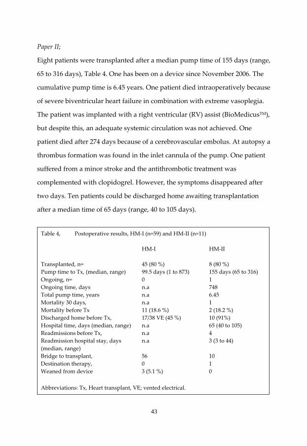
Eight patients were transplanted after a median pump time of 155 days (range,
65 to 316 days), Table 4. One has been on a device since November 2006. The
cumulative pump time is 6.45 years. One patient died intraoperatively because
of severe biventricular heart failure in combination with extreme vasoplegia.
The patient was implanted with a right ventricular (RV) assist (BioMedicusTM),
but despite this, an adequate systemic circulation was not achieved. One
patient died after 274 days because of a cerebrovascular embolus. At autopsy a
thrombus formation was found in the inlet cannula of the pump. One patient
suffered from a minor stroke and the antithrombotic treatment was
complemented with clopidogrel. However, the symptoms disappeared after
two days. Ten patients could be discharged home awaiting transplantation
after a median time of 65 days (range, 40 to 105 days).
Table 4, Postoperative results, HM‐I (n=59) and HM‐II (n=11) HM‐I HM‐II Transplanted, n= 45 (80 %) 8 (80 %) Pump time to Tx, (median, range) 99.5 days (1 to 873) 155 days (65 to 316) Ongoing, n= 0 1 Ongoing time, days n.a 748 Total pump time, years n.a 6.45 Mortality 30 days, n.a 1 Mortality before Tx 11 (18.6 %) 2 (18.2 %) Discharged home before Tx, 17/38 VE (45 %) 10 (91%) Hospital time, days (median, range) n.a 65 (40 to 105) Readmissions before Tx, n.a 4 Readmission hospital stay, days n.a 3 (3 to 44) (median, range) Bridge to transplant, 56 10 Destination therapy, 0 1 Weaned from device 3 (5.1 %) 0
Abbreviations: Tx, Heart transplant, VE; vented electrical.
43

Nine of them were on the waiting list for heart transplantation.
Four patients were re‐admitted to the hospital with a median hospital stay of 3
days (range, 3 to 44 Days). Ten patients were given a LVAD for bridge‐to‐
transplant and one for destination therapy. Three patients had transient right
ventricular failure, responding to pharmacological therapy, when weaning
from CPB. Four patients were reoperated due to bleeding, two patients during
the first 24 hours and two patients later in the postoperative period. The
median postoperative blood loss the first postoperative day was 1 228 mL
(range, 360 to 5 600 mL). The median time on ventilator was 11 days (range,
0.5 to 27 days) postoperatively. One patient required temporary dialysis.
All patients received low molecular weight heparin (LMWH) initially and
acetylsalicylic acid (ASA) from the first postoperative day. Warfarin was
started and administered individually depending on the early postoperative
course. The LMWH was discontinued when international normalised ratio
(INR) reached therapeutic level. Almost all patients recieved triple therapy
with ASA, clopidogrel and warfarin. Dosage was adjusted regarding the
response to the thrombelastogram (TEG). Two patients had late minor
haemorrhagic events with gastrointestinal bleeding. Six patients were
diagnosed with cable infections according to wound cultures and treated
successfully with intravenous antibiotics. No surgical revision was performed.
Two patients also developed septicaemia caused by KNS, Candida albicans
and methicillin‐resistant staphylococci (MRSA). This complication was treated
successfully with antibiotics without sequele or signs of pump endocarditis.
Two patients experienced ventricular arrhythmias necessitating
pharmacological treatment and in one case electro‐conversion. One patient
suffered from increasing abdominal pain prior to planned discharge. He was
44
operated on for gangrenous cholecystitis and made an uneventful
postoperative course.
Cardiac output, filling pressures and mixed venous oxygen saturation
improved significantly when comparing preoperative and postoperative
values.
All patients (n=8) that were transplanted are still alive (December 2008) with a
good life quality. No mechanical errors were recorded. There was one pump‐
stop, probably due to battery change error caused by the patient.
Paper III;
Early mortality in the surgical and non‐surgical groups was 45% and 23%,
respectively, see figure 7. Complications included infection, 36% and right
ventricular failure, 28%.
Surgical group; Cardiac output and cardiac power output postoperatively were
significantly higher among survivors than non‐survivors. The 30‐day
mortality was 45% (15/33 patients). Nine of these 15 patients died within one
week of their operation. The most common cause of death was multiorgan
failure. The 1‐year mortality was 64% (21/33). Patients who received Impella
RD for right ventricular failure had a 30‐day mortality of 75% (6/8), and the
one‐year mortality for these patients was 87% (7/8). In all, the Impella device
was used for a mean of 3.8 days (range, 0.1 to 9 days). The RD was used for a
mean of 4.1 days (range, 0.1 to 8 days) and LD/LP for a mean of 3.9 days
(range, 0.1 to 9 days). Seventeen patients (52%) were reoperated within 24
hours because of excessive bleeding. Nine patients (27%) were reoperated late
in the postoperative period because of bleeding (n=3), sternal infection (n=4),
late sternal closure (n=1) and one for a reason unknown. Device failure was
45

recorded in one case at 3 days. Ten patients (30%) had septicaemia and 13
patients (39%) required dialysis. Fourteen patients (42%) had transient and
pharmacologically treated right ventricular failure, and four of the patients
treated with Impella LVAD had transient right ventricular (RV) failure
necessitating RV‐assist systems. Survival after 30 days was significantly better
for patients with preoperatively placed IABP (p=0.01). Cardiac output (CO) at
12 hours and Cardiac Power Output (CPO) at 6 and 12 hours were also
significantly higher among survivors. Improved survival postoperatively was
indicated by low filling pressures and high mixed venous oxygen saturation
(SvO2) after the first 12 hours, but these observations did not reach statistical
significance. At one‐year follow‐up the mortality was 64% (21/33). The
survivors had significantly higher SvO2 (12 hours) and higher CPO at 12
hours. The preoperative use of IABP was a marker of improved survival
(p=0.01).
Non‐surgical group; 30‐day mortality in this group was 23% (4/17). Excluding
the three patients with LP 2.5 used prophylactically, the 30‐day mortality for
non‐surgical patients with an Impella placed for acute cardiac failure was 21%
(3/14). One of the 30‐days survivors died during the first year after the
treatment. Five of these patients have not yet reached one‐year follow‐up. The
patients in the non‐surgical group were significantly younger than the surgical
patients. No RV‐failure was recorded. Three patients (18%) required dialysis.
In this group of patients the left Impella was used for a mean time of 7.3 days
(range, 2 to 14 days). One patient was bridged‐to‐ECMO after one day and did
not survive. One patient was bridged‐to‐HM‐II after 8 days and later had a
cardiac transplant.
46

Figure 7, Cumulative survival Impella patients
0 6 12 18 24 30 36 42 48 54 60
Survival time (Months)
0,0
0,1
0,2
0,3
0,4
0,5
0,6
0,7
0,8
0,9
1,0
Cin
g
Surgical group
Paper IV;
The mean follow‐up time for the CSD group after cardiac surgery is 3.9 years
(range, 0.9 to 7 years). The one‐year survival for CSD patients was 86% and
after three years 76%. There were three early deaths, two patients with
septicaemia after 6 and 50 days, and one sudden cardiac death after 14 days.
There were three late deaths, two after 2 years (cardiac arrest and multiorgan
umul
ativ
eio
urvi
v
Non-Surgical group
n S
Pro
port
Patients at risk Surgical 33 11 7 Non‐ 17 7 surgical
47

failure), and one after almost five years in progressive heart failure. The
comparison of QoL measured by the SF‐36 between CSD patients and the
control group is similar regarding all eight dimensions and the two summary
parameters PCS(SF‐36) and MCS(SF‐36), Fig 8. Echocardiographic dimensions
(LVEDD, EF) and QoL (MLHF) improved significantly after one and three years
postoperatively for the CSD patients.
Figure 8,
SF-36 QoL CorCap/Norm/HF (mean)
0,0
10,0
20,0
30,0
40,0
50,0
60,0
70,0
80,0
90,0
100,0
PF RP BP GH VT SF RE MH PCS MCS
MeanMean NMean HF
SF‐36 eight dimensions and summary composite for mental and physical health. Mean; CSD‐group. Mean N; SF‐36 general population reference group. Mean HF; Heart failure patients according to Juenger (Heart 2002). PCS and MCS were not calculated in this reference. Abbreviations; PF, physical function; RP, role physical; BP, bodily pain; GH, general health; VT, vitality; SF, social function; RE, role emotional; MH, mental health; PCS, physical component summary; MCS, mental component summary.
48

Discussion
Patients with heart failure are a large group with poor prognosis and they
consume much healthcare. Heart transplantation is still the “gold standard”
for terminal chronic heart failure. Long‐term survival has improved, 10‐year
survival in the ISHLT‐register is about 50% [44]. The problem is that a number
of patients on the waiting list die before a suitable donor organ becomes
available [90]. Mechanical circulatory support (MCS) as a bridge‐to‐transplant
allows the patient to survive, rehabilitate and gain more strength before the
transplantation. Early post‐transplant survival is better in patients treated with
MCS, even if long‐term benefits have been difficult to prove [45]. Different
sorts of MCS for short and long‐term use enable bridging from severe heart
failure of any aetiology to recovery, or time to make a decision on
implantation of another more powerful device before transplantation.
Increasing numbers of patients worldwide is also treated with MCS as an
alternative to cardiac transplantation, i.e. destination therapy [91]. New
devices such as ventricular constraint, aiming to prevent CHF deterioration of
and possibly enabling reverse remodelling are interesting alternative
approaches.
The evaluation of patients with severe CHF is demanding especially when
considering implantation of MCS as bridge to transplant. A critically ill patient
must improve considerably after MCS if multiorgan failure is to recover. High
morbidity and mortality rates are not acceptable for ethical and economical
reasons. Studies report higher mortality and morbidity in critically ill patients
and the optimal patient planned for a long‐term device should be reasonable
stable [92], an INTERMACS level 3 for instance [60], see Appendix. The
identification of risk factors for mortality and morbidity with the help of score
49

systems is important in the issue of patient selection [93, 94]. Our good results
with the use of HM‐I™ in CHF showing 94% survival to transplantation,
became the cornerstone in our continued interest and ability to handle these
sick patients using a multidisciplinary approach.
Over the years we have developed a programme that includes the use of short‐
and long‐term MCS for indications ranging from bridge‐to‐decision in
emergency situations to bridge‐to‐transplantation, and we have even adopted
the concept of “destination therapy” (DT). The use of ventricular constraint fits
this strategy as an adjunct to conventional surgery in selected patients with
enlarged severely depressed hearts. Our department is a medium sized
cardiothoracic centre without transplantation facilities. Based on the size of
our cathment area, we have chosen to limit the number of pump models used
in the clinical practice. The IABP and the Impella for short‐term support and
the HeartMate‐II for intermediate and long‐term support, cover patients with
acute onset heart failure, regardless of cause, to progressively deteriorating
heart failure in patients on the waiting list for heart transplantation. Even with
comparably low numbers of MCS patients over the years we have achieved
outcomes that compare well with others [11, 46, 95]. Participation in an
international network and fruitful national cooperation are key factors for our
success.
The assist devices for short‐term use have been refined since the introduction
of the first clinically useful axial flow pump, the Hemopump, in the eighties
[96]. Our experience with the Impella™ in the treatment of cardiogenic shock
has been favourable due to its minimally invasive and user‐friendly
characteristics.
50
Even if the survival rates have improved with time, cardiogenic shock is still a
disease with high mortality and morbidity, especially after cardiac surgery
[11]. In patients with deteriorating cardiogenic shock our first option is the
IABP, followed by the Impella™ if the clinical situation remains unstable. The
time factor for insertion is very important. Early optimized hemodynamics
(CPO [97], SvO2) are important for long‐term survival, which helps us in the
postoperative decision‐making regarding these patients. This is in accordance
with the prognosis after AHF [76].
The concept of long‐term devices used as bridge‐to‐transplantation has swung
towards “destination therapy” due to donor shortage and an aging group of
patients. Increased mechanical durability, newer technical solutions and
lessons learned in patient selection, timing, pathophysiology and
perioperative strategies are important factors in this development. There are
patients treated with MCS who have survived for over seven years [98]. The
current INTERMACS database includes 15% DT patients [91]. In our clinical
setting, treatment times have increased with the second generation MCS, the
axial flow pumps. One of our HM‐II™ patients, who was implanted in
November 2006, is still ongoing with the device. We have had no mechanical
failures and no pump endocarditis with the current axial flow pumps. MCS
replacement, when required, is associated with acceptably low operative
mortality rates and good intermediate‐term survival [99]. In a recent study
[100], patients treated with MCS for more than one year could spend most of
their time outside the hospital with a reasonable QoL and physical function.
This is our experience as well. The HM‐II™ was recently granted FDA
approval for “destination therapy”.
51

High‐volume centres with great experience are able to conduct large studies
and develop guidelines. Clinical trial networks concerning acute heart failure
have been discussed [86] and are important. A multidisciplinary approach is
essential. With the development of pump reliability and frictionless bearings,
the trend is directed towards long‐term device treatment in older patients, and
destination therapy. The use of continuous axial flow pumps (second
generation) has led to better survival, lower stroke rate, and fewer reoperation
because of mechanical failure, compared to pulsatile pumps after two years
[101].
Complications during Mechanical Circulatory Support (MCS)
Right ventricular failure (RVF) is a major risk factor when using mechanical
assist devices. The definition of RVF is difficult. The need for right ventricular
assist device or inotropic support >14 days, inhaled nitric oxide > 48 hours,
and discharged home with inotropic support are rather rough criteria used by
Matthews et al [94]. The frequency of patients with mild RVF after MCS
implantation is probably greater than believed, but these are disguised by
early aggressive treatment with inotropes and/or inhalation of pulmonary
vasodilators. The cause is multifactorial and related to anatomical and peri‐
operative factors [93]. The authors reported higher mortality and morbidity
pre‐transplant for these patients because of disturbances in volume and
pressure distribution across both systemic and pulmonary circulation. RVF
also predicts mortality after subsequent transplantation [92]. Massive blood
transfusions have a relationship to right ventricular failure, but also infections,
pulmonary insufficiency, allosensitisation and viral transmission [102]. In our
early experience with the HM‐I (paper I) we also found massive transfusion to
be a risk factor for mortality as well. With the use of echocardiography in the
52
operating room and ICU, the diagnosis of RV‐failure and treatment response
can be monitored.
Hemorrhage is a very important issue for mortality and morbidity using any
kind of assist device [61] and is the most common postoperative complication
after LVAD implantation, occurring in up to 60%. In our series with HM‐I and
HM‐II the re‐operation rate for bleeding was lower, 34% and 36%,
respectively, but the rate using the Impella, especially in the failure‐to‐wean
situation, was quite high, 52%. Significant haemorrhage intra‐ and
postoperatively was the major cause of emergency reoperation and reflects the
complexity of the procedure on patients undergoing extensive surgery,
including disturbed coagulation, activated cascade systems with the use of
cardiopulmonary bypass, and the effects of the pump surfaces. Postoperative
bleeding necessitating re‐operation was more common in the early days of
LVAD experience at our centre. Coming over the learning curve and increased
experience with LVAD implantation has led to a reduction in rate. Our
anticoagulation routines have changed over the years, especially for the
failure‐to‐wean short‐time assists. In patients with long term assist devices, the
coagulation status was followed in an increasing number of cases using the
thrombelastogram (TEG). The TEG is a valuable tool when differentiating
responders from non‐responders to antiplatelet therapy in the postoperative
period, in order to avoid thromboembolic and bleeding complications [103].
Thromboembolic events are complications with severe consequences and
responsible for the majority of mortality [92]. There is a balance between
bleeding and embolus. Efforts are being made to control this matter and the
trend with non‐pulsatile devices is to reduce the amount of anticoagulation
[104]. Flow patterns are important, such as the direction of the inflow cannula
53
in the left ventricle where turbulent flow should be avoided. There is a debate
over whether there should be flow over the aortic valve or not, and if so, how
much? Should a previously implanted aortic valve prosthesis be closed or not?
Reports have described fusion of native valves with time [105], as well as
thrombus formation close to the artificial aortic valve [106]. Our intention has
been to have some flow over the aortic ostium, evaluated with intermittent
echocardiography. We have seen no signs of aortic valve fusion. The
important thing is that the ventricle is unloaded. A change in flow patterns
into the pump may be a sign of thrombus formation. A sudden increase in
pump energy consumption indicates the same.
Infection, especially driveline infection, is still the Achilles heel of implanted
pump systems connected to a power supply through the skin. Zierer et al
reported 23% driveline infections with the first generation LVAD [107]. The
REMATCH‐study reported even more, 28% [55]. The appearances of driveline
and/or ascending pump pocket infections are probably a matter of time,
regardless of care taken. The cumulative hazard for developing infection after
one year of pump treatment is 94% according to Zierer et al. Proper
immobilisation of the driveline and exit care is essential. Newer pump systems
have a smoother and more flexible driveline that has decreased the
development of driveline infections [58, 108]. This has also been our
experience. We also find it important to let dedicated patients take
responsibility for their local wound care at home, and patient education is
essential. The axial flow pumps do not twist in their action like the electric
HM‐I, avoiding local irritation of the driveline. There may also be a learning
curve on how to handle these problems. Driveline infections will not
disappear until the entire system is totally implanted. Even though the long‐
54

term prognosis after transplantation is not affected by infections [107],
destination therapy patients will suffer troublesome long‐term morbidity. The
Jarvik 2000 has an interesting solution with a skull‐pedestal‐based power line,
in an attempt to avoid driveline infections thus promoting long‐term
treatment [98]. Local or systemic antibiotic treatment of local driveline
infections is necessary to avoid further migration of the infection, and
aggressive surgical revision must be applied when necessary. Long‐term
antibiotic treatment may be necessary because bacteria are difficult to
eradicate. Bacteria adhere to foreign material and recurrent infections are
common. Systemic infections are more serious and life‐threatening. Early
extubation and mobilisation in the intensive care unit are important factors to
avoid this. Antibiotic treatment immediately prior to transplantation due to
systemic infection is a risk factor for mortality post‐transplant [45], and
surgery should be postponed when possible. With more flexible drivelines and
extended treatment duration, the problem with driveline fractures becomes a
reality [58].
Acute renal failure (ARF) is also common in patients operated with LVAD.
ARF patients have a higher risk for complications and have a worse outcome
regarding mortality while awaiting transplantation [109]. In our material the
occurrence was low using the HM‐devices, and in patients in acute
cardiogenic shock treated with Impella the rate was 39%.
Ventricular constraint (CSD)
The use of ventricular constraint has been questioned because of difficulties in
evaluating its real contribution to reverse remodelling. The study performed
in our department using the CorCap™ (Acorn, Inc) shows significantly
smaller ventricular dimensions (LVEDD), higher contractility (EF) and
55

improved QoL (MLHF) compared to preoperative values. The problem is,
however, that there was no control group for comparison. The mortality in the
group was what would have been expected when comparing with the
logarithmic EuroScore [110, 111]. Interestingly there was no difference in QoL
measured with SF‐36 after more than a 3‐year mean follow‐up period,
compared to an age‐ and sex‐matched control group from the general
population. The groups are small and a study on larger cohorts could possibly
dispute this. Valve surgery and/or coronary artery revascularisation plays a
large part in the prevention or postponement of further deterioration of heart
failure. The 3‐year follow‐up in the Acorn‐study [112] showed no difference in
mortality between the treatment and control group. Improved
echocardiographic dimensions, however, remained after five years in the CSD‐
only group [73]. These authors did not study QoL, brain natriuretic peptide or
functional status. CSD combined with mitral valve surgery have shown
significant improvement that remains at three years [72]. An explanation for
this may be that CSD‐induced reverse remodelling prevents the recurrence of
ischaemic [113] and non‐ischaemic mitral regurgitation [114]. The discussion
introduced by Di Salvo et al, about a multi‐option approach for mitral
regurgitation, applies for chronic heart failure as well. New knowledge that
improves selection of the ideal patient group will come. Our experience with
CorCap CSD™ is promising and supports the findings from the Acorn study,
in the aspect to be used as an additional therapeutic option in the early phase
of severe heart failure, together with medication, CRT‐D and eventually
cardiac surgery. It is not reasonable to believe that the process of remodelling
will be entirely prevented by ventricular constraint. The new approach via a
lateral mini‐thoracotomy is promising, avoiding median sternotomy and
56

possibly the use of CPB. A European multicentre study on the mini‐
thoracotomy technique, in which our department is participating, is under
way.
57

Appendix
INTERMACS profile of advanced heart failure,
Profile Time frame for intervention
1 Critical cardiogenic shock within hours ”crash and burn”
2 Progressive decline days ”sliding on inotropes”
3 Stable but inotrope‐dependant elective, weeks “dependent stability”
4 Resting symptoms weeks to few months
5 Exertion intolerant months
6 Exertion limited variable, months “Walking wounded”
7 Advanced NYHA III not currently indicated
Modifiers for Profiles, Temporary circulatory support Arrhythmias
“Frequent flyer”
ISHLT classification of patients with advanced heart failure allowing optimal selection of patients for the current options of medical and pacing therapies, cardiac transplantation and mechanical circulatory support.
INTERMACS; Interagency Registry of Mechanically Assisted Circulatory Support, ISHLT; International Society for Heart and Lung Transplantation. Copied from [60].
58

Conclusion
We conclude that transfusions per‐ and postoperatively is a risk factor for
mortality. Risk factors for right ventricular failure and postoperative infections
are difficult to define in a retrospective study of HM‐I™ MCS in patients on
the waiting list for heart transplantation. About 80% of the patients could be
bridged to transplantation with an incidence of mortality and morbidity that
correlates well with international reports.
The second generation MCS including the axial flow pump, HM‐II™, has
acceptable low morbidity and mortality rates. It works well as bridge‐to‐
transplantation and for extended long‐term support in patients not eligible for
heart transplantation. The rates of device‐related infection and mechanical
failure are lower than for HM‐I ™.
Treatment of acute heart failure with short‐term devices such as the Impella™
axial flow pump offers a good treatment option in selected patients, even
though mortality and morbidity is high. Long‐term survival was better for the
group of “non‐surgical patients”.
Ventricular constraint, using the CorCap CSD™, in patients with progressive
CHF, shows similar QoL (SF‐36) after more than 3 years, compared to a
general population reference group. Reduction in echocardiographic
dimensions of the left ventricle is sustained with time.
In our department, a non‐transplanting medium‐sized cardiothoracic unit,
short‐ and long‐term MCS with Impella™ and HM™ in patients with acute or
chronic HF, have been used with good results. In our hands the use of
ventricular constraint is a good adjunct in the treatment of progressive chronic
HF patients, early on in the course of the disease.
59

Acknowledgements
The work presented in this thesis was carried out at the Department of
Cardiothoracic and Vascular Surgery, University Hospital in Linköping,
Sweden in cooperation with the Departments of Cardiothoracic Surgery at the
University Hospital of Lund, Gothenburg, Uppsala and Stockholm (Paper I,
III). Financial support was received from the Lions Research Foundation,
Östergötlands Läns Landsting and Linköping Heart Centre.
Special thanks to
Henrik Ahn, Professor at our department, my tutor during all these years.
Thank you for your patience, support and your ability to see things
differently.
Bengt Peterzén, enviable with your never‐ending or failing enthusiasm.
Bo Carnstam, team‐mate 24/7 during all these years.
Stefan Franzén, “The Boss”, head of the Department, open‐minded and
allowing me to do what I do, even though I am younger than you!
The co‐authors, for contributions that made this work possible.
Urban Lönn, the original enthusiast, introducing me into the pump‐world.
The HeartMate team, represented by Kjell Jansson and Barbro Gustafsson,
who makes the cooperation with the Department of Cardiology easy.
The personnel in the OR, THIVA, Avd 6, one of the main reasons why I love
going to work, even on monday mornings or Christmas Eve.
Colleagues and friends at the Department of Cardiothoracic and Vascular
Surgery, and Anaesthesia for creating an excellent environment to work.
Building a team, more ingenious than the first line in LHC.
To my family, Eva, Axel and Erik, because you are the best!
60
References
1. DeBakey, M, The odyssey of the artificial heart. Artif Organs, 2000. 24: p. 405‐411. 2. Dickstein, K, Cohen‐Solal, A, Filippatos, G, McMurray, JJ, Ponikowski, P, Poole‐
Wilson, PA, et al., ESC Guidelines for the diagnosis and treatment of acute and chronic heart failure 2008: the Task Force for the Diagnosis and Treatment of Acute and Chronic Heart Failure 2008 of the European Society of Cardiology. Developed in collaboration with the Heart Failure Association of the ESC (HFA) and endorsed by the European Society of Intensive Care Medicine (ESICM). Eur Heart J, 2008. 29(19): p. 2388‐442.
3. McMurray, JJ Pfeffer, MA, Heart failure. Lancet, 2005. 365(9474): p. 1877‐89. 4. Bleumink, GS, Knetsch, AM, Sturkenboom, MC, Straus, SM, Hofman, A, Deckers, JW,
et al., Quantifying the heart failure epidemic: prevalence, incidence rate, lifetime risk and prognosis of heart failure The Rotterdam Study. Eur Heart J, 2004. 25(18): p. 1614‐9.
5. Hochman, JS, Sleeper, LA, Webb, JG, Sanborn, TA, White, HD, Talley, JD, et al., Early revascularization in acute myocardial infarction complicated by cardiogenic shock. SHOCK Investigators. Should We Emergently Revascularize Occluded Coronaries for Cardiogenic Shock. N Engl J Med, 1999. 341(9): p. 625‐34.
6. Mann, HJ Nolan, PE, Jr., Update on the management of cardiogenic shock. Curr Opin Crit Care, 2006. 12(5): p. 431‐6.
7. Babaev, A, Frederick, PD, Pasta, DJ, Every, N, Sichrovsky, T, Hochman, JS, et al., Trends in Management and Outcomes of Patients With Acute Myocardial Infarction Complicated by Cardiogenic Shock. JAMA, 2005. 294(4): p. 448‐454.
8. Pae, WE, Jr., Miller, CA, Matthews, Y Pierce, WS, Ventricular assist devices for postcardiotomy cardiogenic shock. A combined registry experience. J Thorac Cardiovasc Surg, 1992. 104(3): p. 541‐52; discussion 552‐3.
9. Cardarelli, MG, Young, AJ Griffith, B, Use of extracorporeal membrane oxygenation for adults in cardiac arrest (E‐CPR): a meta‐analysis of observational studies. ASAIO J, 2009. 55(6): p. 581‐6.
10. Shuhaiber, JH, Jenkins, D, Berman, M, Parameshwar, J, Dhital, K, Tsui, S, et al., The Papworth experience with the Levitronix CentriMag ventricular assist device. J Heart Lung Transplant, 2008. 27(2): p. 158‐64.
11. Hernandez, AF, Grab, JD, Gammie, JS, OʹBrien, SM, Hammill, BG, Rogers, JG, et al., A decade of short‐term outcomes in post cardiac surgery ventricular assist device implantation: data from the Society of Thoracic Surgeonsʹ National Cardiac Database. Circulation, 2007. 116(6): p. 606‐12.
12. Greenberg, Be, Cardiac remodeling. Mechanisms and treatment, ed. B., G. 2006: Taylor & Francis.
13. De Keulenaer, GW Brutsaert, DL, Diastolic Heart Failure: A Separate Disease or Selection Bias? Progress in Cardiovascular Diseases, 2007. 49(4): p. 275‐283.
14. Hunt, SA, Abraham, WT, Chin, MH, Feldman, AM, Francis, GS, Ganiats, TG, et al., ACC/AHA 2005 Guideline Update for the Diagnosis and Management of Chronic Heart Failure in the Adult: A Report of the American College of Cardiology/American Heart Association Task Force on Practice Guidelines (Writing Committee to Update the 2001 Guidelines for the Evaluation and Management of Heart Failure): Developed in Collaboration With the American College of Chest
61

Physicians and the International Society for Heart and Lung Transplantation: Endorsed by the Heart Rhythm Society. Circulation, 2005. 112(12): p. e154‐235.
15. Cohn, JN, Structural basis for heart failure. Ventricular remodeling and its pharmacological inhibition. Circulation, 1995. 91(10): p. 2504‐7.
16. Florea, VG, Mareyev, VY, Samko, AN, Orlova, IA, Coats, AJ Belenkov, YN, Left ventricular remodelling: common process in patients with different primary myocardial disorders. Int J Cardiol, 1999. 68(3): p. 281‐7.
17. Pieske, B, Reverse remodeling in heart failure – fact or fiction? Eur Heart J Suppl 2004. 6(Suppl D): p. D66‐D78.
18. Wong, M, Staszewsky, L, Latini, R, Barlera, S, Glazer, R, Aknay, N, et al., Severity of left ventricular remodeling defines outcomes and response to therapy in heart failure: Valsartan heart failure trial (Val‐HeFT) echocardiographic data. J Am Coll Cardiol, 2004. 43(11): p. 2022‐7.
19. Emdin, M, Vittorini, S, Passino, C Clerico, A, Old and new biomarkers of heart failure. Eur J Heart Fail, 2009. 11(4): p. 331‐5.
20. Isaac, DL, Biomarkers in heart failure management. Curr Opin Cardiol, 2008. 23(2): p. 127‐33.
21. Lainchbury, JG, Troughton, RW, Strangman, KM, Frampton, CM, Pilbrow, A, Yandle, TG, et al., N‐Terminal Pro‐B‐Type Natriuretic Peptide‐Guided Treatment for Chronic Heart Failure: Results From the BATTLESCARRED (NT‐proBNP‐Assisted Treatment To Lessen Serial Cardiac Readmissions and Death) Trial. Journal of the American College of Cardiology, 2009. 55(1): p. 53‐60.
22. Jessup, M, Banner, N, Brozena, S, Campana, C, Costard‐Jackle, A, Dengler, T, et al., Optimal pharmacologic and non‐pharmacologic management of cardiac transplant candidates: approaches to be considered prior to transplant evaluation: International Society for Heart and Lung Transplantation guidelines for the care of cardiac transplant candidates‐‐2006. J Heart Lung Transplant, 2006. 25(9): p. 1003‐23.
23. CONSENSUS, Effects of enalapril on mortality in severe congestive heart failure. Results of the Cooperative North Scandinavian Enalapril Survival Study (CONSENSUS). The CONSENSUS Trial Study Group. N Engl J Med, 1987. 316(23): p. 1429‐1435.
24. Hjalmarson, A, Goldstein, S, Fagerberg, B, Wedel, H, Waagstein, F, Kjekshus, J, et al., Effects of Controlled‐Release Metoprolol on Total Mortality, Hospitalizations, and Well‐being in Patients With Heart Failure: The Metoprolol CR/XL Randomized Intervention Trial in Congestive Heart Failure (MERIT‐HF). JAMA, 2000. 283(10): p. 1295‐1302.
25. Pitt, B, Zannad, F, Remme, WJ, Cody, R, Castaigne, A, Perez, A, et al., The Effect of Spironolactone on Morbidity and Mortality in Patients with Severe Heart Failure. N Engl J Med, 1999. 341(10): p. 709‐717.
26. Cohn, JN, Tognoni, G the Valsartan Heart Failure Trial, I, A Randomized Trial of the Angiotensin‐Receptor Blocker Valsartan in Chronic Heart Failure. N Engl J Med, 2001. 345(23): p. 1667‐1675.
27. Boyle, AJ, Schulman, SP Hare, JM, Stem Cell Therapy for Cardiac Repair: Ready for the Next Step. Circulation, 2006. 114(4): p. 339‐352.
28. Gheorghiade, M, Sopko, G, De Luca, L, Velazquez, EJ, Parker, JD, Binkley, PF, et al., Navigating the Crossroads of Coronary Artery Disease and Heart Failure. Circulation, 2006. 114(11): p. 1202‐1213.
62

29. Milavetz, JJ, Giebel, DW, Christian, TF, Schwartz, RS, Holmes, DR, Jr. Gibbons, RJ, Time to therapy and salvage in myocardial infarction. J Am Coll Cardiol, 1998. 31(6): p. 1246‐51.
30. Shanmugam, G Legare, JF, Revascularization for ischaemic cardiomyopathy. Curr Opin Cardiol, 2008. 23(2): p. 148‐52.
31. Braunwald, E Rutherford, JD, Reversible ischemic left ventricular dysfunction: evidence for the ʺhibernating myocardiumʺ. J Am Coll Cardiol, 1986. 8(6): p. 1467‐70.
32. Hochman, JS, Sleeper, LA, Webb, JG, Dzavik, V, Buller, CE, Aylward, P, et al., Early Revascularization and Long‐term Survival in Cardiogenic Shock Complicating Acute Myocardial Infarction. JAMA, 2006. 295(21): p. 2511‐2515.
33. Schinkel, AFL, Poldermans, D, Elhendy, A Bax, JJ, Assessment of Myocardial Viability in Patients with Heart Failure. J Nucl Med, 2007. 48(7): p. 1135‐1146.
34. Sianos, G, Morel, MA, Kappetein, AP, Morice, MC, Colombo, A, Dawkins, K, et al., The SYNTAX Score: an angiographic tool grading the complexity of coronary artery disease. EuroIntervention, 2005. 1(2): p. 219‐27.
35. Authors/Task Force, M, Vahanian, A, Baumgartner, H, Bax, J, Butchart, E, Dion, R, et al., Guidelines on the management of valvular heart disease: The Task Force on the Management of Valvular Heart Disease of the European Society of Cardiology. Eur Heart J, 2007. 28(2): p. 230‐268.
36. Bradley, DJ, Bradley, EA, Baughman, KL, Berger, RD, Calkins, H, Goodman, SN, et al., Cardiac resynchronization and death from progressive heart failure: a meta‐analysis of randomized controlled trials. JAMA, 2003. 289(6): p. 730‐40.
37. Bristow, MR, Saxon, LA, Boehmer, J, Krueger, S, Kass, DA, De Marco, T, et al., Cardiac‐resynchronization therapy with or without an implantable defibrillator in advanced chronic heart failure. N Engl J Med, 2004. 350(21): p. 2140‐50.
38. McAlister, FA, Ezekowitz, J, Hooton, N, Vandermeer, B, Spooner, C, Dryden, DM, et al., Cardiac Resynchronization Therapy for Patients With Left Ventricular Systolic Dysfunction: A Systematic Review. JAMA, 2007. 297(22): p. 2502‐2514.
39. Authors/Task Force, M, Vardas, PE, Auricchio, A, Blanc, J‐J, Daubert, J‐C, Drexler, H, et al., Guidelines for cardiac pacing and cardiac resynchronization therapy: The Task Force for Cardiac Pacing and Cardiac Resynchronization Therapy of the European Society of Cardiology. Developed in Collaboration with the European Heart Rhythm Association. Eur Heart J, 2007. 28(18): p. 2256‐2295.
40. Cleland, JG, Chattopadhyay, S, Khand, A, Houghton, T Kaye, GC, Prevalence and incidence of arrhythmias and sudden death in heart failure. Heart Fail Rev, 2002. 7(3): p. 229‐42.
41. Turer, R, Device Therapy in the Management of Congestive Heart Failure.[Review]. Cardiology in Review, 2005. 13(3): p. 130‐138.
42. Moss, AJ, Hall, WJ, Cannom, DS, Klein, H, Brown, MW, Daubert, JP, et al., Cardiac‐resynchronization therapy for the prevention of heart‐failure events. N Engl J Med, 2009. 361(14): p. 1329‐38.
43. Lahpor, JR, State of the art: implantable ventricular assist devices. Curr Opin Organ Transplant, 2009. 14(5): p. 554‐9.
44. Taylor, DO, Edwards, LB, Aurora, P, Christie, JD, Dobbels, F, Kirk, R, et al., Registry of the International Society for Heart and Lung Transplantation: twenty‐fifth official adult heart transplant report‐‐2008. J Heart Lung Transplant, 2008. 27(9): p. 943‐56.
63

45. Taylor, DO, Stehlik, J, Edwards, LB, Aurora, P, Christie, JD, Dobbels, F, et al., Registry of the International Society for Heart and Lung Transplantation: Twenty‐sixth Official Adult Heart Transplant Report‐‐2009. The Journal of Heart and Lung Transplantation, 2009. 28(10): p. 1007‐1022.
46. Miller, LW, Pagani, FD, Russell, SD, John, R, Boyle, AJ, Aaronson, KD, et al., Use of a continuous‐flow device in patients awaiting heart transplantation. N Engl J Med, 2007. 357(9): p. 885‐96.
47. Christiansen, S, Klocke, A Autschbach, R, Past, present, and future of long‐term mechanical cardiac support in adults. J Card Surg, 2008. 23(6): p. 664‐76.
48. den Uil, CA, Maat, AP, Lagrand, WK, van der Ent, M, Jewbali, LS, van Thiel, RJ, et al., Mechanical circulatory support devices improve tissue perfusion in patients with end‐stage heart failure or cardiogenic shock. J Heart Lung Transplant, 2009. 28(9): p. 906‐11.
49. Gammie, JS, Edwards, LB, Griffith, BP, Pierson III, RN Tsao, L, Optimal timing of cardiac transplantation after ventricular assist device implantation. J Thorac Cardiovasc Surg, 2004. 127: p. 1789‐99.
50. Yacoub, MH, A novel strategy to maximize the efficacy of left ventricular assist devices as a bridge to recovery. Eur Heart J, 2001. 22(7): p. 534‐540.
51. Margulies, KB, Reversal mechanisms of left ventricular remodeling: lessons from left ventricular assist device experiments. J Card Fail, 2002. 8(6 Suppl): p. S500‐5.
52. Klotz, S, Jan Danser, AH Burkhoff, D, Impact of left ventricular assist device (LVAD) support on the cardiac reverse remodeling process. Progress in Biophysics and Molecular Biology, 2008. 97(2‐3): p. 479‐496.
53. Alfieri, O, Maisano, F Schreuder, JJ, Surgical methods to reverse left ventricular remodeling in congestive heart failure. Am J Cardiol, 2003. 91(9A): p. 81F‐87F.
54. Milting, H, A, ELB, Kassner, A, Fey, O, Sarnowski, P, Arusoglu, L, et al., The time course of natriuretic hormones as plasma markers of myocardial recovery in heart transplant candidates during ventricular assist device support reveals differences among device types. J Heart Lung Transplant, 2001. 20(9): p. 949‐55.
55. Rose, EA, Gelijns, AC, Moskowitz, AJ, Heitjan, DF, Stevenson, LW, Dembitsky, W, et al., Long‐term mechanical left ventricular assistance for end‐stage heart failure. N Engl J Med, 2001. 345(20): p. 1435‐43.
56. Fang, JC, Rise of the machines‐‐left ventricular assist devices as permanent therapy for advanced heart failure. N Engl J Med, 2009. 361(23): p. 2282‐5.
57. Lietz, K, Long, JW, Kfoury, AG, Slaughter, MS, Silver, MA, Milano, CA, et al., Outcomes of left ventricular assist device implantation as destination therapy in the post‐REMATCH era: implications for patient selection. Circulation, 2007. 116(5): p. 497‐505.
58. Pagani, FD, Miller, LW, Russell, SD, Aaronson, KD, John, R, Boyle, AJ, et al., Extended mechanical circulatory support with a continuous‐flow rotary left ventricular assist device. J Am Coll Cardiol, 2009. 54(4): p. 312‐21.
59. Leprince, P, Combes, A, Bonnet, N, Ouattara, A, Luyt, CE, Theodore, P, et al., Circulatory support for fulminant myocarditis: consideration for implantation, weaning and explantation. Eur J Cardiothorac Surg, 2003. 24(3): p. 399‐403.
60. Stevenson, LW, Pagani, FD, Young, JB, Jessup, M, Miller, L, Kormos, RL, et al., INTERMACS Profiles of Advanced Heart Failure: The Current Picture. The Journal of Heart and Lung Transplantation, 2009. 28(6): p. 535‐541.
64
61. Goldstein, DJ Beauford, RB, Left ventricular assist devices and bleeding: adding insult to injury. Ann Thorac Surg, 2003. 75(6 Suppl): p. S42‐7.
62. Genovese, EA, Dew, MA, Teuteberg, JJ, Simon, MA, Kay, J, Siegenthaler, MP, et al., Incidence and patterns of adverse event onset during the first 60 days after ventricular assist device implantation. Ann Thorac Surg, 2009. 88(4): p. 1162‐70.
63. Athanasuleas, CL, Buckberg, GD, Stanley, AW, Siler, W, Dor, V, Di Donato, M, et al., Surgical ventricular restoration in the treatment of congestive heart failure due to post‐infarction ventricular dilation. J Am Coll Cardiol, 2004. 44(7): p. 1439‐45.
64. Isomura, T, Horii, T, Suma, H Buckberg, GD, Septal anterior ventricular exclusion operation (Pacopexy) for ischemic dilated cardiomyopathy: treat form not disease. Eur J Cardiothorac Surg, 2006. 29 Suppl 1: p. S245‐50.
65. Dor, V, The endoventricular circular patch plasty (ʺDor procedureʺ) in ischemic akinetic dilated ventricles. Heart Fail Rev, 2001. 6(3): p. 187‐93.
66. Tonnessen, T Knudsen, CW, Surgical left ventricular remodeling in heart failure. Eur J Heart Fail, 2005. 7(5): p. 704‐9.
67. Buckberg, GD Athanasuleas, CL, The STICH trial: misguided conclusions. J Thorac Cardiovasc Surg, 2009. 138(5): p. 1060‐1064 e2.
68. Walsh, RG, Design and features of the Acorn CorCap Cardiac Support Device: the concept of passive mechanical diastolic support. Heart Fail Rev, 2005. 10(2): p. 101‐7.
69. Power, JM, Raman, J, Alferness, CA, Burrell, LM Farish, SJ, Passive ventricular constraint in the treatment of experimental dilated cardiomyopathy in sheep. Cardiovasc Res, 1999. 44: p. 549–555.
70. Sabbah, HN, Effects of cardiac support device on reverse remodeling: molecular, biochemical, and structural mechanisms. J Card Fail, 2004. 10(6 Suppl): p. S207‐14.
71. Mann, DL, Acker, MA, Jessup, M, Sabbah, HN, Starling, RC Kubo, SH, Clinical evaluation of the CorCap Cardiac Support Device in patients with dilated cardiomyopathy. Ann Thorac Surg, 2007. 84(4): p. 1226‐35.
72. Acker, MA, Bolling, S, Shemin, R, Kirklin, J, Oh, JK, Mann, DL, et al., Mitral valve surgery in heart failure: insights from the Acorn Clinical Trial. J Thorac Cardiovasc Surg, 2006. 132(3): p. 568‐77, 577 e1‐4.
73. Mann, D, Starling, R, Sabbah, H, Jessup, M, Acker, M, Oh, J, et al., Long term results of the Acorn CorCap: Five year follow up from the CSD only stratum of the Acorn trial. . J Am Coll Cardiol, 2009. 53(10): p. A194.
74. Jessup, M, Acker, M, Bolling, S, Oh, J, Starling, R, Mann, D, et al., Mitral Valve Repair in Heart Failure: Five‐Year Follow‐Up From the Mitral Valve Repair Stratum of the ACORN Trial. J Am Coll Cardiol, 2009. 53(10, Suppl 1): p. A418.
75. Nieminen, MS, Bohm, M, Cowie, MR, Drexler, H, Filippatos, GS, Jondeau, G, et al., Executive summary of the guidelines on the diagnosis and treatment of acute heart failure: the Task Force on Acute Heart Failure of the European Society of Cardiology. Eur Heart J, 2005. 26(4): p. 384‐416.
76. Cotter, G, Felker, GM, Adams, KF, Milo‐Cotter, O OʹConnor, CM, The pathophysiology of acute heart failure‐‐Is it all about fluid accumulation. American Heart Journal, 2008. 155(1): p. 9‐18.
77. Nieminen, MS, Brutsaert, D, Dickstein, K, Drexler, H, Follath, F, Harjola, VP, et al., EuroHeart Failure Survey II (EHFS II): a survey on hospitalized acute heart failure patients: description of population. Eur Heart J, 2006. 27(22): p. 2725‐36.
65
78. Zannad, F, Mebazaa, A, Juilliere, Y, Cohen‐Solal, A, Guize, L, Alla, F, et al., Clinical profile, contemporary management and one‐year mortality in patients with severe acute heart failure syndromes: The EFICA study. Eur J Heart Fail, 2006. 8(7): p. 697‐705.
79. Sjauw, KD, Engstrom, AE, Vis, MM, van der Schaaf, RJ, Baan, J, Jr., Koch, KT, et al., A systematic review and meta‐analysis of intra‐aortic balloon pump therapy in ST‐elevation myocardial infarction: should we change the guidelines? Eur Heart J, 2009. 30(4): p. 459‐468.
80. Santa‐Cruz, RA, Cohen, MG Ohman, EM, Aortic counterpulsation: a review of the hemodynamic effects and indications for use. Catheter Cardiovasc Interv, 2006. 67(1): p. 68‐77.
81. Kantrowitz, A, Tjonneland, S, Freed, PS, Phillips, SJ, Butner, AN Sherman, JLJ, Initial clinical experience with intraaortic balloon pumping in cardiogenic shock. JAMA, 1968. 203: p. 113‐118.
82. Thiele, H Schuler, G, Cardiogenic shock: to pump or not to pump? Eur Heart J, 2009. 30(4): p. 389‐390.
83. Van de Werf, F, Bax, J, Betriu, A, Blomstrom‐Lundqvist, C, Crea, F, Falk, V, et al., Management of acute myocardial infarction in patients presenting with persistent ST‐segment elevation: the Task Force on the management of ST‐segment elevation acute myocardial infarction of the European Society of Cardiology. Eur Heart J, 2008. 29: p. 2909‐2945.
84. Wampler, RK, Aboul‐Hosn, W, Cleary, M Saunders, M, The Sternotomy Hemopump. A second generation intraarterial ventricular assist device. ASAIO J, 1993. 39(3): p. M218‐23.
85. Lonn, U, Peterzen, B, Granfeldt, H, Babic, A Casimir‐Ahn, H, Hemopump treatment in patients with postcardiotomy heart failure. Ann Thorac Surg, 1995. 60(4): p. 1067‐71.
86. Collins, SP, Levy, PD, Lindsell, CJ, Pang, PS, Storrow, AB, Miller, CD, et al., The Rationale for an Acute Heart Failure Syndromes Clinical Trials Network. Journal of Cardiac Failure, 2009. 15(6): p. 467‐474.
87. Helman, DN, Morales, DLS, Edwards, NM, Mancini, DM, Chen, JM, Rose, EA, et al., Left ventricular assist device bridge‐to‐transplant network improves survival after failed cardiotomy. The Annals of Thoracic Surgery, 1999. 68(4): p. 1187‐1194.
88. Doenst, T Amorim, PA, Metabolic therapy in cardiac surgery ‐ ʺOptimizing the engineʹs fuel supply and more...ʺ. Scandinavian Cardiovascular Journal, 2010. 44(1): p. 4‐8.
89. Svedjeholm, R, Vidlund, M, Vanhanen, I Hakanson, E, A metabolic protective strategy could improve long‐term survival in patients with LV‐dysfunction undergoing CABG. Scand Cardiovasc J, 2010. 44(1): p. 45‐58.
90. Lund, LH, Matthews, J Aaronson, K, Patient selection for left ventricular assist devices. Eur J Heart Fail 2010. 12(5): p. 434‐443.
91. Holman, WL, Pae, WE, Teutenberg, JJ, Acker, MA, Naftel, DC, Sun, BC, et al., INTERMACS: interval analysis of registry data. J Am Coll Surg, 2009. 208(5): p. 755‐61; discussion 761‐2.
92. Holman, WL, Kormos, RL, Naftel, DC, Miller, MA, Pagani, FD, Blume, E, et al., Predictors of death and transplant in patients with a mechanical circulatory support device: a multi‐institutional study. J Heart Lung Transplant, 2009. 28(1): p. 44‐50.
66
93. Dang, NC, Topkara, VK, Mercando, M, Kay, J, Kruger, KH, Aboodi, MS, et al., Right heart failure after left ventricular assist device implantation in patients with chronic congestive heart failure. J Heart Lung Transplant, 2006. 25(1): p. 1‐6.
94. Matthews, JC, Koelling, TM, Pagani, FD Aaronson, KD, The right ventricular failure risk score a pre‐operative tool for assessing the risk of right ventricular failure in left ventricular assist device candidates. J Am Coll Cardiol, 2008. 51(22): p. 2163‐72.
95. Garatti, A, Colombo, T, Russo, C, Lanfranconi, M, Milazzo, F, Catena, E, et al., Left ventricular mechanical support with the Impella Recover left direct microaxial blood pump: a single‐center experience. Artif Organs, 2006. 30(7): p. 523‐8.
96. Frazier, OH, Wampler, RK, Duncan, JM, Dear, WE, Macris, MP, Parnis, SM, et al., First human use of the Hemopump, a catheter‐mounted ventricular assist device. Ann Thorac Surg, 1990. 49(2): p. 299‐304.
97. Mendoza, DD, Cooper, HA Panza, JA, Cardiac power output predicts mortality across a broad spectrum of patients with acute cardiac disease. Am Heart J, 2007. 153(3): p. 366‐70.
98. Westaby, S, Siegenthaler, M, Beyersdorf, F, Massetti, M, Pepper, J, Khayat, A, et al., Destination therapy with a rotary blood pump and novel power delivery. Eur J Cardiothorac Surg, 2010. 37(2): p. 350‐356.
99. Adamson, RM, Dembitsky, WP, Baradarian, S, Chammas, J, Jaski, B, Hoagland, P, et al., HeartMate left ventricular assist system exchange: results and technical considerations. ASAIO J, 2009. 55(6): p. 598‐601.
100. Allen, JG, Weiss, ES, Schaffer, JM, Patel, ND, Ullrich, SL, Russell, SD, et al., Quality of life and functional status in patients surviving 12 months after left ventricular assist device implantation. The Journal of Heart and Lung Transplantation, 2010. 29(3): p. 278‐285.
101. Slaughter, MS, Rogers, JG, Milano, CA, Russell, SD, Conte, JV, Feldman, D, et al., Advanced heart failure treated with continuous‐flow left ventricular assist device. N Engl J Med, 2009. 361(23): p. 2241‐51.
102. Goldstein, DJ, Seldomridge, JA Chen, JM, Use of aprotinin in LVAD recipients reduces blood loss, blood use, and perioperative mortality. Ann Thorac Surg 1995. 59: p. 1063‐8.
103. Tsukui, H, Abla, A, Teuteberg, JJ, McNamara, DM, Mathier, MA, Cadaret, LM, et al., Cerebrovascular accidents in patients with a ventricular assist device. J Thorac Cardiovasc Surg, 2007. 134(1): p. 114‐23.
104. Drews, T, Jurmann, M, Michael, D, Miralem, P, Weng, Y Hetzer, R, Differences in pulsatile and non‐pulsatile mechanical circulatory support in long‐term use. J Heart Lung Transplant, 2008. 27(10): p. 1096‐101.
105. Mudd, JO, Cuda, JD, Halushka, M, Soderlund, KA, Conte, JV Russell, SD, Fusion of aortic valve commissures in patients supported by a continuous axial flow left ventricular assist device. J Heart Lung Transplant, 2008. 27(12): p. 1269‐74.
106. Rose, AG, Connelly, JH, Park, SJ, Frazier, OH, Miller, LW Ormaza, S, Total left ventricular outflow tract obstruction due to left ventricular assist device‐induced sub‐aortic thrombosis in 2 patients with aortic valve bioprosthesis. J Heart Lung Transplant, 2003. 22(5): p. 594‐9.
107. Zierer, A, Melby, SJ, Voeller, RK, Guthrie, TJ, Ewald, GA, Shelton, K, et al., Late‐onset driveline infections: the Achillesʹ heel of prolonged left ventricular assist device support. Ann Thorac Surg, 2007. 84(2): p. 515‐20.
67

68
108. John, R, Kamdar, F, Liao, K, Colvin‐Adams, M, Boyle, A Joyce, L, Improved survival and decreasing incidence of adverse events with the HeartMate II left ventricular assist device as bridge‐to‐transplant therapy. Ann Thorac Surg, 2008. 86(4): p. 1227‐34; discussion 1234‐5.
109. Alba, AC, Rao, V, Ivanov, J, Ross, HJ Delgado, DH, Predictors of acute renal dysfunction after ventricular assist device placement. J Card Fail, 2009. 15(10): p. 874‐81.
110. Nashef, SAM, Roques, F, Michel, P, Gauducheau, E, Lemeshow, S Salamon, R, European system for cardiac operative risk evaluation (EuroSCORE). European Journal of Cardio‐thoracic Surgery, 1999. 16: p. 9‐13.
111. Michela, P, Roquesb, F Nashef, SAM, Logistic or additive EuroSCORE for high‐risk patients? European Journal of Cardio‐thoracic Surgery, 2003. 23: p. 684–687.
112. Starling, RC, Jessup, M, Oh, JK, Sabbah, HN, Acker, MA, Mann, DL, et al., Sustained benefits of the CorCap Cardiac Support Device on left ventricular remodeling: three year follow‐up results from the Acorn clinical trial. Ann Thorac Surg, 2007. 84(4): p. 1236‐42.
113. Hung, J, Papakostas, L, Tahta, SA, Hardy, BG, Bollen, BA, Duran, CM, et al., Mechanism of recurrent ischemic mitral regurgitation after annuloplasty: continued LV remodeling as a moving target. Circulation, 2004. 110(11 Suppl 1): p. II85‐90.
114. Di Salvo, TG, Acker, MA, Dec, GW Byrne, JG, Mitral valve surgery in advanced heart failure. J Am Coll Cardiol, 2010. 55(4): p. 271‐82.