the urachus, its anatomy and associated fasciae
TRANSCRIPT

THE URACHUS, I T S ANATOMY AND ASSOCIATED FASCIAE
GEORGE HAMMOND, LUIS YGLESIAS A N D JAMES E. DAVIS Ann Arbor, Xichigun and Havana, Cuba
T W O TEXT FIUURES AND !ITTO PLATES (SEVEN FIGURES)
The literature concerning the anatomy of the urachus impresses one with the differences of opinion of various investigators. Luschka (1862), Wutz (1883) and more recently Begg ('30) have done careful anatoniical dissections of many specimens and their findings are by no means in complete agreement. This situation is readily understandable in the light of the complex changes occurring in this region after birth and the resultant variability of adult specimens. This work was undertaken in an effort to increase the number of cases carefully studied and to survey the variations which might be expected to occur in this region.
The paper is based on anatomical observations upon thirtp- five autopsy specimens from premature and term fetuses, one 3 year old infant, and over 100 adult cadavers.
I. THE BLADDER AND URACHUS AT BIRTH
The developmental processes leading toward the conditions seen at birth are well covered in the standard text books of embryology (Keibel and Mall, '10-'12 ; Arey, '40, etc.) and more detailed information on the changes in the relations of the bladder are dealt with in a paper by Disse (1892). Accord- ingly, our studies begin with the fetal period.
The fetal and neonatal material consisted of twenty-three premature and twelve full-term fetuses. Of the twenty-three
1 From the Department of Surgery, University o f Michigan Medical School and from the Department o f Pathology, Wayne University College of Medicine.
271
THE ANATOMICAL BEOOBD. VOL. 80, NO. 3 J U L Y , 1841
272 G . HAMMOND, L. YGLESIAS AND J. E. DAVIS
prematures the youngest had a crown-rump measurement of 15.5 em., the oldest a crown-rump measurement of 28 cm. The twelve full-term fetuses had crown-rump measurements vary- ing from 31 em. to 41 em. Each specimen was carefully dis- sected with particular attention to the urachus and its rela- tions with the surrounding structures.
With few exceptions the gross anatomical features of the thirty-five specimens were the same. The bladder dome was usually conical, gradually tapering into a well defined tubular urachus which, in turn, gradually became smaller in size as the umbilicus was approached. The urachus could be traced from the bladder apex to the umbilicus as a tubular structure, independent of either umbilical artery in all but two specimens which showed marked atrophy of the upper half of the urachus. As a rule the umbilical arteries appeared at either side of the bladder and converged toward the umbilical scar where they ended close to one another and to the urachus. The urachus itself was constantly supplied by a small artery which arose from the superior vesical artery and often gave off anasto- motic branches of considerable size to the umbilical arteries. These anastomosing vessels have been mistaken for fibrous strands originating from the urachus and attaching it to the umbilical arteries (Begg, '30).
The musculature of the uraclius appeared directly continu- ous with the bladder musculature and was a well developed coat in all specimens, being thickest at the bladder end and gradually thinning out as the umbilicus was approached.
A careful dissection of the urachus at its bladder end was done in all cases with particular attention to the presence or absence of a communication between the bladder cavity and the urachal lumen. Fifty per cent of specimens revealed a continuity of the two lumina. The opening of one into the other was always minute, being less than 1 mm. in diameter. We were unable to demonstrate the so-called valve of Wutz in any specimen. The urachal opening, as a rule, was on a level with the bladder mucosa and in no instance could we demon- strate this opening occurring on the prominence of a papilla.

THE URACHUS, ANATOMY AND ASSOCIATED PASCLZE 273
Occasionally a small dimple was seen in the bladder mucosa at the site of the opening. In most instances, the openin; 0 was at the apex of the bladder cavity. In the remaining half of the cases, i.e. those revealing no communication with the bladder, the epithelial tube of the urachus could usually be followed through the muscular layer of the bladder to the level of the mucosa. I n other specimens, however, the urachal epithelium ended in the bladder wall, being separated from the bladder mucosa by muscular tissue (fig. 5) .
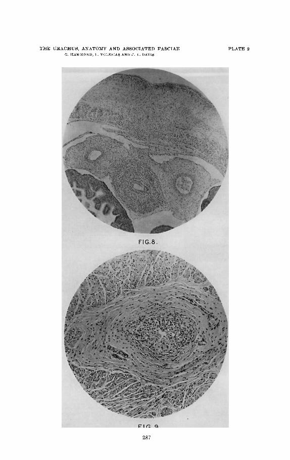
In the fetal and in many of the full term specimens a meso- urachus was demonstrable, the peritoneum being reflected about the urachus, umbilical arteries and bladder dome. This peritoneal reflection joined the peritoneum of the anterior abdominal wall along the midline from the umbilicus to the symphysis pubis. Along this line of peritoneal reflection there was a definite membrane, comparable to a primary ventral mesentery, supporting the urachus and bladder (fig. 8).
11. ADULT SPECIMENS
Following our studies of the urachi of premature and full- term fetuses, we dissected minutely the urachi and associated structures of twenty adults and one 3 year old infant. Although the survey that follows is based primarily on these twenty-one specimens, our experience is much larger, inasmuch as one of us (L.Y.) has studied this region carefully in approximately 100 cadavers.
Our studies indicate that the neonatal relationship of the urachus and the umbilical arteries is maintained essentially unchanged in adults. The adult form of the urachus is deter- mined by the relative amounts of growth and atrophy affect- ing it after birth. If the growth or proliferative changes pres- ent in the fetal urachus do not keep up with the growth of the abdominal wall, as one would expect in a vestigial struc- ture, a variable amount of stretching of the urachus will result and serve to accentuate the atrophy in the area stretched. Since the extent of these changes is different in each case, the urachus exhibits considerable variation in appearance in different specimens.

274 G . HAMMOND, L. YGLESIAS AND J. E. DAVIS
We have divided the twenty-one specimens arbitrarily into four groups between which there is no sharp line of demarca- tion. I n group I are placed the specimens which most nearly approach the fetal type of urachus and in group I V the specimens that exhibit the most marked degree of atrophy. Groups I1 and I11 are merely intermediate.
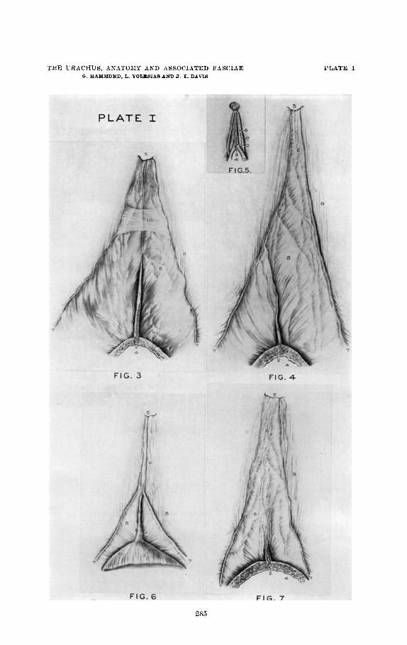
Group 1. This group is comprised of seven adult specimens and the one specimen from a 3 year old infant. I n these cases the urachus, although exhibiting a variable amount of atrophy and growth, could be traced from the bladder apex to the umbilicus as a cord, independent of the cord-like remains of the umbilical arteries, except f o r a relatively short distance near the umbilicus where the urachus and the obliterated arteries blended in a plexus of fibrous bands (fig. 3). The average distance from the bladder apex to the umbilicus was 13.3 em. and the average urachal length was 12 em. in the adult specimens. One specimen, exhibiting a communication between urachal lumen and bladder cavity, was also notable for its lack of atrophy and for the persistence of a definite peritoneal reflection (meso-urachus) which surrounded and supported the urachus and umbilical arteries. The specimen from the 3 year old infant resembled the adult specimens in every respect.
Group 1Z. The distinguishing feature of this group is that the urachus after extending upward for a variable distance from the bladder apex apparently merges with the cord repre- senting one of the umbilical arteries. I t seems probable that the apparent union is due to the close appproximation of two atrophic structures. There are four examples of this type in the present series. Each presents a variable amount of atro- phy without much evidence of changes due to growth (fig. 4). The distance from the bladder apex to the umbilicus averaged 13 em. and the urachal length averaged 6.4 em. The upper end of the urachus apparently joined the. right umbilical artery in three specimens and the left in one specimen. In one in- stance there was a communication between the epithelial canal of the urachus and the bladder cavity. One specimen presented
THE URACHUS, ANATOMY AND ASSOCIATED FASCIAE 275
a mesentery-like peritoneal reflection about the umbilical arteries and urachus.
Group 111. All the four specimens in this group have the same general gross structure. The umbilical arteries converge from either side of the bladder becoming adjacent to the urachus mid-way between the bladder apex and the umbilicus. From here the remains of each artery proceeds upward in rather close approximation to the urachus and t o each other. In its upper half each artery shows considerable scarring and places where its remains are greatly reduced in diameter. The urachus is traceable from the bladder apex up to the region where the umbilical arteries become closely associated. Above this area the urachus has undergone so much atrophy that it can no longer be identified as an independent structure. The average distance from the umbilicus to the bladder apex was 12.7 em. and the urachus averaged 5 em. in length.
Group W. This group is composed of five specimens. Here are assigned those specimens with a very short tubular urachus, exhibiting the greatest amount of regressive changes as compared with the previous groups. From the tubular portion an atrophic prolongation extends upward toward the umbilicus and becomes so small that it is lost from view as a gross structure before the umbilicus is reached. The umbilical arteries show a variable amount of atrophy but can be traced, independent of the urachus, to the umbilicus (fig. 7). There was an average distance of 15 em. from the bladder apex to the umbilicus and the urachus averaged only 3.4 em. in length.
In about a quarter of the twenty-one Bpeciniens in the fore- going groups the urachus presented the gross appearance of a tubular structure with muscular walls. In another quarter it appeared as a smaller cord of fibrous tissue. In the remaining cases the lower half of the urachus was a muscular tube which gradually tapered into a fibrous cord in its upper half. Where the urachus is represented by a muscular tube, a central epi- thelial canal is usually easily dissectable. This canal is small, being 1 mni. or less in diameter, and may pursue a straight or somewhat tortuous course in its supravesical portion as

276 G . HAMMOND, L. YGLESIAS AND J. E. DAVIS
well as in that portion found in the bladder musculature. In some cases t,he epithelial canal shows distinct dilatations which are often filled with epithelial debris. Gross continuity be- tween the bladder cavity and urachal lumen was demonstrated in only two specimens of the entire series. This incidence forms a distinct contrast with that in the fetal specimens. The valve of Wutz could not be demonstrated in any of our speci- mens. I n those cases in which no communication was found the urachal epithelium ended either in the musculature of the bladder (six specimens) or at the bladder mucosa (ten speci- mens). I n three specimens the epithelial canal could not be found.
111. THE UMBILICAL PREVESICAL AND UMBILICAL VESICAL FASCIAE
Between the peritoneal cavity and the transversalis fascia there is an interesting fascial arrangement. I n this paper we are concerned with that particular region which is bounded above by the umbilicus, below by the bladder and laterally by the peritoneal folds covering the obliterated hypogastric (umbilical) arteries. In this area are found two distinct fascial planes, one of peritoneal origin, the other representing the original connective tissue investment of the allantois and the umbilical arteries.
The fascial plane adjacent to the transversalis fascia is called the umbilical prevesical fascia (Delbet, 1895). This fascia originates from the peritoneum which in embryonic life is reflected around the superior portion of the bladder, umbilical arteries and the urachus to form a mesentery-like, supporting membrane. As development proceeds the two peritoneal cul-de-sacs, located between the visceral and parie- tal peritoneum of this reflection gradually become obliterated by fusion of their opposing surfaces, to form this fascial plane. This process of peritoneal obliteration is comparable with that occurring in the mesocolon and mesoduodenum. The umbilical pre-vesical fascia is triangular in shape with its base down- ward and its apex fused with the umbilical scar. Laterally,
THE URACHUS, ANATOMY AND ASSOCIATED FASCIAE 277
this fascia blends with the thin extra-peritoneal tissue along the hypogastric folds of the peritoneum. Inferiorly the base of this triangular fascia extends behind the pubis to fuse with the fascia covering the superior surface of the levator ani muscle. In certain cases the peritoneal fusion may be incom- plete laterally so that evidence of varying degrees of the meso-urachus remain in the adult. Thus from the peritoneal surface we may find portions of these cul-de-sacs remaining along the hypogastric folds of the peritoneum (plicae hypo- gastricae), particularly in their inferior portions at the middle inguinal fossae. I f the opposing surfaces of this peritoneal reflection fail to fuse, the umbilical prevesical fascia will not be present as a triangular fascial layer but will be represented only by the attachment of the meso-urachus at the abdominal wall (fig. 8).
The umbilical vesical fascia is found between the parietal peritoneum and the umbilical prevesical fascia. It is a fascial sheath which surrounds the urachus, obliterated hypogastric arteries, bladder and prostate, and the nerves, blood vessels, and lymphatic vessels to these organs. In its supravesical portion it is triangular in shape, ending laterally as it sur- rounds the obliterated hypogastric arteries.
These fascial planes can be demonstrated in adult specimens by careful dissection (figs. 1 and 2). Very rarely is the umbili- cal prevesical fascia absent. Thus it is seen that the urachus and obliterated hypogastric arteries are separated from the peritoneum posteriorly by the posterior umbilico-vesical fascia and anteriorly from the transversalis fascia by the anterior layer of the umbilical-vesical fascia and the umbilical prevesi- cal fascia.
Clinical sigmificamce of the fascial planes. These two fascial planes are of importance clinically in limiting different patho- logical processes in this region. Urachal infections, cysts, and neoplasms tend to be localized by the umbilical-vcsical fascia if they have spread beyond the urachus. I n such a case a tumescence is often evident in the midline between the pubis and umbilicus. An abscess arising from an acute prostatitis

278 G. H A M M O N D , L. YGLESIAS AND J. E. DAVIS
R&x%m of peritonoum at middle inQuinal
FIG 1.
V r n b i l A p r m M fascra
FIG. 2. Figs. 1 a i d '2 Diagrammatic tracings from photographs of a lower abdominal
wall dissection in an adult cadaver, showjug the urachus and its associated fasciae.
THE URACHUS, ANATOMY AND ASSOCIATED FASCIAE 279
may ascend in the confines of the umbilico-vesical fascia t o present as a lower abdominal mass in the midline. Abscesses spreading upward from the seminal vesicles, or mesially from the broad ligament, occasionally present themselves in the lower abdomen where careful dissection will reveal that the purulent exudate is located between the peritoneum and the posterior layer of the umbilical-vesical fascia. Intra-pelvic suppuration originating from an osteomyelitis of the ilium may form an abscess in this triangular area where it myill be confined between the umbilical prevesical fascia and the ante- rior layer of the umbilical-vesical fascia. With a thorough knowledge of these fascia1 layers, one can readily understand the localization of infections and other diseases of the immedi- ate and more remote neighborhood to this triangular-shaped area between the umbilicus and pubis.
DISCUSSION
A critical survey of this series of adult specimens reveals that each urachus is likely to show minor individual variations from the general underlying plan of structure. As examples of differences in urachal development one needs only to com- pare the fetal type urachi of group I with the atrophic urachi of group IV. In group I, postnatal growth has played a promi- nent role whereas in group IV atrophy has been the dominant factor in the production of the adult form.
Our study of this region in these fetuses and adults leads us to believe that the variations encountered in the adult form of the urachus depend more upon the relative amounts of growth and atrophy affecting it after birth than upon pre- natally established differences in structure. The urachus may grow commensurately with the abdominal wall and exhibit little atrophy, maintaining essentially its fetal form. This is exemplified by the specimens of group I. In contrast the urachus may not grow to an equal degree with the abdominal wall, in which case all or a portion of it becomes stretched out and attenuated, although more o r less of an umbilical con- nection tends to be maintained. The lower portion of the
280 G. HAMMOND, L. YGLESIAS A N D J. E. DAVIS
urachus as a rule exhibits more growth and is less affected by the stretching and atrophy than the upper portion. However, it is not uncommon to find an atrophic urachus extending all the way from the bladder apex to the umbilicus.
Croups 11 and I11 are simply variations in the anatomy of the urachus in its relation to the obliterated hypogastric arteries. I n both there is considerable atrophy of the upper portion of the urachus. I n groups 11,111, and IV there is no indication, as suggested by Begg, that the urachus has de- scended and dragged the obliterated ends of the arteries with it. Our studies indicate that the plexus of Luschka is formed from obliterated blood vessels which at one time anastomosed between the umbilical arteries and the urachal artery. Careful dissection of each plexus demonstrated that it was independent of the urachus and that it was intimately connected with both the umbilical arteries and the urachal artery. This plexus was a prominent finding in only three specimens. The amount of obliteration and atrophy of the umbilical arteries quite con- sistently reflected the amount seen in the urachus. The so- called ligamenturn commune, first described by Peremescko, may be (1) the atrophic upper portion of the urachus or (2) the umbilical arteries closely associated in the midline as in group I11 or (3) a combination of (1) and (2) .
The characteristic histology of the urachus may disappear at any level where atrophy and fibrosis are prominent.
IV. HISTOLOGY O F THE URACHUS
Histologically the average cross section of the urachus shows three layers of tissue. The innermost layer consists of a modified type of transitional epithelium having three fairly well defined layers of cells and surrounding an actual or potential central lumen. The epithelium taken altogether has a circular to oval form. Proceeding outward the nest layer is oval in form and is composed of connective tissue in which a number of blood and lymph vessels are recognized. The last of the three layers is composed of bundles of involuntary muscle arranged in an irregularly circular manner. Consider-

THE URACHUS, ANATOMY AND ASSOCIATED FASCIAE 281
able variation in the quantity of the muscle occurs as well as changes in form ; the amount of muscle tissue is greatest near to the bladder.
Specimens suitable for histological examination have shown considerable variation. The adult urachal specimen in figure 9 averaged 3.15 mm. in diameter. Its epithelial layer was 0.3 mm. thick at the mid-point between the umbilicus and urinary bladder, and the lumen had not been obliterated. The diam- eter of the lumen was equal to one-third of the diameter of the surrounding epithelium. Three layers of epithelial cells were plainly recognized in the mucosa. The surrounding connective tissue, in which were a number of capillaries and some lymph vessels, was oval in form and well nourished. Surrounding this connective tissue was an oval-shaped mass of involuntary muscle bundles continued from the bladder level well up to the umbilicus. A lessened amount of muscle was present in sections near the umbilical level (also see fig. 8).
The authors wish to express their appreciation to Dr. R. E. McCotter, of the Anatomy Department of the University of Michigan Medical School, for access to the anatomical material utilized, and to Dr. Bradley M. Patten for suggestions in the preparation of the manuscript.
V. SUMMARY
1. I n conclusion it may be stated that the gross anatomy of the fetal and full-term urachi revealed striking similarities. I n the majority the urachus and umbilical arteries converged toward the umbilicus as independent structures. The urachal artery was generally found with the urachus and anastomosed with the umbilical arteries. The bladder dome was conical and its cavity communicated with the urachus in about half of the cases.
2. The variations in the structure of the adult urachus appear to be determined by the relative extent of growth and atrophy affecting it after birth. This study has failed to con- firm the so-called descent of the urachus. The urachus, on the

282 G. HAMMOND, L. YGLESIAS AND J. E. DAVIS
contrary, tends to maintain its umbilical connection as in the fetus. The epithelial canal of the urachus tends to be pre- served where the urachus is represented by a muscular tube and to disappear in that part of the urachus showing con- siderable atrophy. A urachal canal communicated with the bladder cavity in 10% of the adult specimens.
3. The umbilical arteries and urachus are surrounded by a fascial sheath, called the umbilical vesical fascia. This fascia is separated from the transversalis fascia by the umbilical pre-vesical fascia. These fascial layers tend to localize disease of the immediate and more remote tissues to a triangular shaped area between the umbilical and pubis. 4. The urachus proper has three layers of tissue: the inner
or epithelial, the submucosal and the muscular.
LITERATURE CITED
Contribution a l’etude du peritoine dam ses rapports avec les arteres ombilicales et l’ouragne. Nancy, No. 14. (Thesis de Nancy.)
AREY, L. B. 1940 Dercloprneiital Anatomy. W. B. Saunders Go., Philadelphia and London, 4th ed.
BEGG, R. C. 1930 The urachus; its anatomy, histology and development. 5. Anat.,
BOUILLY, GEORGES 1880 Des turneurs aignes et chroniques de la eavite prevesicale (cavite de Retzius). Paris. (Thesis de agregacion.)
BEEJIER, J. L. 1936 A Textbook of Histology. Blakistoii’s Son & Go., Inc., Philadelphia, 5th ed.
CUI.IEN, T. 8. 1916 Embryology, Anatomy, and Diseases of the Umbilicus, together with Diseases of the Urachus. W. R. Saunders Co., Philadel- phia and London.
De la signification morpholoque des aponeuroses perivesieals. J. dc Anatomie et la Physiol., vol. 35, pp. 235-245.
1892
ANCEL, ALBERT PAUL 1899
V O ~ . 64; pp. 170-183.
CUNEa, B., AND V. VEAU
DELBET, P. DISSB, I. J.
1899
1895 Anatomie chirurgicale de la vessie, Thesis Paris, No. 167. Untersuehungen uber die Lage der Menschlechen Hornblasc
und ihre Veranderung irn Laufe des Waehstums. Anatomische Hefte,
KEIBEL, F., AND F. P. MALL 1910-1912 Manual of Human Embryo1og;v. J. B.
LUSCHKA 1862 Uebcr den Bau des mmsehlichen Harnstranges. Virch. Arch.,
PIELSOL, G. 9. 1930 Human Anatomy. J. B. Lippineott Co., Philadelphia, 9th ed. WUTZ, J. B. 1883 Ueber Urachus and Urachuscysten. Virch. Arch., vol. 92,
V O ~ . 1, pp. 1-76.
Lippincott Go., Philadelphia and London.
vol. 23, p. 1.
pp. 387-423.

PLATES
PLATE 1
EXPLANA'PION OF FIGURES
Figures 3, 4, 5, 6, 7 reduced one-half. Four adult specimens aud one fetal speci- men of uraclii, umbilical arteries, and bladder apices as seen from the peritoneal surface. These figures represent the general types of urachi f o u d in this study. 1 Urachus. 6 Plexus of obliterated vessels. 2 Uraehal epithelium. 7 Umbilical artery. 3 Bladder wall. 8 Peritoncum. 4 Bladder cavity. 9 Urachal opening into bladder cavity. 5 Umbilical scar. 10 Fascia1 concentration.
THE URACHUS, ASATOJIY A N D ASSOCIATED FASCIAE G. HAYMOXD, L. i -amsIAs AND J. E. DAVIS
PLATE 1
285
PLATE 2
ESPL.!.SATION OF FIGURES
8 Antorior part of a cross section through tlio entire body of a niale fetus 4.4 em. crown-rump length, magnified 150 d's. The urachus flanked by the umbilical artcries is seen in the central portion of the section. The mrsourachus is shown forming the attachment of these structures to tlie anterior abdominal ivall. When the cul-de-sac on either side of the mesouraehus obliterates by fusion of the oppos- ing peritoneal surfaces, the preumhilieal-vesical fascia is formed. I n the abdominal wall portions of the recti inusrles and two layers of cpithelial cells are formed.
9 Cross section of tlie uraclius a t its middle third froiii a 40-year old female. Cross size 3 X 1.5 mor. Epithelial layer 0.3 mm. thick. The lumen is stnall but patent and is surroundcd by epithelium of three cclls in thickness. Out- side of the epithelium is tlie somewhat elongated oval of connective tissue and next outside of this layer is the oval-sh:rped mass of involuntary ninsclo ( x 150).
THY URBCIIUR, BXATOMY AND ASSOCIATED FASCIAE ( 8 , IIAM.MOSI), I Yr.l.ELIAS 4 X U 6. N. D l V I S
PLATE 2
287