the systolic murmur—benign or serious? john michael...
TRANSCRIPT

©2005-6 Blaufuss Multimedia. All rights reserved.
The Systolic Murmur—Benign or Serious?
John Michael Criley, M.D. M.A.C.P. Emeritus Professor of Medicine and Radiological
Sciences, David Geffen School of Medicine at UCLA, Harbor-UCLA Medical Center,
Torrance, California.
David G. Criley, Blaufuss Medical Multimedia, San Francisco, California

©2005-6 Blaufuss Multimedia. All rights reserved.
2
The Systolic Murmur—Benign or Serious?
Introduction
Systolic murmurs are commonly detected during cardiac auscultation. As suggested by the
title, they are often benign, but can indicate serious or life-threatening cardiac conditions or
the need for further evaluation or timely intervention.
All systolic murmurs result from disturbed or “turbulent” flow ultimately resulting from the
pumping action of a ventricle, while all diastolic murmurs occur from ventricular inflow
during ventricular relaxation. Systolic murmurs can be of outflow origin, either from
increased stroke volume (as with anemia, pregnancy, or ventricular overfilling from aortic
valve regurgitation) or flow through a narrowed or distorted outflow channel or conduit. Less
often, systolic murmurs result from backflow either through an atrioventricular valve (mitral
or tricuspid) or through an intracardiac communication, e.g., ventricular septal defect.
Systolic Murmurs (Ventricular Pumping) Diastolic Murmurs (Ventricular Filling) Restricted flow through narrow outflow tract or vessel, or high flow through normal channel
mid systolic
Restricted filling through stenotic A-V valve or disturbed or high volume inflow
mid-diastolic/presystolic Regurgitation through A-V valve holosystolic (occ. early or late systolic)
Regurgitation through semilunar valve early diastolic/holodiastolic
Flow through intracardiac defect (VSD) holosystolic

©2005-6 Blaufuss Multimedia. All rights reserved.
3
Is the murmur systolic?
An approach to the evaluation of a systolic murmur should begin by ascertaining that the
murmur is unequivocally systolic. It is not uncommon to be asked to evaluate a patient
purported to have a systolic murmur only to find that the murmur is not systolic. This
confusion in timing is prompted by the widespread but erroneous assumption that any readily
heard murmur must be systolic. Three techniques can be very helpful in determining that a
murmur is systolic, and it is prudent to employ as many these timing aids as possible in order
to ensure that a murmur is indeed systolic
A. Correlation with visible or palpable central pulses
B. Cadence
C. Post-PVC augmentation
Correlation with visible or palpable pulses can be challenging if the heart rate is rapid, or
if a prominent jugular venous pulsation (JVP) emulates a carotid pulse, so it is important to
palpate as well as observe the respiratory effects on any visible pulse in the neck. Carotid
pulsations, unlike JVPs are minimally affected by respiration. The later arrival and altered
waveform of the radial pulses render them less reliable than the carotid pulse for timing.
Palpable precordial pulsations are usually systolic and are therefore reliable timing aids.

©2005-6 Blaufuss Multimedia. All rights reserved.
4
The cadence of a murmur can be used to establish its timing within the cardiac cycle. If the
heart rate is less than 90, systole will be shorter in duration than diastole, so that the first
sound (S1) is closer to the following second sound (S2), than the interval between S2 and
the following S1. As a result, systolic murmurs are usually shorter than diastolic murmurs. If
the rhythm is irregular, as with atrial fibrillation, the duration of systole varies little, while the
duration of diastole and diastolic murmurs is variable.
Post – PVC (Postextrasystolic) augmentation of a murmur is a reliable indicator that a
murmur is not only systolic in timing, but probably of outflow tract origin. Detection of this
Figure 1. The carotid, jugular (JVP) or apical impulse should be used, if visible or palpable, to establish the timing of sounds and murmurs. The sounds can also be used to establish the timing of pulsations. For example, the JVP a-wave will occur with the first sound (1) while the v-wave will occur with the second sound (2). a = a-wave in JVP and apex beat e = ejection point (onset of ejection) o = time of opening of mitral valve v = v-wave of JVP
Figure 2. The beat following a premature ventricular contraction will usually exhibit an increase in outflow induced systolic murmur intensity because of the increased inotropic drive. The ventricular pressure will usually increase, as will the pressure gradient across a stenotic valve. Murmurs caused by mitral or tricuspid regurgitation are not augmented.

©2005-6 Blaufuss Multimedia. All rights reserved.
5
phenomenon requires comparison of the murmur intensity of the beat following the “dropped
beat” with the murmur heard with the subsequent beats. Postextrasystolic augmentation of
ventricular contractility, especially if the ventricle is hypertrophic, combined with the fall in
arterial pressure caused by increased runoff duration will usually cause more outflow velocity
and a louder, longer murmur.
Continuous murmurs are frequently confused with murmurs confined to systole. Their true
identity can usually be established by moving (“inching”) the stethoscope to an area at which
the murmur as well as the heart sounds can be heard, and noting the murmur continues
through and well beyond S2.
Location, location, location
There are three parameters regarding location that are helpful in establishing the possible
cause(s) of a systolic murmur:
A. Location on precordium of loudest murmur
B. Location(s) of radiation or distribution
C. Location within systole

©2005-6 Blaufuss Multimedia. All rights reserved.
6
The precordial location of the loudest transmission of a cardiac murmur is of help in
determining its origin. Radiation of murmurs from their primary location occurs through
downstream propagation in outflow murmurs as well as through myocardial transmission.
Outflow murmurs (AKA “ejection murmurs”) are usually loudest over the precordial
locations closest to the outflow tract or great artery through which the flow is occurring --
pulmonary outflow murmurs in the second left intercostal space (2 LICS) and aortic in the 2
RICS, and radiate along the course of the great vessels. It should be noted that the pulmonic
valve is close to the chest wall in 2LICS while the aortic valve is buried deep within the heart
behind the right ventricular outflow tract in the 3-4 LICS. The aorta ascends rightward and
ventrally, approaching the chest wall at 2 RICS where aortic outflow murmurs are generally
heard best.
Figure 3. Precordial locations of cardiac valves and the directions of primary sound transmission of sounds and murmurs. Closure sounds (S1 and S2) are best heard at the apex and base respectively. Murmurs are transmitted best downstream from the structure responsible for the disturbed flow producing the murmur, and secondarily transmitted over the ventricular muscle when tensed by systolic contraction.

©2005-6 Blaufuss Multimedia. All rights reserved.
7
Vibrations from murmurs are radiated well through rigid (contracting) myocardium, much as a
cast iron pipe conducts sounds over great distances while a rubber hose dampens sonic
vibrations. The rigidity of contracting myocardium enables outflow murmurs to be well heard
over the ventricle of origin, and may filter out some of the harsher components. A case in
point is the “Gallavardin transformation” of the outflow murmur in aortic stenosis, a pure
musical murmur radiated toward the apex while a rough or harsh murmur is heard over 2
RICS and the carotid arteries downstream.
Regurgitant Murmurs emanating from the tricuspid or mitral valve are usually heard best
over the ventricle of origin, probably because of the rigidity of ventricular myocardium and the
relative laxity of the atria. The left parasternal location of the right ventricle (RV) makes the
murmur of tricuspid regurgitation (TR) well heard along the left sternal border. The murmur
of mitral regurgitation (MR) is best heard where the LV is closest to the chest wall, at the
apex and left axilla. High velocity jets of MR resulting from a flail posterior leaflet may be
directed toward the common atrio-aortic wall and thus emulate an outflow murmur. Flail
anterior leaflet MR may be heard over the dorsal spine and occiput.
The murmur caused by a restrictive ventricular septal defect (VSD) is sometimes classified
as “regurgitant” in that it results from flow counter to LV outflow, increases LV diastolic
volume, and has similar timing. VSD murmurs are best heard over the RV, radiate widely,
and often associated with a systolic thrill over 3-4LICS.

©2005-6 Blaufuss Multimedia. All rights reserved.
8
Location within Systole is dictated by the hemodynamic relationships between the pumping
and recipient chambers. If systole is defined as the interval between the first (S-1) and
second (S2) heart sounds, a murmur that occupies all of systole is termed holosystolic. A
murmur that occupies the mid portion of the space between S1 and S2 is called mid systolic.
An early systolic murmur is initiated by S1 and ends well before S2, while a late systolic
murmur begins well after S1 and blends into or continues through S2. The above
terminology avoids potentially misleading or inappropriate terminology such as “ejection
murmur” and “pansystolic;” the former implies established certainty that the murmur is of
outflow origin (which is not always possible by bedside examination) and the latter literally
means “all of the systoles” rather than all of systole.
A holosystolic murmur (HSM) results from high velocity blood flow from a ventricle into a
recipient chamber (usually an atrium) from the onset of ventricular contraction until
ventricular pressure falls as the ventricle relaxes. Only three conditions can cause a HSM:
TR, MR, and restrictive (small) VSDs. However, not all TR, MR, or VSD murmurs are
holosystolic. Acute MR may cause a shortened murmur if there is marked elevation of left
atrial systolic pressure (v-waves) that stifles late systolic backflow. It is erroneously assumed
that HSMs will always obscure the first and second sounds, and conversely if heart sounds
are heard, the murmur is not holosystolic.

©2005-6 Blaufuss Multimedia. All rights reserved.
9
Mid systolic murmurs are generated by disturbed or turbulent outflow, and often termed
“ejection murmurs,” but as stated above, the timing and not the presumed cause of the
murmur should be applied to these murmurs. Mid systolic murmurs (MSMs) are
characteristic of aortic stenosis (AS), pulmonic stenosis (PS), or a high ventricular stroke
volume in aortic regurgitation (AR) or atrial septal defect (ASD). MSMs are very common
and frequently benign, since they may result from high cardiac output states in normal
adolescents or athletes, pregnancy, anemia, or chest wall configuration (pectus excavatum,
straight back syndrome, asthenia, etc.). Murmur intensity is directly related to velocity and
inversely related to blood viscosity, hence conditions that increase velocity (normal flow
through a stenotic orifice or high flow through a normal channel) may produce a murmur.
Figure 4. Holosystolic murmurs are expected with regurgitant atrioventricular valves and ventricular septal defects because the pressure in the ventricular source is higher than the recipient chamber throughout systole.

©2005-6 Blaufuss Multimedia. All rights reserved.
10
Mitral regurgitation may produce a crescendo-decrescendo murmur that is heard in the
aortic area (2RICS) and can be confused with a murmur of outflow origin, but the holosystolic
or late systolic (see below) timing is not compatible with a murmur produced by outflow. It
should be remembered that outflow cannot occupy all of systole (holosystolic) because aortic
or pulmonic valve closure occurs after cessation of outflow and initiation of backflow,
obligating a pause between the end of the murmur and the second sound.
Figure 5. Mid systolic murmurs caused by ejection through stenotic semilunar valves have a crescendo-decrescendo configuration and end before the closure sound from the valve. Closure of the aortic or pulmonic valves require retrograde flow in the artery beyond the valve, meaning that the forward flow causing the murmur most terminate and reverse, thus ending the murmur prior to the closure sound. In the depiction of Pulmonic Stenosis, the dashed line indicates the closure of the pulmonic valve and P2.

©2005-6 Blaufuss Multimedia. All rights reserved.
11
Late systolic timing of a murmur usually implies mitral valve prolapse (MVP) in which the
mitral valve is competent in early systole but its leaflets lose appostion and become
incompetent in mid and late systole. The acoustic contour of these murmurs may be
crescendo-decrescendo, and these murmurs may spill through the time of aortic valve
closure and obscure the second sound. Late systolic murmurs in MVP often have a
whooping or honking acoustic profile, and may be sufficiently loud to be heard without a
stethoscope.
Figure 6. This patient with Mitral Regurgitation had a holosystolic murmur well heard in the Aortic area that had mid-peaking amplitude, and was recorded from within the aorta by intracardiac phonocardiography. Pressure recording revealed tall v-waves in the left atrium (LA) and no hemodynamic evidence of aortic stenosis. Atrial fibrillation was present. The mitral regurgitant jet impinged on the left atrial wall adjacent to the aorta, causing the murmur to be transmitted to the aorta.

©2005-6 Blaufuss Multimedia. All rights reserved.
12
In Hypertrophic Cardiomyopathy (HCM) Mitral valve dysfunction resembling MVP may
result from the dynamic ventricular cavitary obliteration; the mitral valve may be competent in
early systole but becomes scrunched and rendered incompetent in mid and late systole. In
both of these clinical settings (MVP and HCM) the timing of the murmur may be similarly
altered by maneuvers that affect the left ventricular diastolic volume (e.g. standing and
Figure 7. Mitral Valve Prolapse (MVP) is depicted as a systolic billowing of a portion of the mitral leaflet apparatus that is checked by chordal restraints in mid systole, causing a mid systolic click, followed by a late systolic murmur of mitral regurgitation. The timing of the click and murmur can be altered by perturbations that change the volume and geometry of the left ventricle. A hypothetical prolapse threshold denotes the time when the mitral leaflet cannot be maintained in a competent, sub-annular position. If the volume is abruptly increased by squatting, or reduced by standing, the click and murmur will shift in relation to the other heart sounds.

©2005-6 Blaufuss Multimedia. All rights reserved.
13
squatting, Valsalva strain and release). Maneuvers that increase LV volume (squatting) will
delay murmur onset, while standing or Valsalva strain will decrease LV volume and increase
the length and intensity of the murmur.
A murmur generated by aortic coarctation, or with increased internal mammary arterial flow in
pregnancy (mammary souffle) or lactation may have late systolic timing because the peak
flow rate occurs later in arteries at a distance from the ventricle.
Early systolic murmurs may be generated by TR or MR that is suppressed in late systole.
In a dilated ventricle, the valve leaflets may not be able to meet at the onset of systole but
Figure 8. Severe Mitral Regurgitation with late systolic diminution of the systolic murmur because the “ventriculoid” left atrial (LA) pressure approaches the left ventricular (LV) pressure in late systole, The vertical arrows denote the relationship between S2 and the aortic (Ao) dicrotic notch. There is an S3 associated with the rapid filling wave (RFW) in the left ventricle.

©2005-6 Blaufuss Multimedia. All rights reserved.
14
may appose as the ventricle and annular diameter decrease. As noted above, acute, severe
MR may cause the systolic pressure to rise in the left atrium (LA) and rival LV pressure, and
thus suppress late systolic regurgitant flow. Small muscular VSDs may close when the
septal muscle contracts, terminating the murmur in late systole. The murmur of a muscular
ventricular septal defect may disappear or markedly diminish upon standing.
Sounds Associated with Murmurs
Although often overlooked (or more appropriately, not even listened for) the sounds that
accompany murmurs are often of inestimable value in determining the cause and/or
significance of a murmur, especially if the murmur is mid systolic. Heart sounds are
produced by the abrupt deceleration by impact of a moving column of blood in the
cardiovascular system. In a normal heart, S1 is caused by the abrupt halting of blood pushed
toward the atrium at the onset of ventricular contraction when this retrograde surge is
abruptly halted by the expanding underbellies of the atrioventricular valve leaflets. S2 results
from the abrupt halting of retrograde flow in the aorta or pulmonary artery root when the
ventricle relaxes and the semilunar valve closes.

©2005-6 Blaufuss Multimedia. All rights reserved.
15
When a holosystolic murmur of tricuspid regurgitation is associated with an exceptionally
loud S1 in a patient with pulmonary hypertension, unrecognized mitral stenosis (MS) may
be the underlying cause of the pulmonary hypertension. A soft or absent S1 may indicate
“preclosure” of the mitral valve or an under-powered ventricular contraction in a failing
ventricle. Apparent splitting of S1 caused by an ejection sound (vide infra) can be a clue to
significant outflow tract pathology. Ebstein's anomaly of the tricuspid valve can cause a
Figure 9. Events responsible for the first and second heart sounds and the directions of primary radiation of the sounds are indicated by cartoons with shaded densities representing the pressure in the various chambers. Darkest densities indicate pressures above 80, while the lightest represent less than 10 mmHg.

©2005-6 Blaufuss Multimedia. All rights reserved.
16
loud early systolic sound ("sail sound") generated by late closure of the deformed tricuspid
valve.
Intensity and patterns of splitting of S2 can also provide diagnostic information. Loud S2
implies high closing pressure (systemic or pulmonary hypertension), while appreciation of
abnormal splitting can narrow the differential diagnostic possibilities raised by systolic
murmurs. Physiological splitting of S2 occurs when the duration of right ventricular systole
is increased by augmented right heart inflow, combined with RV outflow into the expanding
lungs that literally vacuum blood out of the right heart with negative pressure, both
contributing to delayed pulmonic valve closure. Persistent splitting of S2 can result from
prolongation right ventricular systolic duration with atrial septal defect (ASD) or pulmonic
stenosis (PS). Persistent splitting occurs when late onset (and end) of right ventricular
systole with right bundle branch block (RBBB), or early closure of the aortic valve when there
is an alternate low impedance outlet such as mitral regurgitation (MR) or ventricular septal
defect (VSD). On the other hand, prolongation of, or late onset of, LV systole results in
paradoxical splitting in which the aortic component of S2, or A2 occurs after the pulmonic
component, P2 during expiration so that the normal inspiratory prolongation of right
ventricular systole closes the P2-A2 gap. If the onset of left ventricular systole is delayed by
left bundle branch block, or the duration of left ventricular systole is prolonged by aortic
stenosis or hypertrophic cardiomyopathy, A2 will follow P2 during expiration or apnea, and
the split will close as P2 is delayed during inspiration.

©2005-6 Blaufuss Multimedia. All rights reserved.
17
Extra diastolic sounds, in chronological order of appearance are termed opening snaps
(OS), third sounds (S3) and fourth sounds (S4). Stenotic mitral valves usually retain
sufficient leaflet flexibility to undergo a partial opening excursion at the base of the valve
while parting of the leaflets at the distal end is abruptly halted by commissural fusion, causing
the blood moving into the valve sleeve to be prevented from flowing freely into the left
ventricle. Blood moving down the mitral sleeve encounters the inability of the funnel-like
terminus to open, with the impact generating a sharp early diastolic OS. The timing of the
S2-OS interval roughly matches the two syllables in the word “butter.”
An S3 is caused by a ventricle resisting inflow as a result of an absolute or a relative increase
in passive ventricular volume. Torrential inflow may occur in high output states or when the
atrium has received a large volume of regurgitant flow in the preceding systole. Conversely,
a small diastolic input may enter a failing ventricle that has not expelled enough blood during
systole to make room for a normal diastolic inflow, abruptly achieving maximal capacity. A
caricature of S3 called a pericardial knock when the ventricles are incased in a fibrotic or
calcified girdle in pericardial constriction.
S4 is generated by a ventricle resisting inflow from augmented atrial contractions, and are
typically heard in conditions in which there is hypertrophy of either the right or left ventricle.
Systolic sounds causing “splitting” of the first heart sound are called ejection sounds (AKA
ejection clicks) that are generated at the time of opening of the aortic or pulmonic valve. An
ejection sound (ES) may result from abrupt checking of the opening excursion of a

©2005-6 Blaufuss Multimedia. All rights reserved.
18
congenitally stenotic aortic or pulmonic valve when the leaflets are mobile but fail to open
properly. As blood in the cylindrical outflow tract gains momentum and the semilunar valve
initiates an opening excursion, the inability of the stenotic valve to open fully abruptly halts
the column of blood, which is then forced through the restricted orifice. Thus an ES can be
heard to initiate the mid systolic murmur (MSM) of aortic stenosis (AS) or pulmonic stenosis
(PS) when the valve leaflets retain mobility. When fibrosis or calcification render the valve
rigid and immobile an ES is not heard. An ES is usually discerned more readily on the
upstream (ventricular) side of the diseased semilunar valve. Since both the ES and S1 are
louder at the apex than at the base, recognition that the first sound is split can be appreciated
better at the apex. An ES can be heard well at the base, where it seems to “jump start” the
murmur.
Ejection sounds do not necessarily indicate the presence of valvar stenosis, because they
are also heard when the aorta or pulmonary artery are dilated and hypertensive, and/or
subjected to high output states when the semilunar valve leaflets are flung open to initiate
ejection. The following conditions are frequently associated with an ES: atrial septal defect
(ASD), pulmonary or systemic hypertension, aortic regurgitation, anemia, and chronic renal
failure with arterio-venous shunt dialysis access.
Mobile systolic clicks (MSC), also called mid systolic clicks or non-ejection clicks, are
virtually diagnostic of mitral valve prolapse (MVP). The floppy, redundant mitral leaflets
inflated by LV systolic pressure billow progressively back into the left atrium, and are
eventually checked by chordal restraint. This abrupt halting of LV pressure’s inflation of the

©2005-6 Blaufuss Multimedia. All rights reserved.
19
leaflets generates a clicking or popping MSC. If the mitral valve is rendered incompetent by
loss of leaflet apposition in MVP, the MSC is followed by a late systolic murmur (LSM) that
may spill through S2. Not all MVP valves are incompetent, so that there may be no murmur
in association with the MSC, and alternatively there may be no MSC in association with the
murmur of MVP.
The mobility of a MSC can be demonstrated by postural maneuvers or other perturbations
that affect ventriculo-valvar proportionality. The valve is demonstrably “too big” for the LV,
and as a result cannot be maintained in a competent sub-annular position when the
ventricular cavity size is reduced during systolic ejection. The misfit between the valve and
LV can be further perturbed by maneuvers that reduce ventricular size (abruptly standing up
from supine or squatting position, Valsalva strain, vasodilators, exercise), or ameliorated by
abruptly increasing ventricular size (squatting after standing). As the MSC moves closer to
S1 the murmur may become longer and louder, and when it moves closer to S2 the murmur
starts later and is often softer.
Visible or Palpable Pulsations
Correlation of auscultation with visible or palpable pulsations is an important aspect of
cardiac bedside examination. Pulsations not only provide timing signals, but frequently have
the added value of providing independent diagnostic information. Although the left
ventricular apical impulse is often obscured by obesity, large breast, or barrel chest
configuration, when visible and/or palpable it provides precise timing of auscultatory events
and a window into LV function. Recordings of the apical pulse, called apexcardiograms

©2005-6 Blaufuss Multimedia. All rights reserved.
20
(ACGs) were utilized by clinical investigators in the 1960s to detect exercise induced
ischemic changes in ventricular function. The apex impulse, as graphically recorded by an
ACG provides an outward thrust during isovolumic left ventricular contraction, the nadir (O
Point) at the time of mitral valve opening, the rapid filling wave and “atrial kick” by the
ventricle, all with little or no time delay. Pathological apical impulse patterns would include a
sustained systolic outward thrust in aortic stenosis or with LV aneurysm, a presystolic
impulse in left ventricular hypertrophy, a rapid filling wave in volume overload lesions (MR,
AR, dilated cardiomyopathy). A “triple ripple” impulse (atrial kick, early and late systolic
outward thrusts) is diagnostic of hypertrophic cardiomyopathy (HCM). A late systolic inward
collapse is often found in MVP when the aneurysmal valve pops into the atrium causing a
sudden tension release in the LV.
Most left parasternal systolic impulses are caused by the RV, although a rapidly
expanding LA from mitral regurgitation (MR) may cause a late systolic heave by pushing the
RV from behind. Left parasternal diastolic impulses can arise from RV diastolic overload
from tricuspid regurgitation, or when being pushed from behind by a rapidly expanding left
ventricle in AR. Hypertensive pulmonary arterial expansion may cause a parasternal impulse
in the 2nd or 3rd left intercostal space and a palpable P2.
Figure 10 Carotid arterial pulse contours may be diagnostic of specific causes of
systolic murmurs, especially the parvus et tardus pulse associated with aortic stenosis, the bisferiens (double- peaked) pulse of aortic regurgitation, and the spike-and-dome pulse of hypertrophic cardiomyopathy. The systolic ejection period is prolonged in hypertension, aortic stenosis, and hypertrophic cardiomyopathy

©2005-6 Blaufuss Multimedia. All rights reserved.
21
Visible and palpable carotid pulsations provide much better timing of auscultatory events
than the radial pulse, particularly at fast heart rates. At rates of 100 or more, the radial
systolic pulse will be felt during ventricular diastole. In addition to the value of the carotid
pulse for timing of heart sounds, there are three “classic” diagnostic carotid pulse contours:
(1) parvus et tardus of AS, bisferiens (double peaked) of AR, and “spike-and-dome” of
HCM. It is important to note that all three of these conditions are commonly associated with
a similar systolic murmur, but can usually be differentiated from one another by the
associated carotid pulse contour. Corrigan’s pulse, bounding and collapsing pulses of the
major subcutaneous arteries, and Quincke’s pulse, elicited by gentle pressure on the digital
nailbeds to produce systolic blushing and diastolic blanching are also virtually diagnostic of
AR.
The jugular venous pulse (JVP) contains valuable diagnostic information that can be
gleaned by correlation with the cardiac auscultation. It should be said that the Internal JVP
emanates from the same area of the neck as the carotid pulse, and this juxtaposition may
lead to considerable confusion when the source of the pulsation is not carefully determined.
The JVP will usually vary more with respiration than the carotid and unlike the carotid often
has 2 widely spaced pulsations per cardiac cycle when sinus rhythm is present. The JVP
pressure declines (x- and y-descents) are more abrupt and therefore more readily seen than
the diastolic decline in carotid arterial pressure. The external JVP is several centimeters
lateral to carotid arterial pulsations, and will usually faithfully transmit the right atrial pressure
contours to the neck, avoiding confusion between arterial and venous pulsations in the

©2005-6 Blaufuss Multimedia. All rights reserved.
22
suprasternal notch or emanating from the common carotid/jugular sheath medial to the
sternocleidomastoid muscle.
The contour of the JVP carries considerable diagnostic information. Large a-waves, almost
coincident with the first heart sound and declining rapidly during systole, indicate RV
hypertrophy, while v-waves, reaching a peak coincident with the second sound indicate
tricuspid regurgitation (TR).
Maneuvers and Perturbations
Some bedside maneuvers are difficult to perform by elderly or infirm patients, but can be very
helpful in more agile patients when heart sounds and murmurs raise several diagnostic
considerations. However the effects of spontaneous or controlled breathing are almost
always possible to observe and can provide important diagnostic information. It is often
useful to use your free hand (the one not holding the stethoscope) to “conduct” the patient’s
breathing pattern by first rehearsing the maneuver with the patient. Have the patient inspire
slowly and quietly by raising your hand palm-up, exhale while lowering your hand palm-down,
and suspend respiration after full exhalation by sweeping your pronated hand back and forth
for a few seconds. The transient hemodynamic effects of inspiration can only be noted
during active inspiration, not by having the patient take and hold a deep breath! If you tell a
patient “hold your breath” the person will almost invariably take a deep breath and bear
down, involuntarily performing a partial Valsalva strain. When the noises associated with
breathing overwhelm the heart sounds, tell the patient to “stop breathing” (as opposed to
“hold your breath”), preferably at end expiration. Two or three seconds of apnea can be

©2005-6 Blaufuss Multimedia. All rights reserved.
23
tolerated by virtually any patient, regardless of respiratory distress, and will permit clear
listening to several heart beats.
A more dramatic negative intrathoracic pressure pull can be affected by a Mueller maneuver
consisting of having the patient attempt to suck on an imaginary pickle or soda straw with the
mouth closed, causing the cheeks to collapse inward. Alternatively, the patient can be told to
breathe in and out through the nose several times and then compressing the nares for 2-3
seconds while listening to the heart during attempted inspiration. There should be a loud
sucking sound upon release of nasal compression if the patient has successfully maintained
negative intrathoracic pressure. Auscultation during the Mueller maneuver will bring out
subtle murmurs from tricuspid regurgitation (TR) that might otherwise be obscured by the
lung sounds.
Post-PVC Augmentation of a systolic murmur is described above, and when present, is
usually indicative of origin in the ventricular outflow tract. Most patients with aortic or
pulmonic stenosis, whether the lesion is valvar, subvalvar, or supravalvar in location, will
have postextrasystolic augmentation of murmur intensity. As previously stated, the
increased inotropic state in the beat following a PVC will generate a more forceful contraction
in the ventricle(s) and will increase the flow velocity causing the murmur. One “special case”
that doesn’t quite fit into this scheme is the murmur of hypertrophic cardiomyopathy
(HCM). Although the murmur is often described as an “outflow murmur” it is very often found
to be holosystolic or late systolic in timing, and thus may be caused by mitral regurgitation.

©2005-6 Blaufuss Multimedia. All rights reserved.
24
The augmentation of the murmur is illustrated in Figure 11, and the murmur is seen to begin
with the first sound and extend to the second sound, and therefore holosystolic.
Auscultation during Valsalva strain and release is an important bedside maneuver that
should be performed correctly to avoid spurious results. The patient should be rehearsed
while being told to breathe in deeply, then out, and then to bear down during peak expiration
for at least 10-15 seconds, and then relax and resume normal breathing. The examiner’s
hand can be placed on the abdomen to counter the outward thrust of the abdomen and
determine if the patient is actually bearing down. The Valsalva strain increases intra-
Figure 11. The post-PVC murmur of Hypertrophic Cardiomyopathy (HCM) is often holosystolic, as shown here, and therefore generated by mitral regurgitation. The brisk initial upstroke of the aortic pressure should be contrasted with the delayed aortic upstroke in Aortic Stenosis (Figures 2 and 5). The left ventricular end diastolic pressure (small arrow) is elevated, and there is a fourth heart sound (4).
©2005-6 Blaufuss Multimedia. All rights reserved.
25
abdominal and intrathoracic pressure and inhibits venous return to the heart, causing the
heart chambers to shrink progressively and the cardiac output to decline. With release, the
pent-up systemic venous blood rapidly returns, first to the right heart and followed 3-4 heart
beats later by the left heart. It is important to have the stethoscope on the chest during the
entire maneuver, especially after release.
Most murmurs will decline in intensity during the Valsalva strain, the heart rate will usually
accelerate and a vasopressor response will ensue as the carotid sinus baroreceptors sense
a decline in pressure. Upon release of the strain there will be a return right heart murmurs
first, followed by left heart murmurs as the chambers progressively and sequentially fill. An
early return suggests that the murmur is from the right heart, and if the murmur intensity
returns after 3-4 heart beats, the murmur is probably of left heart origin. The vasopressor
response during strain may continue into the recovery period, and in turn lead to transient
hypertension and bradycardia.
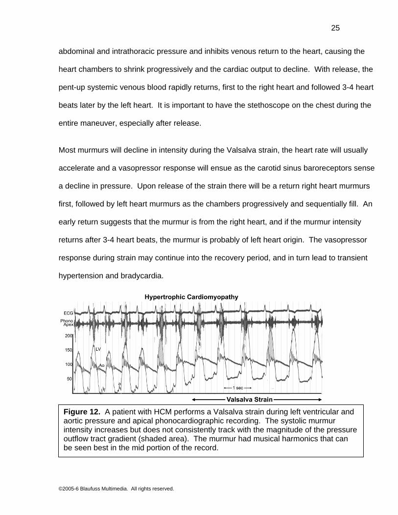
Figure 12. A patient with HCM performs a Valsalva strain during left ventricular and aortic pressure and apical phonocardiographic recording. The systolic murmur intensity increases but does not consistently track with the magnitude of the pressure outflow tract gradient (shaded area). The murmur had musical harmonics that can be seen best in the mid portion of the record.

©2005-6 Blaufuss Multimedia. All rights reserved.
26
There are two conditions in which murmur intensity may actually increase during Valsalva
strain: Mitral valve prolapse (MVP) and hypertrophic cardiomyopathy (HCM), because the
shrinking of the LV exaggerates the disproportionality between the mitral valve and ventricle
mentioned above. As the ventricular diastolic volume shrinks, systolic contractions distort the
valve, causing the systolic murmur to progressively increase in intensity, and often in
duration and pitch as well. Amyloid of the heart may emulate HCM, and will often cause an
increase in murmur intensity with Valsalva strain.
Postural maneuvers can alter the filling and emptying of the ventricles and triggers
autonomic reflexes. Abrupt standing momentarily decreases venous return and may cause
an exaggerated reflex tachycardia in some individuals with MVP. The resulting exaggeration
of the disproportional fit of the mitral valve within the left ventricle will usually cause the
systolic murmur to become louder, longer, and higher in pitch in both MVP and HCM. A
mobile systolic click (MSC) will occur earlier, and then upon squatting, move later as the
murmur is “squashed” into S2 in MVP. Abrupt squatting will pump a bolus of blood into the
central circulation and at the same time increase the impedance to left ventricular ejection.
This double effect benefits patients with Fallot’s tetralogy by promoting increased flow to the
pulmonary arteries, and may transiently increase the intensity of right heart murmurs.
Passive leg raising will cause a sudden volume shift of blood from the periphery into the
central circulation, but is not easily performed because it requires several people to achieve
the maneuver, and the patient may inadvertently defeat the passivity of the maneuver by

©2005-6 Blaufuss Multimedia. All rights reserved.
27
straining. This maneuver may increase the intensity of right heart murmurs and decrease the
murmur of hypertrophic cardiomyopathy.
Sustained isometric handgrip, especially if performed with both hands, increases
impedance to left ventricular ejection and may augment left heart regurgitant murmurs and
reduce the murmur of HCM.
Amyl nitrite, a rapid acting inhaled Vasodilator was traditionally readily available for bedside
evaluation of murmurs, and when used had dramatic and profound effects in enhancing
murmurs induced by forward flow (e.g., mitral and stenosis) and reducing the intensity of
regurgitant murmurs. Although it is no longer available, it served to elucidate the
pathophysiology underlying many murmurs prior to the era of echocardiography.
Vasodilators are now widely used in the management of patients with regurgitant left heart
valves, and although not as rapid-acting as amyl nitrite, will have a similar effect on the
murmurs of mitral and aortic regurgitation.

©2005-6 Blaufuss Multimedia. All rights reserved.
28
Innocent Murmurs are those that are not associated with any demonstrable radiographic,
electrocardiographic, or echocardiographic, abnormality, and are more common in children
than in adults. In some instances, a murmur proclaimed to be “innocent” in childhood proves
later to have been caused by previously unrecognized bicuspid aortic valve stenosis,
ventricular septal defect, or mitral valve prolapse. These murmurs are thought to be of right
heart origin, and usually increase in intensity with inspiration as a result, and often have a
twanging “vibratory” quality.
Is a murmur benign or serious? It is overly simplistic to imply that a given murmur is
always benign or always serious; in real life experience there are infinite shades of gray. In
turns of probability of adverse outcome without timely, specific intervention, the least benign
Figure 13. Summary of cardiac sounds and murmurs.

©2005-6 Blaufuss Multimedia. All rights reserved.
29
conditions are aortic stenosis, hypertrophic cardiomyopathy, and tricuspid regurgitation (if it
results from pulmonary hypertension). Intermediate risk would be asymptomatic mitral
regurgitation, pulmonic stenosis and atrial septal defect. Low risk would include ventricular
septal defect and most patients with mitral valve prolapse.
It is hoped that this exercise will serve to sharpen the astute physician’s bedside evaluation
of patients with systolic murmurs by calling attention to the ancillary visible, palpable, and
auscultatory events that accompany the murmur. Time and space limitations do not permit
any discussion of appropriate diagnostic procedures or therapy.

©2005-6 Blaufuss Multimedia. All rights reserved.
30
Suggested Reading
Heger JW, Niemann JT, Criley JM (eds): Cardiology 5th ed. Lippincott Wiliams and Wilkins,
2004.
Criley JM, Criley DG, Zalace: The Physiological Origins of Heart Sounds and Murmurs (CD-
ROM) Lippincott Williams and Wilkins, 1997
Carabello BA, Crawford FA: Valvular heart disease (review article) N Engl J Med 337:32-41,
1997.