the surgical anatomy of the pulmonary vessels -...
TRANSCRIPT
Thorax (1953), 8, 189.
THE SURGICAL ANATOMY OF THE PULMONARY VESSELSBY
J. C. VAN DER SPUYFrom the Thoracic Surgical Unit, Johannesburg Group of Hospitals, and Department of Surgery,
University of the Witwatersrand, South Africa
(RECEIVED FOR PUBLICATION SEPTEMBER 29, 1952)
Surgical resections of the lung have in recentyears become common and relatively safe pro-cedures. Depending upon the extent and nature ofthe disease process, the whole lung, one or twolobes, or one or more bronchopulmonary segmentsmay require excision. Modern pulmonary re-
section depends to a very large extent upon theprecise identification of the primary, secondary,and tertiary hilar bronchovascular structures, andit has thus become essential to have a detailedknowledge of the anatomy of these structures atthe different hilar levels.The aim of this paper is to describe and to illus-
trate this anatomy. It is based upon a study of 42complete casts of the bronchovascular tree andthe hilar anatomical findings displayed during thecourse of 92 partial and 30 whole lung resectionsperformed by some members of the ThoracicSurgical Unit, Johannesburg, during the past year.The pulmonary vessels and the bronchi are illus-trated chiefly from such approaches as are usedby the surgeon when dissecting the differentprimary, secondary, and tertiary hila.Each lung, anatomically, is composed of 10
bronchovascular or bronchopulmonary segments.These segments are miniature lobes differing fromlobes only in their size and in the usual absence offissures at their anatomical boundaries.A lobar bronchus branches from a main
bronchus at a relatively constant site, and sub-divides in a more or less standard fashion into itssegmental bronchi. The pulmonary artery, how-ever, has not this same lobar distribution, andalthough the segmental pulmonary arteries followthe segmental bronchi somewhat closely, they tendto have a variable and independent origin from thepulmonary artery.The segmental artery, like the segmenital
bronichus, occupies a central position within thebronchopulmonary segment. The blood carriedto the segment by such a centrally situated arteryis collected by perisegmental pulmonary veins; as
such, they are either placed superficially, imme-
diately beneath the pleura, or deeply, in an inter-segmental plane. The intersegmental veins drainalso adjacent segments within a lobe and maydrain adjoining segments of a different lobe. Inthe latter instance the veins pass subpleurallyacross the base of a fissure. The perisegmental(intersegmental or segmental) veins course some-what independently of the segmental bronchi andarteries to reach the subp'eural plane in theprimary hilum and there join a main pulmonaryvein.
PULMONARY ARTERYThe main pulmonary artery divides in front of
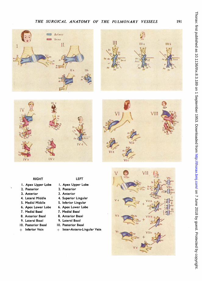
the left main bronchus into a left and a right pul-monary artery (Fig. I).RIGHT PULMONARY ARTERY.-The right pul-
monary artery has to cross the midline to reach theroot of the right lung. This it does by passingtransversely to the right, behind the ascendingaorta and the superior vena cava respectively, infront of the oesophagus and immediately abovethe left atrium and right superior pulmonary veinunder cover of the arch of the azygos vein.Behind the superior vena cava the right pul-
monary artery divides into an upper and a lowertrunk (Fig. I). Of these the superior pulmonarytrunk supplies the anterior and apical broncho-pulmonary segments of the right upper lobe bydividing, as it enters the lung substance in front ofthe right upper lobe bronchus, into the apical (Al)and anterior (A3) segmental arteries (Fig. II).Emerging from behind the superior vena cava, itis, but for the covering given it by the pleura onthe root of the lung, completely exposed.The inferior pulmonary trunk proceeds from
behind the superior vena cava to become sub-pleural, but only to disappear almost immediatelyby passing behind the superior pulmonary vein(Fig. IV). It is the counterpart of the left pul-monary artery. Passing laterally and slightlydownwards, under cover of the superior pul-monary vein (Fig. IV), it crosses, the anterior sur-
on 7 June 2018 by guest. Protected by copyright.
http://thorax.bmj.com
/T
horax: first published as 10.1136/thx.8.3.189 on 1 Septem
ber 1953. Dow
nloaded from

J. C. VAN DER SPUY
face of the right bronchus, between the origins ofthe upper and middle lobe bronchi (Figs. II andIII). Then curving downwards, it runs sub-pleurally in the depths of the oblique fissure (Fig.III), on the antero-lateral aspect of the basalbronchus.The inferior pulmonary trunk supplies the
middle and lower lobes as also, in the majorityof cases, the posterior bronchopulmonary seg-
ment of the upper lobe via the posterior ascend-ing segmental artery (A2, Fig. III) and frequentlyalso the anterior bronchopulmonary segment, viathe anterior ascending segmental artery (A3, Fig.III). In such instances the anterior ascending seg-
mental artery is usually accessory to the anteriorsegmental artery (A3, Fig. II), but in a small per-
centage of these cases it is solely responsible forsupplying the anterior bronchopulmonary seg-
ment. Occasionally, however, the anterior ascend-ing segmental artery may derive its origin fromthe middle lobe artery (A3, Fig. IlIb).The acute angle formed between the oblique
and transverse fissures forms the point of an
arrow which guides one to that area of the sub-pleural pulmonary artery, in the depths of theoblique fissure, from which the pulmonary arterialsupply to the middle and lower lobes and part ofthe upper lobe is derived (Fig. III).
Arising from the postero-lateral surface of theinferior pulmonary trunk, in this area, one findsthe lower lobe apical segmental artery (A6, Fig.III). It descends slightly in a backward direction,lying superior and, as the surgeon sees it, lateralto its corresponding bronchus. To the anatomist,however, the artery is superior and anterior to thebronchus. The reason for this difference is thatthe surgeon, in opening the slit-like oblique fissure,displaces the apex of the lower lobe medially.Similarly also, the middle lobe artery (Figs. III,
IIIa, and IIIb) appears to be superior and lateralto its corresponding bronchus instead of superiorand posterior.The lower lobe apical segmental artery is
usually single (A6, Fig. III), but at times the apicalsegment of the lower lobe summons the aid of a
second segmental artery (A6 i, A6 ii, Fig. IIIa).Arising more or less opposite the lower lobe
apical segmental artery, from the anterior surfaceof the pulmonary artery, is the middle lobe artery.It may be single, when it divides into twobranches, one each for the medial and lateral seg-ments of the middle lobe (A4 5, Fig. Illa). Asfrequently two separate middle lobe segmentalarteries are present (A4, A5, Fig. III).
Arising from the inferior pulmonary trunk,slightly above the lower lobe apical segmentalartery, is the artery to the posterior segment ofthe upper lobe. It passes backwards and upwardsand is termed the posterior ascending artery (A2,Figs. III, Illa, and IVd). Whereas the posteriorsegmental artery of the right upper lobe in themajority of cases arises from the pulmonaryartery in this situation, it frequently, however,arises from the superior pulmonary trunk itselfduring the latter's subpleural course, from either ofits two terminal branches, namely, the apical (Al)or anterior (A3) segmental arteries (Figs. Ila andIlb), from the lower lobe apical segmental artery(A2, Fig. IlIb), or even from the middle lobeartery.The inferior pulmonary trunk breaks up into
the four basal segmental arteries, either just withinthe oblique fissure or just inside the lungparenchyma. Each basal segmental artery followsits respective bronchus very closely, keeping to itsantero-l1teral side. These take off from theinferior pulmonary trunk either singly or via ananterior and a posterior trunk, the former dividinginto the medial (A7) and anterior (A8) basal seg-mental arteries, whereas the latter provides thelateral (A9) and posterior (AIO) basal segmentalarteries (Fig. ILL).LEFT PULMONARY ARTERY.-The left pulmonary
artery, arising in front of the left main bronchus,ascends slightly in a posterior and lateral direc-tion, arches over the left main bronchus, and thenhooks round the left upper lobe bronchus, follow-ing a subpleural course throughout (Figs. V, VI,and VII). It more fully establishes its superiorityover the left superior pulmonary vein (Fig. VIII)than does its right counterpart, the inferior pul-monary trunk, which lies directly behind the rightsuperior pulmonaty vein (Fig. IV).Owing to the different courses taken by the left
and right pulmonary arteries, the antero-posteriorand infero-superior relations in the hilum of theleft lung are vein, bronchus, and artery, whereasthe same relations in the hilum of the right lungare vein, artery, and bronchus.As the left pulmonary artery is about to arch
over the left main bronchus it gives off the first ofa set ies of segmental arteries for the supply of theleft upper lobe. This segmental artery (A3, Fig.V), arising from the antero-superior surface of thepulmonary artery, passes in front of the upperdivision of the left upper lobe bronchus to theanterior bronchopulmonary segment, crossingbehind the apical-posterior tributary (VI, V2, Fig.VIII) of the superior pulmonary vein.
190
on 7 June 2018 by guest. Protected by copyright.
http://thorax.bmj.com
/T
horax: first published as 10.1136/thx.8.3.189 on 1 Septem
ber 1953. Dow
nloaded from
THE SURGICAL ANATOMY OF THE PULMONARY VESSELS
N
2Ir I
-,½IA)~.
I= Arter-.-/'fc 7 r:
11 111 711
R6 A \ 44.5 ; 4n
^,-<R 1At
I I. IIt I
A.,.e4/
11 a
i4 + ,=
r-t 'J
V.e
Stw
/
A,I
..Z ,4
*;St L A-'
1 J a-, \s
V1/'F
AI :
-
, .. .. *..
A-A:
via&
f -9
VibA:
VICn1
;& I*MIV fl.
RIGHT
1. Apex Upper Lobe2. Posterior3. Anterior4. Lateral Middle5. Medial Middle6. Apex Lower Lobe7. Medial Basal8. Anterior Basal9. Lateral Basal10. Posterior Basal+ Inferior Vein
LEFT
1. Apex Upper Lobe2. Posterior3. Anterior4. Superior Lingular5. Inferior Lingular6. Apex Lower Lobe7. Medial Basal8. Anterior Basal9. Lateral Basal
10. Posterior Basal+ Inter-Antero-Lingular Vein
Va I'... ;.
Vc
IV
V '-I.
l'* C 4 f,-) Yll~f,
\Ili
- - A rQt
+V
Xr d W.
111 b
Ai
r;
,.r.
v4V
Y;
191
I
I
v.1," T.kp
-.- Irl,
A ..'Iv O.-, I i'lk
rI
k .-.--
on 7 June 2018 by guest. Protected by copyright.
http://thorax.bmj.com
/T
horax: first published as 10.1136/thx.8.3.189 on 1 Septem
ber 1953. Dow
nloaded from
J. C. VAN DER SPUY
From the postero-superior surface of the leftpulmonary artery the short, sessile, bifurcatingapical-posterior segmental artery (Al, A2, Fig. VI)is derived. The apical-posterior artery may, how-ever, be a clearly defined single trunk, dividinginto its two constituent segmental arteries (Fig.VIb). On the other hand, the apical segment maybe supplied by two separate segmental arteries (Al,Al, Fig. VIa). Similarly, the posterior segmentmay have a dual segmental arterial supply (A2,A2, Fig. VIc).
Arching around the back of the upper lobebronchus, the left pulmonary artery enters theoblique fissure, and, running immediately beneaththe pleura, it comes to lie on the side of the lowerlobe bronchus. From this position, near the upperend of the oblique fissure, the artery sends from itsanterior aspect a single arterial stem to the lin-gular bronchopulmonary segments. The lingularartery is posterior to its corresponding bronchus(but lateral to it as the surgeon sees it) and at asomewhat lower level, and divides into thesuperior and inferior lingular segmental arteries(A4+5, Fig. VII). Commonly the lingular seg-mental arteries arise independently from theanterior surface of the pulmonary artery (A4, A5,Fig. VII i).
Arising from the postero-lateral surface of thepulmonary artery, more or less opposite the lin-gular artery, although usually at a somewhathigher level, is the lower lobe apical segmentalartery (A6, Fig. VII). As on the right side, here toothere may be two separate lower lobe apical seg-mental arteries (A6 i, A6 ii, Fig. VII ii).The level of origin of the lower lobe apical seg-
mental artery in relation to that of the lingularartery is of particular importance (Fig. VII). Inlower lobectomy this segmental artery (A6) re-quires to be ligated separately from the rest of thelower lobe artery, as mass ligation of the pul-monary artery immediately above the origin of thefirst segmental artery to the lower lobe (A6) wouldinclude the segmental arterial supply to the lingula.The anterior segmental artery (A3) commonly
arises from the anterior surface of the pulmonaryartery in the oblique fissure in relation to thelingular artery. It may form with the lingularartery (A4, A5) a common antero-lingular trunk(Fig. VIla), or it may be associated with only theupper of two separate lingular segmental arteries(Fig. VIIa iii). On the other hand, the anteriorsegmental artery may arise independently of andcranial to a bifurcating lingular artery (Fig. VIla i)or two separate lingular segmental arteries (Fig.VIIa ii).
The lingular artery, on the other hand, mayform with the anterior segmental artery a com-mon antero-lingular trunk on the antero-superiorsurface of the pulmonary artery immediately be-fore it loops over the upper surface of the leftmain bronchus (Fig. Vb), leaving the anterior sur-face of the pulmonary artery in the lingular regionof the oblique fissure conspicuously bare (Fig.VIIb). Alternatively, only the superior lingularsegmental artery (A4) may be associated with theanterior segmental artery (Fig. Vc), leaving itspartner (A5) in charge of the oblique fissure (Fig.VIIc).The apical segmental artery (Al) also occasion-
ally arises with the anterior segmental artery (A3)from the antero-superior surface of the left pul-monary artery (Fig. Vd).To ascertain the whereabouts of these segmental
vessels it is advisable to display the segmentalarterial anatomy in the lingular region of theoblique fissure, should this fissure be fullydeveloped. Depending on whether there are three,two, one, or no segmental arteries in the lingulararea, so there will arise no, one, two, or threesegmental arteries from the antero-superior sur-face of the pulmonary artery (Figs. VII, VIla,VIIb, VIIc, and Figs. V, Va, Vb, and Vc). Theremaining arterial stems arising from the postero-superior or posterior surface of the arching pul-monary artery belong to the apical-posteriorbronchopulmonary segment.The four basal segmental arteries follow their
respective bronchi closely, and, like their fellowson the right side, cling to the antero-lateral sidesof these bronchi (Fig. VII). Here also the pul-monary artery may terminate within the obliquefissure, but usually the dissection has to beextended into the lung substance to see that themedial (A7) and anterior (A8) basal segmentalarteries, like their corresponding bronchi, usuallyarise from a common anterior trunk, and that thelateral (A9) and posterior (AIO) basal segmentalarteries either leave singly or via a commonposterior trunk (Fig. VII).
PULMONARY VEINSRIGHT SUPERIOR PULMONARY VEIN.-The right
superior pulmonary vein drains the right upperand middle lobes. On entering the pericardium,this vein lies immediately below the right pul-monary artery. In the hilum of the lung, how-ever, it overlaps the inferior pulmonary trunk,and its uppermost tributary, the apical-anteriorvein (VI, V3, Fig. IV), descends across the anteriorsegmental artery. This subpleural vein drains the
192
on 7 June 2018 by guest. Protected by copyright.
http://thorax.bmj.com
/T
horax: first published as 10.1136/thx.8.3.189 on 1 Septem
ber 1953. Dow
nloaded from
THE SURGICAL ANATOMY OF THE PULMONARY VESSELS
upper and middle lobes via three to five trunks.The uppermost trunk is the apical-anteriorvein, which receives a subpleural segmental veinfrom each of the apical and anterior broncho-pulmonary segments (VI, V3, Fig. IV).
Entering the superior pulmonary vein imme-
diately below the apical-anterior trunk is theinferior vein (+, Fig. IV), so called because itcourses subpleurally on the inferior surface of theupper lobe (anterior segment) in the transversefissure.
Next, the tributaries from the lateral andmedial bronchopulmonary segments of the middlelobe enter the superior pulmonary vein, either as
a common trunk or independently (V4, V5, Fig.IV).One more vein completes the quintet. This is
the posterior segmental vein. But for the course
of this vein the superior pulmonary vein wouldhave been a channel shaped like a hand, all thedigits meeting the palm subpleurally. This pos-
terior vein (V2, Fig. IV), however, comes from thevery depths of the upper lobe to enter incon-spicuously the deep surface of either the pul-monary vein itself, or, more commonly, its inferiortributary, having first catered for the posterior andintersegmental regions of the upper lobe (V2, Fig.IVd). This vein is unique in that, of all the seg-
mental veins, it is the only one which at no stageruns a subpleural course.
One or both middle lobe veins may at timesbecome detached from the superior pulmonaryvein to drain into the inferior pulmonary vein
(Figs. IVb and IVc). Similarly, one or both ofthese middle lobe segmental veins may draindirectly into the left atrium.
RIGHT INFERIOR PULMONARY VEIN.-The rightinferior pulmonary vein, in the root of the lung,courses below the right superior pulmonary vein;but here it occupies a somewhat more posteriorplane. Its tributaries leave the lung by passingbehind their respective bronchi. For this reason,
and also because the inferior pulmonary vein is tosome extent hidden by the lower lobe and by theupward extending anterior layer of the pulmonaryligament, the vein and its tributaries are more
readily dissected from the posterior aspect (Figs.IV and IVa).The inferior pulmonary vein receives all five
segmental veins which drain the lower lobe, andoccasionally, as mentioned above, one or bothmiddle lobe segmental veins. If the superior pul-monary vein may be compared with a hand, thenthe inferior pulmonary vein can most certainly be
Q
said to resemble a shrub (Boyden, 1945), thebranches of which radiate somewhat irregularlyfrom a short stem (Figs. IV and IVa). Its lower-most tributary is the medial basal segmental vein(V7), which lies immediately above a constantlypresent lymph gland at the upper end of the pul-monary ligament.The uppermost, and most easily discernible,
tributary is the lower lobe apical segmental vein,which, descending from the apex of the lowerlobe, crosses behind the basal bronchus, below theorigin of its corresponding bronchus, to end at theupper angle of the receiving vein somewhat pos-teriorly (V6, Figs. IV and IVa).The remaining three tributaries, namely, the
anterior (V8), lateral (V9), and posterior basal(V1O) segmental veins, leave their respective inter-segmental regions (Fig. IV) by crossing behindtheir corresponding bronchi to enter the inferiorpulmonary vein in no fixed manner between thelower lobe apical segmental vein above and themedial basal segmental xein below.
LEFT SUPERIOR PULMONARY VEIN.-The leftsuperior pulmonary vein is formed by threevenous trunks which, to the surgeon, are allantero-inferiorly related to their correspondingbronchi, and which, anteriorly, are all covered bythe pleura on the root of the lung. It receives allthe left upper lobe segmental veins (Fig. VIII).The uppermost tributary is the apical-posterior
trunk, which drains the apical-posterior broncho-pulmonary segment. This apical-posterior trunk(Vl, V2, Fig. VIII) overlies the anterior artery orthe antero-lingular trunk when these arteries arisefrom the antero-superior surface - of the pul-monary artery.
Joining the superior pulmonary vein laterallyand immediately below the apical-posterior veinis the tributary from the anterior segment (V3,Fig. VIII). Before reaching the superior pul-monary vein the anterior segmental vein is joinedby the inter-antero-lingular vein (+, Fig. VIII)which drains along the intersegmental plane de-scribed by its name, thus corresponding somewhatto the inferior vein draining into the right superiorpulmonary vein.The lowermost member of the left superior pul-
monary vein is that which drains the superior andinferior lingular bronchopulmonary segments (V4,V5, Fig. VIII). These two segmental veins may,however, join the superior pulmonary vein separ-ately. One or both lingular veins may, like themiddle lobe veins, empty into the inferior pul-monary vein or directly into the left atrium.
193
on 7 June 2018 by guest. Protected by copyright.
http://thorax.bmj.com
/T
horax: first published as 10.1136/thx.8.3.189 on 1 Septem
ber 1953. Dow
nloaded from

J. C. VAN DER SPUY
The apical-anterior-posterior segmental venouspattern of the left upper lobe may at timesresemble that of the right upper lobe and viceversa.
LEFT INFERIOR PULMONARY VEIN.-The left in-ferior pulmonary vein runs, like the right inferiorpulmonary vein, below the superior pulmonaryvein and at a somewhat more posterior level.Similarly, its five tributaries, crossing behind theirrespective bronchi, can be dissected more readilyfrom behind. Here too the lower lobe apical seg-mental vein (V6) and the medial basal segmentalvein (V7) are the uppermost and lowermost tribu-taries respectively (Fig. VIII).An inferior pulmonary vein frequently re-
sembles not a shrub but a branch of a tree (Boy-den, 1945), the thick end of the branch representingthe inferior pulmonary vein, which, tapering down,becomes the posterior segmental vein (V10). Thebranch has three lateral branches, namely thelower lobe apical segmental vein (V6), the anteriorbasal vein (V8), and the lateral basal vein (V9)respectively, and one medial branch, the medialbasal vein (V7, Fig. VIII).Very occasionally a lung is drained by one pul-
monary vein only.
DISCUSSIONSeveral plates of the pulmonary vessels are
presented showing their relations to each otherand to the bronchi at the different primary, secon-dary, and tertiary hila.
Although, functionally, a lung is composedof " bronchopulmonary " segments, the term" bronchovascular " is sometimes employed, as the
structures met with during hilar dissections are ofprime importance to the surgeon, and the latterterm emphasizes the existence of multiplebronchovascular hila.The usual anatomy as well as different varieties
of the less usual anatomy, as indicated by big andsmall Roman numerals respectively, are illustrated.By indicating the views from which the anatomy isshown, the models, placed immediately above theillustrations, allow one to become readily orien-tated, and explanatory descriptions are therebyconsiderably reduced.
I am extremely grateful to Mr. L. Fatti, SeniorSurgeon, Thoracic Surgical Unit, Johannesburg Groupof Hospitals, and also to Mr. G. R. Crawshaw, ofthe same unit, for communicating to me their observa-tions on the anatomy of the pulmonary vessels inthose resections which I did not personally witness.Of the bronchovascular casts mentioned above, 32
were prepared by Dr. F. Frodl, late of the Depart-ment of Pathology, Utrecht, Holland. His paper onthe detailed anatomy of the pulmonary vessels isnow in the press. I am deeply indebted to ProfessorP. Nieuwenhuijse, head of that department, and toDr. F. Frodl, now of the Department of Radiology,Utrecht, for so generously having granted me accessto these casts.
Mr. P. Marchand, of the Thoracic Surgical Unit,Johannesburg, made 10 similar casts, and to him Iwish to extend my sincerest thanks for having permit-ted me to include them in this series.
Contributions by Mr. L. Fatti and Mr. G. R.Crawshaw made possible the publication of drawingsin colour.
REFERENCE
Boyden, E. A. (1945). Surgery, 13, 706.
194
on 7 June 2018 by guest. Protected by copyright.
http://thorax.bmj.com
/T
horax: first published as 10.1136/thx.8.3.189 on 1 Septem
ber 1953. Dow
nloaded from