the role of the lim1 gene in vertebrate kidney...
TRANSCRIPT

The Role of the Lim1 Gene in Vertebrate Kidney Development
by
Mao Ye
Bachelor of Science, Zhejiang University, 2004
Master of Science, University of Pittsburgh, 2008
Submitted to the Graduate Faculty of
School of Medicine in partial fulfillment
of the requirements for the degree of
Master of Science
University of Pittsburgh
2008
ii
UNIVERSITY OF PITTSBURGH
School of Medicine
This thesis was presented
by
Mao Ye
It was defended on
June 5th, 2008
and approved by
Dr. Martin Schmidt, Associate Professor, Department of Microbiology and Molecular
Genetics
Dr. Thomas Smithgall, Professor, Department of Microbiology and Molecular Genetics
Dr. Neil Hukriede, Assistant Professor, Department of Microbiology and Molecular Genetics
Thesis Advisor: Dr. Martin Schmidt, Associate Professor, Department of Microbiology and
Molecular Genetics

iii
Copyright © by Mao Ye
2008
The role of the Lim1 gene in Vertebrate Kidney Development
Mao Ye, M.S.
University of Pittsburgh, 2008

iv
Lim1 has been reported to regulate the head, nerve system and kidney development in
mouse, zebrafish and Xenopus. In the present study, we demonstrate that Xlim1 plays a pivotal
role in the pronephric specification and development in Xenopus.
In Xenopus embryos at 4-cell and 8-cell stage, loss of function experiments using DEED
depletion showed that down-regulation of Xlim1 expression severely impairs the formation of
pronephros, assessed by pax8 expression. Overexpressing Xlim1 by injecting constitutively
active Lim1 constructs in Xenopus embryos induce remarking enlargement of pronephros at
various stages during development, assessed by both in situ hybridization and
immunohistochemistry staining. Furthermore we studied the working window of Xlim1 on
pronephric specification in mesoderm and observed the peak time point is at stage 10.5 while the
whole working window is between stage 9 and 12.5. To understand the mechanism of Xlim1’s
regulation in kidney development, then we performed the microarray analysis on the induced
pronephric RNA sample extracted from animal caps, targeting couple of genes that either are the
members involved in the lim1 pathway or in the networking that induce the pronephric
specification.
In summary, our data suggest that Lim1 acts as a potent factor that determines the early
specification of pronephros. Its expression levels at the early stage critically influences the
specification of the pronephric tubules. More knowledge about XLim1’s pathway in kidney
development will be studied via examining the gene targets screened from microarray analysis.

v
TABLE OF CONTENTS
PREFACE………………………………………………………………………………………IX
1.0 GENERAL INTRODUCTION………………………………………………………...1
1.1 KIDNEY………………………………………………………………………………...1
1.1.1 Structure and function………………………………………………………….1
1.1.2 Early development of the kidney………………………………………………5
1.1.3 Significance of early kidney development studies…………………………….7
1.2 XENOPUS…………………………………………………………………………….....9
1.2.1 Xenopus as a research model…………………………………………………...9
1.2.2 Xenopus as a research model for kidney study…………………………........13
1.3 LIM1…………………………………………………………………………………...14
1.3.1 Location, expression pattern and function of lim1 gene…………………….14
1.3.2 Lim1's role in kidney development…………………………………………...19
2.0 STUDY OF THE REGULATORY ROLE OF XLIM1 IN KIDENY
DEVELOPMENT IN XENOPUS AND THE IDENTIFICATION OF TARGETS AND
COFACTORS OF LIM1 IN REGULATING KIDNEY DEVELOPMENT………………. 20
2.1 EXPERIMENTAL DESIGN…………………………………………………………20
2.1.1 Cell-target injections and gene-specific knock-down………………………….20
2.1.2 Microarray analysis and gene expression pattern by in situ hybridization….21

vi
2.2 MATERIAL AND METHODS………………………………………………………22
2.2.1 Xenopus laevis embryos manipulation and microinjection…………………22
2.2.2 In situ hybridization…………………………………………………………...23
2.2.3 Expression specificity and ontological analysis……………………………...23
2.2.4 Immunohistochemistry and fluorescien dextran staining…………………..24
2.3 RESULTS……………………………………………………………………………...24
2.3.1 Constitutively active Lim1 expands kidney tissue…………………………..24
2.3.2The potential downstream targets of lim1 retrieved from microarray
analysis……………………………………………………………………………….28
2.3.3 Cell-targeted Xlim1 knockdown affects the formation of the kidney. The
expression of the early kidney marker pax8 is disrupted specifically in kidney
region in Xlim1 knockdown embryos.……………………………………………...39
3.0 CONCLUSIONS AND PROSPECTUS……………………………………………....42
3.1 OBJECTIVES…………………………………………………………………………42
3.1.1 Characterize Xlim1 in Xenopus kidney specification………………………..42
3.1.2 Identify the downstream targets of LIM1……………………………………42
3.2 INTERPRETATION OF THE RESULTS…………………………………………..42
3.3 SIGNIFICANCE AND FUTURE WORK…………………………………………...46
BIBLIOGRAPHY………………………………………………………………………………47

vii
LIST OF TABLES
Table1.The summary of the upregulated genes identified by microarray (lim1-DEED
injected vs. uninjected) in animal caps that are documented to express in kidney………...29
Table2.
Table3. The summary of the upregulated genes identified by microarray( Activin/RA-
treated vs. untreated) in animal caps that are documented to express in kidney…………..35
Table4. The summary of the downregulated genes identified by microarray (Activin/RA
treated vs. untreated) in animal caps that are documented to express in kidney…………..38

viii
LIST OF FIGURES
Figure 1 Presumptive Xlim1 dimers bind to nucleic acid…………………………………….15
Figure 2.The animation of the postulated Xlim-1/Ldb complex and the Ldb1-Xlim1 fusion
protein…………………………………………………………………………………………...17
Figure 3. The constitutively active Xlim1 expands the pronephros………………………….26
Figure 4. The working window of Xlim1 is determined at an early stage…………………...27
Figure 5. The depletion of Xlim1 via DEED injection severely disrupts the formation of
pronephros………………………………………………………………………………………40

ix
PREFACE
It is with great pleasure that I offer my acknowledgement to a number of people for their
help in completing this degree. First I would like to thank all my thesis committee members, Dr.
Hukriede and Dr. Smithgall for excellent guidance throughout my entire studies. A very special
thank you goes to my advisor, Dr. Martin Schmidt. He endured numerous meetings and emails to
provide the ceaseless guidance and support through the whole process. I am indebted to the
current and former faculty and staff in the Department of Microbiology and Molecular Genetics-
Caroline Haldin PhD, Eric De Groh MS, Lisa Antoszewski PhD, Michael Tsang PhD, Gabriella
Molina BS, Richard Chaillet MD, PhD, Bonnie Reinhart PhD, Ceci Cirio MS, Benjamin Shaffer
BS, Leonard D’Aiuto PhD, whose support and involvement made this study possible.
I would like to acknowledge the Interdisciplinary Biomedical Graduate Program, School
of Medicine, University of Pittsburgh. I am grateful to Dr. John Horn, Dr Jennifer Grandis, Dr
Wen Xie, Dr. Wendy Mars and Ms Cindy Duffy for their most precious help.
I am thankful to my friends here at the University of Pittsburgh and the Carnegie Mellon
University for their unwavering support both in my study and life.
My deepest gratitude goes to my parents and my uncles and aunts, my little cousins for
standing by me and giving me understanding as well as the warmth of the family.

1
1.0 GENERAL INTRODUCTION
1.1. KIDNEY
1.1.1 Structure and function
Kidney is an intricate and critical organ that maintains homeostatsis of living creatures,
that is, the stable internal environment for organisms. To maintain the healthy, balanced internal
environment organisms must timely and efficiently remove the daily toxic byproducts generated
from metabolism. Kidney functions largely as a filter for the elimination of the waste products
generated from metabolisms, mainly including creatinine and urea as byproducts from protein
metabolism in liver and some lipids metabolites1. One important function of kidney for all
terrestrial animals is its role in maintaining an optimal and constant osmotic pressure2. Other
functions of kidney include but are not limited to regulating concentration of acid, reabsorption
of lipids, creating hormones that regulate the formation of red blood cells and blood pressure3, 4
.

2
Nephron is the functional unit of vertebrate kidneys5. Each nephron contains two parts: a
filtering component (renal corpuscle) and a tubule part (renal tube). A highly permeable vascular
bed, known as glomerulus, acts as a filter through which allows for the small molecules passing
.Mostly enclosed by the big mouth of Bowman capsule, the glomerulus with Bowman capsule
together constitutes renal corpuscle. The convoluted tubules that connect to the neck of Bowman
capsule are known as renal tubules, which are segmented into several parts for their distinct
functions. The last part of renal tubule, or the collecting duct, leads the waste to the renal pelvis
and to the ureter where via the renal bladder urine will discharge to the exterior6.
The formation of the urine could be divided into two steps. 1) When the blood via the
dorsal artery travels through the capillary network within the glomerulus, the hydrostatic
pressure pushes the fluid and small solute molecules to penetrate the capillary wall into the space
between glomerulus and the Bowman capsule. Meantime, it keeps the large molecules like
proteins to remain in the blood, resulting in a sticky blood, or ultrafilter blood.2) In the next step,
as the glomerulus filtrates pass through the collecting tubes, the cells of the renal tubes
selectively reabsorb the solute molecules and ions. Accompanying the reabsorption, some
particles like lipids are secreted from the blood to the collecting tube alternatively. The solution
that emerges at the end of the collecting tube (urine) thus is very different in composition and
concentration from the solution that enters the tubule (the glomerulus filtrate). The collecting
duct from one nephron and from many others meet up with bladder, from where liquid wastes go
to the urethra and then is excreted from the body6.
3
All three vertebrate kidneys contain nephrons. The complexity of the kidney partially
counts on the number of nephrons the kidney has and the spatial assembly of these
nephrons5(See Dressler’s review Figure 1, 2006). The most anterior and primitive kidney, the
pronephros, has only one nephron7. The adult kidney of fish and amphibians, the mesonephros,
has multiple units similar to nephron7, 8
. The most complex one, the kidney of adult mammals
and birds is called metanephros, which has up to one million nephrons as it has in the medullar
and cortex of human kidney9,10
.
Pronephros, mesonephros and metanephros are the three successive stages in mammalian
kidney development11
. While the pronephros and the mesonephros are transiently present in
embryonic stages, metanephros is the full-fledged organ for mammals. Yet, the presence of
pronephros and mesonephros are indispensable for several species. For fish and amphibia, they
rely on the function of pronephros to survive during the larval period10
. The later replacement of
pronephros, the mesonephros form the adult kidney of lower vertebrates and is also functional in
the fetus of higher vertebrates like elephants, reflecting the aquatic ancestry of this huge
terrestrial animal8, 11
.
The focus of this thesis is pronephros. Pronephros is a very simple organ indeed. The
basic structure of pronephric of the Xenopus is shown in Brandli’s review paper in 1999. The
pronephros is composed of three distinct parts: the pronephric corpuscle, the pronephric tubules
and the pronephric duct. Similar to the structure of the nephron, Xenopus pronephros contains a
single corpuscle, which consists of the pronephric corpuscle and the pronephric glomus. The
pronephric capsule protrudes into the filtration chamber of the pronephros, the nephrocoel, which
4
is partly connecting to coelom. The filtrates diffuse into the coelom and are reabsorbed in the
pronephric tubules. Likewise, pronephric tubules are morphologically segmented into three parts:
ciliated nephrostomes, connecting tubules, and a common tubule. The common tubule joins the
connecting tubules and the connecting duct together in a way of connecting a sieve and a funnel
so that the wastes could be selectively and completely excreted to the end of this filtering system,
the rectal diverticulum, an outgrowth of the cloaca, to maintain the balanced salt and water
environment of the body7, 12
.
1.1.2 Early development of the kidney
The development of urinary system and reproductive organs are closely associated. The
urinary organ is developed from the intermediate mesoderm11
. The permanent organs of
mammals and birds are preceded by a set of embryonic structure, which all atrophy but with the
ducts remained. This evolutionary organ-developing progression occurs rapidly in the mouse
embryos that from the mesonephros to the metanephros it only takes about 1-2 days out of 20-
day gestation13
. In humans, pronephros is quite rudimentary. After the disappearance of the
pronephros, the embryonic pronephric duct, and the initiated Wolffian ducts with the induced
Mullerian ducts still remain and become a part of sex organs14
. Along the Wolffian duct
epithelia, the mesonephron and mesoureter form, which contribute to the excretory and the
development of the reproductive organs in both sex8. Mesonephros undergoes rapid degeneration
whilst the metanephors begins formation as a budding forms from the caudal end of the
mesonephric ducts and invades the loose metanephric mesenchyme8. The specification of various
nephric cells and the spatial assembly of the kidney structure are initiated from the bidirectional

5
inductive interactions between the ductal epithelium and nephrogenic mesenchyme. Signals from
the ureteric bud on one hand promote the survival of the metanephric mesenchyme and, on the
other hand result in the condensement of surrounding metanephric mesenchyme and the
differentiation of the nephron. Interestingly, the metanephric mesenchyme, sends signals back to
the ureteric bud. These signals promote the ureteric epithelium to grow and branch into the
metanephric mesenchyme for the formation of the ultimate metanephric ducts5, 11
. When
nephrogenesis is complete before birth, metanephros not only is the organ with the excretory
function but is an important endocrine organ in utero as well13
.
The early development of kidney is about the early kidney patterning. As
aforementioned, the intermediate mesoderm generates most of the urogenital tract. So to study
the early development of kidney, many efforts should be made to explore the signaling process
that give rise to this mesodermal derivative. The current knowledge in this field reveals a number
of genes, which are expressed both in the lateral plate mesoderm and intermediate mesoderm.,
are likely to involve in the later morphology shaping process5.
For pronephros, the signals that direct patterning of the mesoderm towards pronephric
lineages are unknown. In amphibians the perspective pronephric region could be traced back as
early as the gastrula stage15
. The marginal zone ventrolateral to the blastopore is destined to be
future intermediate mesoderm where pronephros will emerge upon the induction of complicated
signaling interactions16
. The originals of these signals are suspected from Spemann organizer and
ventral side of the embryos17
. However, among the mesoderm formation factors, such as BMP
and activin-like TGF-βfamily members, none of them plays the determinant role for pronephric

6
tissue15, 18
. The involved signals emerge from the evidence that using activin in combination of
retinoic acid can promote pronephric differentiation in explant cultures19, 20
. Additionally, in
Xenopus laevis model, pronephros formation in Xenopus embryos is severely impaired when
retinoic acid signaling is inhibited via different methods. In concert with these results, the ectopic
RA signaling expands the size of the pronephros, indicating RA is crucial in the early steps of
kidney formation. The pronephric defects in Raldh2 knock-out mouse embryos, which are
retinoic-acid deficient types, again suggests the requirement of RA signaling for kidney
formation is evolutionarily conserved21
. However, the role of activin in vivo in kidney
development induction is repressive22
.
Lim1, as one of the earliest and confirmed marker gene for early kidney development,
expresses in the visceral endoderm, the anterior mesendoderm, and the lateral mesoderm that
comprises the lateral plate and intermediate mesoderm23
. The subsequent expression of Lim1 is
found in the nephric duct, mesonephric duct, mesonephric tubules, and parts of the developing
metanephros24
. Studies have shown that kidneys and gonads are missing in the Lim1 null
mutants25
. Concomitant with its expression pattern, it is suggested Lim1 is required for the
correct patterning of the kidney. As a known intermediate mesoderm marker, Odd1’s expression
is also seen in lateral and intermediate mesoderm at about the same time the Lim1 does26
.
Despite the smaller size of the ducts compared to control group, the mouse of mutant Odd1 is
able to generate mesonephric ducts that express pax2 but severely lacks posterior tubules or
evidence of metanephric mesenchyme. These data demonstrate the requirement of Oddl for
metanephros not early ones26
. Other important genes that express during early developmental
stages include pax2 and pax8, both of which have been elaborately studied in various aspects.

7
Pax2 and pax8 have very intriguing relationship. Pax2 mutants can generate a primary nephric
duct but in the absence of renal tubule. The knock-out of pax8 itself will not affect the formation
of kidney, yet it is reported that the double mutant of pax2 and pax8 fail to generate any
epithelial structure within the region of the intermediate mesoderm and do not exhibit Lim1
expression by E9.5 in mouse embryos25, 27, 28
.
Of all the genes mentioned before, pax2 and retinoic acid signaling pathway appear to be
able to specify renal tissue. The convincing role of pax2 in specification stems from the
expansion of the region fated to become renal epithelia by its overexpression. However, in
pronephros, the expression of pax2 is a much later event after the expression of Lim1 and pax85,
arising the suspicion of the conserved role of pax2 in renal early specification in vertebrates. RA
is long known for its vital role in A-P regionalization and new evidence shows that it is also
responsible for pronephros A-P segmentation by targeting cdx genes29
. In addition to
segmentation, as a diffusible morphogen in other tissues, the gradient of RA in intermediate
mesoderm (IM) significantly induces distinct fates of pronephros tubules.21
Moreover, the
heterologous expression of RA enlarges the pronephros. Despite the unknown targets of RA in
IM , its defined role in pronephros development is acknowledged.
1.1.3 Significance of the early development of the kidney
There are many reasons to study kidney development, but the most fascinating prospect
of the kidney development studies is the contribution to clinic application beyond the basic
research. Kidney diseases, no matter, acute or chronic, all have the potential to progress to end-
stage kidney disease. People with end-stage-kidney diseases (ESKD) must undergo dialysis and

8
or transplantation to stay alive. Either dialysis or transplantation is a painful experience and costs
a huge expense30
. Given this situation, the emphasis on the research to treat kidney disease is
repeatedly addressed.
In the research of genetics, researchers supported by the National Insititute of Diabetes
and Digestive and kidney diseases have located two genes that cause the most common form of
PKD, (Polycystic kidney disease) a genetic disorder and learned that owning a defective copy of
either PKD1 gene or PKD2 is sufficient to initiate the phenotypic changes and results in the
development of PKD31
. Researchers have also found homologs of PKDs in C.elegans,
introducing the role of cilia in the polycystin pathway32
. This new knowledge will be used in the
search for effective therapies to prevent or treat PKD. For other inherited kidney diseases, the
search for genes that gives rise to the defective form of kidneys is underway and most of the
disease-related genes play important role in developmental process. Thus the kidney
development is the basis to understand the renal diseases33
.
In the area of transplantation, the mature surgical procedure and the newly developed
drugs help the organs to be adapted by recipients easily and increase the rate of survival. To
solve the gaps between the limited donor organs and the large number of ESKD patients, the
expectation that in the future scientists may develop an artificial kidney for implantation or
repairing is appealing34
. This idea arises closely with the rapid advance of the stem cell research
and biomedical engineering34
. There are several different speculations in taking advantages of
renal stem cells for kidney disease treatment. Kidney contains stem cells with regenerative
capacity35
. The renal papilla is considered niche of adult stem cells that are resistant to apoptosis

9
and that divides slowly under normal circumstance but accelerates the speed of division after the
ischemia-reperfusion36
. Other research alternatively underpins the idea to use bone-marrow-
derived tubule progenitor cells to repair damage because there is evidence that damage caused by
glomerulosclerosis in old mice can be repaired by transplantation of bone marrow from younger
mice, that the speed of repair of renal ischemia–reperfusion injury increases when mesenchymal
stem cells are injected into the suprarenal aorta, and that there is potential to switch the
developmental program of cells37, 38
. Inducing kidney cells in vitro via embryonic stem cells is
another possibility. It is, however, difficult because it heavily depends on the correct
differentiation orientation that could be induced, the purity and the net amount of cells that could
be obtained, and the life cycles of the cells that could be maintained. Compared to pure cell
therapy, currently a wearable dialysis system or a hybrid bioartifical kidney is more pratical34
.
But we expect that eventually we can use biomaterials to cure kidney diseases as the way we do
to conquer bacteria.
1.2 XENOPUS
1.2.1 Xenopus as a research model
Xenopus are a genus of carnivorous frog native to Africa39
. It consists of 15 species and
all have the enormous webbed, five-toed, three-clawed rear feet typical of the group39
. Xenopus
laevis (African clawed frog) has been known to science since 18th
century. Besides Xenopus
laevis was the first vertebrate to be successfully cloned40
.

10
.
Xenopus laevis has benefited numerous vertebrate biological areas, distinguishably for
early embryology, cell biology and electrophysiology. As a matter of fact, Xenopus is a major
vertebrate model for the cellular and developmental biology research that is supported by most of
the Institutes of the NIH41
.
It has several advantages over other model organisms like zebrafish and mouse in
developmental studies. First, the large number of high quality embryos is easy to obtain in most
time of a year. With proper management and culturing, two to three females can provide enough
embryos for experimental needs on a daily basis. Second, the large size of the embryos makes
them easily amenable to micro-surgery and micro-injections. The property that yolk is
partitioned in Xenopus embryos during development and all cells have an autonomous supply of
nutrients, make the embryos ideal for experimental embryological approaches41
.Third,the
external development and the in vitro fertilization expose the whole development process making
easy access to all development stages and tissues. All these advantages have helped to establish
Xenopus as a leading model organism in embryology study42
.
Following the emergence of fate maps, many molecular approaches have been applied
on Xenopus embryology. RNA generated in vitro or from other organism can be injected into the
embryos or oocytes and the results could be studied using molecular methods and
electrophysiological methods43, 44
. Gene expression can be knocked down or splicing modified
using morpholino antisense oligo or DEED injected into Xenopus oocytes or early embryos45-47
.
The development of whole-mount in situ hybridization and immunohistochemistry render the

11
correlation with fate maps and specification maps48, 49
. Other approaches that have been
frequently used for genome-scale gene analysis in early embryo development are RT-PCR and
microarray, both of which provide a high resolution at molecular level49, 50
. With all these
techniques, many well-known signaling pathways are elucidated or validated in Xenopus
embryos, such as TGFβ s, Wnts, hedgehogs and receptor tyrosine kinases pathways51-53
.
Besides, new components are underway of discovery54, 55
56
.
Meanwhile Xenopus oocyte is a widely used system for studying cell biology, especially
the electrophysiology. Xenopus oocytes have been used to study protein posttranslational
modifications, channel ion fluxes with various biophysical approaches, changes in membrane
capacitance, reconstituted transmitter release, channel gating currents, multiprotein subunit
assembly, receptor pharmacology, subcellualr metabolic activity and so on57
(X.Johne Liu,
Xenopus protocol,2006). Eukaryotic chromosomal DNA replication has been extensively studied
in cytoplasmic egg extracts from the frog Xenopus laevis as it is a good cell-free system,
depleted with antibodies57
. Other studies show that the Xenopus egg extracts prove to be an ideal
translation system in which to study the biosynthesis of proteins and present many advantages
for the study of the cell cycle, including the availability of a large quantity of material
synchronized at the particular phase of the cell cycle58, 59
. As a conclusion, Xenopus is a model
full of potential contributing to a variety of research aspects.
The drawback of the Xenopus laevis resides in their pseudotetraploid genome, which is
complicated for genetic studies. So the diploid Xenopus troplicalis is considered as a
complementary system which is amenable to the modern genetic approaches whilst possessing

12
the similar biological advantages of Xenopus laevis. The undergoing genome assembly project of
Xenopus troplicalis carried out by the Department of Energy’s joint Genome Institute
acknowledge the importance of Xenopus troplicalis in the field of genetics. Furthermore, the
knowledge revealed from Xenopus genome probably will enhance our understanding to human
genome granted a number of regulation factors are extraordinarily conserved in vertebrates.
Similarly, making transgenic frogs and obtaining stable lines with reporter gene may
complement the transgenic mouse system considering it is easy, inexpensive and efficient41, 60-63
.
The sequencing of EST and cDNA library of both Xenopus laevis and Xenopus
tropicalis would provide more information about the allelic variants, which will enhance the
accuracy of allele-specific knock out and differential hybridization techniques in gene chip
analysis. Despite the difficulty of sequencing EST from Xenopus laevis, it is still important to
finish the sequencing project because the nature of the pesudotetraploid genome would provide
key information about genome evolution that is divergent from mammals’61
.
While studying Xenopus embryonic early development, there are several convenient
tools that could be taken advantages of. Apart from the aforementioned EST and cDNA library,
Xenopus full length libraries from Kirschner lab at Harvard Medical School and Xenopus micro-
array from the Brivanlou lab at the Rockefeller University are the two enriched Xenopus genetics
resources. These resources provide a plateau for biologists and researchers to efficiently
complete their projects by using these organized and useful information and tools 41
.

13
1.2.2 Xenopus as a model for kidney studies
Xenopus have pronephros, the simplest kidney of all three kidneys in vertebrates.
Xenopus has long been considered as a popular model to study the nephorgenesis because
Xenopus embryos are fertilized in vitro and have a relatively short developmental cycle, which
turn them into a unique subject for organogenesis analysis. Additionally their huge embryos are
easy to manipulate at molecular level and the developmental stage is morphologically visible
as described before. The embryonic explants of Xenopus can be induced to pronephric
differentiation in vitro by adding activin and retinoic acid, providing another interesting system
to study in vitro nephrogenesis19
. This makes Xenopus a nice model to dissect molecular events
relevant to nephrogenesis.
Moreover the available data show that the same bunch of transcription factors and
signaling molecules play a role in Xenopus pronephros differentiation as in mammalian
nephrogenesis. Lim1 and pax2, for example, are essential for correct patterning of kidneys in
many vertebrate model organisms5.Thus, the study of Xenopus pronephros formation is
significant for the general nephrogenesis understanding, and furthermore, the factors identified
may provide insights in genetic renal diseases in humans. One of the genes identified in the
diabetic nephropathy screen is the bone morphogenetic protein antagonist gremlin64
. The
expression pattern of gremlin was analyzed in Xenopus and suggested a requirement for gremlin
activity in kidney development65
. Another study particularly exploits the function of human
HNF1ß.In that study, a frameshift mutation in HNF1ß gene associated with renal ageneses is
shown to have the similar effect on pronephros development in the frog66
.

14
Additionally, with the development of transgenesis in Xenopus, it offers us a real
prospect of characterizing and generating the pattern of interacting genes in
pronephros7.Transgenesis renders the spatial and temporal control of targets genes. More
significantly, it displays a whole picture of its primary and secondary role in the studied
organism. The network drawn from this transgene phenotype provides us with solid evidence of
the real interaction in vivo which might be beneficial for medical field.
1.3 GENE LIM1
1.3.1 Structure, location, expression pattern and function of lim1 gene
Structure
Lim1 (LHX in human and mouse, Xlim1 in Xenopus) encodes a protein with two LIM
domains and one homeodomain67
. It belongs to group 1 LIM protein defined by Dawid’s group
typically having 2 different classes of LIM domains in tandem in the protein68
. As found in
organisms like C-elegans, rat, and drosophila, LIM domain is a conserved motif. Its main
structure is a specialized double-zinc finger motif. Physically they mainly interact with other
domains from various proteins and thus endow proteins possessing LIM domain diverse
functions68, 69
.
Xlim1 is found to show activity in the presence of XLdb1 in Xenopus, inducing the
partial secondary axis formation in embryos69
. The LDB binding domain-fused Xlim1 construct
shows even higher activity in secondary axis induction70
. Thus it is demonstrated that Xlim1’s

15
activity requires the presence of adaptor, Lim domain binding proteins. The homeodomain
implies the nuclear-acid binding ability of LIM1 family proteins. LIM domain-containing protein
is a large and diversified family, Lim1 and its homologs in the known vertebrates and non-
vertebrates only represent a small portion of this family71
. Xlim1/Lhx1’s multiple-domain
structure convenes its existence in both cytoplasm and nuclear, or the ability to shuttle in
between68
.
Figure1. Presumptive lim1 dimers bind to nucleic acid. A, B are the two distinct
cystenine rich Lim domains and HD is the homeodomain that binds to DNA.
Location and expression patterns

16
At early developmental stage in Xenopus, Xlim1 is expressed in the organizer region. At
later time, its expression extends to the future head region, and finally locates in the kidney and
central nervous system, namely, the midbrain, hindbrain and the spinal cord72
. Likewise, Lhx1’s
expression in mouse is detected underlying the region of epiblast for the future primitive streak
and mesodermal wings at the very early stage of gastrulation, the region for the future central
nervous system. With the developmental progression, it expresses from the mesodermal wing, to
the anterior mesoderm, and in the prechordal mesoderm. Shortly afterwards, the area of
presumptive mesonephros is condensed with Lhx1’s expression at day E8.5 in mouse embryos.
Its expression is also detected at the transient pronephros region, but with a quite low level. Yet
unlike in the adult mouse, at early stages, it does not localize in sex organs, such as gonad, ovary,
testis and epididymis, nor in the derivatives of the Wollfian and Mullerian ducts. In the
metanephric kidney, the expression of Lim1 is regionalized in the ureteric bud and the proximal
tissue. Also the distinguished expression could be seen in the renal vesicles and S-shaped body23,
24, 69.
Function
As a summary of previous studies, Lim1 plays multiple roles in developmental process.
Shawlot and Behringer report that targeted mutation of the homeobox gene Lim1 in the mouse
results in embryos that lack anterior head structures but have normal trunk and tail
development.25
. This result indicates the functional analogy between Lim1 and Hox gene family,
both of which are required for organ organization73
. Hence this knock-out evidently support the
existence of an organizing center which is different from the center for tail and trunk proposed
by Hans Spemann74
. Again, injected Lim1 RNA into Xenopus explants induce the formation of

17
the secondary axes, which accounts for its role in the organizing center70
. Lim1-/-
mouse, on the
other hand reveals its role in kidney and sex organ development from the evident loss of kidney
and gonad24
.
Other than transgenesis, the function of Lim1 was revealed via several other approaches.
For example, mRNA microinjection experiments in Xenopus indicate that Xlim1 and Goosecoid
have synergistic effects on notochord formation, and that Chordin, but not Noggin can be
activated by Gsc and Xnot75, 76
. So we can expect to find both parallel and convergent pathways
in the organizer. Loss of Xlim1 via DEED injection in Xenopus severely affects the cell
movements and cell behavior and it is found that the depletion of Xlim1 mRNA results in a
failure to activate the PAPC gene in early gastrulation of frog and zebrafish embryos. These
results suggest that Xlim1 affects cell behavior in the embryo through its regulation of PAPC and
, possibly , other uncharacterized protocadherin genes45
.

18
Figure2. The cartoon of the postulated Xlim-1/Ldb complex and the Ldb1-Xlim1 fusion
protein. LDB-Xlim1 fusion proteins are in a constitutively active form because the homedomains
of the Xlim1 part are always free to bind to DNAs.
Lim1 is a presumed transcription factor but the genes under its regulation hardly have
been reported. One of its targets is gene goosecoid, albeit the complicated regulation pattern with
the aid of Ldb proteins69, 77
. Intriguingly, the transcription ability of Lim1 is maneuvered by LIM
domain. The lack of LIM domain completely abolishes the function of LIM-domain containing
protein, taking example of Apterous, the Lim1 ortholog in Drosophila. These observations could
be reconciled under the model of dimer Lim1s indispensable for functioning (Fig2). This model
hypothesizes that via interacting with LIM domain binding proteins, Xlim1s are dimerized and
stimulated into active form that is able to bind to specific DNAs to regulate gene expressions.
Considering it is true, then the LDB-domain fused Lim1 protein should work as well as the
natural counterparts, the Lim1 and LDB proteins78
.
Other than LIM domain and homeodomain, the function of the C-terminal of Xlim1 has drawn
attention as well78
. Researcher identified two independent activation domains and a negative
regulatory domain involved in axis formation in the arbitrarily designated 5 portions of lim1 C-
terminal. But none of the known coactivators tested interacted with the Xlim-1 transactivation
domain while its own transactivation ability is quite weak. Thus it is anticipated that Xlim-1
requires a specific adaptor protein or transactivators attached to its C-terminal to execute
transactivation78
. It is speculated that the mechanism to switch the inactive state to active state is
to displace some negative repressor and associate with transactivators.

19
1.3.2. Lim1’s role in kidney development
Previously, I mentioned in Lim1-/-
mouse heads, kidneys and gonads are found missing
but all other gut structures, organs and tissues were present and appeared to be normal. As Lim1
is expressed in the developing kidney, the absence of these structures is unlikely due to the
secondary effects of the loss of lim1. In combination of Lim1’s expression pattern, it suggests
that Lim1 is required in multiple tissues during urogenital development.
To dissect Lhx1’s role in early organogenesis, Lhx1-/lacz
mouse strain is generated and the
reduced expression of Pax2 and Hoxb6-lacZ transgenes were found in the intermediate
mesoderm in Lhx1 null line. The severe disorganized structure in intermediate mesoderm
indicates that Lhx1 regulates the differentiation of the intermediate mesoderm in mouse23
.
Another study using mouse chimeras found that Lhx1-deficient cells were unable to contribute to
the Mullerian duct resulted a truncated Mullerian duct , drawing a conclusion that Lim1 is
required cell-autonomously during Mullerian duct development and Lim1 acts by regulating and
/or maintaining the differentiation of the nephric epithelium79, 80
.

20
2.0 STUDY OF THE REGULATORY ROLE OF XLIM1 IN KIDNEY
DEVELOPMENT AND IDENTIFICATION OF THE TARGETS AND COFACTORS OF
XLIM1 IN REGULATING KIDNEY DEVELOPMENT
2.1 EXPERIMENTAL DESIGNS
2.1.1 Cell targeting injection and gene specific knock-down
Rationale:
Previous work has shown that depletion of Xlim1 by early dorsal injection of DEED
antisense oligonucleotides cause truncated embryos. These embryos make an axis, as
demonstrated by the presence of notochord and somatic muscle but pronephric development is
severely disrupted. In consistent with the phenotype shown in Lhx1 knockout mouse, it is
indicated Lim1 and its homologs have conservative role in kidney development. However,
whether the loss of kidney tissue is a direct result of the depletion of Lim1 or is a secondary
effect needs elucidation. Since we know that the Lim1/pax8 mRNA co-injections in Xenopus
embryos induce enlarged and even ectopic nephric structures, it is likely that Lim1/pax8 convert
more cells in intermediate mesoderm into nephric material and may recruit additional cells from
the paraxial mesoderm. So in experiment 1, our goal is to understand the role Xlim1 and pax8
play in kidney specification/morphogenesis. The specific questions we want to address are

21
1) Whether Xlim1 affects the specification of kidney
2) How Xlim1 temporally involves in the specification of kidney tissue.
3) Whether Xlim1 contributes to the cell movement event
Strategy
We used constitutively active Lim1 construct to examine the Xlim1’s role in the
specification of kidney. According to the known division fate of Xenopus embryos we inject
Lim1-DEED into V1 at 4-cell stage and 8-cell stage to investigate the direct effect of Xlim1
depletion on kidney formation. We injected the Dex-regulated Lim1 construct into embryos to
investigate the temporal involvement of Xlim1 in kidney specification. The immunohistology and
fluorescein dextran staining are followed to confirm the kidney formation and cell migration
path.
2.1.2 Microarray and IMAGE clones
Rationale
By utilizing Xenopus in vitro organ culture system, we can induce the ectoderm cells of
Xenopus (animal caps) into a variety of tissues by culturing animal caps in media containing
different sets of growth factors. When animal caps are treated with media containing activin in
combination of retinoic acid, Xlim1, pax8 and wt1, the kidney markers are present, indicating the
differentiation trend towards kidney. This model provides a unique chance to analyze kidney
development without interpreting the influence of other adjacent tissue development in whole
embryos on this process. The previous work completed in our lab shows that over expression of

22
Xlim1 can replace the need of activin presence in the in vitro culture system, that prompt animal
caps to enter into kidney differentiation path. This suggested that Lim1 might be the downstream
factor of endogenous activin. However, whether internal activin actually influences on the
kidney formation still is an enigma. In order to understand Xlim1’s role and other protein
pathways in in vitro kidney differentiation, we took the RNA from Lim1 knock-out samples
treated in activin/retinoic acid condition for microarray analysis and pair-wise comparison of
each sample is conducted. From the microarray data, we expect to target several Lim1
downstream effectors.
Strategy
Based on the microarray data, we select 20-50 potential targets of lim1 based on the fold
change in comparison to the control group. Purchase IMAGE clones to obtain the probes for
expression pattern analyses of interesting targets. The expression pattern will be examined via in
situ hybridization.
2.2 METHODS AND MATERIAL
2.2.1 Xenopus laevis embryo manipulation and microinjection
Embryos were cultured in 0.2 X MMR and staged according to Nieuwkoop and Faber.
Xlim1 DEED-AS synthesis, sequence and specificity have been previously described. 4-8 cell
23
Xenopus embryos were injected with 80-800pg DEED-AS. Synthetic mRNAs were made using
SP6 mMessenger mMachine kit (Ambion, Austin.TX). For overexpression studies 300pg
LLVP16 was injected into the 2 dorsal or ventral blastomeres at the 4-cell stage or 150pg81
. For
temporally controlled overexpression, the glucocorticoid receptor was fused to the C-terminus of
the LLVP16 construct. 50pg of LLVP16-GR mRNA was injected into the left dorsal or ventral
blastomere at the 4-cell stage thus allowing an internal control. Injected embryos were then
treated with 10uM of dexamethasone at indicated stages. Targeted injections used 2-3%
flurorescien dextran as a lineage tracer.
2.2.2 In situ hybridization
Whole-mount in situ hybridization was performed as previously described82
. Xpax8
probe construct (A kind gift from Dr. Tom Carroll from Harvard University) was linearized by
Not I and was transcribed with T7 mMessenge mMachine kit (Ambion, Austin TX). All EST
image clones were purchased from Open Biosystem, Huntsville, AL, verified by sequencing,
blast search similarity and analogous to previously published expression pattern.
2.2.3Expression specificity and ontological analysis
To evaluate pronephros expression specificity of identified genes, gene symbols and
locus numbers were used to retrieve their relevant expression information in the NCBI and Open
Biosystem. The gene chips for Xenopus genes expression were designed by Affymetrix and the

24
quality report and the primary analysis was conducted with the assistance of Dr. Zhao Hui from
NIH.
2.2.4Immunohistochemistry and fluorescein dextran staining
Antibody staining was carried out following Vize’s protocol outlined on the XMMR
website. Fluorescein dextran lineage trace was detected post-fixation using an HRP-linked anti-
fluorescein dextran antibody (mouse monocolonal antibody, Roche Indianapolis, IN,) and DAB
substrate (Sigma, Saint Louis, MO). 3G8 and 4A6 antibodies (a kind gift from Dr. Elizabeth
Jones at the University of Warwick)targeting proximal tubule and distal tubules respectively,
were detected using a goat-anti-mouse AP-linked secondary antibody (Sigma, Saint Louis, MO)
and BM Purple (Roche, Indianapolis, IN) or Fast Red (Sigma, Saint Louis, MO) respectively.
2.3 RESULTS
2.3.1 Constitutively active Xlim1 expands kidney tissues and the precise working window
is between stage 9 to 12, with the most effective time point at 10.5
It has been reported that using a constitutively active construct of Lim1 bearing a
mutation in the negative LIM domain can cause expanded or ectopic tubule formation in
Xenopus embryos79
. However, this phenotype was only observed in a low percentage of the
injected embryos, less than 30% in the injected pool. To test whether we can mimic this
phenotype, we use a differently modified Lim1 construct, LLVP16, that has been described in
Kodjabachian’s paper for injection70
. Briefly, this construct does not contain LIM domain and is

25
fused on to the C-terminus of the dimerization domain of Ldb1.To increase its activation ability,
the transactivation domain of VP16 is fused to the C-terminus of this construct. We found that
after injection of 300pg LLVP16 mRNA into 2 ventral blastomeres at the four-cell stage, the
pronephros region, as assessed by Xpax8 in situ hybridization, at stage 15and 26, conspicuously
enlarged in comparison to the uninjected side. Moreover, the final pronephros as assessed by
immunohistochemistry using the monoclonal antibody 3G8 for tubule detection showed an
increase in either tubule number or length and therefore more coiling at stage 42, while the
staining by 4A6, the duct specific antibody, did not show much difference compared with control
side at the same stage. 12/101 staining of the abdominal muscles was decreased in both sets of
stage 42 embryos, which may due to a developmental delay caused by the injection.

26
Figure 3. The constitutively active Xlim1 expands the pronephros. 300pg LLVP16 was
injected at 4-cell stage into two ventral blastomeres. Xpax8 in situ were carried out at stage 19
and stage 26 and 3G8/4A6 antibody staining at stage 42 ventral overexpression of Xlim1 show
expansion of kidney tissues with no gastrulation defects.12/102 antibody turquoise to mark
somites.3G8 antibody was staining blue and lineage tracer was stained with b-gal red.(Data from
Dr. Hukriede’s lab)
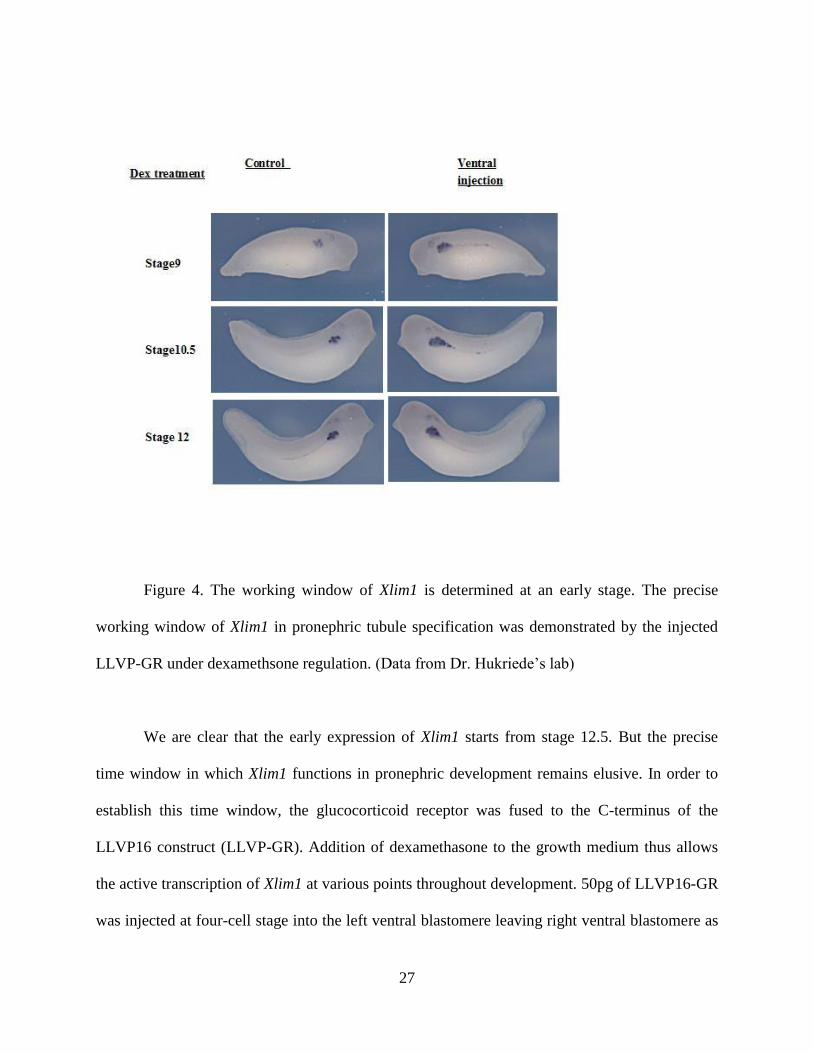
27
Figure 4. The working window of Xlim1 is determined at an early stage. The precise
working window of Xlim1 in pronephric tubule specification was demonstrated by the injected
LLVP-GR under dexamethsone regulation. (Data from Dr. Hukriede’s lab)
We are clear that the early expression of Xlim1 starts from stage 12.5. But the precise
time window in which Xlim1 functions in pronephric development remains elusive. In order to
establish this time window, the glucocorticoid receptor was fused to the C-terminus of the
LLVP16 construct (LLVP-GR). Addition of dexamethasone to the growth medium thus allows
the active transcription of Xlim1 at various points throughout development. 50pg of LLVP16-GR
was injected at four-cell stage into the left ventral blastomere leaving right ventral blastomere as

28
internal control. Figure 6 shows the effects of activating Xlim1 in either the ventral region of the
embryos. Addition of DEX at stage 9 shows that Xlim1 can function in pronephric development
at this stage and overexpression causes formation of enlarged tubules in both ventral injection of
LLVP16-GR. The greatest effect is seen when Xlim1 is activated at stage 10/10.5. At stage 12.5,
when the pronephros looks like a tear shape at the activation by DEX, entails little change in
tubule size. Later treatment does not show obvious effect either. Hence, it suggests around stage
10-10.5, actually is the internal activating time for Xlim1, or the downstream factors are most
effectively expressed at that time. Later than 12.5, it seems like that the networking to specify the
pronephric tubule formation has halted.
2.3.2 The potential downstream targets of lim1 retrieved from microarray analysis
In previous research, our lab discovered that overexpressed Xlim1 transcripts in Xenopus
(animal caps) could replace the need for activin in the culture medium to differentiate explants
into kidney pathway. To identify Lim1’s downstream effectors and the genes involved in the in
vitro kidney differentiation, we carried out microarray analysis to compare the gene expression
profile difference between Xlim1-DEED injected animal caps and uninjected animal caps,
between the retinoic acid/Activin (RA/Ac)treated animal caps and untreated control
ones(Control). All the caps except the control group have been treated in the kidney inducing
condition. We found 216 genes have been significantly upregulated and 576 genes were
significantly downregulated in the DEED-injected vs. uninjected pair. We also found 438
upregulated genes and 266 downregulated genes in AcRA-treated vs. control pair. Among those
upregulated and downregulated genes, we grouped them according to their expression patterns.
29
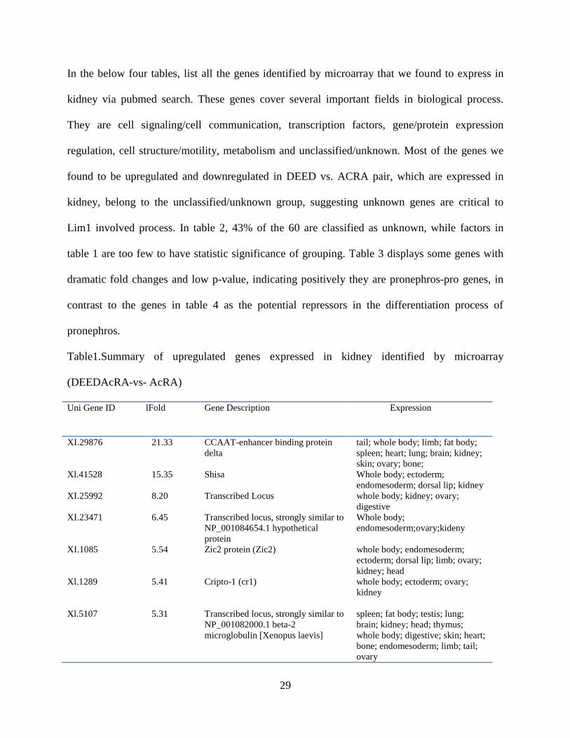
In the below four tables, list all the genes identified by microarray that we found to express in
kidney via pubmed search. These genes cover several important fields in biological process.
They are cell signaling/cell communication, transcription factors, gene/protein expression
regulation, cell structure/motility, metabolism and unclassified/unknown. Most of the genes we
found to be upregulated and downregulated in DEED vs. ACRA pair, which are expressed in
kidney, belong to the unclassified/unknown group, suggesting unknown genes are critical to
Lim1 involved process. In table 2, 43% of the 60 are classified as unknown, while factors in
table 1 are too few to have statistic significance of grouping. Table 3 displays some genes with
dramatic fold changes and low p-value, indicating positively they are pronephros-pro genes, in
contrast to the genes in table 4 as the potential repressors in the differentiation process of
pronephros.
Table1.Summary of upregulated genes expressed in kidney identified by microarray
(DEEDAcRA-vs- AcRA)
Uni Gene ID folFold
Change
Gene Description Expression
XI.29876 21.33 CCAAT-enhancer binding protein
delta
tail; whole body; limb; fat body;
spleen; heart; lung; brain; kidney;
skin; ovary; bone;
Xl.41528 15.35 Shisa Whole body; ectoderm;
endomesoderm; dorsal lip; kidney
XI.25992 8.20 Transcribed Locus whole body; kidney; ovary;
digestive
XI.23471 6.45 Transcribed locus, strongly similar to
NP_001084654.1 hypothetical
protein
Whole body;
endomesoderm;ovary;kideny
XI.1085 5.54 Zic2 protein (Zic2) whole body; endomesoderm;
ectoderm; dorsal lip; limb; ovary;
kidney; head
Xl.1289 5.41 Cripto-1 (cr1) whole body; ectoderm; ovary;
kidney
Xl.5107 5.31 Transcribed locus, strongly similar to
NP_001082000.1 beta-2
microglobulin [Xenopus laevis]
spleen; fat body; testis; lung;
brain; kidney; head; thymus;
whole body; digestive; skin; heart;
bone; endomesoderm; limb; tail;
ovary

30
Xl.23093 5.09 Hypothetical protein MGC53111
(MGC53111)
whole body; kidney; brain; tail;
endomesoderm; digestive
Xl.23425 4.15 Transcribed locus whole body; head; ectoderm; fat
body; digestive; tail; kidney;
endomesoderm
Xl.3201 9.10 Hypothetical protein MGC83260
(MGC83260)
whole body; kidney; testis; ovary
Xl.6392 8.80 Electrogenic sodium
monocarboxylate cotransporter
(smcte)
endomesoderm; whole body;
kidney
Table2 Summary of the downregulated genes expressed in kidney identified by microarray
(DEEDAcRA-vs-AcRA)
Uni Gene
ID
Fold change Description Expression
Xl.12858 4.11 CDNAclone MGC:181766
IMAGE:8550943
whole body; head; fat body; thymus;
kidney
Xl.4083 4.13 MGC83576protein (MGC83576) whole body; spleen; kidney; testis;
endomesoderm
Xl.51341 4.32 Hypothetical LOC495150
(LOC495150)
whole body; head; kidney; ovary
Xl.3820 4.43 Hypothetical protein MGC68533
(MGC68533)
whole body; kidney; ectoderm; testis;
fat body; ovary; endomesoderm;
spleen; head; heart; brain
Xl.3258 4.50 Transcribed locus spleen; heart; kidney; fat body; tail;
bone; whole body
Xl.14521 4.60 Hypothetical protein LOC398688
(LOC398688)
whole body; kidney; ectoderm; lung;
fat body; tail; heart; skin
Xl.11114 4.63 Transcribed locus, moderately similar
to NP_001088258.1 hypothetical
protein LOC495089 [Xenopus laevis]
whole body; kidney; head
Xl.1204 4.79 Xlgv7 gene product (LOC397899) whole body; kidney; ovary;

31
ectoderm; brain; spleen; skin;
endomesoderm; limb
Xl.13880 4.82 MGC82677 protein (MGC82677) whole body; animal cap; ovary;
ectoderm; kidney; dorsal lip
Xl.14515 4.92 Hypothetical protein MGC53361
(MGC53361)
whole body; ectoderm; brain; tail;
skin; kidney; endomesoderm
Xl.5082 5.00 Hypothetical protein MGC68521
(MGC68521)
whole body; kidney; digestive; brain
Xl.46026 5.32 Similar to intergral membrane protein
1 (Itm1)
whole body; ectoderm; kidney; testis;
thymus; skin; endomesoderm; spleen;
lung
Xl.5583 5.37 Hypotheticalprotein LOC100127280
(LOC100127280)
lung; kidney
Xl.4796 5.39 Transcribed locus, strongly similar to
XP_686412.2 PREDICTED:
hypothetical protein, partial [Danio
rerio]
whole body; ovary; digestive; kidney;
ectoderm; spleen; fat body;
endomesoderm
Xl.47404 5.62 MGC80589 protein (MGC80589) lung; ovary; whole body; heart;
spleen; kidney
Xl.28895 5.82 Hypothetical protein MGC82293
(MGC82293)
kidney; testis; ectoderm; whole body
Xl.12498 5.93 Hypothetical protein MGC68533
(MGC68533)
whole body; brain; kidney; thymus
Xl.3820 4.43 Hypothetical LOC494683
(LOC494683)
whole body; kidney; ectoderm; testis;
fat body; ovary; endomesoderm;
spleen; head; heart; brain
Xl.14452 5.96 Transcribed locus whole body; brain; head; kidney; fat
body; limb

32
Xl.14232 6.05 Transcribed locus, moderately similar
to XP_922909.1 PREDICTED:
similar to myosin IXA isoform 2
[Mus musculus]
whole body; ovary; kidney; lung;
testis; fat body
Xl.5611 6.07 Hypothetical protein LOC398440
(LOC398440)
whole body; ectoderm; ovary; dorsal
lip; animal cap; kidney; testis; tail;
endomesoderm
Xl.7178 6.14 Transcribed locus whole body; brain; kidney; thymus
Xl.965 6.26 Epithelial sodium channel, gamma
subunit (gammaxENaC-A)
kidney
Xl.6837 6.41 Hypothetical protein MGC64330
(MGC64330)
whole body; endomesoderm; skin;
ectoderm; ovary; kidney; fat body
Xl.5500 6.53 Hypothetical protein LOC446948
(LOC446948)
whole body; head; skin;
endomesoderm; fat body; spleen;
kidney; testis; lung
Xl.12327 6.60 MGC80468 protein (MGC80468) whole body; lung; spleen; brain;
kidney; ovary
Xl.25327 6.64 Transcribed locus, moderately similar
to NP_003612.1 PTPRF interacting
protein, binding protein 2 (liprin beta
2) [Homo sapiens]
whole body; ovary; kidney; fat body
Xl.19291 6.80 Hypothetical protein LOC398673
(LOC398673)
whole body; dorsal lip; ovary;
kidney; ectoderm; brain
Xl.16481 6.86 Hypothetical protein MGC82240
(MGC82240)
whole body; kidney; head; brain;
testis; spleen; ovary
Xl.24700 7.36 Transcribed locus whole body; kidney
Xl.25179 7.39 Hypothetical protein MGC68635
(MGC68635)
spleen; whole body; thymus; kidney;
brain; limb; testis; skin; fat body;

33
head; digestive
Xl.6133 7.47 XK70 (LOC397756) whole body; ectoderm; animal cap;
head; skin; limb; dorsal lip; digestive;
kidney
Xl.9576 7.55 Carbonic anhydrase II (ca2) whole body; head; brain; kidney;
digestive
Xl.24116 7.56 MGC86445 protein (MGC86445) whole body; testis; ovary; kidney;
spleen; digestive
Xl.2034 7.59 Hypothetical protein LOC443724
(LOC443724)
whole body; spleen; kidney; testis;
endomesoderm; ectoderm
Xl.34077 7.60 Transcribed locus, strongly similar to
NP_001011158.1 SEC31-like 1
[Xenopus tropicalis]
whole body; endomesoderm; kidney
Xl.14521 8.60 Hypothetical protein LOC398688
(LOC398688)
whole body; kidney; ectoderm; lung;
fat body; tail; heart; skin
Xl.695 9.61 Polyprotein, serine proteases and
ovochymase regions (LOC398190)
ovary; whole body; kidney
Xl.16095 9.80 Transcribed locus whole body; kidney
Xl.16255 10.02 Transcribed locus whole body; ectoderm; kidney; dorsal
lip; ovary
Xl.19308 10.82 Hypothetical protein MGC114839
(MGC114839)
kidney; whole body; ovary; dorsal
lip; endomesoderm
Xl.47097 10.96 Hypothetical protein MGC80262
(MGC80262)
whole body; ectoderm; testis; skin;
endomesoderm; ovary; animal cap;
kidney
Xl.206 11.2 Nedd4 protein (nedd4) whole body; dorsal lip; fat body;
kidneymj
Xl.1605 11.32 Transcribed locus, weakly similar to whole body; tail; brain; skin; limb;

34
XP_425376.2 PREDICTED: similar
to envoplakin [Gallus gallus]
spleen; lung; kidney; head
Xl.813 11.96 Hypothetical protein MGC69066
(MGC69066)
spleen; whole body; brain; thymus;
kidney; bone; digestive; lung; head
Xl.21860 14.17 SPARC protein (MGC64258) whole body; brain; limb; fat body;
lung; testis; head; kidney; tail; spleen;
skin; endomesoderm
Xl.15599 15.35 Hypothetical protein MGC68953
(MGC68953)
whole body; kidney; testis; head; fat
body; spleen; skin; limb; brain
Xl.5930 19.83 MGC81015 protein (MGC81015) kidney; whole body; limb
Xl.15997 24.75 Synaptotagmin-like 2 (sytl2) whole body; kidney; endomesoderm;
head
Xl.16272 72.23 Transcribed locus, weakly similar to
XP_221384.4 PREDICTED: similar
to mucin 4 [Rattus norvegicus]
whole body; kidney
Xl.18602 80.05 Hypothetical protein MGC68485
(MGC68485)
whole body; kidney; ectoderm;
ovary; endomesoderm; dorsal lip
Xl.8949 8.53 Ornithine decarboxylase-2
(MGC52527)
whole body; testis; ectoderm; fat
body; skin; animal cap; kidney; head;
endoderm; endomesoderm
Xl.8939 8.73 MGC81907 protein (MGC81907) whole body; kidney; fat body;
thymus
Xl.15221 8.78 Similar to solute carrier family 27
(fatty acid transporter), member 2
(LOC398483)
whole body; kidney; brain
Xl.8759 10.65 Hypothetical protein MGC68473
(MGC68473)
whole body; brain; head; kidney;
testis; spleen; ectoderm
Xl.1500 15.25 Similar to villin (LOC398504) whole body; ectoderm; kidney; limb;

35
skin; brain; testis
Xl.23708 22.25 MGC81939 protein (MGC81939) whole body; brain; ectoderm; kidney;
skin
Shown above are some genes that significantly decreased after the depletion of Lim1 in
explants. Genes that are displayed in these four tables are the genes known to express in kidney
in unigene database. For genes that are expressed in kidneys, their expression time windows and
locations may not overlap pronephric development time window or reside in pronephric region
in reality. Thus it is not wise to only depend on the quantitative evidence of the difference in
expression levels, the actual expression level and location of these genes need to be validated by
in situ hybridization. The same principle applies to the genes that have not been reported to
associate with kidney development or express in kidney region, but reveal to show a difference
in the supplemental materials.
Table3. Summary of the upregulated genes identified by microarray (Activin/RA vs. control) that
have been documented to express in kidney
Unigene ID Fold Change Gene Description Expression
Xl.12667 70.07 transcription factor 2 ///
hypothetical protein MGC68543
whole body; kidney
Xl.15054 48.71 Cnpr03 mRNA, complete
sequence
whole body; ectoderm; fat body; tail;
limb; endomesoderm; brain; kidney;
spleen; lung
Xl.34 40.24 Hypothetical protein MGC68907 whole body; endomesoderm; limb;
thymus; spleen; dorsal lip; lung;
kidney
Xl.23957 38.09 Ami whole body; kidney; testis; spleen; fat

36
body; digestive
Xl.6392 31.17 electrogenic sodium
monocarboxylate cotransporter
endomesoderm; whole body; kidney
Xl.1042 7.41 eural specific DNA binding
protein
heart; whole body; endomesoderm;
kidney; fat body
Xl.899 7.37 pleiotrophic factor-alpha2 endomesoderm; whole body; fat body;
ectoderm; head; kidney; brain; ovary
Xl.29610 6.79 Transcribed locus, weakly similar
to XP_237286.4 PREDICTED:
similar to tensin [Rattus
norvegicus]
lung; kidney; spleen
Xl.34749 6.69 hypothetical protein MGC68579 whole body; ectoderm; kidney; lung;
dorsal lip; brain; tail; limb;
endomesoderm; testis
Xl.23641 6.17 enzymatic glycosylation-
regulating gene
kidney
Xl.216 5.86 Similar to macrophage
stimulating1 (hepatocyte growth
factor-like)
whole body; ectoderm;
endomesoderm; kidney; dorsal lip;
bone
Xl.25327 5.52 Transcribed locus, moderately
similar to XP_426399.1
PREDICTED: similar to coiled-
coil like protein 1 [Gallus gallus]
whole body; ovary; kidney; fat body
Xl.15670 5.52 Transcribed locus, weakly similar
to XP_314118.2
ENSANGP00000003760
[Anopheles gambiae str. PEST]
whole body; kidney
Xl.24700 5.45 Transcribed locus whole body; kidney
Xl.1008 5.30 FGF receptor 4a whole body; endomesoderm;
ectoderm; head; dorsal lip; kidney
Xl.50622 5.03 similar to UDP-glucose
dehydrogenase
whole body; endomesoderm;
ectoderm; ovary; testis; dorsal lip;
kidney
Xl.45046 5.00 MRNA sequence whole body; ectoderm; kidney;
thymus; digestive; endomesoderm
Xl.16655 19.67 solute carrier family 16
(monocarboxylic acid
whole body; kidney; ectoderm;

37
transporters), member 6 endomesoderm
Xl.48471 16.38 hypothetical LOC495093 whole body; ectoderm; kidney; limb;
endomesoderm
Xl.21562 13.46 ras-related protein whole body; ectoderm; dorsal lip;
kidney
Xl.31935 8.31 MAP kinase phosphatase X17C whole body; spleen; kidney; ectoderm;
endomesoderm
Xl.1210 7.66 Hoxb7 protein whole body; brain; kidney; fat body
Xl.1042 7.41 neural specific DNA binding
protein /// zinc finger protein Gli2
heart; whole body; endomesoderm;
kidney; fat body
Xl.2751 4.75 fibroblast growth factor receptor whole body; endomesoderm; testis;
dorsal lip; ectoderm; kidney
Xl.29876 4.73 CCAAT-enhancer binding
protein delta
tail; whole body; limb; fat body;
spleen; heart; lung; brain; kidney;
skin; ovary; bone;
Xl.2243 4.33 Fibronectin protein whole body; endomesoderm; lung;
bone; limb; tail; brain; fat body; testis;
ectoderm; digestive; head; spleen;
kidney
Xl.1130 4.80 Xenopus laevis, Similar to paired
box gene 2, clone
IMAGE:5543325, mRNA ///
Paired box protein
whole body; ectoderm; kidney
Xl.13563 4.07 MGC80391 protein whole body; kidney; digestive
Xl.49562 33.19 hypothetical LOC495088 endomesoderm; whole body; kidney
Xl.15593 13.90 MGC82107 protein whole body; kidney
Xl.1085 10.03 Zic2 protein whole body; endomesoderm;
ectoderm; dorsal lip; limb; ovary;
kidney; head
Xl.6155 12.59 MGC83414 protein whole body; brain; kidney
Xl.24776 10.57 Transcribed locus, weakly similar
to XP_517667.1 PREDICTED:
similar to versican V2 core
protein precursor [Pan
troglodytes]
whole body; ectoderm; ovary; kidney;
brain; spleen; endomesoderm

38
Xl.21860 10.43 SPARC protein whole body; brain; limb; fat body;
lung; testis; head; kidney; tail; spleen;
skin; endomesoderm
Xl.1299
12.58 alkaline phosphatase,
liver/bone/kidney
whole body; endomesoderm; tail;
limb; fat body; kidney; dorsal lip;
brain
Xl.3150 11.34 follistatin-related protein endomesoderm; whole body; bone;
tail; limb; fat body; brain; head; testis;
digestive; lung; kidney
Table4. Summary of the downregulated genes identified by microarray (AcRA vs. Control) that
have been documented to express in kidney
Unigene ID Fold Change Gene Description Expression
Xl.3209 3.46 elongation factor 1-alpha O ectoderm; whole body; ovary; kidney;
endomesoderm; testis
Xl.53426 4.77 Transcribed locus whole body; kidney
Xl.6837 4.87 hypothetical protein MGC64330 whole body; endomesoderm; skin;
ectoderm; ovary; kidney; fat body
Xl.16776 4.99 Hypothetical protein
MGC68565
whole body; brain; kidney
Xl.48740 5.07 MGC81884 protein spleen; whole body; heart; brain; kidney
Xl.12327 5.08 MGC80468 protein whole body; lung; spleen; brain;
kidney; ovary
Xl.931 5.29 T-box transcription factor Tbx2 whole body; kidney; digestive
Xl.5614 5.83 hypothetical protein MGC68646 whole body; kidney
Xl.814 6.38 immunoglobulin light chain spleen; fat body; kidney; brain; whole
body; digestive
Xl.8193 6.90 MGC86499 protein whole body; kidney; brain; testis
Xl.1201 7.91 glucocorticoid receptor protein whole body; lung; kidney; fat body
Xl.2458 8.52 hypothetical LOC496017 whole body; spleen; ovary; brain;
ectoderm; fat body; head; heart; limb;

39
thymus; kidney; skin; digestive
Xl.21648 11.74 Kit receptor tyrosine kinase
homolog
whole body; spleen; kidney
Xl.14126 11.94 Transcribed locus whole body; kidney; endomesoderm
Xl.12819 12.64 MGC81876 protein thymus; kidney
Xl.5158
13.82 Epor mRNA for erythropoietin
receptor homologue
digestive; spleen; lung; kidney; head
Thus, we carried out in situ hybridization for 35 potential targets on Xenopus embryos.
The aim was to investigate whether these targets express in pronephric region and whether any
of them express at the early specification period (data not shown). Our results demonstrated that
some of the targets only expressed at the stage 12.5, the time tubule was specified. Some of them
expressed at stage 19 the time early before the pronephric anlagen is visible. But, for most of the
targets we investigated, they do not display an expression pattern early before the appearing of
the pronephric anlagen. Additionally, among the 7 targets that could be detected at the right time
frame for pronephric specification, none of which showed up in the pronephric region. We also
did in situ hybridization using genes newly identified in microarray analysis on embryos. But
their expression were not clearly associated with the pronephros development process(Data not
shown).
40
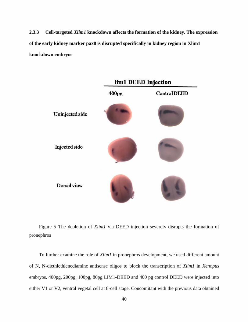
2.3.3 Cell-targeted Xlim1 knockdown affects the formation of the kidney. The expression
of the early kidney marker pax8 is disrupted specifically in kidney region in Xlim1
knockdown embryos
Figure 5 The depletion of Xlim1 via DEED injection severely disrupts the formation of
pronephros
To further examine the role of Xlim1 in pronephros development, we used different amount
of N, N-diethlethlenediamine antisense oligos to block the transcription of Xlim1 in Xenopus
embryos. 400pg, 200pg, 100pg, 80pg LIM1-DEED and 400 pg control DEED were injected into
either V1 or V2, ventral vegetal cell at 8-cell stage. Concomitant with the previous data obtained

41
in the lab at 4-cell stage injection, 200pg DEED disrupts the normal condensement of pronephros
severely. Xpax8 expression is seen around the circumference of the embryos trailing down from
where the tail of the teardrop of typical expression begins. These embryos never show a regular
tear-shape in the correct place in addition to the ventral stain. More critically, when the dose of
DEED was increased to 400pg per cell, Xpax8 was not visible in the pronephros region. The
normal tear-shape staining disappears, which indicates the loss of pronephros tissue in regards of
that Pax8 is the marker of early pronephros. 100pg and 80pg injection, in the contrast, do not
show a phenotype of kidney disruption. All those embryos were fixed at stage 19, the time the
pronephros specification has been established. Xpax8 staining, however is still visible in otic
vesicle, which means cell targeting is correct and the loss of pax8 results from lim1-DEED
injection simply .To rule out the possibility that the ventral expression Xpax8 was in cells that
had been fate-transformed from other tissues, several ventral differentiation markers were
analyzed. XSzl, XVent1, XmyoD and Xalpha T4globin were unaffected.(date not shown).This
result suggests it might be a failure in migration of cells in which Lim1 has been depleted and the
failure of migration will give rise to possible apoptosis of the designated pronephric cells. Other
possibility to result in such a pattern is the secondary effect of gastrulation problem (Discussed
with Dr.Hukriede).

42
3.0 CONCLUSION AND PROSPECTUS
3.1 OBJECTIVES
My work was undertaken with the goal to elucidate the mechanism of Xlim1 in Xenopus
kidney development and it contains two parts. One part is some leftover work of Caroline’s
project which is to explore the regulatory role of Xlim1 in pronephros and the other is the TAP
work with intention to identify the components of Xlim1 complex, which I just started
preliminary experiment and is not included in this thesis. For the mechanism of Xlim1 in kidney
development, this thesis tried to characterize Xlim1’s role in early kidney specification as well as
to identify the downstream factors of Xlim1 pathways
3.2 INTERPRETATION OF THE RESULTS
The early study of Lim1 dated back to 1992 when Taira’s group which was fancied by a
newly identified homeobox class that contains mec-3 and lin-11, started to examine whether the
members of this particular class are involved in the vertebrate embryogenesis. Xlim1 was isolated
and sequenced in Xenopus embryos in that study67
. Soon after another paper came out reporting a
homolog of Xlim1, Lim1 (Lhx1), was identified from the mouse. Since then the role of Lim1 has
been very well investigated. In consistence with several papers published by Behringer’s group

43
describing the Lim1-/-
mouse phenotype, we found that in Xenopus, the specific depletion of
Xlim1 at 8-cell stage in ventral vegetal cell results in the loss of pronephros, assessed by Pax8,
one of the early pronephros markers in Xenopus. But the forming of this phenotype depends on
the dose of Lim1-DEED used in the experiment. A relatively lower dose of Lim1-DEED
injection (200pg/cell at 8-cell stage) gives rise to a tailing down pattern of Xpax8. 100pg or
lower, interestingly, gives a normal phenotype. This indicates that different level of Lim1
transcripts differentially regulates the gene networking that associates with pronephros
specification. Actually, several genes that play critical roles in development show expression
level-dependent effects on cellular differentiation or development. For instance, Oct3/4, an
embryonic transcription factor, was found to be a master regulator of pluripotency that controls
lineage commitment in embryonic stem cells. Three distinct fates of ES cells are governed by the
precise level of Oct-3/4 present in ES cells revealed by the tetracycline–induced system83
.
However, other suggests that the loss of visible pronephros anlagen may be due to secondary
effects of gastrulation problem (Discussed with Dr. Hukriede). So the question remains to be
whether the depletion of Xlim1 gives rise to the loss of pronephros or the depletion of Xlim1
gives rise to the gastrulation defects that consequently disrupts the formation of the pronephros.
Additionally, it raises another question that whether it is the loss of tissue or it is the loss of
inductive signal for pronephros.
To further understand and answer these questions, we took microarray analysis. Our
microarray data indicates the depletion of Xlim1 in animal caps induce couple of genes’
expression going down under the kidney differentiation conditions. Many of these genes play a
role in cell movement, proliferation and differentiation, such as N-cadherin, Marginal Coil, and

44
Annexin et al84-86
. So it implies Lim1 does have close relationship with the molecular networking
involving in cell movement. Consistent with the previous result that PAPC is a downstream
factor of the Xlim1, we would speculate that perhaps more cell-movement factors are regulated
by Xlim1 at some time point and on certain location in embryos45, 87
. Yet, no interesting
hypothesis has been raised to elucidate the mechanism how Xlim1 specifies pronephros fate
because most of these gene expression patterns do not coincide with the pattern of Xlim1 or
Pax8. However, microarray data provide us with a more restricted pool to probe the unknown
genes that may affect the pronephric development.
The precious work completed by Behringer’s group carefully dissected lim1’s role in the
later development of kidney and female reproductive tract50, 88
. But nobody has ever attempted to
regulate the level of Lim1 at a very early developmental stage in mouse by utilizing regulatory
systems like tet-inducible system. If such a system could be established in mouse, the analysis
from that model may provide a better comparison with our results than the current data obtained
from the mouse model of late kidney development and thus might provides more useful
information.
Our results again confirm Carroll and Vize’s results in 1999, that we found the
overexpression of Xlim1 successfully induces an enlarged tubules and glomerulus. The depletion
of Xlim1 in explants does not cause the significant change in Xpax8’s expression level,
admittedly that Xpax8 itself maintains a low expression level below the stringent threshold. So
whether pax8 is regulated by Xlim1 is still elusive.

45
According to our results, the functionally active time window of Xlim1 in manipulating
kidney formation starts as early as stage 9 and ends at stage 12.5, with a peak time at stage 10 to
10.5. Stage 12.5 is the time point that tubule is specified and stage 14 is the time the duct is
specified. In terms of specification, we mean a portion is capable of forming a given tissue when
cultured in a neural environment (Sack, 1987). Clearly we can see that the overexpression of
Xlim1 at early time before stage 12.5 only induced enlarged pronephroi, which means that the
specification of ducts requires other inducers rather than Xlim1 only, while glomerulous and
tubules mostly require Xlim1 involved networking. At the same time, the networking that has
been activated at the stage 10.5 remains to be investigated. It would be helpful if we can screen
EST library finding out all the ESTs expressed during 10.5. However the expression of Xlim1 at
10.5 is not even close to the future pronephros region67
. So there are no practical standards we
can apply to predict where the cofactors should be expressed at an early stage like 10.5 that could
efficiently affect the specification of pronephros. Hence, the molecular work that probes the
factors directly or indirectly via complexes associated with Xlim1 turns to be critical.
Recently compelling evidence from both Xenopus and chicken supports the hypothesis
that pronephros forms by means of secondary induction signals emitted from anterior somites89,
90. In addition, pronephros never forms in the absence of anterior somites. Thus we cannot rule
out the possibility contributed by anterior somites. Possibly Xlim1 also plays some role in
anterior somites formation, at least supports the formation of this tissue but it is not required for
the formation. It is known that Chordin, a factor critical for somitogenesis, induces ectopic
expression of myf5 at 10.5, a coincidence with the peak of Xlim1’s activity. Myf5 expression at
stage 10.5 is an indicator of whether a cell has the potential to form anterior somites47
. This

46
interestingly suggests that Xlim1 and somitogenesis signaling pathway reciprocally induce each
other’s differentiation. In contrast, Seufert’s paper in 1999 reported that reduced somites
compensate for the enlarged pronephros. Whether it is true or not need experimental validation.
I use Unigene database as the main resource to retrieve the cellular location in different
tissues and organs of the interested genes. However, when comparing with the targeted genes
picked by hand by others, some of the genes that are related to kidney were not screened. After
careful examining, those genes that are not listed as expressed in kidney in Unigene database,
generally are annotated as expressed in endomesoderm. Thus, the pool for the potential targets of
Xlim1 may expand by adding the genes that are expressed in endomesoderm too.
3.3 SIGNIFICANCE AND FUTURE WORK
The future direction of this work is to find specific targets of Lim1 as well as to answer
the question that whether lack of lim1 entails the loss of kidney tissue or the loss of signals that
induce kidney development.

47
BIBLIOGRAPHY
1. Fenton, R. A. & Knepper, M. A. Urea and renal function in the 21st century: insights
from knockout mice. J Am Soc Nephrol 18, 679-88 (2007).
2. Nishimura, H. & Fan, Z. Regulation of water movement across vertebrate renal tubules.
Comp Biochem Physiol A Mol Integr Physiol 136, 479-98 (2003).
3. Moestrup, S. K. & Nielsen, L. B. The role of the kidney in lipid metabolism. Curr Opin
Lipidol 16, 301-6 (2005).
4. Bagshaw, S. M. & Gibney, R. T. Conventional markers of kidney function. Crit Care
Med 36, S152-8 (2008).
5. Dressler, G. R. The cellular basis of kidney development. Annu Rev Cell Dev Biol 22,
509-29 (2006).
6. Brenner & Rector’s the kidney (ed. Brenner) (Saunders Elsevier, c2008, philadephila,
2008).
7. Jones, E. A. Xenopus: a prince among models for pronephric kidney development. J Am
Soc Nephrol 16, 313-21 (2005).
8. Ludwig, K. S. & Landmann, L. Early development of the human mesonephros. Anat
Embryol (Berl) 209, 439-47 (2005).
9. Vize, P. D., Seufert, D. W., Carroll, T. J. & Wallingford, J. B. Model systems for the
study of kidney development: use of the pronephros in the analysis of organ induction
and patterning. Dev Biol 188, 189-204 (1997).
10. Drummond, I. Making a zebrafish kidney: a tale of two tubes. Trends Cell Biol 13, 357-
65 (2003).
11. Kuure, S., Vuolteenaho, R. & Vainio, S. Kidney morphogenesis: cellular and molecular
regulation. Mech Dev 92, 31-45 (2000).

48
12. Brandli, A. W. Towards a molecular anatomy of the Xenopus pronephric kidney. Int J
Dev Biol 43, 381-95 (1999).
13. Moritz, K. M. & Wintour, E. M. Functional development of the meso- and metanephros.
Pediatr Nephrol 13, 171-8 (1999).
14. Yin, Y. & Ma, L. Development of the mammalian female reproductive tract. J Biochem
137, 677-83 (2005).
15. Brennan, H. C., Nijjar, S. & Jones, E. A. The specification of the pronephric tubules and
duct in Xenopus laevis. Mech Dev 75, 127-37 (1998).
16. Dale, L. & Slack, J. M. Regional specification within the mesoderm of early embryos of
Xenopus laevis. Development 100, 279-95 (1987).
17. Harland, R. & Gerhart, J. Formation and function of Spemann's organizer. Annu Rev Cell
Dev Biol 13, 611-67 (1997).
18. Dale, L. & Jones, C. M. BMP signalling in early Xenopus development. Bioessays 21,
751-60 (1999).
19. Asashima, M., Ariizumi, T. & Malacinski, G. M. In vitro control of organogenesis and
body patterning by activin during early amphibian development. Comp Biochem Physiol
B Biochem Mol Biol 126, 169-78 (2000).
20. Chan, T. C., Takahashi, S. & Asashima, M. A role for Xlim-1 in pronephros development
in Xenopus laevis. Dev Biol 228, 256-69 (2000).
21. Cartry, J. et al. Retinoic acid signalling is required for specification of pronephric cell
fate. Dev Biol 299, 35-51 (2006).
22. Maeshima, A. et al. Activin A: autocrine regulator of kidney development and repair.
Endocr J 55, 1-9 (2008).
23. Tsang, T. E. et al. Lim1 activity is required for intermediate mesoderm differentiation in
the mouse embryo. Dev Biol 223, 77-90 (2000).
24. Fujii, T. et al. Expression patterns of the murine LIM class homeobox gene lim1 in the
developing brain and excretory system. Dev Dyn 199, 73-83 (1994).
25. Shawlot, W. & Behringer, R. R. Requirement for Lim1 in head-organizer function.
Nature 374, 425-30 (1995).

49
26. James, R. G., Kamei, C. N., Wang, Q., Jiang, R. & Schultheiss, T. M. Odd-skipped
related 1 is required for development of the metanephric kidney and regulates formation
and differentiation of kidney precursor cells. Development 133, 2995-3004 (2006).
27. Narlis, M., Grote, D., Gaitan, Y., Boualia, S. K. & Bouchard, M. Pax2 and pax8 regulate
branching morphogenesis and nephron differentiation in the developing kidney. J Am
Soc Nephrol 18, 1121-9 (2007).
28. Bouchard, M., Souabni, A., Mandler, M., Neubuser, A. & Busslinger, M. Nephric lineage
specification by Pax2 and Pax8. Genes Dev 16, 2958-70 (2002).
29. Wingert, R. A. et al. The cdx genes and retinoic acid control the positioning and
segmentation of the zebrafish pronephros. PLoS Genet 3, 1922-38 (2007).
30. St Peter, W. L. Introduction: chronic kidney disease: a burgeoning health epidemic. J
Manag Care Pharm 13, S2-5 (2007).
31. Chang, M. Y. & Ong, A. C. Autosomal dominant polycystic kidney disease: recent
advances in pathogenesis and treatment. Nephron Physiol 108, p1-7 (2008).
32. Lipton, J. Mating worms and the cystic kidney: Caenorhabditis elegans as a model for
renal disease. Pediatr Nephrol 20, 1531-6 (2005).
33. Eknoyan, G. et al. Proteinuria and other markers of chronic kidney disease: a position
statement of the national kidney foundation (NKF) and the national institute of diabetes
and digestive and kidney diseases (NIDDK). Am J Kidney Dis 42, 617-22 (2003).
34. Fissell, W. H., Fleischman, A. J., Humes, H. D. & Roy, S. Development of continuous
implantable renal replacement: past and future. Transl Res 150, 327-36 (2007).
35. Hensey, C., Dolan, V. & Brady, H. R. The Xenopus pronephros as a model system for the
study of kidney development and pathophysiology. Nephrol Dial Transplant 17 Suppl 9,
73-4 (2002).
36. Braam, B., Verhaar, M. C., Blankestijn, P., Boer, W. H. & Joles, J. A. Technology
insight: Innovative options for end-stage renal disease--from kidney refurbishment to
artificial kidney. Nat Clin Pract Nephrol 3, 564-72 (2007).
37. Lange, C. et al. Administered mesenchymal stem cells enhance recovery from
ischemia/reperfusion-induced acute renal failure in rats. Kidney Int 68, 1613-7 (2005).
38. Feng, Z. et al. Glomerular aging in females is a multi-stage reversible process mediated
by phenotypic changes in progenitors. Am J Pathol 167, 355-63 (2005).

50
39. Faber, N. a. Normal Table of Xenopus laevis (Daudin) ( Garland Publishing Inc., New
York 1994).
40. Gurdon, J. B. From nuclear transfer to nuclear reprogramming: the reversal of cell
differentiation. Annu Rev Cell Dev Biol 22, 1-22 (2006).
41. Klein, S. L. et al. Genetic and genomic tools for Xenopus research: The NIH Xenopus
initiative. Dev Dyn 225, 384-391 (2002).
42. Hazel L.Sive, R. M. G., Richard M Harland. Early Development of Xenopus laevis (Cold
Spring Harbor Laboratory Press, Cold Spring Harbor, New York, 2000).
43. Fournier, F. et al. Expression of calcium channels in Xenopus oocyte injected with crab
skeletal muscle fibre mRNA. Biol Cell 68, 37-42 (1990).
44. Bianchi, L. & Driscoll, M. Heterologous expression of C. elegans ion channels in
Xenopus oocytes. WormBook, 1-16 (2006).
45. Hukriede, N. A. et al. Conserved requirement of Lim1 function for cell movements
during gastrulation. Dev Cell 4, 83-94 (2003).
46. Tahinci, E. et al. Lrp6 is required for convergent extension during Xenopus gastrulation.
Development 134, 4095-106 (2007).
47. Mitchell, T., Jones, E. A., Weeks, D. L. & Sheets, M. D. Chordin affects pronephros
development in Xenopus embryos by anteriorizing presomitic mesoderm. Dev Dyn 236,
251-61 (2007).
48. Kyuno, J., Masse, K. & Jones, E. A. A functional screen for genes involved in Xenopus
pronephros development. Mech Dev 125, 571-86 (2008).
49. Chen, J. A., Voigt, J., Gilchrist, M., Papalopulu, N. & Amaya, E. Identification of novel
genes affecting mesoderm formation and morphogenesis through an enhanced large scale
functional screen in Xenopus. Mech Dev 122, 307-31 (2005).
50. Chen, Y. T., Kobayashi, A., Kwan, K. M., Johnson, R. L. & Behringer, R. R. Gene
expression profiles in developing nephrons using Lim1 metanephric mesenchyme-
specific conditional mutant mice. BMC Nephrol 7, 1 (2006).
51. Saulnier, D. M., Ghanbari, H. & Brandli, A. W. Essential function of Wnt-4 for
tubulogenesis in the Xenopus pronephric kidney. Dev Biol 248, 13-28 (2002).
52. Lopez, S. L. et al. Notch activates sonic hedgehog and both are involved in the
specification of dorsal midline cell-fates in Xenopus. Development 130, 2225-38 (2003).

51
53. Dawid, I. B., Taira, M., Good, P. J. & Rebagliati, M. R. The role of growth factors in
embryonic induction in Xenopus laevis. Mol Reprod Dev 32, 136-44 (1992).
54. de Boer, T. P. et al. Cloning, embryonic expression, and functional characterization of
two novel connexins from Xenopus laevis. Biochem Biophys Res Commun 349, 855-62
(2006).
55. Kishi, Y. A., Funakoshi, H., Matsumoto, K. & Nakamura, T. Molecular cloning,
expression and partial characterization of Xksy, Xenopus member of the Sky family of
receptor tyrosine kinases. Gene 288, 29-40 (2002).
56. Hill, C. S. TGF-beta signalling pathways in early Xenopus development. Curr Opin
Genet Dev 11, 533-40 (2001).
57. Xenopus Protocols: cell biolgy and siganl transduction (ed. Walker, J. M.) (Humana
Press Inc, totowa, New Jersey, 2006).
58. Yu, J., Zhao, Y., Li, Z., Galas, S. & Goldberg, M. L. Greatwall kinase participates in the
Cdc2 autoregulatory loop in Xenopus egg extracts. Mol Cell 22, 83-91 (2006).
59. Groisman, I., Jung, M. Y., Sarkissian, M., Cao, Q. & Richter, J. D. Translational control
of the embryonic cell cycle. Cell 109, 473-83 (2002).
60. Amaya, E., Offield, M. F. & Grainger, R. M. Frog genetics: Xenopus tropicalis jumps
into the future. Trends in Genetics 14, 253-255 (1998).
61. Hirsch, N., Zimmerman, L. B. & Grainger, R. M. Xenopus, the next generation: X.
Tropicalis genetics and genomics. Developmental Dynamics 225, 422-433 (2002).
62. Khokha, M. K. et al. Techniques and probes for the study of Xenopus tropicalis
development. Dev Dyn 225, 499-510 (2002).
63. Morin, R. D. et al. Sequencing and analysis of 10,967 full-length cDNA clones from
Xenopus laevis and Xenopus tropicalis reveals post-tetraploidization transcriptome
remodeling. Genome Research 16, 796-803 (2006).
64. Lappin, D. W., Hensey, C., McMahon, R., Godson, C. & Brady, H. R. Gremlins,
glomeruli and diabetic nephropathy. Curr Opin Nephrol Hypertens 9, 469-72 (2000).
65. Hsu, D. R., Economides, A. N., Wang, X., Eimon, P. M. & Harland, R. M. The Xenopus
dorsalizing factor Gremlin identifies a novel family of secreted proteins that antagonize
BMP activities. Mol Cell 1, 673-83 (1998).
52
66. Bohn, S. et al. Distinct molecular and morphogenetic properties of mutations in the
human HNF1beta gene that lead to defective kidney development. J Am Soc Nephrol 14,
2033-41 (2003).
67. Taira, M., Jamrich, M., Good, P. J. & Dawid, I. B. The LIM domain-containing homeo
box gene Xlim-1 is expressed specifically in the organizer region of Xenopus gastrula
embryos. Genes Dev 6, 356-66 (1992).
68. Dawid, I. B., Breen, J. J. & Toyama, R. LIM domains: multiple roles as adapters and
functional modifiers in protein interactions. Trends Genet 14, 156-62 (1998).
69. Agulnick, A. D. et al. Interactions of the LIM-domain-binding factor Ldb1 with LIM
homeodomain proteins. Nature 384, 270-2 (1996).
70. Kodjabachian, L. et al. A study of Xlim1 function in the Spemann-Mangold organizer.
Int J Dev Biol 45, 209-18 (2001).
71. Bach, I. The LIM domain: regulation by association. Mech Dev 91, 5-17 (2000).
72. Taira, M., Otani, H., Jamrich, M. & Dawid, I. B. Expression of the LIM class homeobox
gene Xlim-1 in pronephros and CNS cell lineages of Xenopus embryos is affected by
retinoic acid and exogastrulation. Development 120, 1525-36 (1994).
73. Duboule, D. The rise and fall of Hox gene clusters. Development 134, 2549-60 (2007).
74. De Robertis, E. M. Developmental biology. Dismantling the organizer. Nature 374, 407-8
(1995).
75. Yasuo, H. & Lemaire, P. Role of Goosecoid, Xnot and Wnt antagonists in the
maintenance of the notochord genetic programme in Xenopus gastrulae. Development
128, 3783-93 (2001).
76. De Robertis, E. M. & Kuroda, H. Dorsal-ventral patterning and neural induction in
Xenopus embryos. Annu Rev Cell Dev Biol 20, 285-308 (2004).
77. Mochizuki, T. et al. Xlim-1 and LIM domain binding protein 1 cooperate with various
transcription factors in the regulation of the goosecoid promoter. Dev Biol 224, 470-85
(2000).
78. Hiratani, I., Mochizuki, T., Tochimoto, N. & Taira, M. Functional domains of the LIM
homeodomain protein Xlim-1 involved in negative regulation, transactivation, and axis
formation in Xenopus embryos. Dev Biol 229, 456-67 (2001).

53
79. Carroll, T. J. & Vize, P. D. Synergism between Pax-8 and lim-1 in embryonic kidney
development. Dev Biol 214, 46-59 (1999).
80. Pedersen, A., Skjong, C. & Shawlot, W. Lim 1 is required for nephric duct extension and
ureteric bud morphogenesis. Dev Biol 288, 571-81 (2005).
81. Kao, K. R. & Elinson, R. P. The entire mesodermal mantle behaves as Spemann's
organizer in dorsoanterior enhanced Xenopus laevis embryos. Dev Biol 127, 64-77
(1988).
82. Kudoh, T. et al. A gene expression screen in zebrafish embryogenesis. Genome Res 11,
1979-87 (2001).
83. Niwa, H., Miyazaki, J. & Smith, A. G. Quantitative expression of Oct-3/4 defines
differentiation, dedifferentiation or self-renewal of ES cells. Nat Genet 24, 372-6 (2000).
84. Frazzetto, G., Klingbeil, P. & Bouwmeester, T. Xenopus marginal coil (Xmc), a novel
FGF inducible cytosolic coiled-coil protein regulating gastrulation movements. Mech
Dev 113, 3-14 (2002).
85. Warga, R. M. & Kane, D. A. A role for N-cadherin in mesodermal morphogenesis during
gastrulation. Dev Biol 310, 211-25 (2007).
86. Seville, R. A., Nijjar, S., Barnett, M. W., Masse, K. & Jones, E. A. Annexin IV (Xanx-4)
has a functional role in the formation of pronephric tubules. Development 129, 1693-704
(2002).
87. Medina, A., Swain, R. K., Kuerner, K. M. & Steinbeisser, H. Xenopus paraxial
protocadherin has signaling functions and is involved in tissue separation. Embo J 23,
3249-58 (2004).
88. Kobayashi, A., Shawlot, W., Kania, A. & Behringer, R. R. Requirement of Lim1 for
female reproductive tract development. Development 131, 539-49 (2004).
89. Mauch, T. J., Yang, G., Wright, M., Smith, D. & Schoenwolf, G. C. Signals from trunk
paraxial mesoderm induce pronephros formation in chick intermediate mesoderm. Dev
Biol 220, 62-75 (2000).
90. Seufert, D. W., Brennan, H. C., DeGuire, J., Jones, E. A. & Vize, P. D. Developmental
basis of pronephric defects in Xenopus body plan phenotypes. Dev Biol 215, 233-42
(1999).

54