the role of prefrontal dopamine d1 receptors in the...
TRANSCRIPT
Neuron
Article
The Role of Prefrontal Dopamine D1 Receptorsin the Neural Mechanisms of Associative LearningM. Victoria Puig1 and Earl K. Miller1,*1The Picower Institute for Learning and Memory and Department of Brain and Cognitive Sciences, Massachusetts Institute of Technology,
Cambridge, MA 02139, USA
*Correspondence: [email protected] 10.1016/j.neuron.2012.04.018
SUMMARY
Dopamine is thought to play a major role in learning.However, while dopamine D1 receptors (D1Rs) in theprefrontal cortex (PFC) have been shown to modu-late working memory-related neural activity, theirrole in the cellular basis of learning is unknown. Werecorded activity from multiple electrodes while in-jecting the D1R antagonist SCH23390 in the lateralPFC as monkeys learned visuomotor associations.Blocking D1Rs impaired learning of novel associa-tions and decreased cognitive flexibility but sparedperformance of already familiar associations. Thissuggests a greater role for prefrontal D1Rs in learn-ing new, rather than performing familiar, associa-tions. There was a corresponding greater decreasein neural selectivity and increase in alpha and betaoscillations in local field potentials for novel thanfor familiar associations. Our results suggest thatweak stimulation of D1Rs observed in aging andpsychiatric disorders may impair learning and PFCfunction by reducing neural selectivity and exacer-bating neural oscillations associated with inattentionand cognitive deficits.
INTRODUCTION
Learning and memory are foundations of advanced cognition.
Their impairment is found, for example, in Parkinson’s disease
and schizophrenia (Owen et al., 1992; Park and Holzman,
1992; Elvevag and Goldberg, 2000; Lewis et al., 2003; Jankovic,
2008; Wang et al., 2011). These disorders also impact the
prefrontal cortex (PFC), a cortical region associated with execu-
tive functions and critical for normal learning (Miller and Cohen,
2001). Profound learning and other cognitive deficits typically
follow PFC damage (Godefroy, 2003; Robbins, 2007; Kehagia
et al., 2010), and neurophysiological studies show learning-
related changes in PFC neural activity (Asaad et al., 1998; Pasu-
pathy andMiller, 2005; Benchenane et al., 2010; Antzoulatos and
Miller, 2011). The widespread inputs the PFC receives from
dopamine axons originating in the ventral tegmental area and
the substantia nigra pars compacta (Williams and Goldman-
Rakic, 1998) are likely to be important. Dopamine neurons fire
874 Neuron 74, 874–886, June 7, 2012 ª2012 Elsevier Inc.
and release dopamine into the PFC to sensory cues that predict
reward (Schultz et al., 1993) and thus provide the reward-
prediction error signals needed for guiding reward-based
learning (Schultz, 2007) and for gating reward-related information
in and out of activeworkingmemory (Cohen et al., 2002; O’Reilly,
2006). In addition, a subset of dopamine neurons is activated by
aversive events. Because these events are nonrewarded, some
dopamine neurons may encode the stimulus salience rather
than its positive value (Matsumoto and Hikosaka, 2009; Brom-
berg-Martin et al., 2010). Thus, dopamine signals in the PFC
could play a role in adapting cognitive function to different
arousal states (e.g., stress or fatigue) (Arnsten et al., 2010).
Neurons in the PFC densely express the dopamine D1-like
family of receptors (D1Rs) (Lidow et al., 1991; de Almeida et al.,
2008; Santana et al., 2009). In monkeys, D1Rs have been shown
to modulate neural activity related to spatial working memory
(Sawaguchi and Goldman-Rakic, 1991, 1994; Williams and
Goldman-Rakic, 1995; Vijayraghavan et al., 2007). Too much or
too little D1R activation induces a decrease in spatial tuning of
sustained PFC activity during a short memory delay (Williams
and Goldman-Rakic, 1995; Vijayraghavan et al., 2007). Deficits
in working memory after D1R manipulation have been shown in
rodents as well (Zahrt et al., 1997; Seamans et al., 1998; Chuda-
sama and Robbins, 2004; Floresco and Magyar, 2006), along
with deficits in attention (Granon et al., 2000; Chudasama
and Robbins, 2004) and cognitive flexibility (Ragozzino, 2002;
Floresco et al., 2006; Floresco and Magyar, 2006). However,
despite the central role dopamine is thought to play in learning,
its involvement in modulating neural correlates of learning in
the PFC is largely unknown.
In addition to understanding D1R function at the single neuron
level, additional insight can be gained from the next level up:
interactions between networks of neurons. This is often studied
by examining oscillations in the local field potentials (LFPs) and
coherence in neural activity, which are thought to reflect com-
munication and interactions between neuron populations. In
the cortex, oscillations at alpha, beta, and gamma frequencies
have been associated with attention and memory (Engel et al.,
2001; Fries et al., 2001, 2008; Jensen et al., 2002; Buschman
and Miller, 2007; Schroeder and Lakatos, 2009; Siegel et al.,
2009; Benchenane et al., 2011; Bollimunta et al., 2011). Impor-
tantly, altered oscillations have been observed in normal and
pathological aging (Lizio et al., 2011) and in a number of neu-
rological and psychiatric disorders, notably Parkinson’s disease
and schizophrenia (Spencer et al., 2003; Cho et al., 2006;
Uhlhaas and Singer, 2006; Basxar and Guntekin, 2008; Wang,
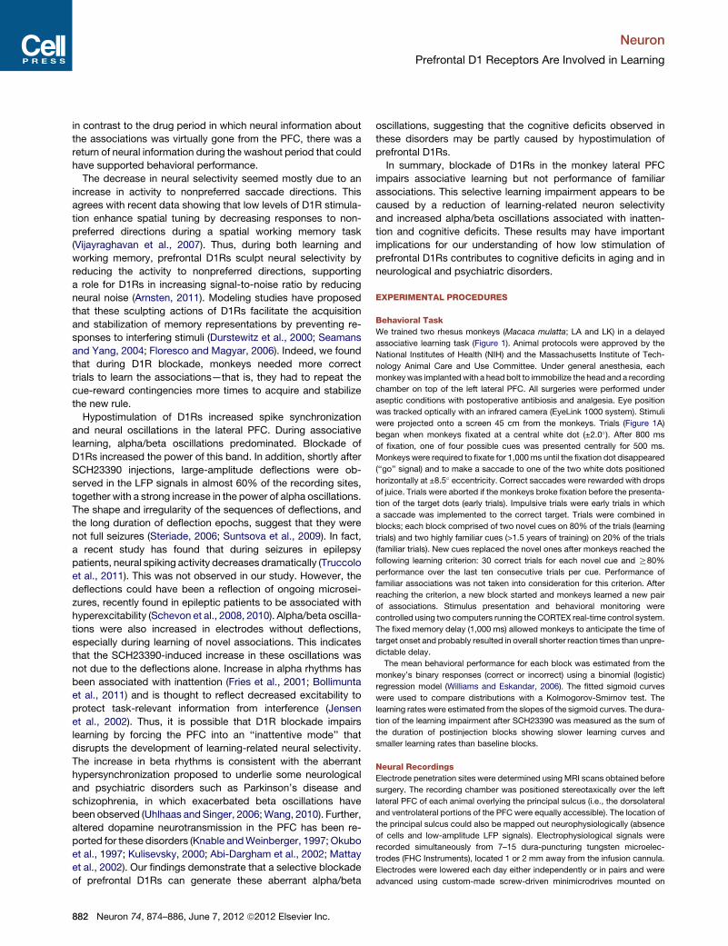
A
B
Novel cues Delay Target dots
Fixation
800 ms
500 ms 1000 ms ResponseTime
Novel associations (80% of trials)
Familiar associations (20% of trials)
Familiar cues Delay Target dots
B1 B2 B3 B4 B5 B6 B7
Baseline Washout
...
.
. . .
. .
.
. .
. .
. .
. .
. .
Block number
S3
S1Inject
S2
Post-inj
Reward
Reward
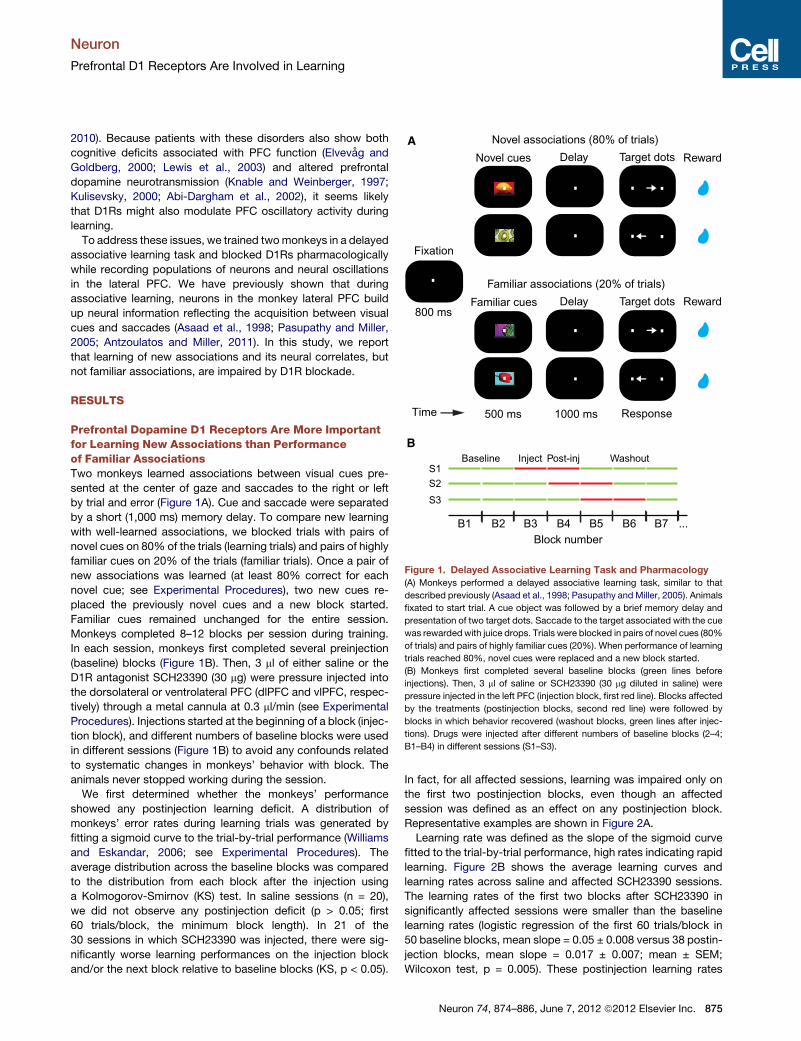
Figure 1. Delayed Associative Learning Task and Pharmacology
(A) Monkeys performed a delayed associative learning task, similar to that
described previously (Asaad et al., 1998; Pasupathy and Miller, 2005). Animals
fixated to start trial. A cue object was followed by a brief memory delay and
presentation of two target dots. Saccade to the target associated with the cue
was rewarded with juice drops. Trials were blocked in pairs of novel cues (80%
of trials) and pairs of highly familiar cues (20%). When performance of learning
trials reached 80%, novel cues were replaced and a new block started.
(B) Monkeys first completed several baseline blocks (green lines before
injections). Then, 3 ml of saline or SCH23390 (30 mg diluted in saline) were
pressure injected in the left PFC (injection block, first red line). Blocks affected
by the treatments (postinjection blocks, second red line) were followed by
blocks in which behavior recovered (washout blocks, green lines after injec-
tions). Drugs were injected after different numbers of baseline blocks (2–4;
B1–B4) in different sessions (S1–S3).
Neuron
Prefrontal D1 Receptors Are Involved in Learning
2010). Because patients with these disorders also show both
cognitive deficits associated with PFC function (Elvevag and
Goldberg, 2000; Lewis et al., 2003) and altered prefrontal
dopamine neurotransmission (Knable and Weinberger, 1997;
Kulisevsky, 2000; Abi-Dargham et al., 2002), it seems likely
that D1Rs might also modulate PFC oscillatory activity during
learning.
To address these issues, we trained twomonkeys in a delayed
associative learning task and blocked D1Rs pharmacologically
while recording populations of neurons and neural oscillations
in the lateral PFC. We have previously shown that during
associative learning, neurons in the monkey lateral PFC build
up neural information reflecting the acquisition between visual
cues and saccades (Asaad et al., 1998; Pasupathy and Miller,
2005; Antzoulatos and Miller, 2011). In this study, we report
that learning of new associations and its neural correlates, but
not familiar associations, are impaired by D1R blockade.
RESULTS
Prefrontal Dopamine D1 Receptors Are More Importantfor Learning New Associations than Performanceof Familiar AssociationsTwo monkeys learned associations between visual cues pre-
sented at the center of gaze and saccades to the right or left
by trial and error (Figure 1A). Cue and saccade were separated
by a short (1,000 ms) memory delay. To compare new learning
with well-learned associations, we blocked trials with pairs of
novel cues on 80% of the trials (learning trials) and pairs of highly
familiar cues on 20% of the trials (familiar trials). Once a pair of
new associations was learned (at least 80% correct for each
novel cue; see Experimental Procedures), two new cues re-
placed the previously novel cues and a new block started.
Familiar cues remained unchanged for the entire session.
Monkeys completed 8–12 blocks per session during training.
In each session, monkeys first completed several preinjection
(baseline) blocks (Figure 1B). Then, 3 ml of either saline or the
D1R antagonist SCH23390 (30 mg) were pressure injected into
the dorsolateral or ventrolateral PFC (dlPFC and vlPFC, respec-
tively) through a metal cannula at 0.3 ml/min (see Experimental
Procedures). Injections started at the beginning of a block (injec-
tion block), and different numbers of baseline blocks were used
in different sessions (Figure 1B) to avoid any confounds related
to systematic changes in monkeys’ behavior with block. The
animals never stopped working during the session.
We first determined whether the monkeys’ performance
showed any postinjection learning deficit. A distribution of
monkeys’ error rates during learning trials was generated by
fitting a sigmoid curve to the trial-by-trial performance (Williams
and Eskandar, 2006; see Experimental Procedures). The
average distribution across the baseline blocks was compared
to the distribution from each block after the injection using
a Kolmogorov-Smirnov (KS) test. In saline sessions (n = 20),
we did not observe any postinjection deficit (p > 0.05; first
60 trials/block, the minimum block length). In 21 of the
30 sessions in which SCH23390 was injected, there were sig-
nificantly worse learning performances on the injection block
and/or the next block relative to baseline blocks (KS, p < 0.05).
In fact, for all affected sessions, learning was impaired only on
the first two postinjection blocks, even though an affected
session was defined as an effect on any postinjection block.
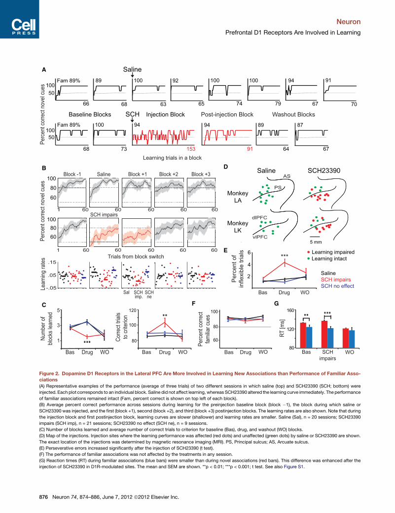
Representative examples are shown in Figure 2A.
Learning rate was defined as the slope of the sigmoid curve
fitted to the trial-by-trial performance, high rates indicating rapid
learning. Figure 2B shows the average learning curves and
learning rates across saline and affected SCH23390 sessions.
The learning rates of the first two blocks after SCH23390 in
significantly affected sessions were smaller than the baseline
learning rates (logistic regression of the first 60 trials/block in
50 baseline blocks, mean slope = 0.05 ± 0.008 versus 38 postin-
jection blocks, mean slope = 0.017 ± 0.007; mean ± SEM;
Wilcoxon test, p = 0.005). These postinjection learning rates
Neuron 74, 874–886, June 7, 2012 ª2012 Elsevier Inc. 875
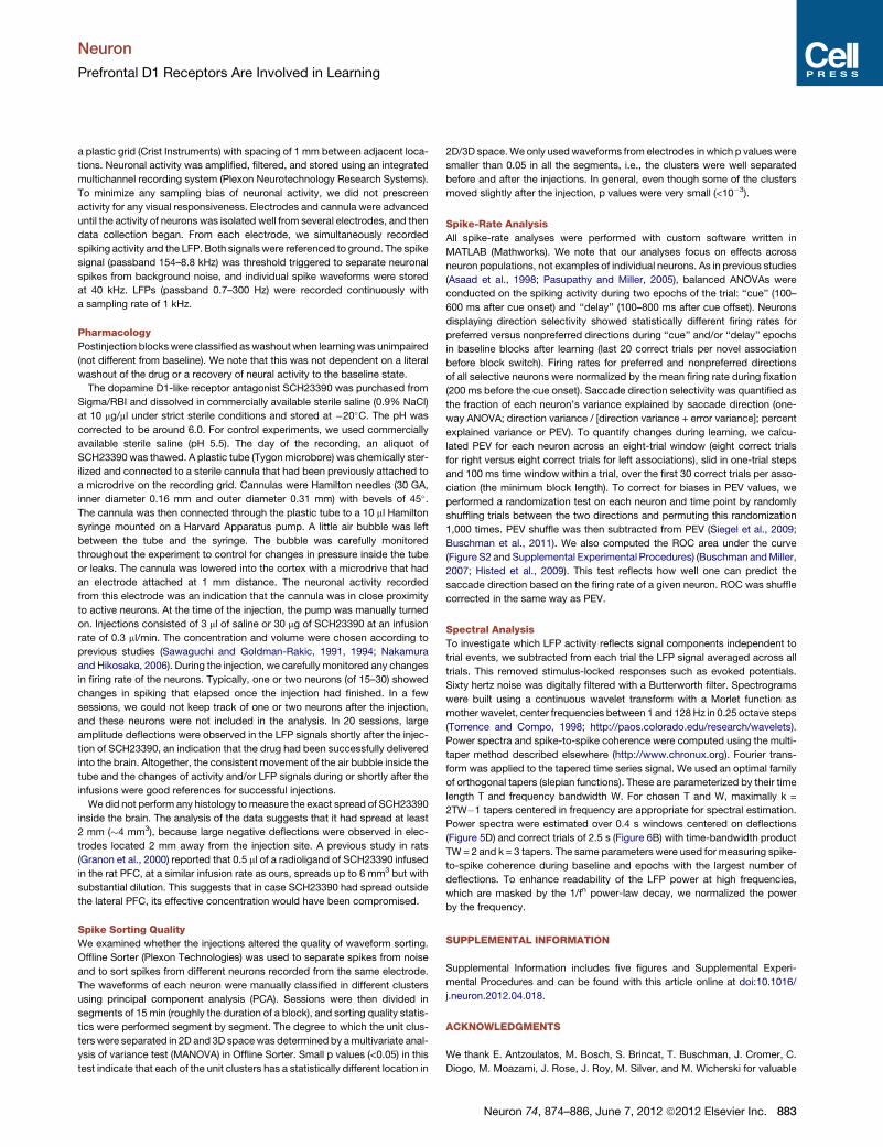
89
64
100
79
100
74
A
B
Learning trials in a block
100
50
Fam 89%
66 100
Saline
100
63
92
65
94
67
100
50
Fam 89%
68
100
73
SCH
94
153
94
91
87
67
FC
68
89
Baseline Blocks Post-injection Block Washout Blocks
5
***
Bas Drug WO
1
3
80
100
120
Bas Drug WO
**
Pe
rce
nt co
rre
ct n
ove
l cu
es
60 60 60 60 601
91
70
Saline
SCH impairs
SCH no effect
G
60 60 60 60 601
Saline Block +1 Block +2 Block +3Block -1
SCH impairs
60
80
100
80
120
160***
Bas SCH
impairs WO
**
E
2
4
Bas Drug WO
Pe
rce
nt
of
infle
xib
le t
ria
ls 6
Injection Block
DSaline SCH23390
dlPFC
vlPFC
PS
AS
Monkey
LK
Monkey
LA
5 mm
***.15
.05
-.05Le
arn
ing
ra
tes
Sal SCH SCHne
Bas Drug WO
Learning impaired
Learning intact
Nu
mb
er
of
blo
cks
lea
rne
d
imp.
Pe
rce
nt co
rre
ct
fam
ilia
r cu
es
RT
[m
s]
Pe
rce
nt co
rre
ct n
ove
l cu
es
100
100
80
80
60
60
Co
rre
ct tria
ls
to c
rite
rio
n
Trials from block switch
Figure 2. Dopamine D1 Receptors in the Lateral PFC Are More Involved in Learning New Associations than Performance of Familiar Asso-
ciations
(A) Representative examples of the performance (average of three trials) of two different sessions in which saline (top) and SCH23390 (SCH; bottom) were
injected. Each plot corresponds to an individual block. Saline did not affect learning, whereas SCH23390 altered the learning curve immediately. The performance
of familiar associations remained intact (Fam, percent correct is shown on top left of each block).
(B) Average percent correct performance across sessions during learning for the preinjection baseline block (block �1), the block during which saline or
SCH23390 was injected, and the first (block +1), second (block +2), and third (block +3) postinjection blocks. The learning rates are also shown. Note that during
the injection block and first postinjection block, learning curves are slower (shallower) and learning rates are smaller. Saline (Sal), n = 20 sessions; SCH23390
impairs (SCH imp), n = 21 sessions; SCH23390 no effect (SCH ne), n = 9 sessions.
(C) Number of blocks learned and average number of correct trials to criterion for baseline (Bas), drug, and washout (WO) blocks.
(D) Map of the injections. Injection sites where the learning performance was affected (red dots) and unaffected (green dots) by saline or SCH23390 are shown.
The exact location of the injections was determined by magnetic resonance imaging (MRI). PS, Principal sulcus; AS, Arcuate sulcus.
(E) Perseverative errors increased significantly after the injection of SCH23390 (t test).
(F) The performance of familiar associations was not affected by the treatments in any session.
(G) Reaction times (RT) during familiar associations (blue bars) were smaller than during novel associations (red bars). This difference was enhanced after the
injection of SCH23390 in D1R-modulated sites. The mean and SEM are shown. **p < 0.01; ***p < 0.001; t test. See also Figure S1.
Neuron
Prefrontal D1 Receptors Are Involved in Learning
876 Neuron 74, 874–886, June 7, 2012 ª2012 Elsevier Inc.
Neuron
Prefrontal D1 Receptors Are Involved in Learning
were also smaller than that of the first two blocks after saline
injections (40 postsaline injection blocks, mean = 0.06 ± 0.007;
p = 1 3 10�4) and smaller than the learning rates of the first
two blocks after SCH23390 in unaffected sessions (18 postinjec-
tion blocks, mean = 0.12 ± 0.04; p = 3 3 10�5). On average, the
impairment lasted 1 hr (59 ± 5 min), during which monkeys
completed fewer numbers of blocks because they needed
more correct trials to learn the associations (Figure 2C). There
was an anatomical dissociation between affected and unaf-
fected sites: most (18 of 21) sessions with a learning deficit
followed vlPFC injections, whereas the sites unaffected by
SCH23390 were mainly in the dlPFC (Figure 2D, proportion of
vlPFC versus dlPFC affected sites; chi-square, p = 9 3 10�5).
We did not observe any anterior versus posterior trend for the
location of affected sites. Performance for each of the two novel
cues was similarly impaired in both animals (see Figure S1 avail-
able online).
The learning impairment was not due to altered eye move-
ments. We did not observe any major changes in the trajectories
or accuracy of the saccades after the injection of SCH23390. The
vast majority of saccades during error trials ended within
the target window around the incorrect target (<4.0�). In fact,
if anything, saccade accuracy somewhat improved after
SCH23390: there was an increase in error trial saccades ending
within the incorrect target window (88% ± 4% to 95% ± 5% of
error trials; t test, p = 0.02). The average eyemovement velocities
(deg/s) also increased after injection of SCH23390 (from 401 ± 3
deg/s to 422 ± 5 deg/s; p = 43 10�4), perhaps due to frustration
from the learning impairment and reduction in reward. Errors
were not caused by increased impulsivity, a premature saccade
toward the correct target before the ‘‘go’’ cue (baseline, 7.4% ±
0.5% of trials; SCH23390, 7.7% ± 0.6%; Wilcoxon test, p = 0.62
versus baseline, p = 0.75 versus saline). But there was a modest
increase in perseveration (the average number of consecutive
repeats of an error), from 1.6% ± 0.2% of trials during baseline
to 4.3% ± 0.6% during the first hour postinjection in affected
sites (Wilcoxon test, p = 4 3 10�5), but not after saline (mean =
1.5%, p = 2 3 10�5 versus affected sites) or SCH23390 in
unaffected sites (mean = 1%, p = 43 10�5 versus affected sites;
Figure 2E).
In contrast to new learning, performance of familiar associa-
tions was unimpaired in all sessions (Figures 2F and S1), and
the proportion of perseverative errors was not different from
baseline (Wilcoxon test, p = 0.59). Reaction times were shorter
for familiar associations than for novel associations during the
baseline blocks (122 ± 5 ms versus 133 ± 1 ms, Wilcoxon test,
p = 0.003), an effect also observed after the injection of
SCH23390 in behaviorally sensitive sites (121 ± 4 ms versus
137 ± 1 ms, p = 6 3 10�4; Figure 2G).
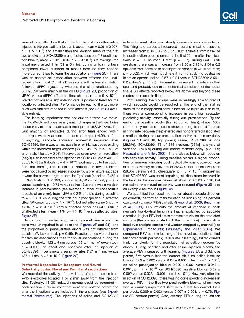
Prefrontal Dopamine D1 Receptors and NeuralSelectivity during Novel and Familiar AssociationsWe recorded the activity of individual prefrontal neurons from
7–15 electrodes located 1 or 2 mm away from the injection
site. Typically, 15–30 isolated neurons could be recorded in
each session. Only neurons that were well isolated before and
after the injections were included in the analyses (see Experi-
mental Procedures). The injections of saline and SCH23390
induced a small, slow, and steady increase in neuronal activity.
The firing rate across all recorded neurons in saline sessions
increased from 2.36 ± 0.2 to 2.57 ± 0.21 spikes/s from baseline
to postinjection epochs (omitting the first 20 min after the injec-
tions; n = 286 neurons; t test, p = 0.07). During SCH23390
sessions, there was an increase from 2.06 ± 0.13 to 2.56 ± 0.2
spikes/s from baseline to postinjection epochs (n = 279 neurons;
p = 0.002), which was not different from that during postsaline
injection epochs (saline: 2.57 ± 0.21 versus SCH23390: 2.56 ±
0.2 spikes/s, p = 0.96). The small increases in firing rate are often
seen and probably due to a mechanical stimulation of the neural
tissue. All effects reported below are above and beyond these
modest increases in firing rate.
With learning, the monkeys were increasingly able to predict
which saccade would be required at the end of the trial as
soon as the cue appeared early in the trial. As in previous studies,
there was a corresponding increase in early trial saccade-
predicting activity, especially during cue presentation. By the
end of the baseline blocks (last 20 correct trials), almost 30%
of randomly selected neurons showed a significant difference
in firing rate between the preferred and nonpreferred associated
directions during the cue presentation and/or the memory delay
(Figures 3A and 3B, top panels; saline, 81 of 286 neurons
[28.3%]; SCH23390, 78 of 279 neurons [28%]; analysis of
variance [ANOVA] during cue and/or memory delay, p < 0.05;
Pasupathy and Miller, 2005). The analyses below will focus on
this early trial activity. During baseline blocks, a higher propor-
tion of neurons showing such selectivity was observed near
sites behaviorally sensitive to SCH23390 than insensitive sites
(28.6% versus 9.4%, chi-square, p = 8 3 10�7), suggesting
that SCH23390 was most impairing at sites more involved in
the task. As the analyses below will show, after SCH23390, but
not saline, this neural selectivity was reduced (Figure 3B; see
an example neuron in Figure S2).
We quantified the neural information about saccade direction
on correctly performed trials for each neuron using the percent
explained variance (PEV) statistic (Siegel et al., 2009; Buschman
et al., 2011). PEV reflects the amount of variance in each
neuron’s trial-by-trial firing rate that is explained by saccade
direction. Higher PEV indicates more selectivity for the predicted
saccade (the one associated with the current cue). It was calcu-
lated over an eight-correct-trial window stepped by one trial (see
Experimental Procedures; Pasupathy and Miller, 2005). We
compared PEV early in learning of the novel associations (first
ten correct trials per block) versus late in learning (last ten correct
trials per block) for the population of selective neurons (as
above). During baseline and after saline injection blocks, the
average PEV increased with learning (Figures 3A and 3B; cue
period, first versus last ten correct trials on saline baseline
blocks: 0.02 ± 0.002 versus 0.04 ± 0.002, t test, p = 1 3 10�6;
on saline postinjection blocks: 0.029 ± 0.001 versus 0.047 ±
0.001, p = 4 3 10�6; on SCH23390 baseline blocks: 0.02 ±
0.002 versus 0.033 ± 0.001, p = 4 3 10�5). However, after the
injection of SCH23390, there was no corresponding increase in
average PEV in the first two postinjection blocks, when there
was a learning impairment (first versus last ten correct trials
per block, 0.009 ± 0.003 versus 0.007 ± 0.001, p = 0.78; Fig-
ure 3B, bottom panels). Also, average PEV during the last ten
Neuron 74, 874–886, June 7, 2012 ª2012 Elsevier Inc. 877
PE
V
Cor
rect
tria
ls 20
15
10
5
1
2
3
4
Cue Delay Cue Delay Sac Cue Delay Sac
Cue Delay Sac Cue Delay Sac Cue Delay Sac
1
2
3
4
0
0.06
0
Cor
rect
tria
ls 20
15
10
5
1
2
3
4
1
2
3
4
1
2
3
4
20
15
10
5
20
15
10
5
20
15
10
5
1
2
3
4
20
15
10
5
0.045
PE
V
1
2
3
Bas Saline WO
*** **
PreferredNon-preferred
1
2
20 60 100 140Time (min)
D
SalineSCH23390
Drug
Novel associations
C
B
ABaseline
Baseline
Saline
SCH23390
Washout
Washout
Saline SCH23390Normalizedfiring rate
1
2
3
4
Bas SCH WO
******1
2
3
Nor
mal
ized
firin
g ra
teN
orm
aliz
edfir
ing
rate
Nor
mal
ized
firin
g ra
te
* *
Sac
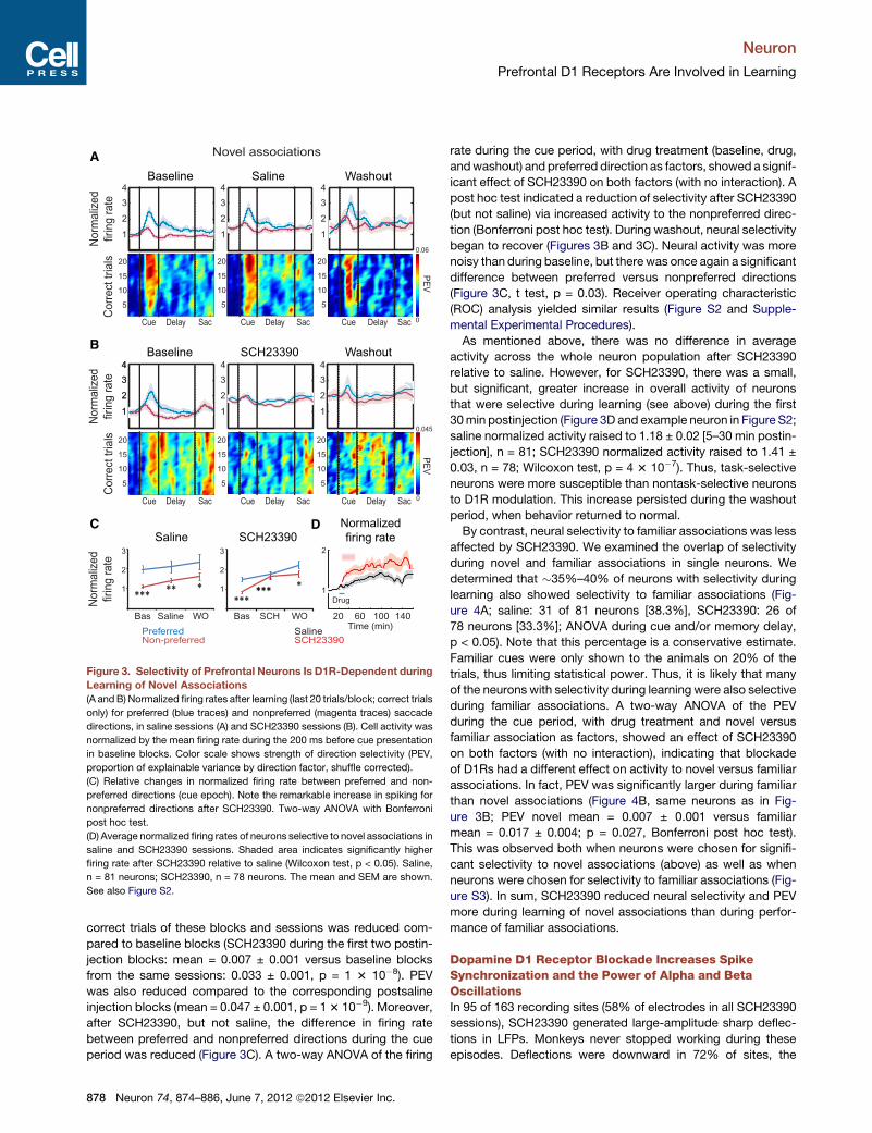
Figure 3. Selectivity of Prefrontal Neurons Is D1R-Dependent during
Learning of Novel Associations
(A andB) Normalized firing rates after learning (last 20 trials/block; correct trials
only) for preferred (blue traces) and nonpreferred (magenta traces) saccade
directions, in saline sessions (A) and SCH23390 sessions (B). Cell activity was
normalized by the mean firing rate during the 200 ms before cue presentation
in baseline blocks. Color scale shows strength of direction selectivity (PEV,
proportion of explainable variance by direction factor, shuffle corrected).
(C) Relative changes in normalized firing rate between preferred and non-
preferred directions (cue epoch). Note the remarkable increase in spiking for
nonpreferred directions after SCH23390. Two-way ANOVA with Bonferroni
post hoc test.
(D) Average normalized firing rates of neurons selective to novel associations in
saline and SCH23390 sessions. Shaded area indicates significantly higher
firing rate after SCH23390 relative to saline (Wilcoxon test, p < 0.05). Saline,
n = 81 neurons; SCH23390, n = 78 neurons. The mean and SEM are shown.
See also Figure S2.
Neuron
Prefrontal D1 Receptors Are Involved in Learning
correct trials of these blocks and sessions was reduced com-
pared to baseline blocks (SCH23390 during the first two postin-
jection blocks: mean = 0.007 ± 0.001 versus baseline blocks
from the same sessions: 0.033 ± 0.001, p = 1 3 10�8). PEV
was also reduced compared to the corresponding postsaline
injection blocks (mean = 0.047 ± 0.001, p = 13 10�9). Moreover,
after SCH23390, but not saline, the difference in firing rate
between preferred and nonpreferred directions during the cue
period was reduced (Figure 3C). A two-way ANOVA of the firing
878 Neuron 74, 874–886, June 7, 2012 ª2012 Elsevier Inc.
rate during the cue period, with drug treatment (baseline, drug,
andwashout) and preferred direction as factors, showed a signif-
icant effect of SCH23390 on both factors (with no interaction). A
post hoc test indicated a reduction of selectivity after SCH23390
(but not saline) via increased activity to the nonpreferred direc-
tion (Bonferroni post hoc test). During washout, neural selectivity
began to recover (Figures 3B and 3C). Neural activity was more
noisy than during baseline, but therewas once again a significant
difference between preferred versus nonpreferred directions
(Figure 3C, t test, p = 0.03). Receiver operating characteristic
(ROC) analysis yielded similar results (Figure S2 and Supple-
mental Experimental Procedures).
As mentioned above, there was no difference in average
activity across the whole neuron population after SCH23390
relative to saline. However, for SCH23390, there was a small,
but significant, greater increase in overall activity of neurons
that were selective during learning (see above) during the first
30min postinjection (Figure 3D and example neuron in Figure S2;
saline normalized activity raised to 1.18 ± 0.02 [5–30 min postin-
jection], n = 81; SCH23390 normalized activity raised to 1.41 ±
0.03, n = 78; Wilcoxon test, p = 4 3 10�7). Thus, task-selective
neurons were more susceptible than nontask-selective neurons
to D1R modulation. This increase persisted during the washout
period, when behavior returned to normal.
By contrast, neural selectivity to familiar associations was less
affected by SCH23390. We examined the overlap of selectivity
during novel and familiar associations in single neurons. We
determined that �35%–40% of neurons with selectivity during
learning also showed selectivity to familiar associations (Fig-
ure 4A; saline: 31 of 81 neurons [38.3%], SCH23390: 26 of
78 neurons [33.3%]; ANOVA during cue and/or memory delay,
p < 0.05). Note that this percentage is a conservative estimate.
Familiar cues were only shown to the animals on 20% of the
trials, thus limiting statistical power. Thus, it is likely that many
of the neurons with selectivity during learning were also selective
during familiar associations. A two-way ANOVA of the PEV
during the cue period, with drug treatment and novel versus
familiar association as factors, showed an effect of SCH23390
on both factors (with no interaction), indicating that blockade
of D1Rs had a different effect on activity to novel versus familiar
associations. In fact, PEV was significantly larger during familiar
than novel associations (Figure 4B, same neurons as in Fig-
ure 3B; PEV novel mean = 0.007 ± 0.001 versus familiar
mean = 0.017 ± 0.004; p = 0.027, Bonferroni post hoc test).
This was observed both when neurons were chosen for signifi-
cant selectivity to novel associations (above) as well as when
neurons were chosen for selectivity to familiar associations (Fig-
ure S3). In sum, SCH23390 reduced neural selectivity and PEV
more during learning of novel associations than during perfor-
mance of familiar associations.
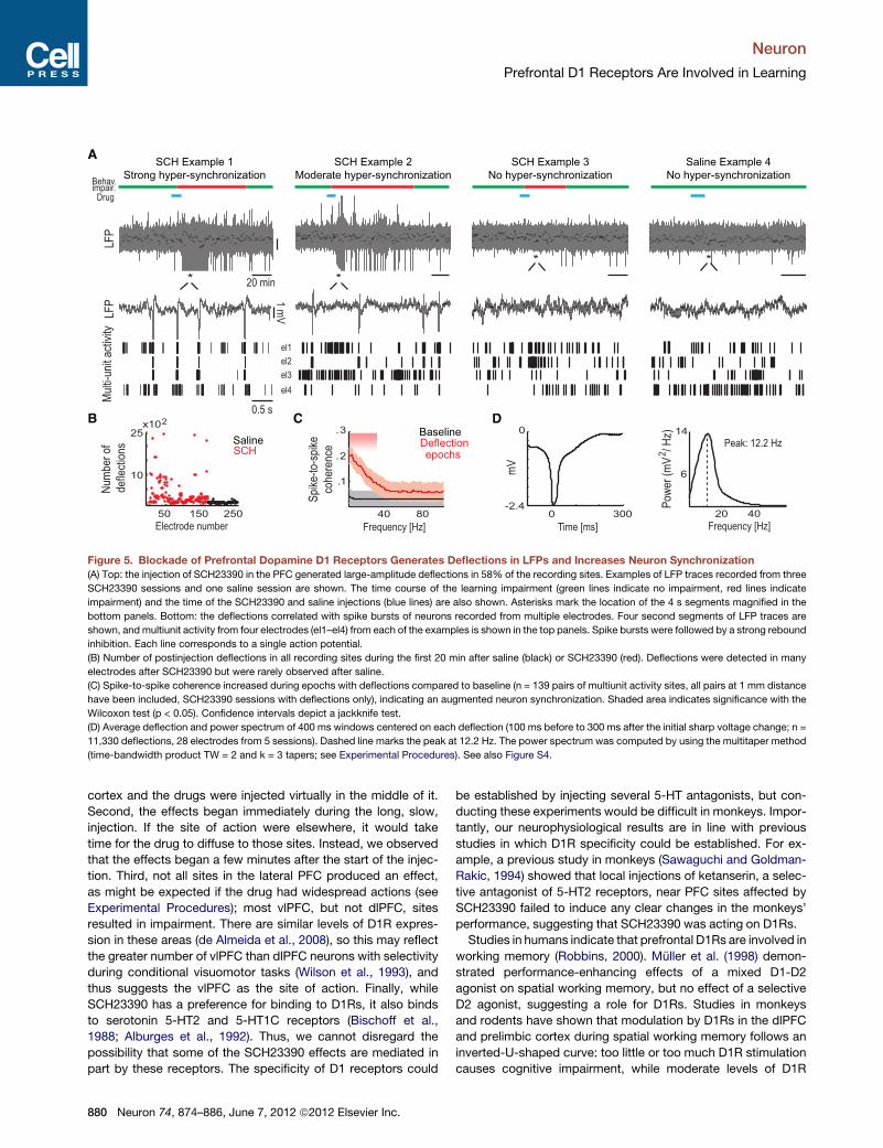
Dopamine D1 Receptor Blockade Increases SpikeSynchronization and the Power of Alpha and BetaOscillationsIn 95 of 163 recording sites (58% of electrodes in all SCH23390
sessions), SCH23390 generated large-amplitude sharp deflec-
tions in LFPs. Monkeys never stopped working during these
episodes. Deflections were downward in 72% of sites, the
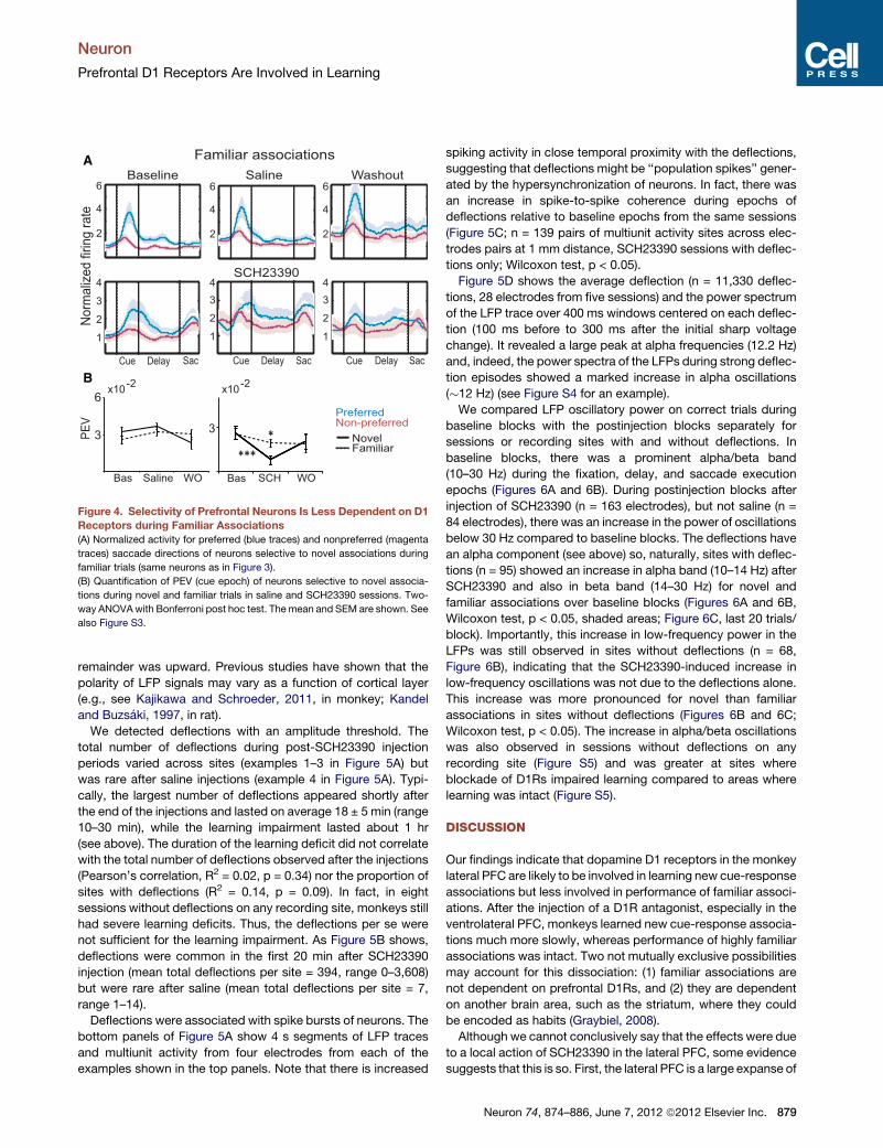
NovelFamiliar
A
B
2
4
6Baseline Saline Washout
SCH23390
Cue Delay Sac Cue Delay Sac Cue Delay Sac
1
2
3
4
1
2
3
4
1
2
3
4
Familiar associations
2
4
6
2
4
6
PE
V
Bas Saline WO
3
6
Bas SCH WO
x10-2
x10-2
3
No
rma
lize
d firin
g r
ate
****
PreferredNon-preferred
Figure 4. Selectivity of Prefrontal Neurons Is Less Dependent on D1
Receptors during Familiar Associations
(A) Normalized activity for preferred (blue traces) and nonpreferred (magenta
traces) saccade directions of neurons selective to novel associations during
familiar trials (same neurons as in Figure 3).
(B) Quantification of PEV (cue epoch) of neurons selective to novel associa-
tions during novel and familiar trials in saline and SCH23390 sessions. Two-
way ANOVAwith Bonferroni post hoc test. Themean and SEM are shown. See
also Figure S3.
Neuron
Prefrontal D1 Receptors Are Involved in Learning
remainder was upward. Previous studies have shown that the
polarity of LFP signals may vary as a function of cortical layer
(e.g., see Kajikawa and Schroeder, 2011, in monkey; Kandel
and Buzsaki, 1997, in rat).
We detected deflections with an amplitude threshold. The
total number of deflections during post-SCH23390 injection
periods varied across sites (examples 1–3 in Figure 5A) but
was rare after saline injections (example 4 in Figure 5A). Typi-
cally, the largest number of deflections appeared shortly after
the end of the injections and lasted on average 18 ± 5 min (range
10–30 min), while the learning impairment lasted about 1 hr
(see above). The duration of the learning deficit did not correlate
with the total number of deflections observed after the injections
(Pearson’s correlation, R2 = 0.02, p = 0.34) nor the proportion of
sites with deflections (R2 = 0.14, p = 0.09). In fact, in eight
sessions without deflections on any recording site, monkeys still
had severe learning deficits. Thus, the deflections per se were
not sufficient for the learning impairment. As Figure 5B shows,
deflections were common in the first 20 min after SCH23390
injection (mean total deflections per site = 394, range 0–3,608)
but were rare after saline (mean total deflections per site = 7,
range 1–14).
Deflections were associated with spike bursts of neurons. The
bottom panels of Figure 5A show 4 s segments of LFP traces
and multiunit activity from four electrodes from each of the
examples shown in the top panels. Note that there is increased
spiking activity in close temporal proximity with the deflections,
suggesting that deflections might be ‘‘population spikes’’ gener-
ated by the hypersynchronization of neurons. In fact, there was
an increase in spike-to-spike coherence during epochs of
deflections relative to baseline epochs from the same sessions
(Figure 5C; n = 139 pairs of multiunit activity sites across elec-
trodes pairs at 1 mm distance, SCH23390 sessions with deflec-
tions only; Wilcoxon test, p < 0.05).
Figure 5D shows the average deflection (n = 11,330 deflec-
tions, 28 electrodes from five sessions) and the power spectrum
of the LFP trace over 400 ms windows centered on each deflec-
tion (100 ms before to 300 ms after the initial sharp voltage
change). It revealed a large peak at alpha frequencies (12.2 Hz)
and, indeed, the power spectra of the LFPs during strong deflec-
tion episodes showed a marked increase in alpha oscillations
(�12 Hz) (see Figure S4 for an example).
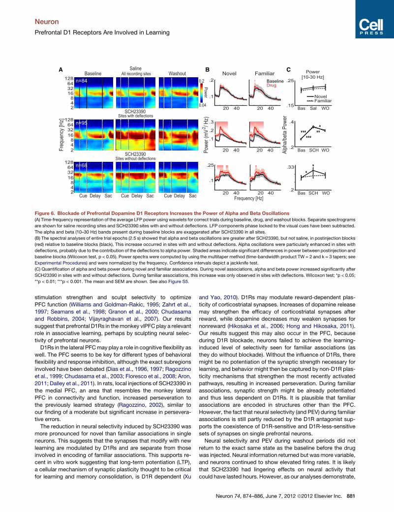
We compared LFP oscillatory power on correct trials during
baseline blocks with the postinjection blocks separately for
sessions or recording sites with and without deflections. In
baseline blocks, there was a prominent alpha/beta band
(10–30 Hz) during the fixation, delay, and saccade execution
epochs (Figures 6A and 6B). During postinjection blocks after
injection of SCH23390 (n = 163 electrodes), but not saline (n =
84 electrodes), there was an increase in the power of oscillations
below 30 Hz compared to baseline blocks. The deflections have
an alpha component (see above) so, naturally, sites with deflec-
tions (n = 95) showed an increase in alpha band (10–14 Hz) after
SCH23390 and also in beta band (14–30 Hz) for novel and
familiar associations over baseline blocks (Figures 6A and 6B,
Wilcoxon test, p < 0.05, shaded areas; Figure 6C, last 20 trials/
block). Importantly, this increase in low-frequency power in the
LFPs was still observed in sites without deflections (n = 68,
Figure 6B), indicating that the SCH23390-induced increase in
low-frequency oscillations was not due to the deflections alone.
This increase was more pronounced for novel than familiar
associations in sites without deflections (Figures 6B and 6C;
Wilcoxon test, p < 0.05). The increase in alpha/beta oscillations
was also observed in sessions without deflections on any
recording site (Figure S5) and was greater at sites where
blockade of D1Rs impaired learning compared to areas where
learning was intact (Figure S5).
DISCUSSION
Our findings indicate that dopamine D1 receptors in the monkey
lateral PFC are likely to be involved in learning new cue-response
associations but less involved in performance of familiar associ-
ations. After the injection of a D1R antagonist, especially in the
ventrolateral PFC, monkeys learned new cue-response associa-
tions much more slowly, whereas performance of highly familiar
associations was intact. Two not mutually exclusive possibilities
may account for this dissociation: (1) familiar associations are
not dependent on prefrontal D1Rs, and (2) they are dependent
on another brain area, such as the striatum, where they could
be encoded as habits (Graybiel, 2008).
Although we cannot conclusively say that the effects were due
to a local action of SCH23390 in the lateral PFC, some evidence
suggests that this is so. First, the lateral PFC is a large expanse of
Neuron 74, 874–886, June 7, 2012 ª2012 Elsevier Inc. 879
1 mV
Mul
ti-un
it ac
tivity
20 min
Drug
LFP
LFP
A
B
el1
el2
el3
el4
SCH Example 1
Strong hyper-synchronization
SCH Example 2
Moderate hyper-synchronization
Saline Example 4
No hyper-synchronization
SCH Example 3
No hyper-synchronization
**
* *
D
Behav.impair.
Num
ber
of
defle
ctio
ns
Electrode number
25
10
50 150 250
x102
SCHSaline
C0
-2.4m
V
0 300
Pow
er (
mV
/ H
z)2
20 40
Frequency [Hz]
14
6
Peak: 12.2 Hz
Time [ms]Frequency [Hz]
Spi
ke-t
o-sp
ike
cohe
renc
e
40 80
1.
2.
3.
DeflectionBaseline
epochs
0.5 s
Figure 5. Blockade of Prefrontal Dopamine D1 Receptors Generates Deflections in LFPs and Increases Neuron Synchronization
(A) Top: the injection of SCH23390 in the PFC generated large-amplitude deflections in 58% of the recording sites. Examples of LFP traces recorded from three
SCH23390 sessions and one saline session are shown. The time course of the learning impairment (green lines indicate no impairment, red lines indicate
impairment) and the time of the SCH23390 and saline injections (blue lines) are also shown. Asterisks mark the location of the 4 s segments magnified in the
bottom panels. Bottom: the deflections correlated with spike bursts of neurons recorded from multiple electrodes. Four second segments of LFP traces are
shown, and multiunit activity from four electrodes (el1–el4) from each of the examples is shown in the top panels. Spike bursts were followed by a strong rebound
inhibition. Each line corresponds to a single action potential.
(B) Number of postinjection deflections in all recording sites during the first 20 min after saline (black) or SCH23390 (red). Deflections were detected in many
electrodes after SCH23390 but were rarely observed after saline.
(C) Spike-to-spike coherence increased during epochs with deflections compared to baseline (n = 139 pairs of multiunit activity sites, all pairs at 1 mm distance
have been included, SCH23390 sessions with deflections only), indicating an augmented neuron synchronization. Shaded area indicates significance with the
Wilcoxon test (p < 0.05). Confidence intervals depict a jackknife test.
(D) Average deflection and power spectrum of 400 ms windows centered on each deflection (100 ms before to 300 ms after the initial sharp voltage change; n =
11,330 deflections, 28 electrodes from 5 sessions). Dashed line marks the peak at 12.2 Hz. The power spectrum was computed by using the multitaper method
(time-bandwidth product TW = 2 and k = 3 tapers; see Experimental Procedures). See also Figure S4.
Neuron
Prefrontal D1 Receptors Are Involved in Learning
cortex and the drugs were injected virtually in the middle of it.
Second, the effects began immediately during the long, slow,
injection. If the site of action were elsewhere, it would take
time for the drug to diffuse to those sites. Instead, we observed
that the effects began a few minutes after the start of the injec-
tion. Third, not all sites in the lateral PFC produced an effect,
as might be expected if the drug had widespread actions (see
Experimental Procedures); most vlPFC, but not dlPFC, sites
resulted in impairment. There are similar levels of D1R expres-
sion in these areas (de Almeida et al., 2008), so this may reflect
the greater number of vlPFC than dlPFC neurons with selectivity
during conditional visuomotor tasks (Wilson et al., 1993), and
thus suggests the vlPFC as the site of action. Finally, while
SCH23390 has a preference for binding to D1Rs, it also binds
to serotonin 5-HT2 and 5-HT1C receptors (Bischoff et al.,
1988; Alburges et al., 1992). Thus, we cannot disregard the
possibility that some of the SCH23390 effects are mediated in
part by these receptors. The specificity of D1 receptors could
880 Neuron 74, 874–886, June 7, 2012 ª2012 Elsevier Inc.
be established by injecting several 5-HT antagonists, but con-
ducting these experiments would be difficult in monkeys. Impor-
tantly, our neurophysiological results are in line with previous
studies in which D1R specificity could be established. For ex-
ample, a previous study in monkeys (Sawaguchi and Goldman-
Rakic, 1994) showed that local injections of ketanserin, a selec-
tive antagonist of 5-HT2 receptors, near PFC sites affected by
SCH23390 failed to induce any clear changes in the monkeys’
performance, suggesting that SCH23390 was acting on D1Rs.
Studies in humans indicate that prefrontal D1Rs are involved in
working memory (Robbins, 2000). Muller et al. (1998) demon-
strated performance-enhancing effects of a mixed D1-D2
agonist on spatial working memory, but no effect of a selective
D2 agonist, suggesting a role for D1Rs. Studies in monkeys
and rodents have shown that modulation by D1Rs in the dlPFC
and prelimbic cortex during spatial working memory follows an
inverted-U-shaped curve: too little or too much D1R stimulation
causes cognitive impairment, while moderate levels of D1R
BA
Fre
quen
cy [H
z]
SCH23390
SalineWashoutBaseline
48
16
32
2
64128
48
1632
2
64128
Pow
er
0.04
0.2
CFamiliarNovel
20 40
Frequency [Hz]
.25
*
Bas Sal WO
Alp
ha/b
eta
Pow
er
.33
Bas SCH WO
***
***.1
.2
.2
.15
Sites with deflections
48
16
32
2
64128
SCH23390Sites without deflections
20 40
.3
.1
.2
20 40
DrugBaseline
20 40
20 4020 40
.1
.25
NovelFamiliar
Bas SCH WO
.4
.2
All recording sites
n=84
n=95
n=68
Power
[10-30 Hz]
Pow
er (
mV
/ H
z)2
Cue Delay Sac Cue Delay Sac Cue Delay Sac
**
*
*
Figure 6. Blockade of Prefrontal Dopamine D1 Receptors Increases the Power of Alpha and Beta Oscillations
(A) Time-frequency representation of the average LFP power using wavelets for correct trials during baseline, drug, and washout blocks. Separate spectrograms
are shown for saline recording sites and SCH23390 sites with and without deflections. LFP components phase locked to the visual cues have been subtracted.
The alpha and beta (10–30 Hz) bands present during baseline blocks are exaggerated after SCH23390 in all sites.
(B) The spectral analyses of entire trial epochs (2.5 s) showed that alpha and beta oscillations are greater after SCH23390, but not saline, in postinjection blocks
(red) relative to baseline blocks (black). This increase occurred in sites with and without deflections. Alpha oscillations were particularly enhanced in sites with
deflections, probably due to the contribution of the deflections to alpha power. Shaded areas indicate significant differences in power between postinjection and
baseline blocks (Wilcoxon test, p < 0.05). Power spectra were computed by using the multitaper method (time-bandwidth product TW = 2 and k = 3 tapers; see
Experimental Procedures) and were normalized by the frequency. Confidence intervals depict a jackknife test.
(C) Quantification of alpha and beta power during novel and familiar associations. During novel associations, alpha and beta power increased significantly after
SCH23390 in sites with and without deflections. During familiar associations, this increase was only observed in sites with deflections. Wilcoxon test; *p < 0.05;
**p < 0.01; ***p < 0.001. The mean and SEM are shown. See also Figure S5.
Neuron
Prefrontal D1 Receptors Are Involved in Learning
stimulation strengthen and sculpt selectivity to optimize
PFC function (Williams and Goldman-Rakic, 1995; Zahrt et al.,
1997; Seamans et al., 1998; Granon et al., 2000; Chudasama
and Robbins, 2004; Vijayraghavan et al., 2007). Our results
suggest that prefrontal D1Rs in themonkey vlPFC play a relevant
role in associative learning, perhaps by sculpting neural selec-
tivity of prefrontal neurons.
D1Rs in the lateral PFCmay play a role in cognitive flexibility as
well. The PFC seems to be key for different types of behavioral
flexibility and response inhibition, although the exact subregions
involved have been debated (Dias et al., 1996, 1997; Ragozzino
et al., 1999; Chudasama et al., 2003; Floresco et al., 2008; Aron,
2011; Dalley et al., 2011). In rats, local injections of SCH23390 in
the medial PFC, an area that resembles the monkey lateral
PFC in connectivity and function, increased perseveration to
the previously learned strategy (Ragozzino, 2002), similar to
our finding of a moderate but significant increase in persevera-
tive errors.
The reduction in neural selectivity induced by SCH23390 was
more pronounced for novel than familiar associations in single
neurons. This suggests that the synapses that modify with new
learning are modulated by D1Rs and are separate from those
involved in encoding of familiar associations. This supports re-
cent in vitro work suggesting that long-term potentiation (LTP),
a cellular mechanism of synaptic plasticity thought to be critical
for learning and memory consolidation, is D1R dependent (Xu
and Yao, 2010). D1Rs may modulate reward-dependent plas-
ticity of corticostriatal synapses. Increases of dopamine release
may strengthen the efficacy of corticostriatal synapses after
reward, while dopamine decreases may weaken synapses for
nonreward (Hikosaka et al., 2006; Hong and Hikosaka, 2011).
Our results suggest this may also occur in the PFC, because
during D1R blockade, neurons failed to achieve the learning-
induced level of selectivity seen for familiar associations (as
they do without blockade). Without the influence of D1Rs, there
might be no potentiation of the synaptic strength necessary for
learning, and behavior might then be captured by non-D1R plas-
ticity mechanisms that strengthen the most recently activated
pathways, resulting in increased perseveration. During familiar
associations, synaptic strength might be already potentiated
and thus less dependent on D1Rs. It is plausible that familiar
associations are encoded in structures other than the PFC.
However, the fact that neural selectivity (and PEV) during familiar
associations is still partly reduced by the D1R antagonist sup-
ports the coexistence of D1R-sensitive and D1R-less-sensitive
sets of synapses on single prefrontal neurons.
Neural selectivity and PEV during washout periods did not
return to the exact same state as the baseline before the drug
was injected. Neural information returned but wasmore variable,
and neurons continued to show elevated firing rates. It is likely
that SCH23390 had lingering effects on neural activity that
could have lasted hours. However, as our analyses demonstrate,
Neuron 74, 874–886, June 7, 2012 ª2012 Elsevier Inc. 881
Neuron
Prefrontal D1 Receptors Are Involved in Learning
in contrast to the drug period in which neural information about
the associations was virtually gone from the PFC, there was a
return of neural information during the washout period that could
have supported behavioral performance.
The decrease in neural selectivity seemed mostly due to an
increase in activity to nonpreferred saccade directions. This
agrees with recent data showing that low levels of D1R stimula-
tion enhance spatial tuning by decreasing responses to non-
preferred directions during a spatial working memory task
(Vijayraghavan et al., 2007). Thus, during both learning and
working memory, prefrontal D1Rs sculpt neural selectivity by
reducing the activity to nonpreferred directions, supporting
a role for D1Rs in increasing signal-to-noise ratio by reducing
neural noise (Arnsten, 2011). Modeling studies have proposed
that these sculpting actions of D1Rs facilitate the acquisition
and stabilization of memory representations by preventing re-
sponses to interfering stimuli (Durstewitz et al., 2000; Seamans
and Yang, 2004; Floresco and Magyar, 2006). Indeed, we found
that during D1R blockade, monkeys needed more correct
trials to learn the associations—that is, they had to repeat the
cue-reward contingencies more times to acquire and stabilize
the new rule.
Hypostimulation of D1Rs increased spike synchronization
and neural oscillations in the lateral PFC. During associative
learning, alpha/beta oscillations predominated. Blockade of
D1Rs increased the power of this band. In addition, shortly after
SCH23390 injections, large-amplitude deflections were ob-
served in the LFP signals in almost 60% of the recording sites,
together with a strong increase in the power of alpha oscillations.
The shape and irregularity of the sequences of deflections, and
the long duration of deflection epochs, suggest that they were
not full seizures (Steriade, 2006; Suntsova et al., 2009). In fact,
a recent study has found that during seizures in epilepsy
patients, neural spiking activity decreases dramatically (Truccolo
et al., 2011). This was not observed in our study. However, the
deflections could have been a reflection of ongoing microsei-
zures, recently found in epileptic patients to be associated with
hyperexcitability (Schevon et al., 2008, 2010). Alpha/beta oscilla-
tions were also increased in electrodes without deflections,
especially during learning of novel associations. This indicates
that the SCH23390-induced increase in these oscillations was
not due to the deflections alone. Increase in alpha rhythms has
been associated with inattention (Fries et al., 2001; Bollimunta
et al., 2011) and is thought to reflect decreased excitability to
protect task-relevant information from interference (Jensen
et al., 2002). Thus, it is possible that D1R blockade impairs
learning by forcing the PFC into an ‘‘inattentive mode’’ that
disrupts the development of learning-related neural selectivity.
The increase in beta rhythms is consistent with the aberrant
hypersynchronization proposed to underlie some neurological
and psychiatric disorders such as Parkinson’s disease and
schizophrenia, in which exacerbated beta oscillations have
been observed (Uhlhaas and Singer, 2006;Wang, 2010). Further,
altered dopamine neurotransmission in the PFC has been re-
ported for these disorders (Knable andWeinberger, 1997; Okubo
et al., 1997; Kulisevsky, 2000; Abi-Dargham et al., 2002; Mattay
et al., 2002). Our findings demonstrate that a selective blockade
of prefrontal D1Rs can generate these aberrant alpha/beta
882 Neuron 74, 874–886, June 7, 2012 ª2012 Elsevier Inc.
oscillations, suggesting that the cognitive deficits observed in
these disorders may be partly caused by hypostimulation of
prefrontal D1Rs.
In summary, blockade of D1Rs in the monkey lateral PFC
impairs associative learning but not performance of familiar
associations. This selective learning impairment appears to be
caused by a reduction of learning-related neuron selectivity
and increased alpha/beta oscillations associated with inatten-
tion and cognitive deficits. These results may have important
implications for our understanding of how low stimulation of
prefrontal D1Rs contributes to cognitive deficits in aging and in
neurological and psychiatric disorders.
EXPERIMENTAL PROCEDURES
Behavioral Task
We trained two rhesus monkeys (Macaca mulatta; LA and LK) in a delayed
associative learning task (Figure 1). Animal protocols were approved by the
National Institutes of Health (NIH) and the Massachusetts Institute of Tech-
nology Animal Care and Use Committee. Under general anesthesia, each
monkeywas implantedwith a head bolt to immobilize the head and a recording
chamber on top of the left lateral PFC. All surgeries were performed under
aseptic conditions with postoperative antibiosis and analgesia. Eye position
was tracked optically with an infrared camera (EyeLink 1000 system). Stimuli
were projected onto a screen 45 cm from the monkeys. Trials (Figure 1A)
began when monkeys fixated at a central white dot (±2.0�). After 800 ms
of fixation, one of four possible cues was presented centrally for 500 ms.
Monkeys were required to fixate for 1,000ms until the fixation dot disappeared
(‘‘go’’ signal) and to make a saccade to one of the two white dots positioned
horizontally at ±8.5� eccentricity. Correct saccades were rewarded with drops
of juice. Trials were aborted if the monkeys broke fixation before the presenta-
tion of the target dots (early trials). Impulsive trials were early trials in which
a saccade was implemented to the correct target. Trials were combined in
blocks; each block comprised of two novel cues on 80% of the trials (learning
trials) and two highly familiar cues (>1.5 years of training) on 20% of the trials
(familiar trials). New cues replaced the novel ones after monkeys reached the
following learning criterion: 30 correct trials for each novel cue and R80%
performance over the last ten consecutive trials per cue. Performance of
familiar associations was not taken into consideration for this criterion. After
reaching the criterion, a new block started and monkeys learned a new pair
of associations. Stimulus presentation and behavioral monitoring were
controlled using two computers running the CORTEX real-time control system.
The fixed memory delay (1,000 ms) allowed monkeys to anticipate the time of
target onset and probably resulted in overall shorter reaction times than unpre-
dictable delay.
The mean behavioral performance for each block was estimated from the
monkey’s binary responses (correct or incorrect) using a binomial (logistic)
regression model (Williams and Eskandar, 2006). The fitted sigmoid curves
were used to compare distributions with a Kolmogorov-Smirnov test. The
learning rates were estimated from the slopes of the sigmoid curves. The dura-
tion of the learning impairment after SCH23390 was measured as the sum of
the duration of postinjection blocks showing slower learning curves and
smaller learning rates than baseline blocks.
Neural Recordings
Electrode penetration sites were determined using MRI scans obtained before
surgery. The recording chamber was positioned stereotaxically over the left
lateral PFC of each animal overlying the principal sulcus (i.e., the dorsolateral
and ventrolateral portions of the PFC were equally accessible). The location of
the principal sulcus could also be mapped out neurophysiologically (absence
of cells and low-amplitude LFP signals). Electrophysiological signals were
recorded simultaneously from 7–15 dura-puncturing tungsten microelec-
trodes (FHC Instruments), located 1 or 2 mm away from the infusion cannula.
Electrodes were lowered each day either independently or in pairs and were
advanced using custom-made screw-driven minimicrodrives mounted on
Neuron
Prefrontal D1 Receptors Are Involved in Learning
a plastic grid (Crist Instruments) with spacing of 1 mm between adjacent loca-
tions. Neuronal activity was amplified, filtered, and stored using an integrated
multichannel recording system (Plexon Neurotechnology Research Systems).
To minimize any sampling bias of neuronal activity, we did not prescreen
activity for any visual responsiveness. Electrodes and cannula were advanced
until the activity of neurons was isolated well from several electrodes, and then
data collection began. From each electrode, we simultaneously recorded
spiking activity and the LFP. Both signals were referenced to ground. The spike
signal (passband 154–8.8 kHz) was threshold triggered to separate neuronal
spikes from background noise, and individual spike waveforms were stored
at 40 kHz. LFPs (passband 0.7–300 Hz) were recorded continuously with
a sampling rate of 1 kHz.
Pharmacology
Postinjection blockswere classified aswashoutwhen learningwas unimpaired
(not different from baseline). We note that this was not dependent on a literal
washout of the drug or a recovery of neural activity to the baseline state.
The dopamine D1-like receptor antagonist SCH23390 was purchased from
Sigma/RBI and dissolved in commercially available sterile saline (0.9% NaCl)
at 10 mg/ml under strict sterile conditions and stored at �20�C. The pH was
corrected to be around 6.0. For control experiments, we used commercially
available sterile saline (pH 5.5). The day of the recording, an aliquot of
SCH23390 was thawed. A plastic tube (Tygonmicrobore) was chemically ster-
ilized and connected to a sterile cannula that had been previously attached to
a microdrive on the recording grid. Cannulas were Hamilton needles (30 GA,
inner diameter 0.16 mm and outer diameter 0.31 mm) with bevels of 45�.The cannula was then connected through the plastic tube to a 10 ml Hamilton
syringe mounted on a Harvard Apparatus pump. A little air bubble was left
between the tube and the syringe. The bubble was carefully monitored
throughout the experiment to control for changes in pressure inside the tube
or leaks. The cannula was lowered into the cortex with a microdrive that had
an electrode attached at 1 mm distance. The neuronal activity recorded
from this electrode was an indication that the cannula was in close proximity
to active neurons. At the time of the injection, the pump was manually turned
on. Injections consisted of 3 ml of saline or 30 mg of SCH23390 at an infusion
rate of 0.3 ml/min. The concentration and volume were chosen according to
previous studies (Sawaguchi and Goldman-Rakic, 1991, 1994; Nakamura
and Hikosaka, 2006). During the injection, we carefully monitored any changes
in firing rate of the neurons. Typically, one or two neurons (of 15–30) showed
changes in spiking that elapsed once the injection had finished. In a few
sessions, we could not keep track of one or two neurons after the injection,
and these neurons were not included in the analysis. In 20 sessions, large
amplitude deflections were observed in the LFP signals shortly after the injec-
tion of SCH23390, an indication that the drug had been successfully delivered
into the brain. Altogether, the consistent movement of the air bubble inside the
tube and the changes of activity and/or LFP signals during or shortly after the
infusions were good references for successful injections.
We did not perform any histology tomeasure the exact spread of SCH23390
inside the brain. The analysis of the data suggests that it had spread at least
2 mm (�4 mm3), because large negative deflections were observed in elec-
trodes located 2 mm away from the injection site. A previous study in rats
(Granon et al., 2000) reported that 0.5 ml of a radioligand of SCH23390 infused
in the rat PFC, at a similar infusion rate as ours, spreads up to 6 mm3 but with
substantial dilution. This suggests that in case SCH23390 had spread outside
the lateral PFC, its effective concentration would have been compromised.
Spike Sorting Quality
We examined whether the injections altered the quality of waveform sorting.
Offline Sorter (Plexon Technologies) was used to separate spikes from noise
and to sort spikes from different neurons recorded from the same electrode.
The waveforms of each neuron were manually classified in different clusters
using principal component analysis (PCA). Sessions were then divided in
segments of 15 min (roughly the duration of a block), and sorting quality statis-
tics were performed segment by segment. The degree to which the unit clus-
terswere separated in 2D and 3D spacewas determined by amultivariate anal-
ysis of variance test (MANOVA) in Offline Sorter. Small p values (<0.05) in this
test indicate that each of the unit clusters has a statistically different location in
2D/3D space.We only usedwaveforms from electrodes in which p valueswere
smaller than 0.05 in all the segments, i.e., the clusters were well separated
before and after the injections. In general, even though some of the clusters
moved slightly after the injection, p values were very small (<10�3).
Spike-Rate Analysis
All spike-rate analyses were performed with custom software written in
MATLAB (Mathworks). We note that our analyses focus on effects across
neuron populations, not examples of individual neurons. As in previous studies
(Asaad et al., 1998; Pasupathy and Miller, 2005), balanced ANOVAs were
conducted on the spiking activity during two epochs of the trial: ‘‘cue’’ (100–
600 ms after cue onset) and ‘‘delay’’ (100–800 ms after cue offset). Neurons
displaying direction selectivity showed statistically different firing rates for
preferred versus nonpreferred directions during ‘‘cue’’ and/or ‘‘delay’’ epochs
in baseline blocks after learning (last 20 correct trials per novel association
before block switch). Firing rates for preferred and nonpreferred directions
of all selective neurons were normalized by the mean firing rate during fixation
(200 ms before the cue onset). Saccade direction selectivity was quantified as
the fraction of each neuron’s variance explained by saccade direction (one-
way ANOVA; direction variance / [direction variance + error variance]; percent
explained variance or PEV). To quantify changes during learning, we calcu-
lated PEV for each neuron across an eight-trial window (eight correct trials
for right versus eight correct trials for left associations), slid in one-trial steps
and 100 ms time window within a trial, over the first 30 correct trials per asso-
ciation (the minimum block length). To correct for biases in PEV values, we
performed a randomization test on each neuron and time point by randomly
shuffling trials between the two directions and permuting this randomization
1,000 times. PEV shuffle was then subtracted from PEV (Siegel et al., 2009;
Buschman et al., 2011). We also computed the ROC area under the curve
(Figure S2 and Supplemental Experimental Procedures) (Buschman andMiller,
2007; Histed et al., 2009). This test reflects how well one can predict the
saccade direction based on the firing rate of a given neuron. ROC was shuffle
corrected in the same way as PEV.
Spectral Analysis
To investigate which LFP activity reflects signal components independent to
trial events, we subtracted from each trial the LFP signal averaged across all
trials. This removed stimulus-locked responses such as evoked potentials.
Sixty hertz noise was digitally filtered with a Butterworth filter. Spectrograms
were built using a continuous wavelet transform with a Morlet function as
mother wavelet, center frequencies between 1 and 128Hz in 0.25 octave steps
(Torrence and Compo, 1998; http://paos.colorado.edu/research/wavelets).
Power spectra and spike-to-spike coherence were computed using the multi-
taper method described elsewhere (http://www.chronux.org). Fourier trans-
form was applied to the tapered time series signal. We used an optimal family
of orthogonal tapers (slepian functions). These are parameterized by their time
length T and frequency bandwidth W. For chosen T and W, maximally k =
2TW�1 tapers centered in frequency are appropriate for spectral estimation.
Power spectra were estimated over 0.4 s windows centered on deflections
(Figure 5D) and correct trials of 2.5 s (Figure 6B) with time-bandwidth product
TW = 2 and k = 3 tapers. The same parameters were used for measuring spike-
to-spike coherence during baseline and epochs with the largest number of
deflections. To enhance readability of the LFP power at high frequencies,
which are masked by the 1/fn power-law decay, we normalized the power
by the frequency.
SUPPLEMENTAL INFORMATION
Supplemental Information includes five figures and Supplemental Experi-
mental Procedures and can be found with this article online at doi:10.1016/
j.neuron.2012.04.018.
ACKNOWLEDGMENTS
We thank E. Antzoulatos, M. Bosch, S. Brincat, T. Buschman, J. Cromer, C.
Diogo, M. Moazami, J. Rose, J. Roy, M. Silver, and M. Wicherski for valuable
Neuron 74, 874–886, June 7, 2012 ª2012 Elsevier Inc. 883
Neuron
Prefrontal D1 Receptors Are Involved in Learning
discussions on the manuscript. We also thank B. Gray, K. MacCully, M. Noble,
and D. Ouellette for technical assistance and R. Marini for surgical assistance
and veterinary care. This work was supported by CELEST, a National Science
Foundation Science of Learning Center (NSF OMA-0835976), NIH-NINDS
R01-NS035145, and the Human Frontiers Science Program Organization (to
M.V.P). M.V.P conceived of and designed the experiment. M.V.P performed
(and E.K.M supervised) training, electrophysiological recording, and data anal-
ysis. M.V.P and E.K.M wrote the paper.
Accepted: April 3, 2012
Published: June 6, 2012
REFERENCES
Abi-Dargham, A., Mawlawi, O., Lombardo, I., Gil, R., Martinez, D., Huang, Y.,
Hwang, D.R., Keilp, J., Kochan, L., Van Heertum, R., et al. (2002). Prefrontal
dopamine D1 receptors and working memory in schizophrenia. J. Neurosci.
22, 3708–3719.
Alburges, M.E., Hunt, M.E., McQuade, R.D., and Wamsley, J.K. (1992).
D1-receptor antagonists: comparison of [3H]SCH39166 to [3H]SCH23390.
J. Chem. Neuroanat. 5, 357–366.
Antzoulatos, E.G., and Miller, E.K. (2011). Differences between neural activity
in prefrontal cortex and striatum during learning of novel abstract categories.
Neuron 71, 243–249.
Arnsten, A.F.T. (2011). Catecholamine influences on dorsolateral prefrontal
cortical networks. Biol. Psychiatry 69, e89–e99.
Arnsten, A.F.T., Paspalas, C.D., Gamo, N.J., Yang, Y., and Wang, M. (2010).
Dynamic Network Connectivity: A new form of neuroplasticity. Trends Cogn.
Sci. (Regul. Ed.) 14, 365–375.
Aron, A.R. (2011). From reactive to proactive and selective control: developing
a richer model for stopping inappropriate responses. Biol. Psychiatry 69,
e55–e68.
Asaad, W.F., Rainer, G., and Miller, E.K. (1998). Neural activity in the primate
prefrontal cortex during associative learning. Neuron 21, 1399–1407.
Basxar, E., and Guntekin, B. (2008). A review of brain oscillations in cognitive
disorders and the role of neurotransmitters. Brain Res. 1235, 172–193.
Benchenane, K., Peyrache, A., Khamassi, M., Tierney, P.L., Gioanni, Y.,
Battaglia, F.P., andWiener, S.I. (2010). Coherent theta oscillations and reorga-
nization of spike timing in the hippocampal- prefrontal network upon learning.
Neuron 66, 921–936.
Benchenane, K., Tiesinga, P.H., and Battaglia, F.P. (2011). Oscillations in the
prefrontal cortex: a gateway to memory and attention. Curr. Opin. Neurobiol.
21, 475–485.
Bischoff, S., Heinrich, M., Krauss, J., Sills, M.A., Williams, M., and Vassout, A.
(1988). Interaction of the D1 receptor antagonist SCH 23390 with the central
5-HT system: radioligand binding studies, measurements of biochemical
parameters and effects on L-5-HTP syndrome. J. Recept. Res. 8, 107–120.
Bollimunta, A., Mo, J., Schroeder, C.E., and Ding, M. (2011). Neuronal
mechanisms and attentional modulation of corticothalamic a oscillations.
J. Neurosci. 31, 4935–4943.
Bromberg-Martin, E.S., Matsumoto, M., and Hikosaka, O. (2010). Dopamine in
motivational control: rewarding, aversive, and alerting. Neuron 68, 815–834.
Buschman, T.J., and Miller, E.K. (2007). Top-down versus bottom-up control
of attention in the prefrontal and posterior parietal cortices. Science 315,
1860–1862.
Buschman, T.J., Siegel, M., Roy, J.E., and Miller, E.K. (2011). Neural
substrates of cognitive capacity limitations. Proc. Natl. Acad. Sci. USA 108,
11252–11255.
Cho, R.Y., Konecky, R.O., and Carter, C.S. (2006). Impairments in frontal
cortical gamma synchrony and cognitive control in schizophrenia. Proc.
Natl. Acad. Sci. USA 103, 19878–19883.
884 Neuron 74, 874–886, June 7, 2012 ª2012 Elsevier Inc.
Chudasama, Y., and Robbins, T.W. (2004). Dopaminergic modulation of visual
attention and working memory in the rodent prefrontal cortex.
Neuropsychopharmacology 29, 1628–1636.
Chudasama, Y., Passetti, F., Rhodes, S.E.V., Lopian, D., Desai, A., and
Robbins, T.W. (2003). Dissociable aspects of performance on the 5-choice
serial reaction time task following lesions of the dorsal anterior cingulate,
infralimbic and orbitofrontal cortex in the rat: differential effects on selectivity,
impulsivity and compulsivity. Behav. Brain Res. 146, 105–119.
Cohen, J.D., Braver, T.S., and Brown, J.W. (2002). Computational perspec-
tives on dopamine function in prefrontal cortex. Curr. Opin. Neurobiol. 12,
223–229.
Dalley, J.W., Everitt, B.J., and Robbins, T.W. (2011). Impulsivity, compulsivity,
and top-down cognitive control. Neuron 69, 680–694.
de Almeida, J., Palacios, J.M., andMengod, G. (2008). Distribution of 5-HT and
DA receptors in primate prefrontal cortex: implications for pathophysiology
and treatment. Prog. Brain Res. 172, 101–115.
Dias, R., Robbins, T.W., and Roberts, A.C. (1996). Primate analogue of the
Wisconsin Card Sorting Test: effects of excitotoxic lesions of the prefrontal
cortex in the marmoset. Behav. Neurosci. 110, 872–886.
Dias, R., Robbins, T.W., and Roberts, A.C. (1997). Dissociable forms of inhib-
itory control within prefrontal cortex with an analog of the Wisconsin Card Sort
Test: restriction to novel situations and independence from ‘‘on-line’’ process-
ing. J. Neurosci. 17, 9285–9297.
Durstewitz, D., Seamans, J.K., and Sejnowski, T.J. (2000). Dopamine-medi-
ated stabilization of delay-period activity in a network model of prefrontal
cortex. J. Neurophysiol. 83, 1733–1750.
Elvevag, B., and Goldberg, T.E. (2000). Cognitive impairment in schizophrenia
is the core of the disorder. Crit. Rev. Neurobiol. 14, 1–21.
Engel, A.K., Fries, P., and Singer, W. (2001). Dynamic predictions: oscillations
and synchrony in top-down processing. Nat. Rev. Neurosci. 2, 704–716.
Floresco, S.B., and Magyar, O. (2006). Mesocortical dopamine modulation of
executive functions: beyond working memory. Psychopharmacology (Berl.)
188, 567–585.
Floresco, S.B., Magyar, O., Ghods-Sharifi, S., Vexelman, C., and Tse, M.T.L.
(2006). Multiple dopamine receptor subtypes in the medial prefrontal cortex
of the rat regulate set-shifting. Neuropsychopharmacology 31, 297–309.
Floresco, S.B., Block, A.E., and Tse, M.T.L. (2008). Inactivation of the medial
prefrontal cortex of the rat impairs strategy set-shifting, but not reversal
learning, using a novel, automated procedure. Behav. Brain Res. 190, 85–96.
Fries, P., Reynolds, J.H., Rorie, A.E., and Desimone, R. (2001). Modulation of
oscillatory neuronal synchronization by selective visual attention. Science 291,
1560–1563.
Fries, P.,Womelsdorf, T., Oostenveld, R., andDesimone, R. (2008). The effects
of visual stimulation and selective visual attention on rhythmic neuronal
synchronization in macaque area V4. J. Neurosci. 28, 4823–4835.
Godefroy, O. (2003). Frontal syndrome and disorders of executive functions.
J. Neurol. 250, 1–6.
Granon, S., Passetti, F., Thomas, K.L., Dalley, J.W., Everitt, B.J., and Robbins,
T.W. (2000). Enhanced and impaired attentional performance after infusion of
D1 dopaminergic receptor agents into rat prefrontal cortex. J. Neurosci. 20,
1208–1215.
Graybiel, A.M. (2008). Habits, rituals, and the evaluative brain. Annu. Rev.
Neurosci. 31, 359–387.
Hikosaka, O., Nakamura, K., and Nakahara, H. (2006). Basal ganglia orient
eyes to reward. J. Neurophysiol. 95, 567–584.
Histed, M.H., Pasupathy, A., and Miller, E.K. (2009). Learning substrates in the
primate prefrontal cortex and striatum: sustained activity related to successful
actions. Neuron 63, 244–253.
Hong, S., and Hikosaka, O. (2011). Dopamine-mediated learning and switch-
ing in cortico-striatal circuit explain behavioral changes in reinforcement
learning. Front Behav Neurosci 5, 15.
Neuron
Prefrontal D1 Receptors Are Involved in Learning
Jankovic, J. (2008). Parkinson’s disease: clinical features and diagnosis.
J. Neurol. Neurosurg. Psychiatry 79, 368–376.
Jensen, O., Gelfand, J., Kounios, J., and Lisman, J.E. (2002). Oscillations in the
alpha band (9-12 Hz) increase with memory load during retention in a short-
term memory task. Cereb. Cortex 12, 877–882.
Kajikawa, Y., and Schroeder, C.E. (2011). How local is the local field potential?
Neuron 72, 847–858.
Kandel, A., and Buzsaki, G. (1997). Cellular-synaptic generation of sleep spin-
dles, spike-and-wave discharges, and evoked thalamocortical responses in
the neocortex of the rat. J. Neurosci. 17, 6783–6797.
Kehagia, A.A., Murray, G.K., and Robbins, T.W. (2010). Learning and cognitive
flexibility: frontostriatal function and monoaminergic modulation. Curr. Opin.
Neurobiol. 20, 199–204.
Knable, M.B., and Weinberger, D.R. (1997). Dopamine, the prefrontal cortex
and schizophrenia. J. Psychopharmacol. (Oxford) 11, 123–131.
Kulisevsky, J. (2000). Role of dopamine in learning and memory: implications
for the treatment of cognitive dysfunction in patients with Parkinson’s disease.
Drugs Aging 16, 365–379.
Lewis, S.J.G., Dove, A., Robbins, T.W., Barker, R.A., and Owen, A.M. (2003).
Cognitive impairments in early Parkinson’s disease are accompanied by
reductions in activity in frontostriatal neural circuitry. J. Neurosci. 23, 6351–
6356.
Lidow, M.S., Goldman-Rakic, P.S., Gallager, D.W., and Rakic, P. (1991).
Distribution of dopaminergic receptors in the primate cerebral cortex: quanti-
tative autoradiographic analysis using [3H]raclopride, [3H]spiperone and [3H]
SCH23390. Neuroscience 40, 657–671.
Lizio, R., Vecchio, F., Frisoni, G.B., Ferri, R., Rodriguez, G., and Babiloni, C.
(2011). Electroencephalographic rhythms in Alzheimer’s disease. Int. J.
Alzheimers Dis. 2011, 927573.
Matsumoto, M., and Hikosaka, O. (2009). Two types of dopamine neuron
distinctly convey positive and negative motivational signals. Nature 459,
837–841.
Mattay, V.S., Tessitore, A., Callicott, J.H., Bertolino, A., Goldberg, T.E., Chase,
T.N., Hyde, T.M., and Weinberger, D.R. (2002). Dopaminergic modulation
of cortical function in patients with Parkinson’s disease. Ann. Neurol. 51,
156–164.
Miller, E.K., and Cohen, J.D. (2001). An integrative theory of prefrontal cortex
function. Annu. Rev. Neurosci. 24, 167–202.
Muller, U., von Cramon, D.Y., and Pollmann, S. (1998). D1- versus D2-receptor
modulation of visuospatial working memory in humans. J. Neurosci. 18, 2720–
2728.
Nakamura, K., and Hikosaka, O. (2006). Role of dopamine in the primate
caudate nucleus in reward modulation of saccades. J. Neurosci. 26, 5360–
5369.
O’Reilly, R.C. (2006). Biologically based computational models of high-level
cognition. Science 314, 91–94.
Okubo, Y., Suhara, T., Suzuki, K., Kobayashi, K., Inoue, O., Terasaki, O.,
Someya, Y., Sassa, T., Sudo, Y., Matsushima, E., et al. (1997). Decreased
prefrontal dopamine D1 receptors in schizophrenia revealed by PET. Nature
385, 634–636.
Owen, A.M., James, M., Leigh, P.N., Summers, B.A., Marsden, C.D., Quinn,
N.P., Lange, K.W., and Robbins, T.W. (1992). Fronto-striatal cognitive deficits
at different stages of Parkinson’s disease. Brain 115, 1727–1751.
Park, S., and Holzman, P.S. (1992). Schizophrenics show spatial working
memory deficits. Arch. Gen. Psychiatry 49, 975–982.
Pasupathy, A., and Miller, E.K. (2005). Different time courses of learning-
related activity in the prefrontal cortex and striatum. Nature 433, 873–876.
Ragozzino, M.E. (2002). The effects of dopamine D(1) receptor blockade in the
prelimbic-infralimbic areas on behavioral flexibility. Learn. Mem. 9, 18–28.
Ragozzino, M.E., Detrick, S., and Kesner, R.P. (1999). Involvement of the
prelimbic-infralimbic areas of the rodent prefrontal cortex in behavioral flexi-
bility for place and response learning. J. Neurosci. 19, 4585–4594.
Robbins, T.W. (2000). Chemical neuromodulation of frontal-executive func-
tions in humans and other animals. Exp. Brain Res. 133, 130–138.
Robbins, T.W. (2007). Shifting and stopping: fronto-striatal substrates, neuro-
chemical modulation and clinical implications. Philos. Trans. R. Soc. Lond. B
Biol. Sci. 362, 917–932.
Santana, N., Mengod, G., and Artigas, F. (2009). Quantitative analysis of the
expression of dopamine D1 and D2 receptors in pyramidal and GABAergic
neurons of the rat prefrontal cortex. Cereb. Cortex 19, 849–860.
Sawaguchi, T., and Goldman-Rakic, P.S. (1991). D1 dopamine receptors in
prefrontal cortex: involvement in working memory. Science 251, 947–950.
Sawaguchi, T., and Goldman-Rakic, P.S. (1994). The role of D1-dopamine
receptor in working memory: local injections of dopamine antagonists into
the prefrontal cortex of rhesus monkeys performing an oculomotor delayed-
response task. J. Neurophysiol. 71, 515–528.
Schevon, C.A., Ng, S.K., Cappell, J., Goodman, R.R., McKhann, G., Jr., Waziri,
A., Branner, A., Sosunov, A., Schroeder, C.E., and Emerson, R.G. (2008).
Microphysiology of epileptiform activity in human neocortex. J. Clin.
Neurophysiol. 25, 321–330.
Schevon, C.A., Goodman, R.R., McKhann, G., Jr., and Emerson, R.G. (2010).
Propagation of epileptiform activity on a submillimeter scale. J. Clin.
Neurophysiol. 27, 406–411.
Schroeder, C.E., and Lakatos, P. (2009). Low-frequency neuronal oscillations
as instruments of sensory selection. Trends Neurosci. 32, 9–18.
Schultz, W. (2007). Behavioral dopamine signals. Trends Neurosci. 30,
203–210.
Schultz, W., Apicella, P., and Ljungberg, T. (1993). Responses of monkey
dopamine neurons to reward and conditioned stimuli during successive steps
of learning a delayed response task. J. Neurosci. 13, 900–913.
Seamans, J.K., and Yang, C.R. (2004). The principal features andmechanisms
of dopamine modulation in the prefrontal cortex. Prog. Neurobiol. 74, 1–58.
Seamans, J.K., Floresco, S.B., and Phillips, A.G. (1998). D1 receptor modula-
tion of hippocampal-prefrontal cortical circuits integrating spatial memory with
executive functions in the rat. J. Neurosci. 18, 1613–1621.
Siegel, M., Warden, M.R., and Miller, E.K. (2009). Phase-dependent neuronal
coding of objects in short-term memory. Proc. Natl. Acad. Sci. USA 106,
21341–21346.
Spencer, K.M., Nestor, P.G., Niznikiewicz, M.A., Salisbury, D.F., Shenton,
M.E., and McCarley, R.W. (2003). Abnormal neural synchrony in schizo-
phrenia. J. Neurosci. 23, 7407–7411.
Steriade, M. (2006). Grouping of brain rhythms in corticothalamic systems.
Neuroscience 137, 1087–1106.
Suntsova, N., Kumar, S., Guzman-Marin, R., Alam, M.N., Szymusiak, R., and
McGinty, D. (2009). A role for the preoptic sleep-promoting system in absence
epilepsy. Neurobiol. Dis. 36, 126–141.
Torrence, C., and Compo, G.P. (1998). A practical guide to wavelet analysis.
Bull. Am. Meteorol. Soc. 79, 61–78.
Truccolo, W., Donoghue, J.A., Hochberg, L.R., Eskandar, E.N., Madsen, J.R.,
Anderson, W.S., Brown, E.N., Halgren, E., and Cash, S.S. (2011). Single-
neuron dynamics in human focal epilepsy. Nat. Neurosci. 14, 635–641.
Uhlhaas, P.J., and Singer, W. (2006). Neural synchrony in brain disorders: rele-
vance for cognitive dysfunctions and pathophysiology. Neuron 52, 155–168.
Vijayraghavan, S., Wang, M., Birnbaum, S.G., Williams, G.V., and Arnsten,
A.F.T. (2007). Inverted-U dopamine D1 receptor actions on prefrontal neurons
engaged in working memory. Nat. Neurosci. 10, 376–384.
Wang, X.J. (2010). Neurophysiological and computational principles of cortical
rhythms in cognition. Physiol. Rev. 90, 1195–1268.
Wang, M., Gamo, N.J., Yang, Y., Jin, L.E., Wang, X.J., Laubach, M., Mazer,
J.A., Lee, D., and Arnsten, A.F.T. (2011). Neuronal basis of age-related working
memory decline. Nature 476, 210–213.
Williams, Z.M., and Eskandar, E.N. (2006). Selective enhancement of associa-
tive learning by microstimulation of the anterior caudate. Nat. Neurosci. 9,
562–568.
Neuron 74, 874–886, June 7, 2012 ª2012 Elsevier Inc. 885
Neuron
Prefrontal D1 Receptors Are Involved in Learning
Williams, G.V., and Goldman-Rakic, P.S. (1995). Modulation of memory fields
by dopamine D1 receptors in prefrontal cortex. Nature 376, 572–575.
Williams, S.M., and Goldman-Rakic, P.S. (1998). Widespread origin of the
primate mesofrontal dopamine system. Cereb. Cortex 8, 321–345.
Wilson, F.A., Scalaidhe, S.P., and Goldman-Rakic, P.S. (1993). Dissociation of
object and spatial processing domains in primate prefrontal cortex. Science
260, 1955–1958.
886 Neuron 74, 874–886, June 7, 2012 ª2012 Elsevier Inc.
Xu, T.X., and Yao, W.D. (2010). D1 and D2 dopamine receptors in separate
circuits cooperate to drive associative long-term potentiation in the prefrontal
cortex. Proc. Nat. Acad. Sci. USA 107, 16366–16371.
Zahrt, J., Taylor, J.R., Mathew, R.G., and Arnsten, A.F. (1997). Supranormal
stimulation of D1 dopamine receptors in the rodent prefrontal cortex impairs
spatial working memory performance. J. Neurosci. 17, 8528–8535.