article · cb1 receptors on cortical glutama tergic afferen ts. dopamine increases driven by...
TRANSCRIPT

Article
Article
Article
Article
Article
Article
ArticleArticle
Endocannabinoid
Endocannabinoid
Endocannabinoid
Endocannabinoid
Endocannabinoid
Endocannabinoid
EndocannabinoidEndocannabinoid Actions
Actions
Actions
Actions
Actions
Actions
ActionsActions on
on
on
on
on
on
onon Cortical
Cortical
Cortical
Cortical
Cortical
Cortical
CorticalCortical Terminals
Terminals
Terminals
Terminals
Terminals
Terminals
TerminalsTerminals
Orchestrate
Orchestrate
Orchestrate
Orchestrate
Orchestrate
Orchestrate
OrchestrateOrchestrate Local
Local
Local
Local
Local
Local
LocalLocal Modulation
Modulation
Modulation
Modulation
Modulation
Modulation
ModulationModulation of
of
of
of
of
of
ofof Dopamine
Dopamine
Dopamine
Dopamine
Dopamine
Dopamine
DopamineDopamine Release
Release
Release
Release
Release
Release
ReleaseRelease
in
in
in
in
in
in
inin the
the
the
the
the
the
thethe Nucleus
Nucleus
Nucleus
Nucleus
Nucleus
Nucleus
NucleusNucleus Accumbens
Accumbens
Accumbens
Accumbens
Accumbens
Accumbens
AccumbensAccumbens
Highlights
d Cannabinoids and endocannabinoids inhibit dopamine
release in nucleus accu mbens
d This involves CB1 recept ors on affe rents from prefro ntal
cortex
d Cholinergic and gluta matergic synapses drive CB 1-sensitive
dopamine releas e
d CB1 receptors on prefronto-acc umbens afferen ts modulate
reward-driven behavior
Authors
Yolanda Mateo, Kari A. Johnso n,
Dan P. Covey, ..., Marisela Morales ,
Joseph F. Ch eer, David M. Loving er
Correspondence
[email protected] land.edu (J.F.C. ),
[email protected] (D.M.L.)
In Brief
Mateo et al. demonstrate that glutama te
and acetylcholine- driven dopamin e
release in the nucleus accumbens is
modulated by CB1 receptors on
prefrontal cortical afferen ts. Endogenous
activation of these receptors modifies
dopamine-depend ent reward-driven
behavior sustained by optical activation
of prefrontal cortical terminals.
Mateo et al., 2017, Neuron , 1112–112696
December 6, 2017 Published by Elsevier Inc.
https://doi.org/10.1016/j.neuron.2017.11.012

Neuron
Article
Article
Article
Article
Article
Article
ArticleArticle
Endocannabinoid
Endocannabinoid
Endocannabinoid
Endocannabinoid
Endocannabinoid
Endocannabinoid
EndocannabinoidEndocannabinoid Actions
Actions
Actions
Actions
Actions
Actions
ActionsActions o
o
o
o
o
o
oon
n
n
n
n
n
nn Cortical
Cortical
Cortical
Cortical
Cortical
Cortical
CorticalCortical Terminals
Terminals
Terminals
Terminals
Terminals
Terminals
TerminalsTerminals
Orchestrate
Orchestrate
Orchestrate
Orchestrate
Orchestrate
Orchestrate
OrchestrateOrchestrate Local
Local
Local
Local
Local
Local
LocalLocal Modulation
Modulation
Modulation
Modulation
Modulation
Modulation
ModulationModulation
of
of
of
of
of
of
ofof Dopamine
Dopamine
Dopamine
Dopamine
Dopamine
Dopamine
DopamineDopamine Release
Release
Release
Release
Release
Release
ReleaseRelease in
in
in
in
in
in
inin the
the
the
the
the
the
thethe Nucleus
Nucleus
Nucleus
Nucleus
Nucleus
Nucleus
NucleusNucleus Accumbens
Accumbens
Accumbens
Accumbens
Accumbens
Accumbens
AccumbensAccumbens
Yolanda Mateo, 1 Kari A. Johnson,1 Dan P. Covey,2 Brady K. Atwood,1 Hui-Ling Wang, 3 Shiliang Zhang,3 Iness Gildish,2
Roger Cachope,2 Luigi Bellocchio, 4 Manuel Guzman, 4 Marisela Morales,3 Joseph F. Cheer,2 5 6, , ,*
and David M. Lovinger 1 6 7, , ,*
1Section on Synaptic Pharmacology, Laboratory for Integrative Neuroscience, National Institute on Alcohol Abuse and Alcoholism,
US National Institutes of Health, Rockville, MD, USA2Department of Anatomy and Neurobiology, University of Maryland School of Medicine, Baltimore, MD, USA3
Neuronal Networks Section, National Institute on Drug Abuse, US National Institutes of Health, Baltimore, MD, USA4Department of Biochemistry and Molecular Biology I, Centro de Investigacio ´ n Biome dica en Red sobre Enfermedades Neurodegenerativas
(CIBERNED), Instituto Ramo n y Cajal de Investigacio n Sanitaria (IRYCIS) and Instituto Universitario de Investigacio´ n Neuroquı mica (IUIN),
Complutense University, Madrid, Spain5Department of Psychiatry, University of Maryland School of Medicine, Baltimore, MD, USA6These authors contributed equally7Lead Contact
*Correspondence: (J.F.C.), (D.M.L.)[email protected] [email protected]
https://doi.org/10.1016/j.neuron.2017.11.012
SUMMARY
Dopamine (DA) transmission mediates numerous as-
pects of behavior. Although DA release is strongly
linked to firing of DA neurons, recent developments
indicate the importanc e of presynaptic modu lation
at striatal dopaminergic terminals. The endocanna bi-
noid (eCB) system regulates DA release and is a ca-
nonical gatekeeper of goal-directed behavior. Here
we report that extracellular DA increas es induced
by selective optogenetic activation of cholinergi c
neurons in the nucl eus accumbens (NA c) are inhibited
by CB1 agonists and eCB s. This modulation requires
CB1 receptors on cortical glutama tergic afferen ts.
Dopamine increases driven by optogenetic activation
of prefrontal cortex (PFC) terminals in the NAc are
similarly modulated by activation of these CB1 recep-
tors. We further demonstrate that this same popula-
tion of CB1 receptors modulates optical self-stimula-
tion sustained by activation of PFC afferents in the
NAc. These results establish local eCB actions on
PFC terminals within the NAc that inhibit me solimbic
DA release and constrain rewar d-driven behavior.
INTRODUCTION
The role of dopamine (DA) in adaptive behaviors such as motiva-
tion, action control, and learning has been extensively studied.
Regulation of DA release at axon terminals is governed by
changes in firing rate of DA neurons. However, new evidence
supporting the relevance of alterations in DA release from termi-
nals in the striatum is emerging ( ) withCachope and Cheer, 2014
the interplay between glutamatergic and cholinergic systems
drawing most attention ( Cachope et al., 2012; Threlfell et al.,
2012; Zhou et al., 2001). Endogenous cannabinoids (eCBs) are
critical modulators of motivated behaviors, partly through their
actions on rostral dopaminergic projections to the nucleus ac-
cumbens (NAc) ( ). However, dopaminergicOleson et al., 2012
neurons do not express cannabinoid type 1 (CB1) receptors (Ju-
lian et al., 2003). Therefore, the actions of eCBs and exogenous
cannabinoids at the level of the dopaminergic cell bodies in the
ventral tegmental area (VTA) are indirect and likely involve disin-
hibition of dopaminergic neurons via decreased GABA release at
CB1-expressing inhibitory afferents (Cheer et al., 2000b; Lupica
et al., 2004; Riegel and Lupica, 2004). This disinhibition likely
produces enhanced phasic DA concentrations in the extracel-
lular space within the NAc (Cheer et al., 2004; Oleson et al.,
2012). On the other hand, it is not clear how CB1 receptors
locally regulate DA release in terminal regions such as the NAc,
due to the large number of convergent neural messengers
involved. The NAc is crucial for the generation of motivated be-
haviors and integrates dopaminergic reinforcement signals
with glutamate-encoded environmental stimuli to produce motor
sequences that underlie goal-directed actions ( ).Floresco, 2015
Importantly, medium spiny neurons (MSNs) of the NAc contain
a molecular arrangement consistent with the on-demand pro-
duction of eCBs and retrograde signaling via CB1 receptor acti-
vation ( ).Uchigashima et al., 2007
Prefrontal glutamatergic afferents to the NAc have long been
theorized to carry information about exteroceptive triggers and
contextual information ( ), and charac-Sesack and Grace, 2010
terization of the behavioral effects of activating these afferents
has been reported ( ). However, there is virtuallyBritt et al., 2012
no information regarding the presynaptic modulation of gluta-
mate release by CB1 receptor activation as it pertains to rapid
DA dynamics within the NAc. Nevertheless, studies demonstrate
that several forms of excitatory synaptic plasticity in the NAc

1112 Neuron , 1112–1126, December 6, 2017 Published by Elsevier Inc.96
( ), as well as inMato et al., 2005, 2008; Robbe et al., 2002a, 2002b
the dorsal striatum, require eCB signaling (Atwood et al., 2014;
Gerdeman et al., 2002; Kreitzer and Malenka, 2005; Ronesi
et al., 2004). These effects have predomi nantly involved CB1 re-
ceptor activation on glutamatergic terminals impinging upon
MSNs and in some cases require D2 DA (Gerdeman et al.,
2002) or metabotropic glutamate receptor 5 (mGluR5) activation
( ).Kreitzer and Malenka, 2005
Another possible target for CB1 receptor modulation of phasic
DA release within the NAc is the cholinergic interneuron (CIN)
population, which exerts a profound influence on DA terminals,
as its selective activation potently increases DA release indepen-
dent of cell bodies in the VTA (Cachope et al., 2012; Threlfell
et al., 2012). Indeed, endogenous acetylcholine (ACh) release
promotes depolarization-induced mobilization of eCBs from
MSNs ( ). Addition-Narushima et al., 2007; Narushima et al., 2006
ally, NAc CINs co-release glutamate, and the cooperative action
of DA and glutamate generates plastic changes in synaptic
strength that underlie contrast enhancement of cues and action
selection ( ).Cachope et al., 2012; Higley et al., 2011
To clarify how CB1 receptors within the NAc modulate DA
release independently from cell bodies in the midbrain, we uti-
lized a combination of electron microscopy, immunohistochem-
istry, pharmacology, electrophysiology, electrochemistry, and
optogenetics. We also utilized cre-lox recombination to generate
conditional knockout mouse models to test our hypotheses. We
report that accumbal CB1 receptors are present in prefrontal ter-
minals and that their specific genetic ablation eliminates CB1 re-
ceptor agonist-mediated inhibition of CIN-driven DA release. We
further provide evidence that CIN activation recruits production
of the eCB 2-arachidonoylglycerol (2-AG) to activate CB1 recep-
tors on cortical glutamatergic terminals to modulate DA release.
Critically, 2-AG mobilization influences behavior reinforced by
optical activation of prefrontal cortical terminals in the NAc.
These results uncover the intricate and precise nature of eCB
signaling within different nodes of a common anatomical frame-
work and provide another crucial target for the modulation of
mesolimbic DA release in reinforcement.
RESULTS
CB1 Receptor Activation Modulates DA Release Elicited
by Selective Activation of Cholinergic Interneurons
Previous studies have suggested that cholinergic transmission
can be potently modulated by CB1 receptor activation (Tzavara
et al., 2008) and our published work has shown that selective CIN
excitation enhances DA release in the NAc both andin vivo
in vitro ( ). Thus, CB1 receptor activationCachope et al., 2012
may alter ACh release and thus indirectly modify CIN-driven
DA release. To examine this possibility, we monitored the effect
of WIN 55,212-2 (WIN) a CB1 receptor agonist on NAc DA
release triggered by selective optical stimulation of accumbal
CINs ( A and 1B). First, we replicated our previous find-Figures 1
ings ( ) and determined that DACachope et al., 2012 in vivo
release is triggered by endogenous release of ACh ( A).Figure 1
Next, we found that WIN (1.5 mg/kg, i.p.) significantly decreases
CIN-driven DA release ( B and 1C; p < 0.05). Wein vivo Figures 1
then aimed to further study this phenomenon mechanistically in a
brain slice preparation ( D). We observed that bathFigure 1
application of WIN (1 M) significantly decreased DA peak
levels evoked by CIN activation by an average of 42% ± 3% rela-
tive to pretreatment levels (p < 0.001; n = 7; E), recapit-Figure 1
ulating the findings. Pretreatment with the CB1 receptorin vivo
antagonist AM251 (3 M) prevented WIN-elicited decreases in
CIN-driven DA release (n = 5; D–1F), confirming theFigures 1
involvement of CB1 receptors in this modulation.
CB1 Receptors Are Not Localized on Cholinergic
Interneurons within the Nucleus Accumbens
The above-described data could be explained most parsimoni-
ously by the presence of CB1 receptors on cholinergic terminals.
Therefore, we used CB1 radioactive antisense riboprobes to
examine the expression of CB1 mRNA in the NAc. We closely in-
spected individual ChAT immunoreactive neurons in the core
and shell portions of the NAc to determine whether ChAT
immunoreactivity co-localized with CB1 mRNA, but did not
observe significant expression of CB1 mRNA in ChAT-express-
ing neurons in any of the NAc sub-fields studied ( A).Figure S1
This finding is none too surprising given previous data indi-
cating no CB1 receptor expression in CINs within the NAc (Hoh-
mann and Herkenham, 2000). As a complementary functional
approach to determine if CB1 receptors on cholinergic terminals
contribute to the inhibition of DA release, we generated condi-
tional mutant mice in which CB1 was removed from cholinergic
neurons by crossing CB1flox/flox
mice with ChAT::cre mice. We
confirmed that this approach was successful by measuring a
significant decrease in CB1 mRNA in ChAT positive cells of
ChAT cre_CB1flox/flox
mice within the lateral septum ( ).Figure S2
Next, we optically evoked CIN-driven DA release in the NAc
and applied WIN (1 M) to the slice. Recordings from
ChAT::cre/CB1flox/flox mice showed a WIN-induced depression
of CIN-evoked DA release that was indistinguishable from that
observed in ChAT::cre mice, consistent with a lack of CB1 recep-
tor expression on CINs (p < 0.0001; n = 5; B). Therefore,Figure S1
the regulation of DA release evoked by selective activation of
CINs likely involves CB1 receptors at sites other than CINs
themselves.
CB1 Receptor Modulation of Cholinergic Interneuron-
Driven DA Release Requires Glutamatergic
Transmission
Our prior work demonstrated that glutamate signaling at AMPA
receptors contributes to CIN-evoked DA release (Cachope
et al., 2012). As CIN-driven DA release is sensitive to CB1 recep-
tor activation and CINs do not express CB1 receptors, we next
tested whether the effect of WIN on CIN-evoked DA involves
an indirect mechanism whereby CB1 receptors dampen gluta-
matergic drive to CINs. We first assessed whether optical activa-
tion of CINs elicited oEPSCs recorded from MSNs. Optical
activation of CINs produced measureable oEPSCs ( AFigures 2
and 2B), as seen in previous work (Cachope et al., 2012; Higley
et al., 2011; Threlfell et al., 2012). oEPSC amplitude remained
stable throughout the length of the recordings ( B;Figure 2
104% ± 4% baseline). Moreover, these oEPSCs appeared to
have two components and a relatively long latency to peak,
consistent with a disynaptic effect ( A). A 10-min bathFigure S3

Neuron , 1112–1126, December 6, 201796 1113
Figure 1. CB1 Receptor Activation Inhibits DA Release Driven by Selective Activation of Choliner gic Interneurons
(A) Anesthetized preparation for recording CIN-evoked DA release. Light stimulation (473 nm at 20 Hz) was delivered to NAc CINs through an optical fiber placed
at 15 degrees relative to a carbon-fiber FSCV electrode.
(B) Repre sentative recordings showing that the cannabinoid receptor agonist WIN55, 212-2 (WIN) attenuates CIN-evoked DA release. DA wasin vivo
electrochemically identified based on ‘‘cyclic voltammogra ms’’ (inset) displaying changes in current (y axis) across the applied potential (x axis, E(V)) and
pseudo-color plots (bot tom panel) of serial cyclic voltammograms plotted across time (x axis), displaying changes in current (z axis) across the applied potential
(y axis).
(C) Average (±SEM) CIN-evoked DA release recorded (top panel) at baseline and following vehicle (Veh, left panel; n = 5) and WIN (right panel; n = 5)in vivo
treatment. Time course of peak CIN-driven [DA] O (bottom panel) prior to ( 10 to 0 min) and following VEH or WIN treatment (treatment time: F (8,55) = 2.45,
p = 0.02).

(legend continued on next page)
1114 Neuron , 1112–1126, December 6, 201796
application of WIN (1 M) reduced the amplitude of recorded
MSN oEPSCs ( A–2C; reduction to 74% ± 3% baseline).Figures 2
When AM251 (3 M) was present throughout the recording,
application of WIN no longer decreased MSN oEPSC amplitude
( A–2C; 92% ± 6% baseline), demonstrating that theFigures 2
effect of WIN occurs through CB1 receptor activation. To deter-
mine whether AMPA and CB1 receptors share a common neural
substrate underlying the effects of WIN on CIN-driven DA
release, we utilized a combined pharmacological and voltam-
metric approach. To achieve this, we initially activated CINs op-
togenetically and elicited a significant decrease in DA release in
the presence of the AMPA receptor antagonist, NBQX (5 M;
(D) Average (±SEM) CIN-driven DA release recorded following single pulse optical stimulation was similarly decreased by WIN (left panel, n = 7) and thein vitro
decrease was blocked by AM251, a CB1 receptor antagonist (right panel, n = 5).
Figure 2. CB1 Receptor Modulation of DA
Release Driven by Cholinergic Interneurons
Requires Glutamatergic Transmission
(A–E) CB1 receptor activation decreases the
amplitude of excitatory postsynaptic currents
(EPSCs) elicited by activation of cholinergic in-
terneurons (CINs). Representative trac es (A) and
summary data (B and C) demonstrating that
470 nm light stimulation of CINs produced meas-
ureable EPSCs recorded in MSNs. Light alone
(control) did not produce a decrease in oEPSC
magnitude (n = 5, p > 0.05 versus baseline). The
CB1 receptor agonist, WIN55,212-2 (1 M; WIN)
decreased the magnitude of EPSCs (n = 6;
p < 0.001 versus Control) and this effect was
blocked by the CB1 recepto r antagonist AM251
(3 M AM251; WIN + AM251; n = 5; p < 0.05 versus
WIN; p > 0.05 versus Control). Representative
traces are the average of the first 10 min (first of
each pair) and the final 10 min (second of each
pair) of recording and statistical analyses were
performed on these same time windows. Scale
bars represent 50 pA, 50 ms. All error bars indicate
SEM. Optically activated CINs elicit a significant
increase in DA release that is decreased in the
presence of the AMPA receptor antagonist, NBQX
(5 M; D and E). Co-application of WIN does not
decrease DA levels further when applied in the
presence of NBQX (D and E).
(F) Gaba zine did not significantly modify CIN-
driven DA release in the nucleus accumbens. Blue
rectangles (not to scale) repres ent time of optical
stimulation.
All data are expressed as mean ± SEM.
Figure 2D), as in our prior publication
( ). The effects ofCachope et al., 2012
NBQX were recapitulated with the use of
the non-competitive AMPA receptor
antagonist GYKI 52466 ( B).Figure S3
Next, we observed that application of
WIN did not decrease DA levels further
when applied in the presence of NBQX ( D). We alsoFigure 2
observed this same effect when NBQX was applied in the pres-
ence of WIN ( E). This occlusion of the effect of WIN byFigure 2
NBQX suggests that CB1 receptors are localized upstream of
AMPA effector sites.
Consistent with previous reports (Sidlo et al., 2008 ), we also
found that DA release evoked by single pulse electrical stimula-
tion was unaffected by WIN ( C and S3D) in agreementFigures S3
with a lack of CB1 receptor expression on dopaminergic
terminals within the NAc. Interestingly, AMPA receptors are
similarly uninvolved in DA release induced by single electrical
pulses ( ). These findings indicate thatAvshalumov et al., 2003
(E) Time course of peak CIN-driv en [DA] O showing the effect of WIN (1 M, red trace; F(12,90
) = 31.27; p < 0.0001).
(F) Time course showing that pretreatment with AM251 (3 M, red trace, n = 5) prevents the reduction elicited by WIN (1 M) on DA levels elicited by optical
stimulation of CINs. Blue rectangles (not to scale) represent time of optical stimulation.
Neuron , 1112–1126, December 6, 201796 1115
glutamatergic contributions to DA release and CB1 receptor
modulation of release differ when release is driven by electrical
stimulation versus direct CIN activation. A possible contribution
of GABA to regulation of CIN-evoked DA release was ruled out
because bath application of gabazine, a GABAA receptor antag-
onist, (5 M) did not change DA release induced by optical CIN
activation ( F).Figure 2
Nicotinic Acetylcholine Receptors Stimulate
Glutamatergic Transmission Presynaptically in the
Nucleus Accumbens
Previous reports suggest that activation of presynaptic 7-con-
taining nAChRs can facilitate glutamate release in the NAc
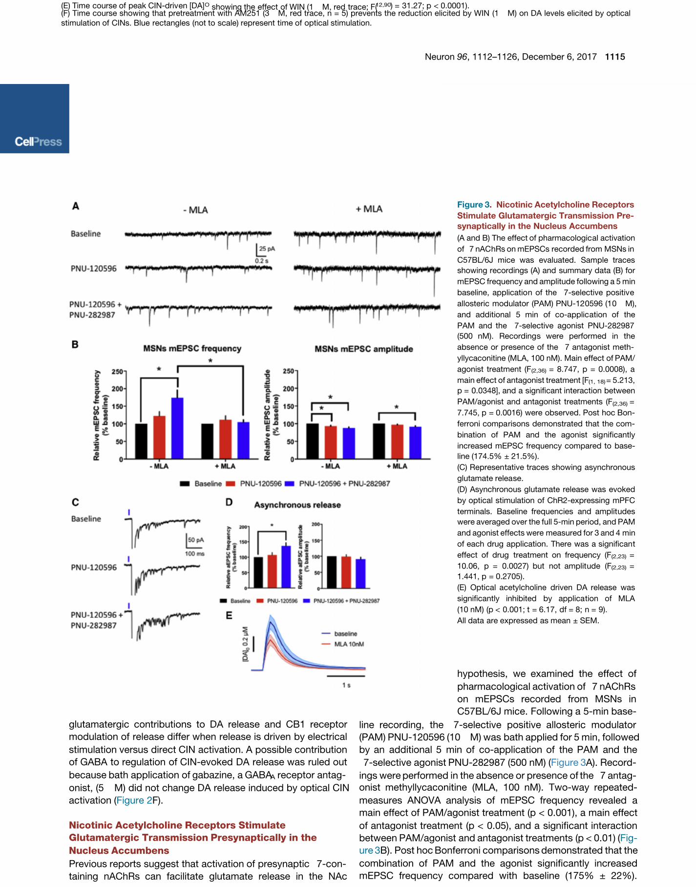
Figure 3. Nicotinic Acetylcholine Receptors
Stimulate Glutamatergic Transmission Pre-
synaptically in the Nucleus Accumbens
(A and B) The effect of pharmacologic al activation
of 7 nAC hRs on mEPSCs recorded from MSNs in
C57BL/6J mice was evaluated. Sample traces
showing recordings (A) and summary data (B) for
mEPSC frequency and amplitude following a 5 min
baseline, applicatio n of the 7-selective positiv e
allosteric modulator (PAM) PNU-12 0596 (1 0 M),
and additional 5 min of co-applic ation of the
PAM and the 7-selective agonist PNU-282987
(500 nM). Recording s were performed in the
absence or presence of the 7 antagonist meth-
yllycaconitine (MLA, 100 nM). Main effect of PAM/
agonist treatment (F (2,36) = 8.747, p = 0.0008), a
main effect of antagonist treatment [F(1, 18) = 5.213,
p = 0.0348], and a significant interaction between
PAM/agonist and antagonist treatments (F(2,36) =
7.745, p = 0.0016) were observed. Post hoc Bon-
ferroni compa risons demonstrated that the com-
bination of PAM and the agonist sig nificantly
increased mEPSC frequency compared to base-
line (174.5% ± 21.5%).
(C) Represen tative traces showing asynchronous
glutamate relea se.
(D) Asynchronous glutamate release was evoked
by opt ical stimulation of ChR2-expressing mPFC
terminals. Baseline frequencies and amplit udes
were averaged over the full 5-min period, and PAM
and agonist effects were measured for 3 and 4 min
of each drug application. There was a significan t
effect of drug treatment on frequency (F(2,23) =
10.06, p = 0.0027) but not amplit ude (F(2,23) =
1.441, p = 0.2705).
(E) Optical acetylcholine driven DA release was
significantly inhibited by applica tion of MLA
(10 nM) (p < 0.001; t = 6.17, df = 8; n = 9).
All data are expr essed as mean ± SEM.
hypothesis, we examined the effect of
pharmacological activation of 7 nAChRs
on mEPSCs recorded from MSNs in
C57BL/6J mice. Following a 5-min base-
line recording, the 7-selective positive allosteric modulator
(PAM) PNU-120596 (10 M) was bath applied for 5 min, followed
by an additional 5 min of co-application of the PAM and the
7-selective agonist PNU-282987 (500 nM) ( A). Record-Figure 3
ings were performed in the absence or presence of the 7 antag-
onist methyllycaconitine (MLA, 100 nM). Two-way repeated-
measures ANOVA analysis of mEPSC frequency revealed a
main effect of PAM/agonist treatment (p < 0.001), a main effect
of antagonist treatment (p < 0.05), and a significant interaction
between PAM/agonist and antagonist treatments (p < 0.01) (Fig-
ure 3B). Post hoc Bonferroni comparisons demonstrated that the
combination of PAM and the agonist significantly increased
mEPSC frequency compared with baseline (175% ± 22%).
(Jones et al., 2001; Kaiser and Wonnacott, 2000; Zhang andWarren, 2002) and this mechanism could link CIN stimulation
to glutamate release that subsequently facilitates increases in
extracellular DA concentration. To examine the viability of this
Moreover, MLA inhibited the effect of the PAM/agonist onmEPSC frequency ( A and 3B). We also observed aFigures 3
small but statistically significant decrease in mEPSC amplitude
that was not inhibited by MLA ( B). We commonlyFigure 3
1116 Neuron , 1112–1126, December 6, 201796
observe this small decrease in mEPSC amplitude over the
course of control mEPSC recordings in MSNs; therefore, this
result is unlikely to be related to activation of 7 nAChRs. In addi-
tion, to directly assess whether 7 nAChRs can regulate gluta-
mate release onto CINs, we also measured mEPSC frequency
from CINs with the 7 PAM accompanied by the agonist PNU-
282987 and found a significant increase in mEPSC frequency
( ). Lastly, to further demonstrate that mPFC terminalsFigure S4
contain functional nicotinic receptors, we examined asynchro-
nous glutamate release. Briefly, increased frequency of asyn-
chronous mEPSCs would indicate a presynaptic effect, most
likely an increase in probability of glutamate release, while a
change in amplitude would indicate a change in postsynaptic
responsiveness. For this experiment, MSNs were recorded in
aCSF containing 0 mM Ca2+ and 4 mM Sr2+. Asynchronous
glutamate release was evoked by optical stimulation of ChR2-
expressing mPFC terminals ( C and 3D). Repeated-Figures 3
measures one-way ANOVA shows significant effect of drug
treatment on frequency (p < 0.01) but not amplitude. Post hoc
Dunnett’s test of frequency shows a significant increase in fre-
quency in the PAM plus agonist condition compared with contro l
(138% ± 10%), but no effect of PAM alone. Having found that
activation of 7 nAChRs modulates excitatory synaptic trans-
mission through changes in the probability of glutamate release,
we posited that blockade of these receptors could inhibit the
glutamate-dependent component of CIN-evoked DA release.
Indeed, the application of MLA at a concentration that has selec-
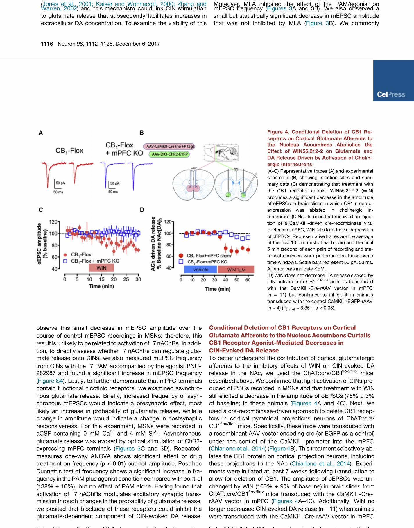
Conditional Deletion of CB1 Receptors on Cortical
Glutamate Afferents to the Nucleus Accumbens Curtails
CB1 Receptor Agonist-Mediated Decreases in
CIN-Evoked DA Release
To better understand the contribution of cortical glutamatergic
afferents to the inhibitory effects of WIN on CIN-evoked DA
release in the NAc, we used the ChAT::cre/CB1flox/flox mice
described above. We confirmed that light activation of CINs pro-
duced oEPSCs recorded in MSNs and that treatment with WIN
still elicited a decrease in the amplitude of oEPSCs (78% ± 3%
of baseline; in these animals ( A and 4C). Next, weFigures 4
used a cre-recombinase-driven approach to delete CB1 recep-
tors in cortical pyramidal projections neurons of ChAT::cre/
CB1flox/flox
mice. Specifically, these mice were transduced with
a recombinant AAV vector encoding cre (or EGFP as a control)
under the control of the CaMKII promoter into the mPFC
(Chiarlone et al., 2014) ( Figure 4B). This treatment selectively ab-
lates the CB1 protein on cortical projection neurons, including
those projections to the NAc ( ). Experi-Chiarlone et al., 2014
ments were initiated at least 7 weeks following transduction to
allow for deletion of CB1. The amplitude of oEPSCs was un-
changed by WIN (100% ± 9% of baseline) in brain slices from
ChAT::cre/CB1flox/flox
mice transduced with the CaMKII -Cre-
rAAV vector in mPFC ( A–4C). Additionally, WIN noFigures 4
longer decreased CIN-evoked DA release (n = 11) when animals
were transduced with the CaMKII -Cre-rAAV vector in mPFC
but still inhibited DA release in animals transduced with the
Figure 4. Conditional Deletion of CB1 Re-
ceptors on Cortical Glutamate Afferents to
the Nucleus Accumbens Abolishes the
Effect of WIN55,212-2 on Glutamate and
DA Release Driven by Activation of Cholin-
ergic Interneurons
(A–C) Representative traces (A) and experimental
schematic (B) showing injection sites and sum-
mary data (C) demons trating that treatment with
the CB1 receptor agonist WIN55,212-2 (WIN)
produces a significan t decrease in the amplit ude
of oEPSCs in brain slices in which CB1 receptor
expression was ablated in cholinergic in-
terneurons (CINs). In mice that received an injec-
tion of a CaMKII -driven cre-recombinase viral
vector into mPFC, WIN fails to induce a depressio n
of oEPS Cs. Represen tative traces are the average
of the first 10 min (first of each pair) and the final
5 min (second of each pair) of recording and sta-
tistical analyses were performed on these same
time windows. Scale bars represent 50 pA, 50 ms.
All error bars indicate SEM.
(D) WIN does not decrease DA release evoked by
CIN activation in CB1flox/flox
animals transduced
with the CaMKII -Cre- rAAV vector in mPFC
(n = 11) bu t continues to inhibit it in animals
transduced with the control CaMKII -EGFP-rAAV
(n = 4) (F (1,13) = 8.851; p < 0.05).

Indeed, the application of MLA at a concentration that has selec-
tivity for 7 versus other nAChR subtypes (10 nM, Mogg et al.,2002) significantly inhibited CIN-driven DA release (p < 0.001,
Figure 3E). These results suggest that functional nicotinic recep-
tors exist on prefrontal glutamate terminals and augment DA
release.
but still inhibited DA release in animals transduced with thecontrol CaMKII -EGFP-rAAV vector (n = 4) (p < 0.0001; Fig-
ure 4D). These data suggest that optical CIN stimulation recruits
CB1 receptor-expressing glutamatergic afferents that enhance
DA release and that CB1 activation modulates these glutamater-
gic afferents.
Neuron , 1112–1126, December 6, 201796 1117
AMPA Receptor Activation on Accumbal DA Terminals
Elicits DA Release
One mechanism that could underlie the above findings would be
that glutamate release from cortical terminals directly depolar-
izes dopaminergic axons. To test this, we initially prepared cor-
onal brain sections for immunohistochemical analysis, which
revealed that the GluA1 and A2 subunits of the AMPA receptor
are clearly detectable within tyrosine hydroxylase (TH)-positive
axons in the NAc ( A, top). To validate these findings,Figure S5
Figure 5. Selective Activation of Excitator y
Prefrontocortical Projec tions to the Nucleus
Accumbens Enhances DA Release and Is
Modulated by Cortical CB1 Receptors
(A) In the nucleus accumbens, axon terminals (AT ,
green outline) co-expressing CB1 (gold particles,
green arrowheads) and VGluT1 (scattered dark
material) made asymmetric syn apses (green ar-
rows) with a dendrite (De, orange outline). Scale
bar represents 200 nm. Bottom panel shows
quantification of the different classes of axon ter-
minals observed.
(B) C57BL/6J mice were transduced with a re-
combinant AAV vector under the control of the
CaMII promoter into the mPF C (AA V1.CamKIIa.
hChR2(II134R)-eYFP; blue trace). Single light
pulse activation of mPFC afferents in the nucleus
accumbens elicits DA release (n = 6), which is
prevented by NBQX.
(C) When the CB1 receptor agonist, WIN55,212-2
(WIN) was added to the slice, a marked reduction
in cortically evoked DA release was obser ved (red
trace, approximately 60%, p < 0.0001; n = 4), an
effect blocked in the prese nce of the CB1 receptor
antagonist AM251. Blue rectang les (not to sca le)
represent time of optical stimulation.
All data are expressed as mean ± SEM.
This manipulation produced robust levels
of DA release ( B, left) comparedFigure S5
to micro-pressure-applied aCSF (Fig-
ure S5B, right).
Excitatory Prefrontocortical
Projections to the Nucleus
Accumbens Enhance DA Release
and Are Modulated by Cortical CB1
Receptors
Given the absence of CB1 receptors on
CINs and DA axons, as discussed above,
another site for CB1 regulation of CIN-
evoked DA release could be glutamater-
gic afferents from mPFC. We verified
that CB1 receptors were expressed on mPFC glutamate affer-
ents in the NAc by examining terminals expressing the vesicular
glutamate transporter type I (VGlut1, a protein mainly found on
cortical afferents) using immuno-electron microscopy. Dense
expression of the CB1 receptor was detected in approximately
one quarter of the VGlut1 axon terminals ( A). In lightFigure 5
of our data suggesting that activation of AMPA receptors con-
tributes to DA release ( B), including a contributionFigure S5
following CIN activation ( D and 2E), we sought to deter-Figures 2

we performed immuno-electron microscopy analysis of accum-
bal tissue and confirmed that AMPA receptors are indeedlocated on the longitudinal axis of DA terminals ( A, bot-Figure S5
tom). To further determine whether depolarization of DA termi-
nals by AMPA receptor activation is sufficient to induce DA
release, we micro-pressure applied AMPA (100 M) onto
slices from C57BL/6J mice in the presence of TTX (100 nM).
mine whether this could be achieved by cortically released gluta-mate as well. Specifically, we transduced C57BL/6J mice with arecombinant AAV vector expressing ChR2 under the control of
the CaMKII promoter injected into the mPFC. Seven weeks
after transduction, NAc brain slices were prepared for voltam-
metric recordings. Single light pulse activation of mPFC afferents
in the NAc elicited DA release, which was tetrodotoxin (TTX)
1118 Neuron , 1112–1126, December 6, 201796
sensitive (n = 6; A and S6B), suggesting that glutama-Figures S6
tergic inputs from cortical terminals indeed drive DA release in
the NAc in support of recent work ( ). Applica-Kosillo et al., 2016
tion of NBQX (5 M) for 10 min significantly decreased DA
release evoked by optogenetic stimulation of PFC afferents
(90%). Following a 30-min washout period, DA signals recovered
( B). Previous work suggests that cortically driven DAFigure 5
release is mediated by activation of CINs ( ).Kosillo et al., 2016
In an attempt to isolate the effects of ChR2-expressing mPFC
inputs directly to dopaminergic terminals, we added 4-amino-
pyridine (4-AP, 100 M) following TTX (100 nM). TTX prevents
synaptic transmission, and 4-AP is used to allow more efficient
ChR2-mediated depolarization of axonal and presynaptic mem-
branes. This technique has been used in past studies to isolate
monosynaptic responses driven by ChR2 activation (Cruikshank
et al., 2010; Petreanu et al., 2007). Application of TTX for
Figure 6. Raising Tissue Levels of Endoge-
nous Cannabinoids Mimics the Effec t of
Full CB1 Receptor Agon ism
(A and B) The monoacylglycerol lipase inhibitor
JZL184 (2 M) decreases DA release evoked by
activation of chol inergic interneurons (CINs). Vol-
tammetric average traces showing the effect of 35
perfusion of JZL184 without blockin g CB1 re-
ceptors (A; p < 0.001; t = 5.246, df = 12; n = 7) and
pretreating slices with the CB1 receptor antago-
nist AM251 at 3 M (B; n = 6).
(C) Average data for JZL184 and AM25 1
(**p < 0.001).
(D) Raising tissue levels of anandamide wit h
URB597 (1 M), a FAAH inhibitor, similarly
decreased DA release evoked by CINs (p < 0.0001;
t = 8.753, df = 8).
(E) Pretrea tment with AM251 (3 M) blocked the
decrease of DA relea se induced by URB595.
(F) Average data for URB597 and AM251
(***p < 0.0001). Blue rectangles (not to scale)
represent time of optical stimulation.
All data are expressed as mean ± SEM.
facilitation of DA release induced by
4 2 nicotinic receptors may occur in
the absence of action potential genera-
tion, similar to what we observed
following pressure application of AMPA.
Of course, this new finding indicates
that the TTX/4-AP procedure did not
isolate a truly monosynaptic response
contrary to our initial plans, and additional
work is needed to determine the mecha-
nisms underlying the ACh role in PFC-
stimulated release under these conditions. Nonetheless, these
data support the idea that PFC-induced DA release requires
the activity of CINs.
W e n e x t a i m e d t o f u n c t i o n a l l y p r o b e t h e r o l e o f C B 1
r e c e p t o r s o n c o r t i c a l a f f e r e n t s . T o d o t h i s , w e b a t h a p p l i e d
W I N ( 1 M ) a n d o b s e r v e d a m a r k e d r e d u c t i o n o f c o r t i c a l l y
e v o k e d D A r e l e a s e ( a p p r o x i m a t e l y 6 0 % ; n = 4 ; F i g u r e 5 C,
l e f t ) . M o r e o v e r , t h e a c t i o n s o f W I N w e r e c o m p l e t e l y p r e v e n t e d
b y a d d i t i o n o f t h e C B 1 r e c e p t o r a n t a g o n i s t A M 2 5 1 ( 3 M ) ( F i g -
u r e 5 C , r i g h t ) . T h e r e f o r e , P F C - d r i v e n N A c D A r e l e a s e i s
m o d u l a t e d p r e s y n a p t i c a l l y b y C B 1 r e c e p t o r s a n d g l u t a m a t e r -
g i c s y n a p t i c t r a n s m i s s i o n o n t o C I N s i s s i m i l a r l y m o d u l a t e d
t h r o u g h C B 1 r e c e p t o r - m e d i a t e d r e g u l a t i o n a s s h o w n i n
F i g u r e S 7 .
Raising Tissue Levels of Endogenous Cannabinoids
6–12 min blocked selective mPFC terminal ChR2-evoked DA re-
sponses completely. Addition of 4-AP for 6 min (during TTX
application) allowed for laser-evoked responses to recover (Fig-
ures S6 C–S6E). Somewhat surprisingly, these responses were
still sensitive to blockade by 4 2-containing nAChR receptor
antagonist ( D and S6E), consistent with the findingsFigures S6
of in the absence of TTX. Thus, our observa-Kosillo et al. (2016)
tions extend these previous findings by providing evidence that
Mimics the Effect of WIN55,212-2
Because exogenous activation of CB1 receptors modulates
cortical and glutamate-sensitive CIN-evoked DA release, we
next investigated whether raising tissue levels of eCBs
would elicit inhibitory effects similar to those observed with
WIN. Signaling by the eCB 2-AG, one of the better-
characterized eCBs released from membrane phospholipid pre-
cursors, is metabolically regulated by its degradative enzyme,
Neuron , 1112–1126, December 6, 201796 1119
monoacylglycerol lipase (MAGL). Thus, we used the MAGL in-
hibitor JZL184 (2 M) to raise tissue levels of 2-AG. When
JZL184 was bath applied, DA levels evoked by single pulse
the NAc. To this end, we used URB597, an inhibitor of fatty acid
amide hydrolase (FAAH) the primary degradative enzyme for
anandamide. Addition of URB597 to the bath decreased CIN-
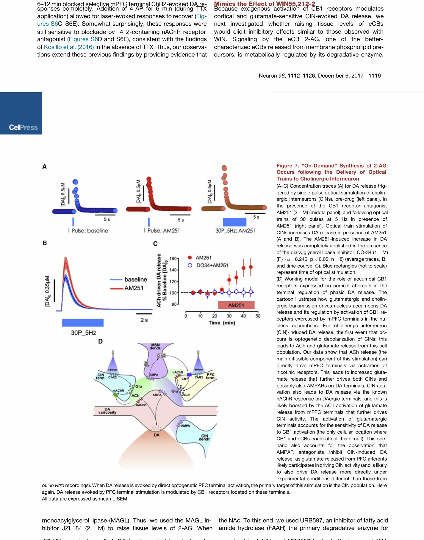
Figure 7. ‘‘On-Demand’’ Synthesis of 2-AG
Occurs following the Delivery of Optical
Trains to Cholinergic Inte rneuron
(A–C) Concentration traces (A) for DA release trig-
gered by single pulse opt ical stim ulation of cholin-
ergic interneurons (CIN s), pre-drug (left panel), in
the presence of the CB1 receptor antagonist
AM251 (3 M) (middle panel), and following optical
trains of 30 pulses at 5 Hz in presence of
AM251 (right panel). Optical train stimulation of
CINs increases DA release in presence of AM251
(A and B). The AM251-induced increase in DA
release was completely aboli shed in the presence
of the diacylglycerol lipase inhibitor, DO-34 (1 M)
(F(1,14)
= 8.246; p < 0.05; n = 8) (average traces , B,
and time course, C). Blue rectangles (not to scale)
represent time of optical stimulation.
(D) Working model for the role of accumbal CB1
receptors expres sed on cortical afferents in the
terminal regulation of phasic DA release. The
cartoon illustrat es how glutamatergic and cholin-
ergic transmis sion drives nucleus accumbens DA
release and its regulation by activatio n of CB1 re-
ceptors expressed by mPFC terminals in the nu-
cleus accumbens. For cholinergic interneuron
(CIN)-induced DA release, the first event that oc-
curs is optogenetic depolarization of CINs; this
leads to ACh and glutamat e release from this cell
population. Our data show that ACh relea se (the
main diffusible component of this stimulation) can
directly dri ve mPFC terminals via activation of
nicotinic receptors. This leads to increase d gluta-
mate release tha t further dri ves both CINs and
possibly also AMPA Rs on DA terminals. CIN acti-
vation also leads to DA release via the known
nAChR response on DAergic terminals, and this is
likely boosted by the ACh activation of glutamate
release from mPF C terminals that further drives
CIN activity. The activation of glutamatergic
terminals accounts for the sensitivity of DA release
to CB1 activation (the only cellular locati on where
CB1 and eCBs could affect this circuit). This sce-
nario also accounts for the observation that
AMPAR antagonists inhibit CIN -induced DA
release, as glutamat e released from PFC afferents
likely participates in driving CIN activity (and is likely
to also drive DA release more directly under
experimental conditions different than those from
our recordings). When DA release is evoked by direct optogenetic PFC terminal activation, the primary target of this stimulation is the CIN popula tion. Herein vitro
again, DA release evoked by PF C terminal stimulation is modulated by CB1 receptors located on these terminals.
All data are expressed as mean ± SEM.

JZL184 was bath applied, DA levels evoked by single pulseCIN optical stimulation were reduced by 46% ± 8% (p < 0.001;n = 7; Figures 6A and 6C). When slices were pretreated with
the CB1 receptor antagonis t AM251 (3 M) for 45 min and the
antagonist was then bath applied together with JZL184, the
reduction elicited by JZL184 was prevented ( B andFigures 6
6C). We next determined whether anandamide, another eCB
released under specific conditions in the striatum (Gerdeman
et al., 2002; Giuffrida et al., 1999; Zhang et al., 2015), would simi-
larly depress DA release caused by optical activation of CINs in
anandamide. Addition of URB597 to the bath decreased CIN-
evoked DA release ( D and 6F) in a CB1 receptor-depen-Figures 6dent fashion ( E and 6F). These results suggest that eCBFigures 6
signaling within the NAc has the potential to modulate DA release
evoked by activation of CINs.
‘‘On-Demand’’ Synthesis of 2-AG Occurs following the
Delivery of Optical Trains to CINs
The results above show that eCBs can potentially limit CIN-
evoked DA release. In our next experiments, we asked whether
1120 Neuron , 1112–1126, December 6, 201796
eCBs could actively be recruited to shape patterns of CIN-
evoked DA release. 2-AG is the primary eCB that mediates
retrograde synaptic signaling at central synapses (Tanimura
Figure 8. CB1 Recept or Activation Modu-
lates Reinforcem ent Driven by Cortical
Glutamate Afferents into the Nucleus Ac-
cumbens
(A) Schematic of viral transductio n in the prefrontal
cortex (PFC) and optic fiber placement in the
striatum (Str; top, left). Fluorescent tag (eYFP;
green) and DAPI (blue) in the PFC (top, right) and
fibers in the NAc (bot tom). LV, lateral ventricle;
NAcC, NAc core; ac, anterior commis sure.
(B) Conditional deletion of the CB1 recepto r on
PFC afferents potentiated self-stimulation (2-way
repeated-measures ANOVA; session: F (12,54) =
18.57, p < 0.001; group: F (1,84) = 4.90, p < 0.05;
group x session interaction: F (7,84) = 1.90, p = 0.08).
(C) Representative cumulative response records
following eCB manipulations.
(D and E) JZL-attenu ated active presses com-
pared to the prior behavioral session (Pre) when
data are expressed as (D) the number of active
presses (two- way repeated-measures ANOVA;
drug session interaction: F (4,25) = 5.14,
p < 0.001) and (E) percent change (one-way
ANOVA: F (4, 25) = 6.29, p = 0.001). JZL effects were
reversed by AM-251 (AM) and absent following
conditional CB 1 receptor deletion. Data are rep-
resented as mean + SEM. p 0.001, **p 0.01*** < <
(Tukey’s post hoc test).
(F) The D1 receptor antagonist SCH2339 0 (SCH,
1 g/0.5 L) and the non-selective nACh recepto r
mecamylamine (MEC, 10 g/0.5 L) potently
attenuate responding in ICSS when injected into
the NAc compared to saline (0.5 L).
(G) Pooled behavioral effects of intracere bral
administration of SCH and mecamylamine versus
saline during ICS S maint ained by optical stimula-
tion of cortical terminals in the NAc (one-way
repeated-measures ANOV A: F(3,12) = 10.80,
p < 0.001. **p < 0.01 versus saline).
range for CIN-evoked DA release in the
NAc ( ). Under theseCachope et al., 2012
conditions, DA release increased by
45% (p < 0.001; n = 8; A, right,Figures 7
7B, and 7C) relative to train-induced
release in the absence of the antagonist.
Importantly, the increase in DA release
induced by AM251 was completely abolished in the presence
of the DGL inhibitor DO34 (1 M; ;Ogasawara et al., 2016
p < 0.05) as depicted in C), suggesting that applica-Figure 7

et al., 2010). On-demand 2-AG biosynthesis is catalyzed bydiacylglycerol lipase (DGL ) via calcium- and G q -protein-
coupled receptor-dependent mechanisms (Tanimura et al.,
2010). Here, we show that AM251 does not modify DA release
in response to single pulse optical stimulation ( A), sug-Figure 7
gesting that this pattern of stimulation is not sufficient to pro-
duce 2-AG release. Therefore, to investigate the conditions
better suited for 2-AG mobilization following CIN optical activa-
tion, we incubated slices with or without AM251 (3 M) and
optically activated CINs with 30 pulses delivered at 5 Hz, a
frequency previously shown to be in the preferential pulse
tion of optical trains to CINs, but not stimulation with singlepulses, mobilizes 2-AG to limit CIN-driven DA release (Fig-ure 7D). Addition of AM251 during train stimulation prevents
activation of CB1, presumably by displacing 2-AG released
during stimulation. The CB1 receptors activated by 2-AG are
almost certainly those on cortical terminals given our evidence
that the receptor is not expressed by other relevant neurons
within the circuit as shown in the diagram on D.Figure 7
Through this indirect mechanism, CB1 blockade ultimately pre-
vents 2-AG actions that would normally inhibit extracellular DA
levels.
Neuron , 1112–1126, December 6, 201796 1121
2-AG Binding to CB1 Receptors Expressed on mPFC
Terminals in the Nucleus Accumbens Constrains
Reinforcement Sustained by Optical Activation of These
Cortical Axons
It has previously been demonstrated that optical activation of
cortical terminals in the NAc reinforces instrumental behavior
( ). Here, we assessed whether CB1 receptors in-Britt et al., 2012
fluence reinforcement driven by mPFC glutamate afferents in the
NAc. Specifically, we measured self-stimulation induced by op-
togenetic activation of ChR2-expressing accumbal mPFC axons
( A; ) and its sensitivity to eCBs, DA, andFigure 8 Britt et al., 2012
nACh receptor manipulations. We observed robust self-stimula-
tion behavior ( B) as previously described (Figure 8 Britt et al.,
2012). Conditional knockout of the CB1 receptor on PFC affer-
ents potentiated self-stimulation compared to controls, support-
ing the idea that eCB signaling can modulate functional effects of
PFC activation ( B and ). Conversely,in vivo Figure 8 Figure S8
self-stimulation was attenuated by increasing tissue levels of
the eCB 2-AG by inhibiting its degradation with JZL184
(18 mg/kg, i.p.). Moreover, the effects of JZL184 were reversed
by co-administration of AM251, which did not have any effects
per se at the relatively low dose we used (0.75 mg/kg. i.p.,
Figures 8C–8E). The effects of JZL184 were abolished in mice
lacking CB1 receptors on PFC terminals ( C–8E), an ef-Figures 8
fect that cannot be explained by an indiscriminate higher rate of
lever pressing in animals with an ablation of CB1 receptors on
PFC terminals ( ). Altogether, these data suggest thatFigure S8
this population of receptors mediates the pharmacological ef-
fects of JZL. Next, we demonstrate a link between self-stimula-
tion of cortical terminals in the NAc and D1 receptors as shown
previously by our group and others; namely, a decrease in the
vigor of self-stimulation behavior induced by a D1-like receptor
antagonist ( ;Figure 8 Nowend et al., 2001; Yun et al., 2004; Cheer
et al., 2007). D1 receptors were targeted, with a D1 antagonist
(SCH 23390), because of their low-affinity state, which would
be primarily tuned to the phasic, high concentration release of
DA, documented here and (also seein vitro in vivo Cachope
et al., 2012). Finally, behavior was also potently disrupted
when the non-selective nACh receptor antagonist mecamyl-
amine was infused bilaterally into the NAc at a concentration
devoid of locomotor confounds ( ), supportingCollins et al., 2016
an important role for these receptors on PFC-driven instrumental
behavior, a finding that aligns with observations from our in vitro
experiments and our prior work ( ).Cachope et al., 2012
DISCUSSION
CB1 receptor activation does not inhibit DA release following sin-
gle pulse electrical stimulation as shown by others and by us
here (Sidlo et al., 2008; Szabo et al., 1999). Additionally,
GABAergic transmission does not appear to contribute to the
CIN-driven DA release, ruling out a role for the other major
CB1-expressing neuronal subtype in striatum (i.e., the parvalbu-
min-positive fast-spiking interneurons and the MSNs them-
selves). Our results show a previously unseen level of specificity
for actions of CB1 receptors in the terminal modulation of DA
release and of behavior reinforced by specific activation of
cortical glutamate terminals in the NAc ( D). These find-Figure 7
ings extend earlier work showing that CB1 receptors are
powerful modulators of striatal DA release and motivated
behavior due to their indirect disinhibitory actions on dopami-
nergic neurons in the midbrain (Cheer et al., 2000a, 2004;
Lupica et al., 2004; Riegel and Lupica, 2004; Wallmichrath and
Szabo, 2002).
The different neural substrates involved in DA release evoked
by a single electrical versus optical pulse stimulation may be
numerous, but differential involvement of glutamatergic synaptic
transmission appears to be crucial to the explanation of our find-
ings. Glutamate receptor antagonists have no effect on single
pulse electrical stimulus-induced DA release (Avshalumov
et al., 2003). Threlfell et al. showed no effect of an NMDA recep-
tor antagonist plus a low concentration of an AMPA receptor
antagonist on CIN-evoked DA release in dorsal striatum except
when CINs are activated by thalamic inputs (Threlfell et al.,
2012). In the NAc, antagonists of AMPA and NMDA ionotropic
glutamate receptors inhibit DA release following CIN activation
( ). This discrepancy could be due to regionalCachope et al., 2012
differences although a detailed pharmacological characteriza-
tion of the involvement of all ionotropic glutamate receptors in
DA release in both regions is warranted. Accordingly, we
observed that optical CIN activation produces EPSCs in
MSNs, responses regulated by CB1 receptors. Additionally,
cortical glutamate drive onto CINs themselves is also regulated
presynaptically by CB1 receptors. Glutamate release can be
stimulated directly by activation of PFC afferents or indirectly
through activation of 7 nAChRs on PFC terminals. The released
glutamate could regulate DA release in at least two ways. It is
known that cortical activation stimulates CINs and this enhances
DA release through activation of nAChRs ( ).Kosillo et al., 2016
This appears to be the predominant mechanism by which opto-
genetic PFC activation increases DA release given the sensitivity
of this process to nAChR blockade ( ;Figure S6 Kosillo et al.,
2016). The AMPA receptors that we find on DA terminals may

Here we show that activation of CB1 receptors on cortical termi-
nals decreases DA release evoked by CIN activation andin vivo
in brain slices. It is unlikely that this reduction is due to decreased
probability of ACh release involving activation of CB1 receptors
present on cholinergic terminals, because our own data as well
as that of others ( ) demonstrateHohmann and Herkenham, 2000
that CB1 mRNA is not expressed by CINs in the NAc and thus
CB1 receptors do not direc tly modulate ACh release. Dopami-
nergic terminals do not express CB1 receptors (Julian et al.,
2003), and thus these neurons are not the site of the CB1 recep-
tor-mediated effects reported here. Consistent with this idea,
also contribute to glutamatergically driven DA release, althoughit remains to be determined under which circumstances they
play a role. When CINs are activated optogenetically, glutamate
release driven by ACh could act directly on AMPARs located on
DA terminals, although this will have to be determined in future
studies. The other possibility is that glutamate released following
CIN stimulation and subsequent 7 nAChR activation feeds back
onto CINs to further enhance ACh release. This scenario,
although admittedly complicated, cannot be ruled out at this
time. Importantly, the finding that AMPA receptor blockade oc-
cludes the effects of WIN on CIN-evoked release is consistent
with our results showing that CB1 receptors act by decreasing
1122 Neuron , 1112–1126, December 6, 201796
glutamate release and subsequent AMPA receptor activation.
This process may involve AMPA receptors on CINs as well as
those receptors on dopaminergic terminals (Kaiser and Wonna-
cott, 2000; Kendrick et al., 1996; Smolders et al., 1996). Thus,
CB1 receptors on glutamatergic afferents modulate DA release
only under conditions where these afferents are activated and
glutamate contributes to DA release. Together, these data lead
us to propose that CB1 receptors on cortical glutamatergic ter-
minals modulate DA release via mechanisms upstream of
AMPA effector sites.
Because some studies have shown that AMPA receptor
blockade actually enhances DA release evoked by electrical
stimulation under specific conditions ( ),Avshalumov et al., 2003
a provocative thought arises. If AMPA receptor blockade only in-
hibits CIN or mPFC-driven release of DA as shown in the present
study, this implies that electrical stimulation recruits additional
DA release mechanisms that are not AMPA receptor dependent.
This idea is consistent with a recent study demonstrating that
AMPA receptor blockade does not affect DA release induced
by direct stimulation of VTA afferents ( ). Thus,Zhang et al., 2015
direct activation of at least some dopaminergic terminals may
bypass sensitivity to AMPA receptor activation (Zhang et al.,
2015). It is also possible that electrical stimulation activates glu-
tamatergic transmission to a degree that 7-type nAChRs cannot
further influence release. Nevertheless, our findings are most
consistent with the idea that CIN activation primarily evokes
ACh release, which likely depolarizes nearby CB1 receptor-
expressing glutamatergic terminals via volume transmission (Ca-
chope and Cheer, 2014). Our finding that activation of 7-type
nAChRs increases the frequency of glutamatergic mEPSCs as
well as mPFC afferent-specific asynchronous glutamate release
in the NAc is aligned with this hypothesis. Modulation of
glutamate release by 7 nAChRs is also supported by previous
studies showing that nAChRs on glutamate terminals powerfully
enhance glutamate neurotransmission in NAc synaptosomes
and slices (Jones et al., 2001; Kaiser and Wonnacott, 2000;
Zhang and Warren, 2002). Single pulse optical CIN activation,
as opposed to electrical stimulation, may depolarize CB1 (and
nicotinic) receptor-expressing glutamatergic terminals, and
glutamate released from these afferents activates AMPA recep-
tors on CINs (and probably on DA terminals) to facilitate DA
release (in addition to their direct depolarization by 2-expressing
nicotinic receptors [ ]).Cachope et al., 2012; Threlfell et al., 2012
Another possibility is that ACh has a tonic effect on glutamate
release through 7-type nAChRs, but there is no evidence for
mechanistic link between transduction at D2 receptors and
anandamide synthesis has yet to be established. However,
and in agreement with the ability of CB1 receptors to respond
to both eCBs ( ), we observed thatMathur and Lovinger, 2012
pharmacological elevation of 2-AG or anandamide mimic the
inhibitory effects of WIN on CIN-evoked DA release in the NAc.
We further provide evidence that 2-AG is released ‘‘on demand’’
following optical trains but not single pulse stimulation of CINs.
Prevention of AM251-induced facilitation of DA release by
blockade of 2-AG synthesis indicates that 2-AG is mobilized
following sustained CIN depolarization and limits dopaminergic
output. Given the sampling rate of FSCV, it may not be possible
to precisely determine when the AM251 effect begins. However,
it is known that eCB release can occur within hundreds of
milliseconds ( ). Therefore, optical trainsHeinbockel et al., 2005
that mimic the physiological activity of CINs are likely to initiate
the production of 2-AG (possibly by MSNs but other sources
cannot be ruled out); this ‘‘on demand’’ mobilization of 2-AG
can be followed by retrograde signaling onto CB1 receptors pre-
sent on cortical glutamatergic terminals to lessen glutamate
release probability and thus decrease overall CIN-evoked DA
release. Unfortunately, due to the lack of specific anandamide
synthesis inhibitors, it remains to be determined whether optical
trains similarly mobilize this eCB.
Prefrontocortical glutamatergic afferents to the NAc are crit-
ical for the integration of contextual information related to the
pursuit of rewards ( ). Moreover, this interfaceFloresco, 2015
has been extensively studied with regard to eCB-mediated
changes in synaptic plasticity, where CB1 receptor activation
decreases strength of glutamatergic synapses onto accumbal
MSNs ( ). The findings that WIN-Robbe et al., 2002a, 2002b
mediated effects on CIN-evoked changes in MSN synaptic
excitation and DA release are dramatically diminished in ani-
mals with a conditional deletion of CB1 receptors specifically
in this projection, suggest that cholinergic and dopaminergic
signals are more subtly intertwined than previously thought.
Indeed, eCB tone may be a key activity-dependent mediator
of release from terminals within these micro-circuits. Our
behavioral results suggest that this is the case, at least when
behavior is reinforced by optical activation of cortical terminals
in the NAc ( ). A key observation is that, underBritt et al., 2012
these circumstances, raising 2-AG levels shows an effect that
is opposite to what we previously described for motivated
behavior driven by stimulation of the midbrain (Oleson et al.,
2012), in strong support of the different mechanisms of action
release through 7-type nAChRs, but there is no evidence forthis to date.
The eCBs anandamide and 2-AG are mobilized by receptor
activation and depolarization of MSNs and they change synaptic
strength depending on the particular signal necessary for eCB
production ( ). In particular, 2-AG syn-Hashimotodani et al., 2013
thesis is thought to arise mainly from activation of postsynaptic
G q
/11-coupled receptors such as group I metabotropic gluta-
mate receptors and M1/M3 muscarinic cholinergic receptors
that recruit the activity of several phospholipases that produce
diacylglycerol, the precursor for 2-AG (Narushima et al., 2007;
Uchigashima et al., 2007). On the other hand, anandamide
biosynthesis has been linked to D2 receptor activation (Gerde-
man et al., 2002; Giuffrida et al., 1999), although the precise
2012), in strong support of the different mechanisms of actionfor 2-AG; e.g., inhibition of cortically driven excitation in theNAc versus disinhibition of DA neuron cell bodies in the
midbrain. These opposite effects of JZL184 are likely to occur
due to the different substrates utilized by 2-AG (i.e., activation
of CB1 receptors on glutamate terminals in NAc versus CB1 re-
ceptors on GABA terminals in the VTA). We further show that
behavior maintained by optical stimulation of PFC terminals in
the NAc is reliant upon DA D1 (Yun et al., 2004; Cheer et al.,
2007 Crespo et al., 2008; Feduccia) and nicotinic receptors (
et al., 2014), suggesting that it may recruit, at least in part,
the mechanisms uncovered in our experiments for itsin vitro
maintenance. It is notable that nicotinic receptor blockade
can, under different experimental condit ions, enhance behavior
Neuron , 1112–1126, December 6, 201796 1123
as well as phasic DA concentrations in the NAc (Collins et al.,
2016), thereby highlighting a precise level of influence of these
neurotransmitters that is critically dependent on the neural
mechanisms that motivate instrumental behavior. Thus, a
possible function of eCB signaling at PFC to NAc synapses
when cholinergic activity is elevated, for example when rewards
are obtained ( ), may be to promote the se-Joshua et al., 2008
lection of goal-directed actions toward available reinforcers in
a context- and neural substrate-specific manner. These find-
ings are relevant to neuropsychiatric conditions where aberrant
responses to contextual triggers are observed clinically, as is
the case of relapse to drug seeking.
STAR METHODS
Detailed methods are provided in the online version of this paper
and include the following:
KEY RESOURCES TABLE
CONTACT FOR REAGENTS AND RESOURCE SHARING
EXPERIMENTAL MODEL AND SUBJECT DETAILS
Animals
METHOD DETAILS
Stereotaxic virus injection
In vivo optical stimulation and fast-scan cyclic vol-
tammetry
In vitro optical stimulation and fast-scan cyclic vol-
tammetry
Electrophysiology
In situ hybridization and ChAT immunolabeling
Fluorescence microscopy and image analysis for im-
munodetection of AMPA receptors on DA terminals
Electron microscopy
Behavioral experiments
QUANTIFICATION AND STATISTICAL ANALYSIS
Data analysis for immunohistochemistry
Ultrastructural analysis of brain tissue
SUPPLEMENTAL INFORMATION
Supplemental Information includes eight figures and can be found with this
article online at .https://d oi.org/10.1016/j.neuron.2017.11.012
AUTHOR CONTRIBUTIONS
Received: Septembe r 6, 2016
Revised: April 8, 2017
Accepted: Novembe r 9, 2017
Published: Decembe r 6, 2017
REFERENCES
Atwood, B.K., Kupferschmidt , D.A., and Lovinger, D.M. (2014). Opioids induce
dissociable forms of long-term depression of excitatory inputs to the dorsal
striatum. Nat. Neurosci. , 540–54817 .
Avshalumov, M.V., Chen, B.T., Marshall, S.P., Pen a, D.M. , and Rice , M.E.
(2003). Glutamate-dependent inhibition of dopamine release in striatum
is mediated by a new diffusible mess enger, H2O2. J. Neurosci. ,23
2744–2750.
Britt, J.P., Benaliouad, F., McDevitt, R.A., Stuber, G.D., Wise, R.A., and Bonci,
A. (2012). Synaptic and behavioral profile of multiple glutamatergic inputs to
the nucleus accumbens. Neuron , 790–80376 .
Cachope, R., and Cheer, J.F. (2014). Local contro l of striatal dopamine
release. Fro nt. Behav. Neurosci. , 1888 .
Cachope, R., Mateo, Y., Mathur, B.N., Irving, J., Wang, H.L., Morales , M.,
Lovinger, D.M., and Cheer, J.F. (2012 ). Selective activation of cholinergic inter-
neurons enhan ces accumbal phasic dopamine release: setting the tone for
reward processing. Cell Rep. , 33–412 .
Cheer, J.F., Kendall, D.A., and Marsden, C.A. (2000a). Cannabinoid recep-
tors and reward in the rat: a conditioned place preference study.
Psychopharmacology (Berl.) , 25–30151 .
Cheer, J.F., Marsden, C.A., Kendall, D.A., and Mason, R. (2000b). Lack
of response suppres sion follows repeated ventral tegmental cannabinoid
administration: an in vitro electrophys iological study. Neuroscience ,99
661–667.
Cheer, J.F., Wassum, K.M., Heien, M.L., Phillips, P.E., and Wightman, R.M .
(2004). Cannabinoids enhance subsecond dopamine release in the nucleus
accumbens of awake rats. J. Neurosci. , 4393–44 0024 .
Cheer, J.F., Aragon a, B.J., Heien, M.L., Seipel, A.T., Carelli, R.M., and
Wightman, R.M. (2007). Coordinated accumbal dopamine release and neural
activity drive goal-directed behavior. Neuron , 237–24454 .
Chiarlone, A., Bellocc hio, L., Blazquez, C., Resel, E., Soria-Go mez, E.,
Cannich, A., Ferrero, J.J., Sagredo, O., Benito, C., Romero, J., et al. (2014).
A restricted population of CB1 cannabinoid receptors with neuroprotective ac-
tivity. Proc. Natl. Acad. Sci. US A , 8257–8262111 .
Collins, A.L., Aitken, T.J., Greenfield, V.Y., Ostlund, S.B., and Wassum, K.M.
(2016). Nucleus accumbens acetylcholine receptors modulate dopamine
and motivatio n. Neuropsychopharmaco logy , 2830–283841 .
Crespo, J.A., Stockl , P., Zorn, K., Saria, A., and Zernig, G. (2008). Nucleus
accumbens core acetylcholine is preferentially activated during acquisition
of drug- vs food-reinforced behavior. Neuropsychopharma cology ,33
3213–3220.

AUTHOR CONTRIBUTIONS
Y.M., K.A.J., B.K.A., H.W., D.P.C., I.G., and S.Z. conceived and performed ex-
periments; Y.M., K.A.J., B.K.A., D.P.C., J.F.C., and D.M.L. wrote the manu-
script; J.F.C., D.M.L., and M.M. conceived experiments and secured funding;
L.B. and M.G. provided viral reagents; R.C. and M.M. provided expertise and
feedback.
ACKNOWLEDGMENTS
We acknowledge funding from the Division of Intramural Clinical and Biological
Research of NIAAA, ZIA AA000416 (Y.M., B.K.A., K.A.J., and D.M.L.), the
Intramural Research Program of NIDA ZIA DA000511 (H.W., S.Z., and M.M.),
the Spanish Ministerio de Economı a y Competitividad (grant number
SAF2015-64945-R to M.G.), NIH grant numbers DA022340 and DA042595
(J.F.C.) and DA041827 (D.P.C.) and a Postdocto ral Resear ch Associate
(PRAT) Fell owship from NIGMS (K.A. J.).
3213–3220.Cruikshank, S.J., Urabe, H., Nurmikko, A.V., and Connors, B.W. (2010).
Pathway-specific feedforwa rd circuits between thalamus and neocortex re-
vealed by selective opt ical stimulation of axons. Neuron , 230–24565 .
Floresco, S.B. (2 015). The nucle us accumbens: an interface betwee n cogni-
tion, emotio n, and action. Annu. Rev. Psychol. , 25–5266 .
Feduccia, A.A., Simms , J.A., Mill, D., Yi, H.Y., and Bartlett, S.E. (2014).
Varenicline decreases ethanol intake and increases dopamine release via
neuronal nicotinic acetylcholine receptor s in the nucleus accumbens . Br. J.
Pharmacol. , 3420–343117 1 .
Gerdeman, G.L., Ronesi, J., and Lovinge r, D.M. (2002). Po stsynaptic endo-
cannabinoid release is critical to long-term depression in the striatum . Nat.
Neurosci. , 446–4515 .
Giuffrida, A., Parson s, L.H., Kerr, T.M., Rodrı guez de Fonseca, F., Navarro, M.,
and Piomelli, D. (1999). Dopamine activation of endogenous cannabinoid
signaling in dorsal striatum. Nat. Neurosci. , 358–3632 .
1124 Neuron , 1112–1126, December 6, 201796
Hashimotodani, Y., Ohno-Shosaku, T., Tani mura, A., Kita, Y., Sano, Y.,
Shimizu, T., Di Marzo, V., and Kano, M. (2013). Acute inhibition of diacylgly-
cerol lipase blocks endoc annabinoid-mediated retrograde signalling: evi-
dence for on-demand biosynthesis of 2-a rachidono ylglycerol. J. Physiol.
591, 4765–4776.
Heinbockel, T., Brager, D.H., Reich, C.G., Zhao, J., Muralidharan, S., Alger,
B.E., and Kao, J.P. (2005). Endocannabinoid signaling dynamics probed
with optical tools. J. Neurosci. , 9449–945925 .
Higley, M.J., Gittis, A.H., Oldenburg, I.A., Balthasar, N., Seal, R.P., Edwards,
R.H., Lowell, B.B., Kreitzer, A.C., and Sabatini, B.L. (2011). Cholinerg ic inter-
neurons mediate fast VGlu T3-dependent glutamatergic transmission in the
striatum. PLoS ONE , e191556 .
Hohmann, A.G., and Herkenham, M. (2000). Localization of cannabinoid CB(1)
receptor mRNA in neuro nal subpopulations of rat striatum: a double -label
in situ hybridization study. Synapse , 71–803 7 .
Jennings, J.H., Rizzi, G., Stamatakis, A.M., Ung, R.L., and Stuber, G.D. (2013).
The inhibitor y circuit architecture of the lateral hypothalamus orchestrates
feeding. Science , 1517–15 21341 .
Jones, I.W., Bolam, J.P., and Wonnacott, S. (2001). Presynaptic localisation of
the nicotinic acetylcholine receptor beta2 subunit immun oreactivity in rat
nigrostriatal dopamine rgic neurones. J. Comp. Neurol. , 235–247439 .
Joshua, M., Adler, A., Mitelman, R., Vaadia, E., and Bergman, H. (2008).
Midbrain dopaminergic neurons and striatal cholinergic interneurons
encode the difference between reward and aversive events at different
epochs of probabilistic classical conditioning trials. J. Neurosci. ,28
11673–11684.
Julian, M.D., Martin, A.B., Cuellar, B., Rodriguez De Fonseca, F., Navarro, M.,
Moratalla, R., and Garcia-Segura, L.M. (2003). Neuroanatomical relationship
between type 1 cannabinoid receptors and dopaminergic systems in the rat
basal ganglia . Neuroscienc e , 309–318119 .
Kaiser, S., and Wonna cott, S. (2000). alpha-b ungarotoxin-sensitive nicotinic
receptors indirectly modulate [(3)H]dopamine release in rat striat al slices via
glutamate release . Mol. Pharmacol. , 312–31858 .
Kendrick, K.M., Guevara -Guzman, R., de la Riva, C., Christensen, J.,
Ostergaard, K., and Emson, P.C. (1996). NMDA and kainate-evoked release
of nitric oxide and classical transmitters in the rat striatum: in vivo evidence
that nitric oxide may play a neuroprotective role. Eur. J. Neurosci. ,8
2619–2634.
Kosillo, P., Zhang, Y.F., Threlfell, S., and Cragg, S.J. (2016). Cortical Control of
Striatal Dopamine Transmission via Striatal Cho linergic Interneuro ns. Cereb.
Cortex. Publis hed online August 27, 2016. https://doi.org/10.1093/c ercor/
bhw252.
Kreitzer, A.C., and Malenka, R.C. (2005). Dopamine modulatio n of st ate-
dependent endocanna binoid release and long-term depression in the stria-
tum. J. Neurosci. , 10537–1054525 .
otoxin-MII-sensitive presynaptic nicotinic acetylcholine receptors in rat stria-
tum. J. Pharmacol. Exp. Ther. , 197–204302 .
Morales, M., and Wang, S.D. (2002). Differential composition of 5-hydroxy tryp-
tamine3 receptors synthesized in the rat CNS and peripheral nervous system.
J. Neurosci. , 6732–674122 .
Narushima, M., Uchiga shima, M., Hashimo to, K., Watanabe, M., and Kano, M.
(2006). Depolarization-induced suppression of inhibition mediated by endo-
cannabinoids at synapse s from fast-spiking inte rneurons to medium spiny
neurons in the striatum. Eur. J. Neurosci. , 2246–225224 .
Narushima, M., Uchigashima, M., Fukaya, M., Matsui, M., Manabe, T.,
Hashimoto, K., Wata nabe, M., and Kano, M. (2007). Tonic enhancement of en-
docannabinoid-mediated retrograde suppression of inhibition by cholinergic
interneuron activity in the striatum. J. Neurosci. , 496–50627 .
Nowend, K.L., Arizzi, M., Carl son, B.B., and Salamone, J.D. (2001). D1 or D2
antagonism in nucleus accum bens core or dorsomedial shell suppres ses lever
pressing for food but leads to compensatory increa ses in chow consumption.
Pharmacol. Biochem. Behav. , 373–38269 .
Ogasawara, D., Deng, H., Viader, A., Baggelaar, M.P., Breman, A., den Dulk,
H., van den Nieuwendijk, A.M., Soethoudt, M., van der Wel, T., Zhou, J.,
et al. (2016). Rapid and profound rewiring of brain lipid signaling networks
by acute diacylglycerol lipas e inhib ition. Proc. Natl. Acad. Sci. USA
113, 26–33.
Oleson, E.B., Beckert, M.V., Morra, J.T., Lansink, C.S., Cachope, R., Abdulla h,
R.A., Loria ux, A.L., Schetters, D., Pattij, T., Roitman, M.F., et al. (2012).
Endocannabinoids shape accum bal encoding of cue-motivated behavior via
CB1 receptor activation in the ventral tegmentum. Neuron , 360–37373 .
Peters, J.M., Harris, J.R., and Kleins chmidt, J.A. (1991). Ultrastructure of the
approximately 26S complex containing the approximately 20S cylinder parti-
cle (multicat alytic proteinase/prote asome). Eur. J. Cell Biol. , 422–43256 .
Petreanu, L., Huber, D., Sobczyk, A., and Svoboda, K. (2007). Channelrhodopsin-
2-assisted circuit mapping of long-range callosal projections. Nat. Neurosci. ,10
663–668.
Riegel, A.C., and Lupica, C.R. (2004). Independe nt presynaptic and postsyn-
aptic mechanisms regulate endocannab inoid signaling at multiple synapses
in the ventral tegmental area. J. Neurosci. , 11070–1107824 .
Robbe, D., Bock aert, J., and Manzoni, O.J. (2002a). Metabotropic glutamate
receptor 2/3-dep endent long-term depression in the nucleus acc umbens is
blocked in morphine withdrawn mice. Eur. J. Neurosci. , 2231–223516 .
Robbe, D., Kopf, M., Remaury, A., Bockaert, J., and Manzoni, O.J. (2002b).
Endogenous cannabinoids mediate long-term synaptic depression in the nu-
cleus accumbens. Proc. Natl. Acad. Sci. USA , 8384–838899 .
Ronesi, J., Gerdeman, G.L., and Lovinger, D.M. (2004). Disrupt ion of endocan-
nabinoid release and striatal long-term depression by postsynaptic blockade
of endocanna binoid membrane transport. J. Neurosci. , 1673–167924 .

Long, J.Z., Li, W., Booker, L., Burston, J.J., Kinsey, S.G., Schlosburg, J.E.,Pavon, F.J., Serrano, A.M., Selley, D.E., Parsons, L.H ., et al. (2009).
Selective blockade of 2-arachidono ylglycerol hydrolysis produces cannabi-
noid behavioral effects. Nat. Chem. Biol. , 37–445 .
Lupica, C.R., Riegel, A.C., and Hoffman, A.F. (2004). Marijuana and cannab i-
noid regulation of brain reward circuits. Br. J. Pharmacol. , 227–234143 .
Mathur, B.N., and Lovinger, D.M. (2012). Endocannabinoid-dopamine interac-
tions in striatal synaptic plasticity. Front. Pharmacol. 3, 66.
Mato, S., Robbe, D., Puente, N., Grandes, P., and Manzoni, O.J. (2005).
Presynaptic homeostatic plasticity rescues long-term depression after chronic
Delta 9-tetrahydrocannabino l exposure. J. Neurosci. , 11619–1162725 .
Mato, S., Lafourcade, M., Robbe, D., Bakiri, Y., and Manzoni, O.J. (2008).
Role of the cyclic-AMP/PKA cascade and of P/Q-type Ca++ channels in endo-
cannabinoid-mediated long-term depression in the nucleus accumbens.
Neuropharmacology , 87–9454 .
Mogg, A.J., Whiteaker, P., McIntosh, J.M., Marks, M., Collins, A.C., and
Wonnacott, S. (2002). Methyllycaconitine is a potent antagonist of alpha-con-
Sesack, S.R., and Grace, A.A. (2010). Cortico-Basal Ganglia reward network:
microcircuitry. Neuropsychopharmacology , 27–4735 .
Sidlo, Z., Reggio, P.H., and Rice, M.E. (2008). Inhibition of striatal dopamine
release by CB1 receptor activation requires nonsynaptic communication
involving GABA, H2O2, and KATP channels. Neurochem. Int. , 80–8852 .
Smolders, I., Sarre, S., Vanhaesendonck, C., Ebinger, G., and Michotte, Y.
(1996). Extracellular striatal dopamine and glutamate after decortication and
kainate receptor stimulation, as meas ured by microdialysis. J. Neuroch em.
66, 2373–2380.
Szabo, B., M
uller, T., and Koch, H. (1999). Effects of cannabinoids on dopa-
mine release in the corpus striatum and the nucleus accumbens in vitro.
J. Neurochem. , 1084–10 8973 .
Tanimura, A., Yamazaki, M., Hashimotodani, Y., Uchigashima, M., Kawata, S.,
Abe, M., Kita, Y., Hashimoto , K., Shimizu, T., Watanabe, M., et al. (2010). The
endocannabinoid 2-arachidon oylglycerol produced by diacylglycerol lipase
alpha mediates retrograde suppression of synaptic transmission. Neuron ,65
320–327.
Neuron , 1112–1126, December 6, 201796 1125
Threlfell, S., Lalic, T., Platt, N.J., Jenning s, K.A., Deisseroth, K., and Cragg, S.J.
(2012). Striatal dopamine release is triggered by syn chronized activity in
cholinergic interneurons. Neuron , 58–6475 .
Tzavara, E.T., Wade, M.R., Davis, R.J., El Khoury, M.A., and Nomikos, G.G.
(2008). Role of metab otropic glutamate receptor 5 in the procholinergic effects
of neuro psychotherap eutic compounds. Synapse , 940–94362 .
Uchigashima, M., Narushima, M., Fukaya, M., Katona, I., Kano, M., and
Watanabe, M. (2007). Subcellular arrangement of molecules for 2-arachido-
noyl-glycerol-mediated retrograde signaling and its physiological contribution
to synaptic modulation in the striatum. J. Neurosci. , 3663–367627 .
Wallmichrath, I., and Szabo , B. (2002). Cannabinoids inhibit striatonigral
GABAergic neuro transmission in the mous e. Neuroscience , 6 71–682113 .
Wang, H.L., and Morales, M. (2008). Corticotropin-rele asing factor binding
protein within the ventral tegmental area is expressed in a subset of dopami-
nergic neurons. J. Com p. Neurol. , 302–318 509 .
Yorgason, J.T., Espana, R.A., and Jones, S.R. (2011). Demon voltammetry and
analysis software: analysi s of cocaine-indu ced alterations in dopamine
signaling using multiple kinetic measures. J. Neurosci. Methods , 158–164202 .
Yun, I.A., Nicola, S.M., and Fields, H.L. (2004). Contrasting effects of dopamine
and glut amate rece ptor antagonist injection in the nucleus accumbens sug-
gest a neural mechanism underlying cue-evoked goal-directed behavior.
Eur. J. Neurosci. , 249–26320 .
Zhang, L., and Warren, R.A. (2002). Muscarinic and nicotinic presynapt ic mod-
ulation of EPSCs in the nucleus accum bens during postnatal development.
J. Neurophysiol. , 3315–33 3088 .
Zhang, S., Qi, J., Li, X., Wang, H.L., Britt, J.P., Hoffman, A.F., Bonci, A., Lupica,
C.R., and Morales, M. (2015). Dopaminergic and glutamatergic microdomains
in a subset of rodent mesoaccumb ens axons. Nat. Neurosci. , 386–39218 .
Zhou, F.M., Liang, Y., and Dani, J.A. (2001). Endogeno us nicotinic cholinergic
activity regulates dopamine release in the striatum. Nat. Neurosci. ,4
1224–1229.

1126 Neuron , 1112–1126, December 6, 201796
STAR METHODS
KEY RESOURCES TABLE
REAGENT or RESOURCE SOURCE IDENTIFIER
Antibodies
Goat polyclonal anti-ChAT EMD Millipore Cat# ab144P, RRID: AB_207 9751
Mouse monoclonal anti-TH EMD Millipore Cat# MAB318, RRID: AB_2201528
Rabbit polyclonal anti-GluR1 Frontier Institute Cat# GluA1-Rb-Af690, RRID: AB_2571752
Rabbit polyclonal anti-GluR2 Frontier Institute Cat# GluR2C-Rb-Af1050, RRID:
AB_2201528
Rabbit polyclonal anti-CB1 Frontier Institute Cat# CB1-Rb-Af380, RRID: AB_257 1591
Guinea pig polyclonal anti-VGluT1 Frontier Institute Cat# VGluT1-GP-Af570, RRID:
AB_2571618
Alexa Fluor-488-rabbit Jackson Immunoresearch Cat#711-545-152, RRID: AB_2313584
Alexa Fluor-594-mouse Jackson Immunoresearch Cat# 715-585-150, RRID: AB_234 0854
Anti-rabbit-Ig coupled to 1.4-nm gold Nanoprobes Cat# 2003
Bacterial and Virus Strains
AAV1.CamKIIa.hChR2(H134R)-eYFP.WPRE.hGH Penn Vector Core Addgene26969P
rAAV hybrid serotype 1/2 rAAV plasmid CamkP-EGF P Universidad Complutense, Madrid Virus 8 Guzman lab
rAAV hybrid serotype 1/2 rAAV plasmid CamkP-CRE Universidad Complutense, Madrid Virus 9 Guzman lab
AAV5.EF1.DIO.hChR2(H134R)-eYFP.WP RE.hGH Penn Vector Core Addgene20298P
Chemicals, Peptides, and Recombinant Proteins
PNU 120596 Tocris Bioscience Cat#2498; CAS: 501925-31-1
PNU 282987 Tocris Bioscience Cat#2303; CAS: 123464-89-1
Picrotoxin Sigma-Aldrich P1675; CAS: 124-87-8
[35S]- and [33P]-labeled single-stranded antisense
or sense of CB1 receptor probes
NIDA N/A
R(+)-SCH-23390 hydrochloride Sigma-Aldrich 125941-87-9
Mecamylamine Sigma-Aldrich 826-39-1
JZL-184 Cayman 13158
AM-251 Cayman 71670
WIN55-212-2 Sigma-Aldrich W102-25MG
URB597 Cayman CAS 546141-08-6
Methyllycaconitine Sigma-Aldrich 112825-05-5DO34 Laboratory of Dr. Benjamin Cravatt N/A
WIN 55,212-2 Tocris 131543-23-2
Experimental Models: Organisms/Strains
Mouse: C57BL/6J Jackson Laboratories Cat#000664; RRID: IMSR_JAX:000664
Mouse: Ai14: Gt(ROSA)26Sortm14(CAG-
tdTomato)Hze
Jackson Laboratories Cat#007914; RRID: IMSR_JAX:007914
Mouse: CB1flox/flox
NIAAA N/A
Mouse: Chat::cre Jackson Laboratories Cat#006410; RRID: IMSR_JAX:006410
Software and Algorithms
Mini Analysis Synaptosoft N/A
Clampex/Clampfit 10.3 Molecular Devices N/A
Demon EChem Software Custom N/A
Neuron , 1112–1126.e1–e5, December 6, 201796 e1
CONTACT FOR REAGENTS AND RESOURCE SHARING
Further information and requests for reagents and resources should be directed to, and will be fulfilled by the Lead Contact, Dr. David
Lovinger ( )[email protected]
EXPERIMENTAL MODEL AND SUBJECT DETAILS
Animals
Group housed (4 per cage) in a 12 hr cycle adult male ChAT-IRES-Cre mice, B6;129S6-Chattm1(cre)Lowl /J (Jackson Laboratory,
ME), weighing an average of 27 g were used unless otherwise stated. Animals were housed in a temperature-controlled vivarium
(24
C) and fed regular laboratory chow with access to water. These animals were also used to generate a selectivead libitum
knockout of the CB1 receptor (CB1R) from cholinergic neurons. First, CB1flox/flox
were obtained from the Fisher’s Lane Animal Center
maintained by the NIAAA-NIH. CB1flox/flox mice have the endogenous Cnr1 gene, which encodes for the CB1 receptor protein, re-
placed with a version that has exon 2 flanked on both sides by one pair of loxP sites. Next, homozygous ChAT::Cre mice were mated
with C57BL/6J mice to generate heterozygous ChAT::Cre mice (ChAT+/cre). ChAT+ ::cre mice were then mated with CB1flox/flox , re-
sulting in a 1:1 ratio of ChAT+
::cre CB1+
/flox
: ChAT+/+
CB1+/flox
pups. The generated ChAT+
::creCB1+/flox
animals were crossed with
CB1flox/flox
mice, resulting in a 1:1:1:1 ratio of the following genotypes: ChAT+/+CB1 +/flox ; ChAT+ ::cre/CB1 +/flox; ChAT +/+/CB1flox/flox
and ChAT+
::Cre/CB1flox/flox
. This mating scheme was continued, as both required breeder genotypes (ChAT+
::Cre/CB1+/flox
and
CB1flox/flox ) are produced via this mating scheme. ChAT+ ::Cre/CB1flox/flox were used as experimental mice. After surgery, mice
were single housed in a room under a 12-hr light/dark cycle and food/water continued to be available .ad libitum
To enable whole cell patch recordings from CINs, ChAT+ ::cre mice were crossed with Ai14 mice (Jackson Laboratory), which ex-
press the fluorescent reporter TdTomato under the control of Cre recombinase. Male C57BL/6J mice (10-15 weeks old, Jackson
Laboratory) were used for experiments in which ChR2 was expressed in mPFC projections and for miniature EPSC (mEPSC) record-
ings. All experiments and procedures were approved by the University of Maryland School of Medicine’s Institutional Animal Care
and Use Committee protocols and the NIH Guide for the Care and Use of animals.
METHOD DETAILS
Stereotaxic virus injection
Mice were positioned in a stereotaxic frame under anesthesia with isoflurane (3% induction, 2% maintenance). AAV-ElF1a-DIO-
hChR2-EYFP was injected bilaterally (300 nl/side) into the NAc (+ 1.2 anteroposterior, ± 1.2 mediolateral and 3.8 dorsoventral, relative
to bregma) using a Hamilton 800 series syringe. For some experiments mice were additionally transfected with a recombinant AAV
vector encoding cre (or EGFP as a control) under the control of the CaMKII promoter into the mPFC (+ 1.9 anteroposterior, ± 0.5
mediolateral and 2.5 dorsoventral).
optical stimulation and fast-scan cyclic voltammetry
At least 4-8 weeks after virus injection (AAV-ElF1a-DIO-hChR2-EYFP), ChAT::Cre mice were anaesthetized with urethane
(1.5 g/kg i.p.) and placed in an stereotaxic frame. A glass-encas ed cylindrical carbon fiber (Goodfellow, PA) (7 m diameter, 150-

200 m exposed length) was aimed to the NAc (AP +1.0 mm; L +1.2 mm; V 3.7-4.2 mm). The optical fiber was lowered with a lateralangle of 15
(AP +1.0; L +2.2 mm; V 3.8 mm). DA was evoked with optical stimulation triggered with 20 pulses at 20 Hz trains.
optical stimulation and fast-scan cyclic voltammetry
At least 4-8 weeks after virus injection, mice brain slices were prepared ( ). Expression of ChR2-eYFP was alwaysCachope et al., 2012
verified by YFP visualization using an epifluorescence microscope (Olympus MVX10) for each slice before recording. A glass-en-
cased cylindrical carbon fiber (Goodfellow, PA) (7 m diameter, 100-130 m exposed length) was placed into the NAc at a location
expressing eYFP. For the majority of experiments performed for this study, optical stimulation was delivered by placing an optical
fiber (105 m core diameter, 0.22 NA (Thorlabs, NJ) in apposition to the brain slice. Optical stimulations are represented on the figures
as blue rectangles, depicting the time of application. When electrical stimulation was applied; stimuli were delivered by a constant
current isolated stimulator (A-M Systems, WA) through a bipolar tungsten electrode in contact with the slice. Extracellular DA release
was monitored by FSCV by applying a triangular input waveform from 0.4V to +1.2V and back to 0.4V (versus an Ag/AgCl refer-
ence electrode immersed in the bath solution) through the carbon fiber electrode. Cyclic voltammograms were acquired at 100 ms
intervals. Ten cyclic voltammograms recorded before stimulation were averaged and subtracted from post-stimulus voltammograms
to measure changes in DA levels using Demon acquisition and analysis program ( ).Yorgason et al., 2011
Electrophysiology
Mouse brain slices were transferred to a recording chamber and placed on an elevated nylon mesh platform. Artificial cerebral spinal
fluid (aCSF: 124 mM NaCl, 4.5 mM KCl, 2 mM CaCl2, 1 mM MgCl2 , 26 mM NaHCO3, 1.2 mM NaH 2PO4 and 10 mM glucose, raised to
320-330 mOsm with sucrose) was superfused across the slices at a rate of 1–2 mL min-1
at 29–32 C. Whole-cell, voltage-clamp
e2 Neuron , 1112–1126.e1–e5, December 6, 201796
recordings of excitatory postsynaptic currents (EPSCs) from medium spiny neurons (MSNs) or CINs were carried out using a Multi-
clamp 700A amplifier (Axon Instruments). Slices were visualized on an Olympus BX50W microscope (Olympus Corporation of Amer-
ica) using a 40x/0.80 n.a. water-immersion objective for localizing cells for whole-cell recordings. Recording pipettes of 2.0–4.0 M
were filled with a CsMeSO3-based solution of 300–315 mOsm containing 120 mM CsMeSO3 , 5 mM NaCl, 10 mM TEA-Cl, 10 mM
HEPES, 5 mM lidocaine bromide, 1.1 mM EGTA, 0.3 mM Na-GTP and 4 mM Mg-ATP. Picrotoxin (50 M) was added to the aCSF
for recordings to isolate excitatory transmission. For mEPSC recordings, tetrodotoxin (TTX, 0.5 M) was also included in the
aCSF. MSNs were identified based on their capacitance and membrane resistance. CINs were identified by online visualization of
cre-dependent TdTomato expression. Cells were held at 60 mV throughout the course of the experiments. Optically evoked EPSCs
(oEPSCs) in MSNs or CINs were elicited in brain slices using 470-nm blue light (5-ms exposure time) delivered via field illumination
using a High-Power LED Source (LED4D067, Thor Labs). Light intensity was adjusted to produce a maximal oEPSC amplitude of
400 pA (< 100 mW). oEPSCs were evoked once per minute. Asynchronous EPSC (aEPSC) events were recorded in aCSF in which
Ca2+
was replaced with 4 mM Sr2+, and were analyzed between 50 and 450 ms following the onset of the light pulse. For mEPSC and
aEPSC experiments, baselines were recorded for five minutes, followed by sequential five-minute drug applications. Baseline fre-
quencies and amplitudes were averaged over the full 5-minute period, and drug effects were measured for minutes 3 and 4 of
each drug application.
hybridization and ChAT immunolabeling
Three ChAT::cre mice were anesthetized with chloral hydrate (35 mg/100 g) and perfused transcardially with 4% (W/V) paraformal-
dehyde (PF) in 0.1 M phosphate buffer (PB), pH 7.3. Brains were left in 4% PF for 2 hr at 4 C, rinsed with PB and transferred sequen-
tially to 12%, 14% and 18% sucrose solutions in PB. Coronal serial sections of 20 m in thickness were prepared.
in situ
Coronal free-floating sections were processed as described previously ( ). SectionsMorales and Wang 2002; Wang and Morales 2008
were hybridized for 16 hr at 55 C in hybridization buffer containing [35S]- and [33P]-labeled single-stranded antisense or sense of rat
CB1 receptor probes at 20 107 cpm/ml. Sections were treated with 4 g/ml RNase A at 37 C for 1 hr, washed with 1 x SSC, 50%
formamide at 55 C for 1 hr, and with 0.1 x SSC at 68
C for 1 hr. After the last SSC wash, sections were rinsed with PB and incubated
for 1 hr in PB supplemented with 4% bovine serum albumin and 0.3% Triton X-100. This was followed by the overnight incubation
at 4 C with an anti-ChAT goat antibody (1:50). After rinsing 3 10 min in PB, sections were processed with an ABC kit (Vector
Laboratories, Burlingame, CA). The material was incubated for 1 hr at room temperature in a 1:200 dilution of the biotinylated
secondary antibody, rinsed with PB, and incubated with avidin-biotinylated horseradish peroxidase for 1 hr. Sections were rinsed
and the peroxidase reaction was then developed with 0.05% 3, 3 diaminobenzidine-4 HCl (DAB) and 0.03% hydrogen peroxide
(H2O2). Free-floating sections were mounted on coated slides. Slides were exposed in the dark at 4 C for four weeks prior to
development.
Fluorescence microscopy and image analysis for immunodetection of AMPA receptors on DA terminals
Fixed mouse coronal brain sections (40 m) were incubated for 1 hr in PB supplemented with 4% BSA and 0.3% Triton X-100. Sec-
tions were then incubated with cocktails of primary antibodies: rabbit-anti-GluR1 (GluR1C-Rb-Af692-1; Frontier Institute, 1:500
tions were then incubated with cocktails of primary antibodies: rabbit-anti-GluR1 (GluR1C-Rb-Af692-1; Frontier Institute, 1:500dilution) + mouse-anti-TH (MAB318; EMD Millipore, 1:1000 dilution); or rabbit-anti-GluR2 (GluR2C-Rb-Af286; Frontier Institute,
1:500 dilution) + mouse-anti-TH (MAB318; EMD Millipore, 1:1000 dilution) overnight at 4 C. Fluorescent images were collected
with Olympus FV1000 Confocal System (Olympus, Center Valley, PA).
Electron microscopy
Vibratome brain sections were rinsed with 0.1 M PB (pH 7.3), incubated with 1% (w/v) sodium borohydride in PB for 30 min to inac-
tivate free aldehyde groups, rinsed in PB, and then incubated with blocking solution [1% (v/v) normal goat serum (NGS), 4% (w/v) BSA
in PB supplemented with 0.02% (w/v) saponin] for 30 min. Sections were incubated with primary antibodies rabbit-anti-CB1 (1:200,
CB1-Rb-Af380, Frontier Institute) + guinea pig-anti-VGluT1 (1:400, VGluT1-GP-Af570, Frontier Institute). Sections were rinsed and
incubated overnight at 4 C in the corresponding secondary antibodies: biotinylated goat-anti-guinea pig (VGluT1 detection) +
anti-rabbit-IgG coupled to 1.4-nm gold (2003; 1:100 dilution, CB1 detection). Next, sections were processed with an ABC kit, fixed
with 0.5% (v/v) osmium tetroxide, and contrasted in 1% (w/v) uranyl acetate. Sections were dehydrated and flat embedded in Dur-
cupan ACM epoxy resin (EMS). Sections were examined and photo graphed using a Tecnai G2
12 transmission electron microscope
(Fei). Spec ificity of primary antibodies has been previous described ( ).Zhang et al., 2015
Behavioral experiments
Male C57BL/6J mice (n = 7) were transduced with a recombinant AAV vector under the control of the CaMKII alpha promoter
(AAV1.CamKII .hChR2(II134R)-eYFP; 500 nl) bilaterally into the mPFC. CB1flox/flox
(n = 7) transduced with a recombinant AAV vector
expressing Cre recombinase under the control of the CaMKII promoter (AAV1.CamKII .4.Cre.SV40; 300 nl) along with a Cre-depen-
dent ChR2 vector (AAV5.EF1a.DIO.hChR2(H134R)-eYFP.WPRE.hGH; 200 nl) bilaterally into the mPFC as described above. Optical
Neuron , 1112–1126.e1–e5, December 6, 201796 e3
fibers (105 m core diameter, 0.22 NA; Thorlabs, NJ) were implanted bilaterally into the NAc 5-6 weeks following viral transduction.
Stereotaxic coordinates for the mPFC and NAc are relative to bregma as follows: mPFC, +1.9 anterioposterior, ± 0.5 mediolateral,
2.5 dorsoventral; NAc, +1.2 anterioposterior, ± 1.2 mediolateral, 3.8 dorsoventral. Mice were tested for optogenetic intracranial
self-stimulation (ICSS) of CamKII -expressing inputs to the NAc 1 week following fiber implants.
For self-stimulation experiments, mice were placed in operant chambers (21.6 chm x 17.8 cm x 14 cm; Med Associates, St Albans,
VT) equipped with retractable active and inactive levers. An LED light above the lever indicated the active lever. Active lever presses
produced 30, 5 ms light pulses delivered at 30 Hz. Inactive lever presses were recorded but had no programmed consequence. Ses-
sions lasted 30 min. Light was delivered by a diode-pumped solid-state laser (473 nm, 150 mW) coupled to 62.5 m core, 0.22 NA
optical fiber (Thor Labs), and was split with a fused optical coupler. Light output was 10 mW per split fiber.
For self-stimulation experiments, mice were treated with vehicle, AM251 (0.75 mg/kg), JZL184 (18 mg/kg), or JZL (18 mg/kg) +
AM251 (0.75 mg/kg). Injections were administered intraperitoneally and assigned using a Latin square design with a minimum of
3 days between treatments. Pretreatment times were 30 min for AM251 and vehicle, and 120 min for JZL ( ). DrugsLong et al., 2009
were prepared in a 1:1:18 vehicle consisting of emulphor, ethanol, and saline, respectively. For ICSS and local pharmacology exper-
iments, a 26-gauge guide cannula was coupled to an optical fiber ( ) and implanted bilaterally in the NAc. SalineJennings et al., 2013
(0.5 l), SCH 23390 (1 g/0.5 l) or mecamylamine (10 g/0.5 l) were infused at a rate of 0.25 l/min. Cannulae were left in place for an
additional 2 min to allow drug diffusion. Subjects were placed in the behavioral chamber 10 min later. Drug concentrations were cho-
sen based on studies that have previously demonstrated a lack of non-specific locomotor impairments (Nowend et al., 2001; Yun
et al., 2004; Collins et al., 2016).
QUANTIFICATION AND STATISTICAL ANALYSIS
Animals were randomly allocated to experimental or control groups and coded key of all specimens was used when possible to blind
investigators performing the experiments until data analysis was completed. The primary end of point for each experiment was pro-
spectively assessed and mice that did not survive until this primary end point were not included data analysis. Appropriate parametric
statistics were utilized to test our hypotheses. If the data did not meet the assumptions of the intended parametric test (normality,
heterogeneity of variance tests) appropriate non-parametric tests were implemented used. Power analysis assumptions were:
power = 0.9; alpha = 0.5; two-tailed and an expected difference 50% greater than the observed standard deviation. Unless otherwise
indicated, 2-way ANOVA followed by Bonferroni corrections was performed using Prism (GraphPad Software, CA) for analysis of vol-
tammetry recordings as in . Measurements of EPSCs were made using pClamp software (Molecular Devices).Cachope et al. (2012)
Graphs and statistical analyses were generated using GraphPad Prism 4.0 software (Hearne Scientific Software). EPSC data are re-
ported as percent of baseline (mean ± SEM). Percent baseline measurements of EPSCs reported in the text are the average of the
time window (from 31 to 40 min for 40-min recordings in C and 5D, and for 26-30 min for 30-min recordings in B andFigures 5 Figures 6
6C). mEPSC frequency and amplitude were analyzed using Mini Analysis (Synaptosoft). mEPSC data are reported for 2 min windows
during baseline and drug recordings. Two-way repeated-measures ANOVA followed by post hoc Bonferroni tests were performed onmEPSC data. Statistical analyses were performed on these same time windows. N values for all figures represent numbers of re-
corded cells; only one cell was used from each slice, and at least two animals were used for each experimental group. No statistical
methods were used to pre-determine sample sizes, but our sample sizes are similar to those reported previously ( Atwood et al.,
2014). Comparisons of treatments and controls were performed using Student’s t tests and one-way ANOVA with Tukey’s multiple
comparisons post-test.
Data analysis for immunohistochemistry
Single and double-labeled neurons were observed within each traced region at high power (20X objective lens). Double-labeled ma-
terial was analyzed under bright field and by using epiluminescence to increase the contrast of silver grains (neither dark-field nor
bright field optics allow clear visualization of silver grains when co-localized with high concentration of immunoproducts). A cell
was considered to express transcripts encoding CB1 receptor when its soma contained concentric aggregates of silver particles
above background levels. A neuron was considered to express ChAT immunoreactivity (IR) when its soma was clearly labeled
as brown.
Ultrastructural analysis of brain tissue
Serial ultrathin sections of the nucleus accumbens (bregma 2.76 mm to 0.96 mm) from 4 male ChAT::cre mice. Synaptic contacts
were classified according to their morphology and immunolabel, and photographed at a magnification of 6,800–13,0 00 . The
morphological criteria used for identification and classification of cellular components observed in these thin sections were as pre-
viously described ( ). Type I synapses, here referred as asymmetric synapses, were defined by the presence ofPeters et al., 1991
contiguous synaptic vesicles in the presynaptic AT and a thick postsynaptic density (PSD) greater than 40 nm (Peters et al.,
1991). Type II synapses, here referred as symmetric synapses, were defined by the presence of contiguous synaptic vesicles in
the presynaptic AT and a thin PSD ( ). Serial sections were obtained to determine the type of synapse. In the serialPeters et al., 1991
e4 Neuron , 1112–1126.e1–e5, December 6, 201796
sections, a terminal containing greater than five immunogold particles were considered as an immunopositive terminal. Pictures
were adjusted to match contrast and brightness by using Adobe Photoshop (Adobe Systems). This experiment was successfully
repeated three times. Electron microscopy and confocal analysis quantification occurred blindly. No statistical methods were
used to predetermine sample sizes but our sample sizes are similar to those reported in previous publications (Zhang et al.,
2015). Behavioral data used Shapiro-Wilk normality tests and 1 or 2-way ANOVA followed by Scheffe and Bonferroni corrections.
Alpha was set at 0.05.

Neuron , 1112–1126.e1–e5, December 6, 201796 e5