the role of parasite – derived arginase in the infection of leishmania...
TRANSCRIPT

THE ROLE OF PARASITE – DERIVED ARGINASE IN THE INFECTION OF LEISHMANIA MAJOR
Alicia Palfi
Supervisor: Dr. Jude Uzonna
A thesis submitted in partial fulfillment of the Honours Thesis (05.4116/6) Course
Department of Biology
The University of Winnipeg 2006

ii
II Abstract Leishmaniasis is a disease caused by an intracellular parasitic protozoan
belonging to the genus Leishmania. Leishmania species contain a gene that encodes for
the enzyme arginase, which is important for polyamine synthesis and subsequently
parasite proliferation. Arginase has a role in both polyamine synthesis and the Th II
immune-response pathway. In this study, it was investigated if the presence of this
enzyme shifts the Th I/Th II immune response in the parasite’s favor. The species
Leishmania major was used to test in vitro infection of macrophages, as well as nitric
oxide production from the infected cell, using an arginase knockout version of the
parasite. Knockout parasites were unable to proliferate inside the host cells and showed a
significant difference in the number of parasites per infected macrophage (p < 0.005) in
comparison to the wild type parasites. However, there were no significant differences in
nitric oxide production between the macrophage cells infected with either type of
parasite. This, along with further testing of cytokine production in the infected cells, leads
to the conclusion that the presence of arginase does not alter the immune system of the
host, but is present solely to provide the required polyamines for proliferation of the
parasites.
iii
III Acknowledgments
I would first like to acknowledge Dr. Jude Uzonna, of the Immunology
Department of the University of Manitoba, for being a terrific supervisor and allowing
me to work with him in his new lab. I would also like to thank the other students working
in his lab, Dong Liu and Ifeoma Okwor, for helping to teach me the research procedures
and helping me with some of my experiments. Finally, I wish to express my sincere
appreciation to Dr. Adkins and Dr. Holloway for being on my committee, and Dr.
Moodie for all his help this year as course coordinator.

iv
IV Table of Contents I Title page i
II Abstract ii
III Acknowledgements iii
IV Table of Contents iv
V List of figures v
1 Introduction 1
2 Materials and Methods 9 2.1 Mice 9 2.2 Parasites and infections 9 2.3 Cytokine Determination 11 2.4 Measurement of nitric oxide production 12 2.5 Statistical Evaluation 13 3 Results 14 3.1 Infection of bone marrow-derived macrophages with wild type, 14
arginase knockout and arginase add back Leishmania major parasites.
3.2 Infection of peritoneal macrophages with wild type, arginase 16 knockout and arginase add back Leishmania major parasites.
3.3 In vivo studies with wild type and arginase knockout parasites. 18 3.4 In vivo immune response of Balb/c mice infected with wild type 18
and arginase knockout parasites. 3.5 Cytokine (IL-10, IL-4 and IFN-γ) responses of mice infected 21
with wt and arg- L. major parasites. 3.6 Nitric oxide production in the supernatants of tissue cultures 24
infected with the wild type, knockout, or add back parasites, then stimulated with either LPS, IFNγ, or both.
4 Discussion 26 5 Conclusion 29 6 References 30

v
V List of figures
1. Life cycle of Leishmania species. 2 2. Infectivity and growth (proliferation) pattern of wild type (WT), 15 arginase knockout (KO), and arginase add-back (AB) Leishmania major parasites in bone marrow-derive macrophages (BMDM). BMDM were infected with parasites (see materials and methods) and the proliferation of parasites was determined at indicated times by staining cytospin preparations with Giemsa stain. A) The number of parasites present for each observed infected macrophage. B) The total amount of parasites present per 100 (non-infected and infected) observed macrophages. 3. Infectivity and growth (proliferation) pattern of WT and arginase 17 KO Leishmania major parasites in non-elicited peritoneal (primary) macrophages from BALB/c mice. Peritoneal macrophages obtained by flushing the peritoneum of mice with medium were infected with WT and KO parasites (see materials and methods) and the proliferation of parasites was determined at indicated times by staining cytospin preparations with Giemsa stain. A) Average number of parasites per infected cells. B) The total amount of parasites present per 100 (non- infected and infected). C) Total percentage of infected cells at the indicated times. 4. Kinetics of footpad lesion progression (A) and parasite burden (B) in 19 BALB/c mice infected with WT, KO and AB L. major. BALB/c mice (6 mice per group) were infected in the right hind feet with 5 million stationary phase promastigotes of each parasite and footpad swelling was measured weakly with calipers. Eight weeks after infection, mice were sacrificed and parasite burden in the infected feet was determined by limiting dilution. 5. Antigen-specific proliferation of cells from the draining lymph node 20 (dLN) of mice infected with wild type (WT) and knockout (KO) parasites. BALB/c mice were infected with 5 million stationary phase promastigotes from either type of parasite and sacrificed 3 days later. The dLN cells were stimulated with freeze-thawed Leishmania antigen (Ag) for 4 days then pulsed with 3[H] thymidine. Proliferation was measured by measuring the number of cells using a scintillation counter. 6. In vivo early cytokine responses in mice infected with WT and arginase 22 KO parasites. Cytokine detection in Balb/c infected mice with wild type and arginase knock out parasites using ELISA detection method. A) Cells were not stimulated with anything, but left in their medium and analyzed

vi
after three and seven days of infection for the presence of IFN�. B) Cells were stimulated with SLA and analyzed after three and seven days of infection for the presence of IFN�. C) Cells were not stimulated with anything, but left in their medium and analyzed after three and seven days of infection for the presence of IL-4. D) Cells were stimulated with SLA and analyzed after three and seven days of infection for the presence of IL-4. E) Cells were not stimulated with anything, but left in their medium and analyzed after three and seven days of infection for the presence of IL-10. F) Cells were stimulated with SLA and analyzed after three and seven days of infection for the presence of IL-10. 7. Analysis of cytokine production and parasite load in mice infected with 23 either the wild type, arginase knock out or arginase add back parasites, after 4 weeks of infection. Cells from dLNs were cultured for 3 days in the presence of 50 µg/ml soluble Leishmania antigen (SLA). The supernatant fluids were then harvested and assayed for IFN-γ (A), IL-10 (B) and IL-4 (C) by ELISA. At the time of sacrifice, parasite burden in the infected feet was also determined by limiting dilution (D). 8. Nitric oxide detection in bone marrow derived macrophage cell cultures 25 infected with wild type (WT), arginase knockout (KO), and arginase add back (AB) parasites using the Greiss assay method. Cultures were \stimulated with LPS, IFNγ or both. A) Cells were incubated for 24 hours after simultaneous infection and stimulation. B) Cells were incubated for 48 hours after simultaneous infection and stimulation. C) Cells were incubated for 72 hours after simultaneous infection and stimulation.

1
1 Introduction
Leishmania spp are unicellular, parasitic protozoa that typically cause the vector-
borne zoonotic disease called leishmaniasis. The disease is primarily transmitted by
various species of phlebotomine sand flies that are indigenous to the five continents
leishmaniasis is currently prevalent in, namely Africa, Asia, Europe, North America and
South America. According to the World Health Organization (WHO), over 12 million
people are currently infected with the disease with a population of over 350 million at
risk (Reiner and Locksley 1995).
Leishmania infections can produce diverse symptoms in the mammalian host,
depending on the species of parasites, and the host genetic makeup. L. donovani and L.
infantum cause visceral leishmaniasis, the most severe form of the disease. Leishmania
braziliensis causes mucocutaneous leishmaniasis, infecting the mucous membranes of the
host. Lastly, L. tropica and L. major cause simple cutaneous leishmaniasis, producing
skin ulcers, and is the most common type of the disease. Again depending on the host
genetics, the cutaneous leishmaniasis are characterized by localized skin ulcers on the
exposed parts of the body, (simple cutaneous leishmaniasis), or serious widespread
lesions and ulcers all over the body (diffuse cutaneous leishmaniasis). While the simple
cutaneous form is self-limiting (healing with strong immunity to re-infection), the diffuse
cutaneous form never heals and tends to relapse after treatment.
The parasites are transmitted from animal to human, from human to human, and
plausibly from human to animal as well. Leishmania species are digenetic, existing in
two different life forms (Figure 1).

2
Figure 1: Life cycle of Leishmania species. (CDC: http://www.dpd.cdc.gov/dpdx)

3
In the infective stage, the promastigotes are spindle-shaped motile cells, which
exist extracellularly inside the digestive tract of the sand fly (the insect vector). When the
sand fly bites a mammalian host, the promastigotes are injected into the skin of the
human, rodent or domestic animal. Once inside the host, the promastigotes quickly infect
the monocytes, macrophages and dendritic cells, where they rapidly differentiate into
amastigotes. Amastigotes are non-motile and oval or round-shaped, and undergo
extensive proliferation inside infected cells, such as macrophages, leading to their lysis.
This lysis of infected cells results in release of the parasites, which go on to infect new
cells. The infected cells are introduced back into the sand fly when the sand fly acquires
its blood meal. Once back inside the gut of the sand fly, the amastigotes differentiate into
the promastigotes again.
Due to the varied and extremely hostile environments that they reside in during
their lifecycle, the parasites have adapted innovative methods of survival. For example,
the host cell (macrophage) that the parasites inhabit in the mammalian host is also
important in the protection of the body against infection and other noxious substances.
Because of their intracellular life style inside the mammalian host, antibodies made
against the parasites are ineffective in controlling the infection, and might actually be
exploited by the parasite for evasion of host killing mechanisms (Padigel et al. 2003).
Complete understanding of the Leishmania spp survival in the human host has not yet
been accomplished.
Macrophages are white blood cells, more specifically phagocytes, acting in the
nonspecific defense as well as the specific defense system of humans and other vertebrate
animals. Their role is to phagocytize cellular debris and pathogens, either as stationary or
4
mobile cells. The attraction of wandering macrophages to a damaged site occurs through
chemotaxis, triggered by a number of things. Primarily, damaged cells and pathogens
release chemical substances, which macrophages are attracted to and activated by.
Activation of macrophages is an important step for the killing of intracellular pathogens,
including Leishmania. Activated macrophage cells produce two key enzymes that
regulate the killing ability of macrophages, inducible nitric oxide synthase (iNOS) and
arginase (Iniesta et al. 2002). Both enzymes metabolize arginine but with different
results.
Arginine is oxidized by iNOS to produce nitric oxide (NO) and citrulline. The NO
is then used as an important effector molecule in cell-mediated immune reactions for
killing and/or inhibiting the proliferation of intracellular pathogens including leishmania
(Reiner and Locksley 1995). The cellular and molecular mechanism in which nitric oxide
applies its cytotoxic activity is not well understood; however, recent studies have found
numerous targets of NO in Leishmania parasites (Holzmuller et al. 2002). These targets
include metabolic enzymes, such as glyceraldehyde-3-phosphate dehydrogenase,
aconitase and cysteine proteinase, as well as extensive nuclear DNA fragmentation
(Holzmuller et al. 2002). The overall toxic effect of NO in Leishmania parasites is the
induction of necrosis as well as apoptosis.
Arginase metabolizes arginine by two, separately encoded, distinct isoforms of
the enzyme in mammalian hosts (Tapiero et al. 2002). These isoforms differ in their
location and tissue expression, as well as their function. Type one arginase (arginase I) is
usually found in the cytosol, and is expressed in the liver as a component of the urea
cycle. Type-two arginase (arginase II) is a mitochondrial enzyme that is wide spread in

5
numerous tissues of the body. It is involved in biosynthetic functions, such as the
synthesis of ornithine. Both isoforms of arginase hydrolyze arginine to produce urea, a
waste product, and ornithine, which is the substrate for the key enzyme in polyamine
synthesis (Bansal and Ochoa 2003). Because polyamines are essential to the growth of
leishmania, this pathway is responsible for the survival of the parasites, and the
promotion of their growth (Roberts et al. 2004). It has been found that activated
macrophages up-regulate arginase I, thus making this enzyme important in the
pathogenesis of leishmaniasis (Munder et al. 1998).
The activities of arginase and iNOS in vivo can be profoundly regulated by the
cytokines produced by CD4+ T cells (T helper cells). Murine T helper cells can be
classified into Th1 or Th2 subsets based on their cytokine patterns. Th1 cells secrete
interleukin-2 (IL-2) and interferon-� (IFN-γ) and mediate cell-mediated immunity, while
Th2 cells produce IL-4, IL-5 and IL-10 and predominantly favor the production of
antibodies (Liew et al. 1997; Modolell et al. 1995). Healing in resistant mice
infected with L. major is associated with the development of Th1 cells producing IFN-γ,
which activates macrophages to produce nitric oxide, an effector molecule for killing
intracellular Leishmania parasites ( Iniesta et al. 2001; Munder et al. 1999; Munder et al.
1998; Reiner and Locksley 1995). In contrast, susceptible mice produce IL-4 early, which
promotes the development and expansion of Th2 cells that produce IL-4 and IL-10. These
are cytokines that deactivate macrophages and inhibit intracellular parasite killing (Iniesta
et al. 2001; Munder et al. 1998; Reiner and Locksley 1995). Activation of macrophages
to produce NO (via the iNOS pathway) is mediated by cytokines IFN-γ and TNF-α
produced by Th1 cells. In contrast, IL-4 and IL-10 produced by Th2 cells enhance

6
arginase activities and downregulate the iNOS pathway. Conversely, while cytokines
produced by Th2 cells enhance the activity of arginase, the Th1 cytokines downregulates
arginase (Liew et al. 1997). Thus, the balance between Th1 and Th2 immune responses
in vivo directly or indirectly regulate the preferential activation of macrophages via the
arginase or iNOS pathways with distinct and opposing results (Munder et al. 1998; Kropf
et al. 2005).
It has been shown that the limiting factor that regulates the activities of arginase
and iNOS is the availability of their substrate, arginine (Tapiero et al. 2002). The
competitive regulation between the two pathways is enhanced by the limited coupling of
iNOS. This particular dimer enzyme is only able to dimerize in the presence of arginine.
In its absence, the enzyme is inactive. As well, inhibitors of each pathway can be found
in the alternative pathway as a product or intermediate. For example, Nw-Hydroxy-L-
Arginine (NOHA), an intermediate product in the conversion of arginine to nitric oxide
by iNOS, is the most effective physiological inhibitor of arginase, while the polyamines
produced by the arginase pathway can act as inhibitors of NO production (Bansal and
Ochoa 2003).
Several studies have been carried out to investigate the role of these alternative
pathways of arginase metabolism in the pathogenesis of Leishmania major in mice.
When infected with L. major, the iNOS protein is apparent in the cells earlier and in
higher quantity in clinically resistant mice (C57BL/6) in comparison to the non-healing
BALB/C mice (Stenger et al. 1994). In addition, fewer parasites were found in sections
taken from the infection site from the resistant C57BL/6 mice and this was directly
correlated with high in situ expression of iNOS protein. This suggests that there is a

7
correlation between the amount of iNOS in the host cell and the susceptibility of the host
to the parasites.
In addition, mice having a disrupted gene encoding for the iNOS enzyme, and
hence unable to produce the iNOS protein, are significantly more vulnerable to infection
from the parasites than wild type controls. This is in spite of having high levels of the
Th1 cytokines. This is not surprising because the iNOS gene activation that leads to
resistance is downstream of Th1 response in the cascade of events (i.e. it is activated by
IFN-�).
In contrast, treating L. major-infected mice with the Th2 cytokines, IL-4 and IL-
10, activates arginase, leading to a significant increase in parasite burden (Iniesta et al.
2002). Thus, the activity of arginase I promotes pathology and uncontrolled proliferation
of amastigotes (Kropf et al. 2005). Inhibiting the activity of this enzyme causes a reduced
pathology and a control of parasite proliferation. Because the Th2 response is not altered
in these mice, the results indicate that arginase regulates parasite growth by affecting
polyamine synthesis. Consistent with this, it has been found that addition of large
amounts of ornithine or smaller amounts of putrescine promotes parasite proliferation
(Iniesta et al. 2002).
Like mammals, Leishmania have a gene that encodes for an arginase enzyme. The
reason why Leishmania species have conserved this gene is not known but it is thought to
relate to their intracellular survival. The presence of additional arginase activity (parasite-
derived) in the host cell could lead to an overall increase in the production of polyamines,
which can then be used by the parasites for nutritional requirements needed to survive. In
addition, parasite-derived arginase may also be used by the parasite to influence the
8
competitive outcome between arginase and iNOS. Additional arginase enzymes, and
hence increased activities, will further deplete the available arginine in the cell, making
the competition for arginine greater for iNOS. The aim of this project was to investigate
the role of Leishmania-derived arginase on survival and immunopathogenesis of
Leishmaniasis in mice.
Hypothesis:
Leishmania-derived arginase is vital for disease pathogenesis and it influences the host
immune response.
Objectives:
1. To determine the role of parasite-derived arginase in infectivity (in vitro and in
vivo) of L. major;
2. To determine the influence of parasite-derived arginase in production of nitric
oxide by infected macrophages; and
3. To determine the influence of parasite-derived arginase on CD4+ T helper cell response in vivo.

9
2 Materials and Methods
2.1. Mice
Female BALB/c mice (6-8 weeks old) were purchased from the GMC mouse colony of
the Central Animal Care Services (CACS), University of Manitoba. Mice were
maintained in the Level 2 facilities of the CACS according to the standards recommended
by the Canadian Council for Animal Care.
2.2 Parasites and infections
Leishmania major strain LV39 clone 5 promastigotes (Rho/SU/59/P; [WT]) were
grown in M199 medium supplemented with 20% fetal bovine serum (FBS), 2 mM L-
glutamine, 100 U/mL penicillin, and 100 µg/mL streptomycin, 2 µg/mL of biopterin, 5
µg/mL of hemin, 1.0 µg/mL of biotin, 0.1mM of adenine, and 40 mM of HEPES (pH 7.4;
complete M199 medium). The homozygous arg- (arginase knockout) mutant and
parasites complemented with Arginase, termed arg-/+ (arginase add back), all derived
from the WT clone, were kindly provided by Dr. Steve Beverley (Washington University,
St Louis MO). The parasites were also maintained in complete M199 medium
supplemented with putrescine (arg-). For infection of mice, parasites were washed three
times in phosphate-buffered saline (PBS) and 50 µL containing 5 x 10⁶ parasites were
injected into the right hind footpad. Infected mice were monitored for development of
footpad lesion, which was measured with calipers.
Parasite quantification
Parasite burden in the infected footpad was quantified by limiting dilution. Briefly,

10
infected feet were cut just above the ankle joint, rinsed in 70% alcohol and homogenized
with a tissue grinder. The homogenates were plated in 10-fold serial dilutions in Grace’s
insect medium starting at a 1/100 dilution. Each sample was plated in quadruplicates,
and the mean of the negative log parasite titer was calculated after 7 days. Results were
presented as means + SEM for 3-5 mice per group.
Cell culture and Cytokine production
At different times after infection, groups of mice were sacrificed and single cell
suspensions from draining lymph nodes of infected mice were made. Cells (4 x 10⁶/mL)
were plated in 24- or 96-well plates (1 mL or 200 µL, respectively) in complete tissue
culture medium (DMEM supplemented with 10% FBS, 5 x 10⁻⁵ µM 2-Methoxyethanol
(2-ME), 2 mM L-glutamine, 100 U/mL penicillin, and 100 µg/mL streptomycin) and
stimulated with 50 µg/mL soluble leishmanial antigen (SLA), freezed-thawed antigen
(FTAg) or 1 µg/mL anti-CD3 and anti-CD28 monoclonal antibody. Cells were incubated
at 37oC for 72 hr and supernatants assayed for cytokines by ELISA.
Proliferation assays by [3H] thymidine incorporation
Lymph node cells, obtained from sacrificed mice as above, were cultured in complete
medium and stimulated with soluble anti-CD3 (1 µg/mL) and anti-CD28 (1 µg/mL)
antibodies or SLA (50 µg/mL). After 72 hr, the cultures were pulsed with [3H] thymidine
(0.5 µCi) overnight and incorporation of thymidine assessed by a scintillation counter.

11
Bone marrow derived macrophages
Bone marrow cells were obtained by flushing out the bone marrow of murine femur
bones. Bone marrow derived macrophages (BMDM) were derived from bone marrow
cells by growing them on Petri dishes in complete tissue culture medium supplemented
with 30% L929 cell-conditioned medium. After 7 days of incubation at 37°C, adherent
cells were harvested from the plates using disposable cell scrapers, washed twice, and
suspended at a concentration of 1 x 10⁶ cells/mL in 5-mL polypropylene tubes. Some
tubes were primed with 10 U/mL IFN-γ and 100 ng/mL LPS (L6143; Sigma-Aldrich, St.
Louis, MO) for 4 hr and infected in suspension cultures with stationary phase L. major
promastigotes at a 4:1 parasite:cell ratio. After 3 hr, the cells were washed twice to
remove excess parasites and incubated at 34°C. At the indicated times, the percentage of
infected cells and number of parasites per 100 macrophages were enumerated from
staining cytospin preparations with Wright Giemsa. The supernatant fluid was collected
and assayed for the presence of cytokines by ELISA (see below) and nitric oxide (NO) by
the Greiss assay.
Peritoneal macrophage cells
Peritoneal macrophage cells were obtained by injecting about 10 mL of RPMI
medium into the peritoneal cavity using a 21-gauge needle. The cell suspension was
washed, counted and re-suspended in medium for use in experiments.

12
2.3 Cytokine Determination
Cytokines (IL-4, IL-10 and IFNγ) in the supernatants of the tissue culture
experiments were determined by ELISA using paired antibodies according to the
manufacturers’ suggested protocols (Groovy Blue Genes Biotech Ltd, Vineland, ON).
The measuring range of the ELISA tests for all three cytokines was 31.25 pg/mL to 2000
pg/mL.
Intracellular cytokine staining was also performed to detect the presence of IL-4,
IL-10 and IFNγ by flow cytometry, using a fluorescence-activated cell scanner (FACs
machine) and Cellquest pro program. After incubation, the tissue culture cells were
harvested and washed in FACS buffer (PBS with 0.1% bovine serum albumin (BSA) and
0.1% Na azide). The cells were incubated for five minutes on ice with Fc Block (1µg/test
tube) then surface stained (CD4-PE at 1.25 µL/20µL). After a 25 minute incubation
period on ice, the cells were washed and fixed with 0.5 mL of four percent
pariformaldehyde in PBS. They were incubated on ice for 15 mins, washed, and then 1
mL of saponin buffer (FACS buffer containing 0.1% saponin) was added. After another
ten minute incubation period, the cells were spun down and the labeled anti-cytokine
antibodies (Ab) at pre-determined concentrations were added at 20 µL/test tube. The Ab
were diluted with saponin buffer at the following concentrations: IL4-APC at 1.25
µL/tube, IFNγ-FITC at 0.5 µL/tube, and in a separate tube, IL10-APC at 1.25 µL/tube.
The cells were incubated on ice with the antibodies for 30 mins, and then washed with
saponin buffer. The cells were subsequently washed with 2 mL of FACs buffer, and re-
suspended in 0.3 mL of FACs buffer for acquisition.

13
2.4 Measurement of nitric oxide production
Nitric oxide rapidly reacts with oxygen to form nitrite and nitrate which are
relatively stable in vitro and thus can be used for quantification of NO (Moncada et al.
1991). Nitrite concentration in the culture supernatant was measured by the Griess
reaction according to a previously described microassay (Ding et al. 1988). Briefly, 50
µL of the supernatants from the cultures was added to a fresh 96 well plate, and a
standard curve was prepared with a NaNO2 stock solution, starting at 125 µM
concentration. The testing solution was made immediately before addition and was
comprised of a one to one ratio of sulfanilamide (1%) in 2.5% H3PO4 (Sigma) and
napthylethylenediamine dihydrochloride (0.1%) in 2.5% H3PO4. One hundred µL of this
testing solution was added to each well in the 96-well plate. The reaction was allowed to
proceed for a minimum of ten minutes before reading at 538nm on an ELISA plate
reader. The sensitivity of this assay was 2 µM.
2.5 Statistical Evaluation
Results were analyzed by analysis of variance (ANOVA) tests, and Student’s
paired t-test at the stated confidence levels.

14
3 Results
3.1 Infection of bone marrow-derived macrophages with wild type, arginase knockout and arginase add back Leishmania major parasites. Three independent experiments were performed in vitro with bone marrow-
derived macrophages to investigate the effect of deletion of the gene encoding arginase
enzyme on infectivity and proliferation of Leishmania major. Stationary phase
promastigotes of wild type, arginase gene deficient (arg-) and arg- complemented with
arginase gene (add back) were used to infected bone marrow-derived macrophages and
the numbers of intracellular amastigotes were counted at 24, 48 and 72 hours. The results
show both the wt and arg- parasites can infect macrophages with initial similar efficiency
(Figure 2), suggesting that deletion of this gene has no effect in the entry of parasites into
macrophages. While the number of parasites inside infected macrophages increased over
time in cells infected with wt, indicative of proliferation, the number of parasites
remained fairly stable in cells infected with arg- parasites. The number of parasites inside
infected cells was significantly lower in cells infected with arg- than in those infected
with wt at 24, 48, and 72 hr. As expected, the behavior of add back parasites was similar
to that with wt, suggesting that the inability of the arg- to proliferate was due to specific
deletion of this gene and not as a result of non-specific effects resulting from the genetic
manipulation process. These results suggest that parasite-derived arginase plays an
important role in survival of leishmania inside infected macrophages.

15
A)
B)
Figure 2: Infectivity and growth (proliferation) pattern of wild type (WT), arginase knockout (KO), and arginase add-back (AB) Leishmania major parasites in bone marrow-derive macrophages (BMDM). BMDM were infected with parasites (see materials and methods) and the proliferation of parasites was determined at indicated times by staining cytospin preparations with Giemsa stain. A) The number of parasites present for each observed infected macrophage. B) The total amount of parasites present per 100 (non-infected and infected) observed macrophages.
16
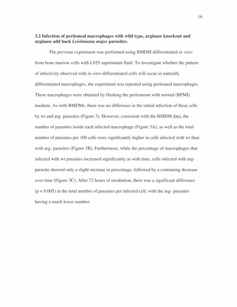
3.2 Infection of peritoneal macrophages with wild type, arginase knockout and arginase add back Leishmania major parasites. The previous experiment was performed using BMDM differentiated in vitro
from bone marrow cells with L929 supernatant fluid. To investigate whether the pattern
of infectivity observed with in vitro differentiated cells will occur in naturally
differentiated macrophages, the experiment was repeated using peritoneal macrophages.
These macrophages were obtained by flushing the peritoneum with normal (RPMI)
medium. As with BMDMs, there was no difference in the initial infection of these cells
by wt and arg- parasites (Figure 3). However, consistent with the BMDM data, the
number of parasites inside each infected macrophage (Figure 3A), as well as the total
number of parasites per 100 cells were significantly higher in cells infected with wt than
with arg- parasites (Figure 3B). Furthermore, while the percentage of macrophages that
infected with wt parasites increased significantly as with time, cells infected with arg-
parasite showed only a slight increase in percentage, followed by a continuing decrease
over time (Figure 3C). After 72 hours of incubation, there was a significant difference
(p < 0.005) in the total number of parasites per infected cell, with the arg- parasites
having a much lower number.

17
A) B) C) Figure 3: Infectivity and growth (proliferation) pattern of WT and arginase KO Leishmania major parasites in non-elicited peritoneal (primary) macrophages from BALB/c mice. Peritoneal macrophages obtained by flushing the peritoneum of mice with medium were infected with WT and KO parasites (see materials and methods) and the proliferation of parasites was determined at indicated times by staining cytospin preparations with Giemsa stain. A) Average number of parasites per infected cells. B) The total amount of parasites present per 100 (non-infected and infected). C) Total percentage of infected cells at the indicated times.

18
3.3 In vivo studies with wild type and arginase knockout parasites
BALB/c mice were infected with 5 million stationary phase promastigotes of wt,
arg- or add back, and the lesion sizes were monitored weekly. As shown in Figure 4A,
mice infected with wt parasites developed progressive lesions that began to ulcerate
around 8 weeks and hence mice were euthanized. This development of progressive lesion
was associated with massive parasite proliferation in the infected footpad (Figure 4B). In
contrast, mice infected with arg- parasites showed delayed lesion development that was
non-progressive (Figure 4A). These mice also had significantly lower parasites in their
footpad (Figure 4B). The lesion progression and parasite numbers in mice infected with
add back parasites were similar to those obtained with wt parasites. These results indicate
that deletion of arginase gene in L. major is associated with reduced pathology in vivo in
mice.
3.4 In vivo immune response of Balb/c mice infected with wild type and arginase knockout parasites. To investigate the influence of parasite-derived arginase on the host early immune
response, infected mice were sacrificed at different times after infection. The draining
popliteal lymph nodes were collected, made into single cell suspension and cultured in
vitro in the presence or absence of SLA (antigen). After 72 hr, the cells were pulsed with
3H-thymidine to measure proliferation. Infection with L. major caused a marked increase
in the number of cells in the draining popliteal lymph node three days after infection

19
1 2 3 4 5 6 7 80
1
2
3
4
5
6
7
WTArgABArgKO
WT Add back KO5
6
7
8
9
10
A)
Figure 4: Kinetics of footpad lesion progression (A) and parasite burden (B) in BALB/c mice infected with WT, KO and AB L. major. BALB/c mice (6 mice per group) were infected in the right hind feet with 5 million stationary phase promastigotes of each parasite and footpad swelling was measured weekly with calipers. Eight weeks after infection, mice were sacrificed and parasite burden in the infected feet was determined by limiting dilution.
B)
Weeks post-infection

20
Figure 5: Antigen-specific proliferation of cells from the draining lymph node (dLN) of mice infected with wild type (WT) and knockout (KO) parasites. BALB/c mice were infected with 5 million stationary phase promastigotes from either type of parasite and sacrificed 3 days later. The dLN cells were stimulated with freeze-thawed Leishmania antigen (Ag) for 4 days then pulsed with 3[H] thymidine. Proliferation was measured by measuring the number of cells using a scintillation counter.

21
(Figure 5). However, there was no significant difference in cell numbers in lymph nodes
(LNs) from mice infected with wt and arg- parasites, suggesting that the absence of
parasite-derived arginase does not influence the early inflammatory response against L.
major. Similarly, there was no difference in antigen-specific proliferative response of
cells taken from mice infected with wt and arg- parasites.
3.5 Cytokine (IL-10, IL-4 and IFN-γγγγ) responses of mice infected with wt and arg- L. major parasites. The level of arginase activity has been shown to influence the nature of CD4+ T
cell response in vivo. We speculated that parasite-encoded arginase could raise the overall
level of arginase in infected cells and hence influence the cytokine response. Therefore
the levels of cytokines were measured in the supernatant fluids of cells from mice
infected with wt or arg- parasites. As shown in Figure 6, there was a strong induction of
antigen-specific IL-4, IL-10 and IFN-g responses following infection with L. major,
compared with uninfected, or naïve, controls. Surprisingly, there was no significant
difference in the production of these cytokines by cells from mice infected with wt and
arg- parasites at all points analyzed. Similar results were obtained following polyclonal
stimulation of the cells with anti-CD3/anti-CD28. Paradoxically, this trend was
maintained even at 4 weeks despite the fact that mice infected with arg- parasites had
several fold lower number of parasites than those infected with wt or add back parasites
(Figure 7). These results suggest that parasite-derived arginase has no significant
influence on the host cellular immune response. However, they suggest that reduction in
parasite numbers observed in arg- may be related to alternative mechanism(s) that

22
A) IFN� detection in cells in medium B) IFN� detection in cells stimulated with SLA
C) IL-4 detection in cells in medium D) IL-4 detection in cells stimulated with SLA
0
5
10
15
20
25
30
35
40
3 7
Days infection
IL-4
(pg/
ml) WT
KO
E) IL-10 detection in cells in medium F) IL-10 detection in cells stimulated with SLA
Figure 6: In vivo early cytokine responses in mice infected with WT and arginase KO parasites. Cytokine detection in Balb/c infected mice with wild type and arginase knock out parasites using ELISA detection method. A) Cells were not stimulated with anything, but left in their medium and analyzed after three and seven days of infection for the presence of IFN�. B) Cells were stimulated with SLA and analyzed after three and seven days of infection for the presence of IFN�. C) Cells were not stimulated with anything, but left in their medium and analyzed after three and seven days of infection for the presence of IL-4. D) Cells were stimulated with SLA and analyzed after three and seven days of infection for the presence of IL-4. E) Cells were not stimulated with anything, but left in their medium and analyzed after three and seven days of infection for the presence of IL-10. F) Cells were stimulated with SLA and analyzed after three and seven days of infection for the presence of IL-10.

23
A) B)
C) D)
Figure 7: Analysis of cytokine production and parasite load in mice infected with either the wild type, arginase knock out or arginase add back parasites, after 4 weeks of infection. Cells from dLNs were cultured for 3 days in the presence of 50 µg/ml soluble Leishmania antigen (SLA). The supernatant fluids were then harvested and assayed for IFN-γ (A), IL-10 (B) and IL-4 (C) by ELISA. At the time of sacrifice, parasite burden in the infected feet was also determined by limiting dilution (D).
IL-10
WT KO Add Back0.0
2.5
5.0
7.5
10.0
12.5IFN-g
WT KO Add Back0
1000
2000
3000
4000
5000
6000
WT KO Add back4
6
8
10IL-4
WT KO Add Back0
1000
2000
3000
4000
Week 4.5

24
regulate parasite proliferation that is independent of the classical Th1/Th2 cytokine
regulation of immunity to L. major.
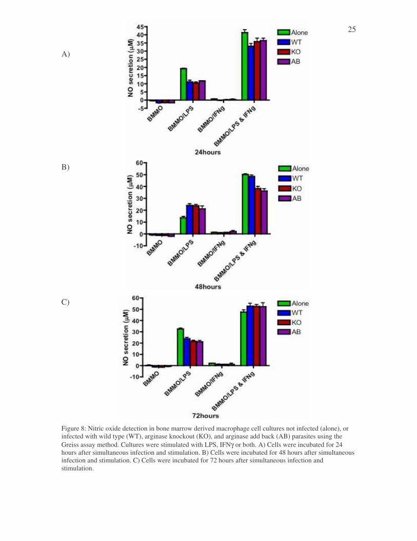
3.6 Nitric oxide production in the supernatants of tissue cultures infected with the wild type, knockout, or add back parasites, then stimulated with either LPS, IFNγγγγ, or both. Parasite-derived arginase may also be used to influence the competitive outcome
between arginase and iNOS. Additional arginase enzymes, and hence increased activities,
could deplete the available arginine in the cell, making the competition for arginine
greater for iNOS. Hence we speculated that in the absence of parasite-derived arginase,
the activities of iNOS may be enhanced. Bone marrow derived macrophages were
infected with wt or arg- L. major and stimulated with IFN-g, LPS or both (to enhance
iNOS activity) and the production of NO (nitrite) was measured by Griess assay. The cell
cultures that were not stimulated, or were only stimulated with IFNγ, did not have any
measurable nitric oxide production after 24, 48 or 72 hours of infection (Figure 8A, 8B,
8C, respectively). In contrast, cells stimulated with LPS or LPS + IFN-g produced
significant amounts of NO. There seem to be a trend that infection with L. major
decreases the amount of NO produced by macrophages. However, there were no
differences in NO production between cells infected with wt and arg- parasites at any
point tested.
25
A)
B)
C) Figure 8: Nitric oxide detection in bone marrow derived macrophage cell cultures not infected (alone), or infected with wild type (WT), arginase knockout (KO), and arginase add back (AB) parasites using the Greiss assay method. Cultures were stimulated with LPS, IFNγ or both. A) Cells were incubated for 24 hours after simultaneous infection and stimulation. B) Cells were incubated for 48 hours after simultaneous infection and stimulation. C) Cells were incubated for 72 hours after simultaneous infection and stimulation.

26
4 Discussion
Killing of L. major parasites inside infected cells is mediated by nitric oxide,
enhanced by IFN-g produced by activated CD4+ Th1 cells. The production of nitric
oxide in macrophages is regulated by several factors including the availability of the
substrate, arginine, and competition by arginase, another enzyme whose substrate is
arginine. This study focuses on determining the effect of parasite-derived arginase on the
host immune response leading to control of parasite proliferation. The effects of arginase
enzyme on Leishmania major growth have been addressed by a number of previous
studies. The synthesis of polyamines from ornithine following the degradation of
arginine by arginase is essential for Leishmania proliferation (Iniesta et al. 2001). Thus,
the availability of arginase in the host cell is crucial for parasite viability. Leishmania
organisms have a gene that encodes arginase. Interestingly, parasite-derived arginase has
been shown to metabolize host arginine to provide polyamine precursors (Roberts et al.
2004). Alternatively, parasite-derived arginase may also be used to suppress Th1
response and favor the production of Th2 cytokines, thereby allowing the parasite to
evade the host’s immune response (Iniesta et al. 2001). Here, it was shown that parasite-
encoded arginase is vital for optimum parasite survival inside infected host cells.
Interestingly, infected host cells contained detectable numbers of parasites at all times
investigated suggesting that there may be a parasite encoded independent pathway to
salvage polyamines. Similar results were obtained, when mice were infected with arg-
parasites, where there was a persistence of parasites at infected sites with minimal
pathology. The mechanisms by which arg- parasites salvage polyamines were not tested
in this study.

27
The expression level of arginase in host cells is regulated by the balance between
Th1 and Th2 cytokines produced by activated CD4+ T helper cells. It was found that in
spite of lacking arginase activity, arg- induced early inflammatory and cytokine responses
comparable to wt parasites. Furthermore, there was no difference in the induction of key
CD4+ T cell cytokines ((IL-4, IL-10 and IFN-g) following infection with wt and arg-
parasites. Because it was previously shown that arginase levels could also influence Th1
and Th2 response, it was speculated that parasite-derived arginase could raise the overall
cellular concentration and activity of this enzyme and therefore regulate the cytokine
responses in vivo. However, it was shown that this does not seem to be the case.
Furthermore, we found no significant differences in NO production by macrophages
infected with wt or arg- parasites. These results refute the hypothesis that additional
arginase enzyme shifts the immune response in the favor of the arginase pathway.
Although the proliferation of arg- parasites in macrophages was severely
impaired, these parasites were not completely destroyed. The levels of the parasites inside
infected cells were found to plateau with time. These results would suggest that lack of
proliferation is possibly related to diminished polyamine synthesis, rather than enhanced
NO-mediated killing. This would be the main reason for the observed reduction in
proliferation of arg- compared to wt parasites. The implication of the lingering parasites
in macrophages in vitro and in infected mice in vivo supports the view that the additional
arginase activity (parasite-derived) does not enhance parasite infection by helping to
evade the host’s immune system, but rather does so by allowing the required optimal
amounts of nutrients for optimum parasite proliferation to be produced. It will therefore
be highly interesting to investigate the growth and disease pathology of arg- parasites in

28
iNOS deficient mice, which are incapable of metabolizing arginine via the iNOS
pathway.

29
5 Conclusion
1. The arginase knock out parasites were found to be unable to proliferate inside the
infected macrophage, indicating that the parasite-derived arginase has a role in
parasitic survival.
2. There was no significant difference in NO production between macrophage cells
infected with either the wild type, knock out, or add back parasites, indicating that
the immune pathway of the cell was not altered by the presence of the parasite-
derived arginase.
3. Though the knock out parasites were not able to proliferate inside the infected
macrophage, they were also not completely destroyed, further indicating that the
parasite derived arginase does not alter the immune response of the cell, but is
solely present to provide nutrients for the parasite.

30
References
Bansal, V. and J.B. Ochoa. 2003. Arginine availability, arginase and the immune response. Curr Opin Clin Nutr Metab Care. 6: 223 – 228. Corraliza, I. M., Soler, G., Eichmann, K. and M. Modolell. 1994. Arginase induction by suppressors of nitric oxide synthesis (IL-4, IL-10 and PGE2) in murine bone-marrow-derived macrophages. Biochem. Biophys. Res. Commun. 206: 667 – 673. Holzmuller, P., Sereno, D., Cavaleyra, M., Mangot, I., Daulouede, S., Vincendeau, P., and J-L Lemesre. 2002. Nitric oxide-mediated proteasome-dependent oligonucleosomal DNA fragmentation in Leishmania amazonensis amastigotes. Infect. Immun. 7: 3727 – 3735. Iniesta, V., Gomez-Nieto, L. C. and I. Corraliza. 2001. The inhibition of arginase by Nw-hydroxy-L-arginine controls the growth of Leishmania inside macrophages. J. Exp. Med. 193: 777 – 784. Iniesta, V., Gomez-Nieto, L. C., Molano, I., Mohedano, A., Carcelen, J., Miron, C., Alonso, C., and I. Corraliza. 2002. Arginase I induction in macrophages, triggered by Th2-type cytokines, supports the growth of intracellular Leishmania parasites. Parasite Immunol. 24: 113 – 118. Kropf, P., Fuentes, J. M., Fahnrich, E., Arpa, L., Herath, S., Weber, V., Soler, G., Celada, A., Moddell, M., and I. Muller. 2005. Arginase and polyamine synthesis are key factors in the regulation of experimental leishmaniasis in vivo. FASEB J. 19: 1000 – 1002. Liew, F. Y., Qing-Xiao, W., and L. Proudfoot. 1997. Cytokines and nitric oxide as effector molecules against parasitic infections. Phil. Trans. R. Soc. Lond. 352: 1311 – 1315. Miles, S.A., Conrad, S.M., Alves, R.G., Jeronimo, S.M. and D. M. Mosser. 2005. A role for IgG immune complexes during infection with the intracellular pathogen Leishmania. J. Exp. Med. 201(5): 747 – 754. Modolell, M., Corraliza, I. M., Link, F., Soler, G., and K. Eichmann. 1995. Reciprocal regulation of the nitric oxide synthase/arginase balance in mouse bone marrow-derived macrophages by Th1 and Th2 cytokines. Eur. J. Immunol. 25: 1101 – 1104. Munder, M., Eichmann, K., Moran, J. M., Centeno, F., Soler, G. and M. Modolell. 1999. Th1/Th2-regulated expression of arginase isoforms in murine macrophages and dendritic cells. J. Immunol. 163: 3771 – 3777.

31
Munder, M., Eichmann, K. and M. Modolell. 1998. Alternative metabolic states in murine macrophages reflected by the nitric oxide synthase/arginase balance: competitive regulation by CD4+ T cells correlates with Th1/Th2 phenotype. J. Immunol. 160: 5347 – 5354. Padigel, U. M., Alexander, J. and J. P. Farrell. 2003. The role of interleukin-10 in susceptibility of BALB/c mice to infection with Leishmania mexicana and Leishmania amazonensis. J. Immunol. 171: 3705 – 3710. Reiner, S. L., and R. M. Locksley. 1995. The regulation of immunity to Leishmania major. Annu. Rev. Immunol. 13: 151 – 177. Roberts, S. C., Tancer, M. J., Polinsky, M. R., Gibson, K. M., Heby, O. and B. Ullman. 2004. Arginase plays a pivotal role in polyamine precursor metabolism in Leishmania. J. Biol. Chem. 279: 23668 – 23678. Stenger, S., Donhauser, N., Thuring, H., Rollinghoff, M., and C. Bogdan. 1996. Reactivation of latent leishmaniasis by inhibition of inducible nitric oxide synthase. J. Exp. Med. 183: 1501 – 1514. Stenger, S., Thuring, H., Rollinghoff, M., and C., Bogdan. 1994. Tissue expression of inducible nitric oxide synthase is closely associated with resistance to Leishmania major. J. Exp. Med. 180: 783 – 793. Stout, R. D, 1993. Macrophage activation by T cells: cognate and non-cognate signals. Curr. Opin. Immunol. 5: 398 – 403. Tapiero, H., Mathe, G., Couvreur, P., and K. D. Tew. 2002. Arginine. Biomed. Pharmacother. 56: 439 – 445.