the role of ivus in contemporary endovascular treatment
TRANSCRIPT

The role of IVUS in contemporary endovascular treatment
Osamu Iida, MD, FACC
Kansai Rosai Hospital, Cardiovascular Center
Amagasaki, Hyogo, Japan

DisclosureSpeaker name:
.................................................................................
I have the following potential conflicts of interest to report:
Consulting
Employment in industry
Stockholder of a healthcare company
Owner of a healthcare company
Other(s)
I do not have any potential conflict of interest
Osamu Iida, MD
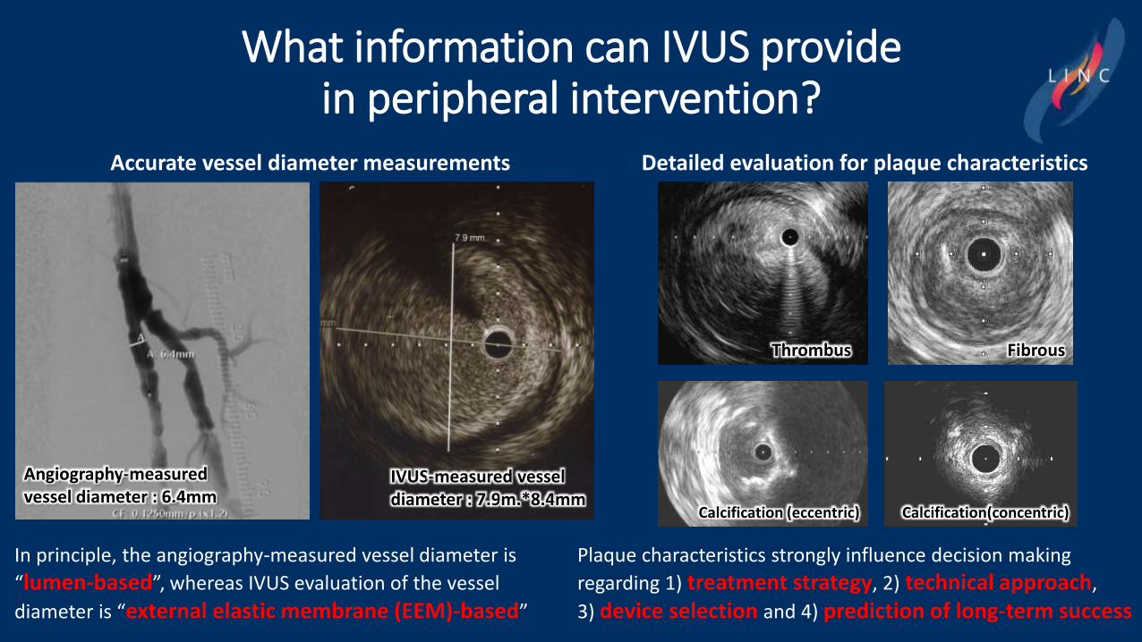
What information can IVUS provide in peripheral intervention?
Accurate vessel diameter measurements
In principle, the angiography-measured vessel diameter is
“lumen-based”, whereas IVUS evaluation of the vessel
diameter is “external elastic membrane (EEM)-based”
Angiography-measured vessel diameter : 6.4mm
IVUS-measured vessel diameter : 7.9m.*8.4mm
Detailed evaluation for plaque characteristics
Plaque characteristics strongly influence decision making
regarding 1) treatment strategy, 2) technical approach,
3) device selection and 4) prediction of long-term success
Thrombus
Calcification (eccentric) Calcification(concentric)
Fibrous
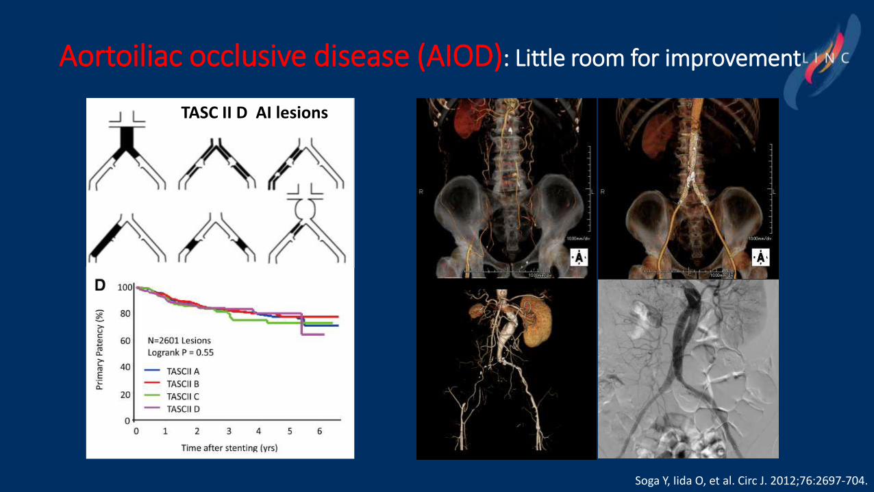
Aortoiliac occlusive disease (AIOD): Little room for improvement
TASC II D AI lesions
Soga Y, Iida O, et al. Circ J. 2012;76:2697-704.

IVUS Imaging During Aortoiliac Stenting: No Impact on Outcomes at 1 Year
Tsujimura T, Takahara M, Iida O, et al. JEVT 2020;1526602820949872
Propensity score matching analysis revealed that duration and fluoroscopy time during IVUS-supported procedures were SIGNIFICANTLY LONGER than in cases without IVUS use, whereas the 12-month restenosis rate was NOT significantly different between the groups.
IVUS No IVUS P value
Procedure time ≤1 hour 46.9% (42.0% to 51.9%)
71.6% (63.3% to 78.7%)
P<0.001
12-month restenosis 10.2% (6.9% to 14.9%)
10.3% (5.4% to 18.6%)
0.99
×
Study design: Pre-specified analysis for prospective registry
Study subjects: 545 patients underwent IVUS-supported stent implantation versus 258 patients treated without IVUS
Outcomes: Technical and long-term success

Below-the-knee disease (BTKD): IVUS could make something
IVUSQVA

IVUS-guided “EEM approach” for BTK angioplasty
QVA
ATA prox2.01mm
ATA distal1.65mm
Pre
Soga Y et al. Catheter Cardiovasc Interv. 2020 Oct 21. doi: 10.1002/ccd.29347. Online ahead of print.
The IVUS-guided group was treated with a larger balloon size for all types of below-the-knee vessel (p < .001), although lesion characteristics, including the QVA-measured vessel diameter, were similar between the two groups.
PTA 2.5x150mm PTA 4.0x150mmAngio-guide IVUS-guide

IVUS-guided “EEM approach” for BTK angioplasty
Soga Y et al. Catheter Cardiovasc Interv. 2020 Oct 21. doi: 10.1002/ccd.29347. Online ahead of print.
•.•.
Wound healing was significantly earlier and the time to wound healing was significantly shorter in the IVUS-guided group. Total number of EVT to achieve complete wound healing for the index limb was also significantly lower in the IVUS-guided group.

18
9
30
43
13
31
0
10
20
30
40
50
DEBATE-BTK IN.PACT DEEP BIOLUX-PII
TLR
@ 1
2 m
on
ths
DCB PTA
Herten M et al. Vasc Health Risk Manag. 2016;12:341-56.
P≤0.01
NS
NS
BTK RCT DEBATE-BTK IN.PACT DEEP BIOLUX-PII IN.PACT BTK 2020DCB type used IN.PACT™ Amphirion IN.PACT™ Amphirion Passeo-18 Lux IN.PACT 014 PTX-coated balloon
DCB PTA DCB PTA DCB PTA DCB PTANumber of patients (n) 65 67 239 119 36 36 23 27Diabetics (%) 100 100 76 69 61 72 74 96CLI (%) 100 100 100 99 78 78 100 100Lesion length (mm) 129±83 131±79 101±91 129±95 113±88 115±87 215±83 218±80De novo lesion (%) 100 100 93 96Total occlusion (%) 77 82 39 46Calcified lesion (%) 75 78 36 11Severe calcification (%) 14 11 42 8
RVD (mm) 2.91±0.27 2.87±0.29 2.46±0.69 2.41±0.56 2.28±0.54, 2.19±0.57 2.80±0.54 2.71±0.39
RVDs are different among studies, leading that selection of DCB size also differ from each DCB trial. Use of larger DCB size has more advantage to reduce restenosis rate.
Larger DCB size selection would lead better outcome?
0,59
1,26
0,0
0,5
1,0
1,5
2,0
IN.PACT BTK 2020
LLL
@ 9
mo
nth
s P=0.017

0%
20%
40%
60%
80%
100%
0 50 100 150 200 250 300
Prim
ary
Pate
ncy
Lesion Length (mm)
Ohki T, et al,JEVT 2014
Ohki T, et al,JEVT 2014
Gouëffic Y, et al,Trials 2014
Dake M, et al,JEVT 2011
Mori S, et al,JEVT 2015
Zeller T, et al,JACC 2013
Fujjihara M, et al,HV2014
Zeller T, et al,CVS 2013
Listky J, et al,Dove press 2014
Iida O, et al,JACC 2014
Yokoi H, et al,JACC 2016
Leopardi M, et al,JACC(Torino) 2013
Dake M, et al,Circ interv 2011
Femoropopliteal disease (FPD): IVUS plays an important roleDES era: Primary patency after Zilver PTX implantation for SFA

Efficacy of IVUS in FP stenting for PAD with TASC II class A to C lesion
Iida O, Soga Y, et al. J Endovasc Ther. 2014;21:485-492.
IVUS use (+)
IVUS use (-)
0
20
40
60
80
100
12m 24m
Pri
mar
y P
ate
ncy
(%)
OVERALL
IVUS User
Non IVUS User
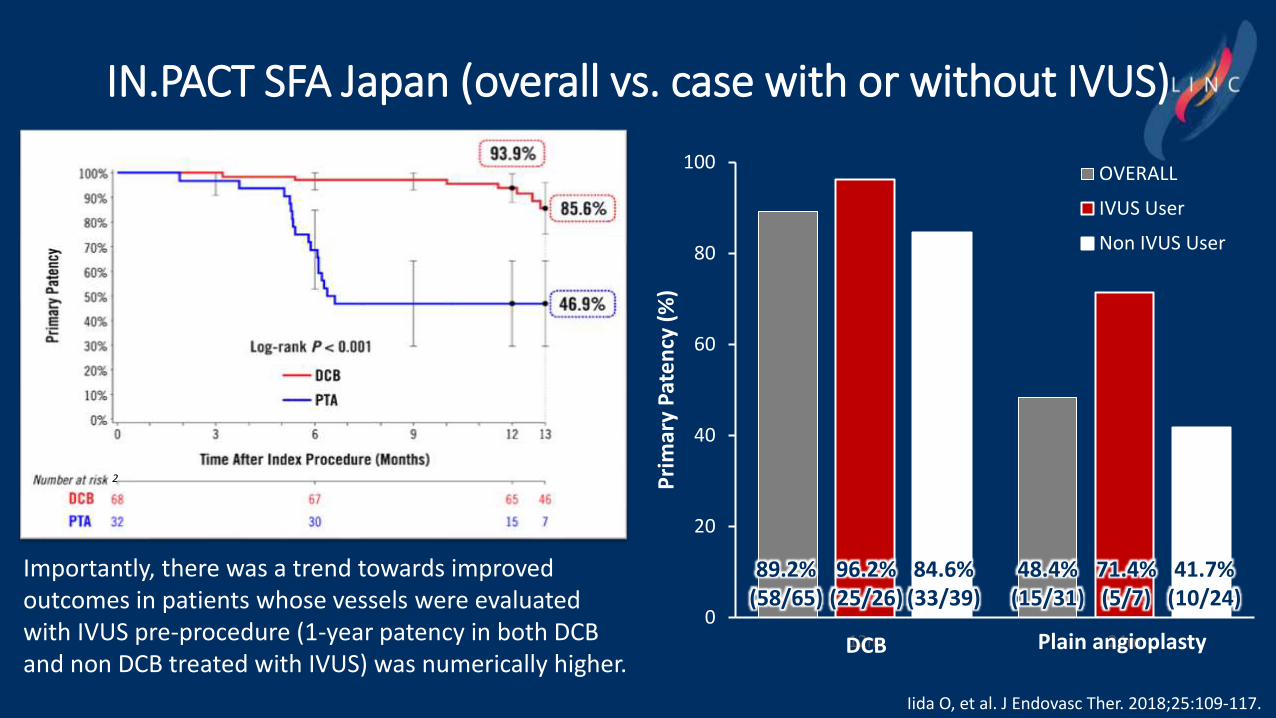
IN.PACT SFA Japan (overall vs. case with or without IVUS)
2
89.2%(58/65)
96.2%(25/26)
84.6%(33/39)
48.4%(15/31)
71.4%(5/7)
41.7%(10/24)
DCB Plain angioplasty
Importantly, there was a trend towards improved outcomes in patients whose vessels were evaluated with IVUS pre-procedure (1-year patency in both DCB and non DCB treated with IVUS) was numerically higher.
Iida O, et al. J Endovasc Ther. 2018;25:109-117.

Apparent difference between the vessel diameter (VD) assessed by angiography and that by IVUS in FP-EVT
Association of angiography-assessed VD with the difference between angiography- and IVUS-assessed VD (n=1725)
IVUS-assessed VD was significantly larger than angiography-assessed RVD (6.0 ± 1.0 mm versus 5.0 ± 1.0 mm; P<0.001). The difference of IVUS- versus angiography-assessed VD was more marked in cases with a smaller angiography-assessed VD.
Work in progress

Association of anatomical factors with IVUS- minus angiography-assessed RVD of 1 mm or larger
A smaller angiography-assessed RVD, a lesion without chronic total occlusion, angiography-assessed bilateral calcification, and history of stent implantation were significantly associated with ΔRVD ≥1 mm.
Work in progress
Unadjusted odds ratio Adjusted odds ratio
Popliteal lesion 1.61 [1.31 to 1.96] (P<0.001) 1.20 [0.95 to 1.51] (P=0.13)
Angiography-assessed RVD (per 1 mm) 0.38 [0.34 to 0.43] (P<0.001) 0.38 [0.34 to 0.43] (P<0.001)
Chronic total occlusion 0.78 [0.65 to 0.94] (P=0.008) 0.63 [0.50 to 0.80] (P<0.001)
Lesion length (per 10 cm) 1.06 [0.97 to 1.16] (P=0.19) 1.01 [0.90 to 1.14] (P=0.81)
Angiography-assessed calcification (versus none) 1.00 (Ref) 1.00 (Ref)
Unilateral calcification 1.20 [0.97 to 1.50] (P=0.099) 1.18 [0.93 to 1.51] (P=0.18)
Bilateral calcification 1.34 [1.08 to 1.67] (P=0.008) 1.36 [1.06 to 1.74] (P=0.014)
History of revascularization (versus never) 1.00 (Ref) 1.00 (Ref)
History of plain angioplasty 0.90 [0.50 to 1.62] (P=0.73) 0.66 [0.34 to 1.27] (P=0.21)
History of stent implantation 1.86 [1.39 to 2.51] (P<0.001) 1.72 [1.23 to 2.41] (P=0.001)
the association of anatomical factors with ΔRVD ≥1 mm (n=1725)

Take home message
✓ During aorto-iliac stenting, IVUS had No Impact on Outcomes at 1 Year.
✓ In FP and BTK treatment, IVUS guided EVT would improve clinical outcomes.
✓ IVUS-assessed VD was significantly larger than angiography-assessed RVD (6.0 ± 1.0 mm versus 5.0 ± 1.0 mm; P<0.001).
✓ The difference of IVUS- versus angiography-assessed VD was more marked in cases with a smaller angiography-assessed VD.
✓ A smaller angiography-assessed RVD, a lesion without chronic total occlusion, angiography-assessed bilateral calcification, and history of stent implantation were significantly associated with ΔRVD ≥1 mm.

The role of IVUS in contemporary endovascular treatment
Osamu Iida, MD, FACC
Kansai Rosai Hospital, Cardiovascular Center
Amagasaki, Hyogo, Japan