the pharmacology of ginexin – f (ginkgo biloba) and its clinical uses by professor kamal eldin...
Post on 20-Dec-2015
218 views
TRANSCRIPT
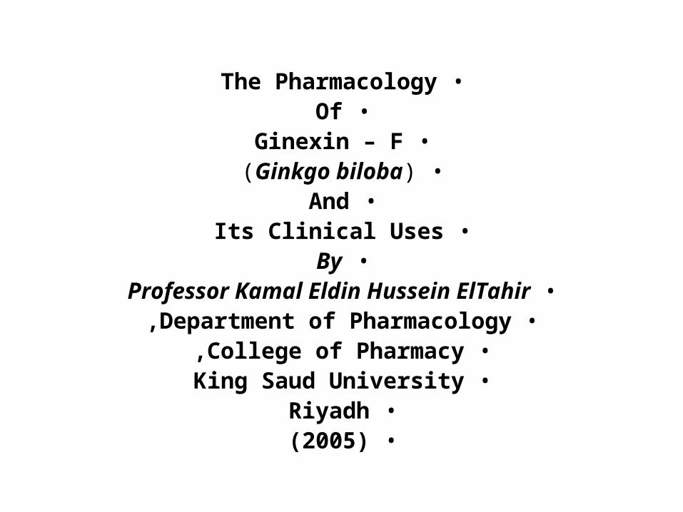
•The Pharmacology•Of
•Ginexin – F•(Ginkgo biloba)
•And•Its Clinical Uses
•By•Professor Kamal Eldin Hussein ElTahir
•Department of Pharmacology,•College of Pharmacy,•King Saud University
•Riyadh•(2005)

1
IntroductionGinkgo biloba )Family:
Common Name: Maidenhair tree.Height : 30 – 40 meters.Circumference : 4 meters.The Leaves : Fan – shaped
Green to Golden YellowIndigenous to : Korea, China & Japan.Known in China for: Over 5000 Years.Old Chinese uses : Circulation disorders
Memory Disturbance Asthma, Bronchitis
Introduction in Europe: 1730 in America : 1784
Start of Interest in it : 1965 In Germany

2
•Part of the Plant used: Leaves•Constituents:
• a( Flavonoidal Glycosides•( Mono –, bio – and tri – osides of):
• Quercetin, Kaempferol, Isorhamnetin and 3´-O methyl Myristicin .
• b( Bioflavonoids:• Bilobetin, Amentoflavone, Ginkgetin, 5-Methyl
Bilobtin and Isoginkgetin .• c( Tri Lactonic diterpenes:• Ginkgolides A, B and C
• d( Tri Lactonic Sesquiterpene:• Bilabolide
• e( Anthocyanin

3•Ginkgo Extraction: Acetone: Water and Spray Drying.
•Standardization of Extraction:•Standardized to Contain:
• 24% Flavonoid glycosides• 6% Lactonic di- and sesqui-Terpenes
• Lessthan 0.6 mg Ginkgolic Acid per 100gm• 1Kg Extract = 70 Kg dried Leaves
Pharmaceutical Forms: Capsules & Tablets, 40; 60; and 120 mg .Trade Names: Ginexin – F; EGB 761;
• Ginkgold; Ginkoba; GBE 24; Tebonin•Status Classification:
• In USA: Dietry Supplement• In Canada: Food Additive
• In France and Germany:As over–the Counter Drugs •( No Prescription )
•Availability : In more than 50 Countries
4
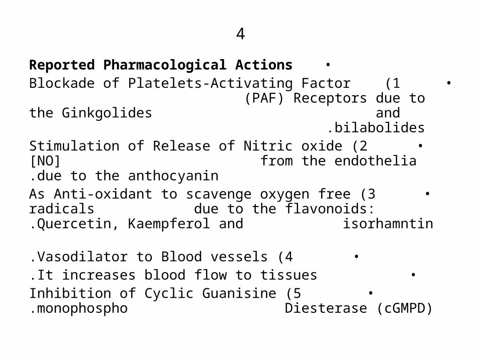
•Reported Pharmacological Actions• 1 )Blockade of Platelets-Activating Factor
)PAF( Receptors due to the Ginkgolides and bilabolides .
• 2 )Stimulation of Release of Nitric oxide [NO] from the endothelia due to the anthocyanin.
• 3 )As Anti-oxidant to scavenge oxygen free radicals due to the flavonoids: Quercetin, Kaempferol and
isorhamntin .• 4 )Vasodilator to Blood vessels.
• It increases blood flow to tissues.• 5 )Inhibition of Cyclic Guanisine monophospho
Diesterase )cGMPD( .
5
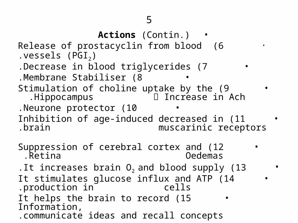
•Actions )Contin.(• 6 )Release of prostacyclin from blood vessels )PGI2(.
• 7 )Decrease in blood triglycerides.• 8 )Membrane Stabiliser.
• 9 )Stimulation of choline uptake by the Hippocampus ༜ Increase in Ach .
• 10 )Neurone protector.• 11 )Inhibition of age-induced decreased in
brain muscarinic receptors .• 12 )Suppression of cerebral cortex and Retina
Oedemas .• 13 )It increases brain O2 and blood supply.
• 14 )It stimulates glucose influx and ATP production in cells.
• 15 )It helps the brain to record Information, communicate ideas and recall concepts.
6
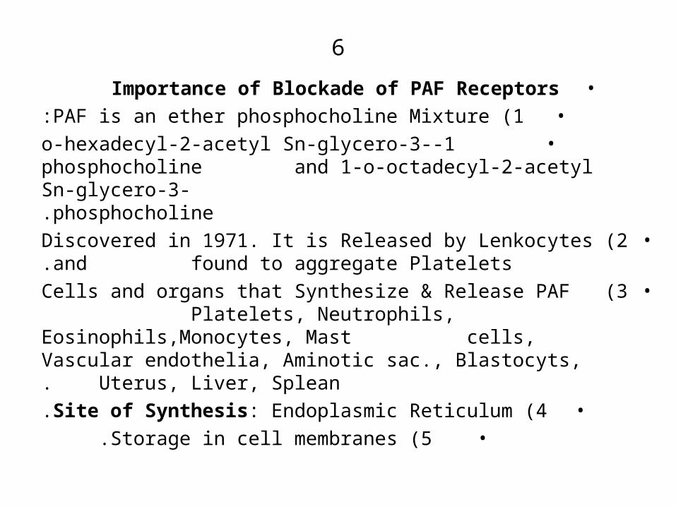
•Importance of Blockade of PAF Receptors•1 )PAF is an ether phosphocholine Mixture:
• 1-o-hexadecyl-2-acetyl Sn-glycero-3-phosphocholine and 1-o-octadecyl-2-acetyl Sn-glycero-3- phosphocholine.
•2 )Discovered in 1971. It is Released by Lenkocytes and found to aggregate Platelets.
•3 )Cells and organs that Synthesize & Release PAF Platelets, Neutrophils, Eosinophils,Monocytes, Mast cells, Vascular endothelia, Aminotic sac., Blastocyts, Uterus, Liver, Splean.
•4 )Site of Synthesis: Endoplasmic Reticulum.•5 )Storage in cell membranes .
7
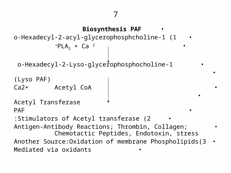
•Biosynthesis PAF•1 )1-o-Hexadecyl-2-acyl-glycerophosphcholine
• PLA2 + Ca 2+
• 1-o-Hexadecyl-2-Lyso-glycerophosphocholine •( Lyso PAF)
• Ca2+ Acetyl CoA• Acetyl Transferase
• PAF•2 )Stimulators of Acetyl transferase:
• Antigen-Antibody Reactions; Thrombin, Collagen; Chemotactic Peptides, Endotoxin, stress
•3)Another Source:Oxidation of membrane Phospholipids• Mediated via oxidants

8
•Known Pharmacological Actions of PAF•1 )Aggregation of both plateletes and PMNL)Neutrophil,
Eosinophils, Monocytes(.
•2 )Release of TXA2 from platelets.
•3 )Release of PMNL contents. i.e. Degranulation e.g Release of Lysosomal enzymes + Eicosanoids )PGs + LTs( + oxygen free radicals.
•4 )Vasodilation leading to decrease in arteial blood pressure .
•5 )But decreases Coronary blood flow due to its induced aggregation and release of TXA2 .
•6 )Decrease in Pulmonary blood flow due to platelets aggregation and Release of TXA2.

9
•Action of PAF )Contin.(
•7 )Increase in Capillary permeability leading to Oedemas .•8 )Chemotaxis of Leukocytes.
•9 )Stimulation of Adherence of neutrophils to endothelial cells leading to Leukocytopenia .
•10 )Bronchoconstriction and induction of pulmonary oedemas.Its Bronchoconstriction is direct via release of intracellular Ca2+ via IP3. and due to Release of Histamine + Eicosanoids.
•11 )Induction of Hyper-responsiveness )Reactivity( of Pulmonary relaxant epithelia and the Non-Adrenergic Non-cholinergic autonomic innervation via its induced release of Basic and Cationic Proteins from
Eosinophils .
10
•Actions of PAF )Contin(
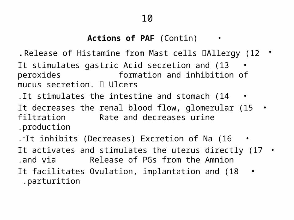
•12 )Release of Histamine from Mast cells ༜Allergy.•13 )It stimulates gastric Acid secretion and peroxides
formation and inhibition of mucus secretion. ༜ Ulcers
•14 )It stimulates the intestine and stomach.•15 )It decreases the renal blood flow, glomerular
filtration Rate and decreases urine production.•16 )It inhibits )Decreases) Excretion of Na+.
•17 )It activates and stimulates the uterus directly and via Release of PGs from the Amnion.
•18 )It facilitates Ovulation, implantation and parturition .

11
•PAF-induced Diseases & Disorders: It is involved in
•1 )Inflammation 2( Asthma•3 )Anginas 4( Atherosclerosis
•5 )Rejection of transplanted organs.•6 )Anuria 7( Gastric ulcers
•8 )Anaphylactic and Endotoxin shocks: )It decreases blood pressure, cardiac output, heart rate, plasma
volume, coronary blood flow and the haematocrit( •9 )Pulmonary Hypertension.
12
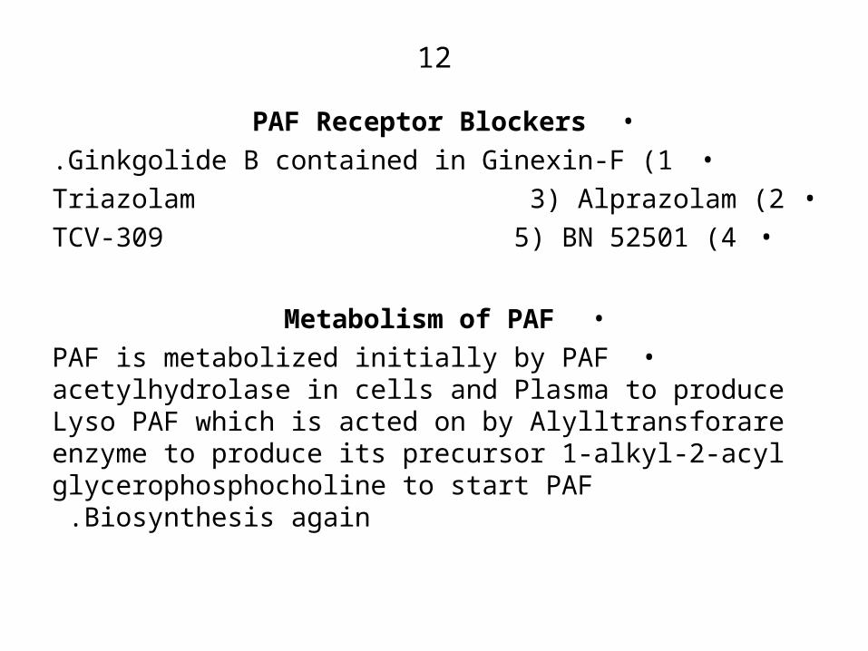
•PAF Receptor Blockers•1 )Ginkgolide B contained in Ginexin-F.•2 )Triazolam 3( Alprazolam
•4 )TCV-309 5( BN 52501
•Metabolism of PAF•PAF is metabolized initially by PAF acetylhydrolase in
cells and Plasma to produce Lyso PAF which is acted on by Alylltransforare enzyme to produce its precursor 1-alkyl-2-acyl glycerophosphocholine to start PAF
Biosynthesis again .
13
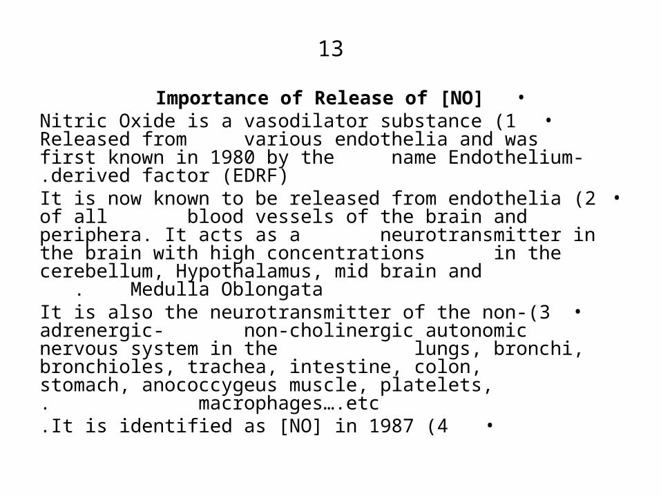
•Importance of Release of [NO]•1 )Nitric Oxide is a vasodilator substance Released from
various endothelia and was first known in 1980 by the name Endothelium-derived factor )EDRF(.
•2 )It is now known to be released from endothelia of all blood vessels of the brain and periphera. It acts as a neurotransmitter in the brain with high concentrations in the cerebellum, Hypothalamus, mid brain and
Medulla Oblongata .•3)It is also the neurotransmitter of the non-adrenergic-
non-cholinergic autonomic nervous system in the lungs, bronchi, bronchioles, trachea, intestine, colon, stomach, anococcygeus muscle, platelets, macrophages….etc.
•4 )It is identified as [NO] in 1987.
14
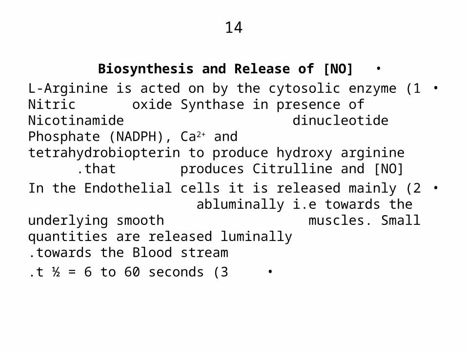
•Biosynthesis and Release of [NO]•1 )L-Arginine is acted on by the cytosolic enzyme Nitric
oxide Synthase in presence of Nicotinamide dinucleotide Phosphate )NADPH(, Ca2+ and tetrahydrobiopterin to produce hydroxy arginine that
produces Citrulline and [NO] .•2 )In the Endothelial cells it is released mainly
abluminally i.e towards the underlying smooth muscles. Small quantities are released luminally towards the Blood stream.
•3 )t ½ = 6 to 60 seconds.

15
•NO Synthases [NO]•1 )Types: a( Constitutive Enzyme always present in its
synthesizing organs. b( Inducible Enzyme: Induced by Bacterial
endotoxins )Lipopolysaccherides(, the Cytokines: TNF, IL-1, INF-⍺ in
Macrophages, Liver, Tumor cells .2 )Brain NO Synthase is Identical to Cytochrome
P450 Reductase .3 )The Inducible NOS is independent of Ca2+ but for
its activity it requires both NADPH & Tetrahydrobiopterin .
4 )The Inducible NOS is present both in the Cytoplasm and attached to cell membranes .

16
•NO Synthesis Inhibitors•1 )L-N-Nitromethyl arginine )LNMA(.
•2 )GN- Nitoarginine.•3 )L-GN-Nitroarginine Methyl ester )L-NAME( .
•4 )L- Canavanine )In neutrophils only(.•NO Synthesis Stimulators
•1)Ginexin-F; 2( Arginine 3( ACh 4( BK•5 )Histamine 6(Substance 7(Kainic Acid )Brain(
•8)N-Methyl D-aspartate )NMDA( in Brain.•9)IP3 )Inositol tri Phosphate( 10(Cromakalin 11(ADP •12)ATP 13(5-HT 14(VIP )Vaso Active intestinal
peptide(•15)Endothelin 16(Thrombin 17(Blood flow
18(UV Rays.

17
•Known NO Pharmacological Actions•1 )It mediates all inhibitory neurotransmission in:
Respiratory system, Gastrointestinal tract, ….etc.•2 )Potent Vasodilator in all blood vessels.
•3 )Potent Inhibitor of platelet aggregation induced by ADP,AA, Collagen …etc.
•4 )It inhibits adhesion of platelets to vascular endothelial cells.
•5 )Cytotoxicity Towards Tumor cells, bacteria, viruses …etc. )It is released by Macrophages during their attack to foreign bodies(.
•6 )It is a cytotoxic tool released by T-Killer lymphocytes )CD8+( .

18
•NO Actions )Contin.(
•7 )Decreases Pulmonary Vascular Resistance.•8 )Enhances Blood flow to the penis leading to erection.•9 )It is involved in pain transmission within the spinal
cord. It enhances transmission.•10)It is involved in inflammation via its vasodilation.
•11)It increases the bleeding time.

19
•NO Antagonists or Inhibitors•1 )Oxy haemoglobin
•2 )Methylene Blue.•3 )Mepacrine.
•4 )Nordihydroguiaretic Acid
•Mechanism of Action•NO Activates the enzyme Guanylyl Cyclase leading to
stimulation of production of Cyclic guanisine mono-phosphate. The latter activates protein Kinases that act in presence of ATP to phosphorylate cellular proteins that bind intracellular Ca2+ and inhibit influx of extracellular Ca2+ together with stimulation of K+ eflux resulting in hyperpolarization of the cells.
20
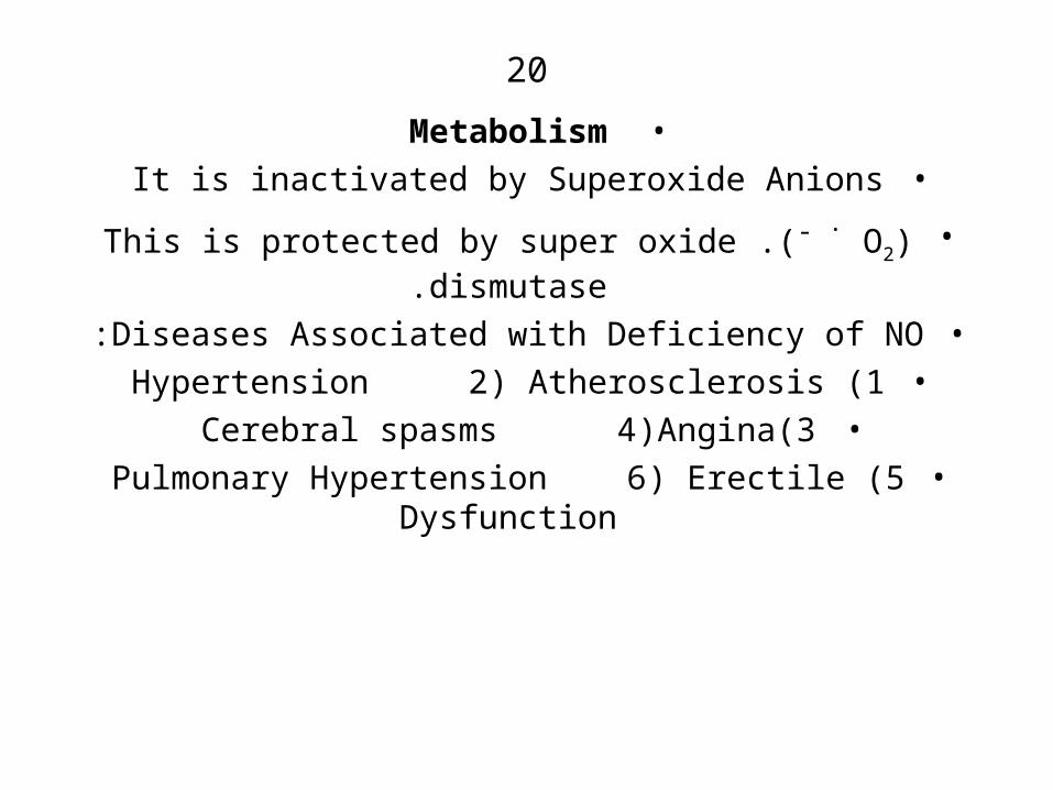
•Metabolism•It is inactivated by Superoxide Anions
•(O2 .- .)This is protected by super oxide dismutase.
•Diseases Associated with Deficiency of NO:•1 )Hypertension 2( Atherosclerosis
•3)Cerebral spasms 4(Angina•5 )Pulmonary Hypertension 6( Erectile Dysfunction

21
•Importance of Scavenging Free Radicals•1 )Definition: A free radical is any atom or molecule that
contains one or more unpaired )unshared( electron. They are very unstable and reactive .
•2 )Examples are the oxygen free Radicals: The superoxide anion [O2
. -];) - . OH(;)- . O-O-H( and )NO . –(
•3)The superoxide anion interacts with NO to give the more toxic peroxynitrite radical N-O-O . –
•4 )Sites of production:• a( In Mitochondria during ATP production.
• b( In the Endoplasmic Reticulum.

22
•Initiation of Production of free - . OH Radicals• a( Interaction of Fe3+ )iron( with superoxide anion :
• O2. - + Fe3+ Fe2+ + O2
• Then Fe2+ + H2O2 Fe3+ + OH + - . OH
• b( Interaction of superoxide anion with H2O2
• O2
. - + H2O2 O2 + OH + - . OH• c( Acceptance of a proton by H2O2
• H2O2 e + H+ O2 + OH + - . OH
• d( Interaction of O2. - or - . O-O-H with Cuprous
Cu2+ Or Ferrous Fe2 +
•

23
•Factors that Increase Free Radicals Production
•1 )Ischaemia•2 )Excessive metabolism of Arachidonic acid via COXes
Enzymes to produce Prostoglandins and via Lipoxygenases )5-Lipoxygenase and 12- Lipoxygenase( to produce Leukotrienes

24
•Actions of Free Radicals
•1 )They are the weapons of Macrophages during Phagocytosis and T-killer Lymphocytes in attacking transplanted foreign organs.
•2 )However, they attack unsaturated fatty acids, Proteins and DNA resulting in necrosis of various cells and
damage to the neurones .

25
•Actions of free Radicals )Contin.(•3 )Attack of Polyunsaturated fatty acids e.g. Linolenic or
Linoleic to produce peroxy acids )Fatty acid radicals( which are also damaging to enzymes, proteins, ion channel, receptors and even DNA. They inhibit
prostacyclin synthesis .• O2
. - + R – C = C = C → R – O – O .–
• Fatty Acid Radical•4)The fatty acid Radical then interacts with O2 to
produce a Peroxide Radical • R – O – O . - + O2 → Fatty Acid + – . O – O – H
•5 )The peroxyl Radical then attacks a new unsaturated fatty acid to produce a fatty acid radical & the cycle
goes on .
26
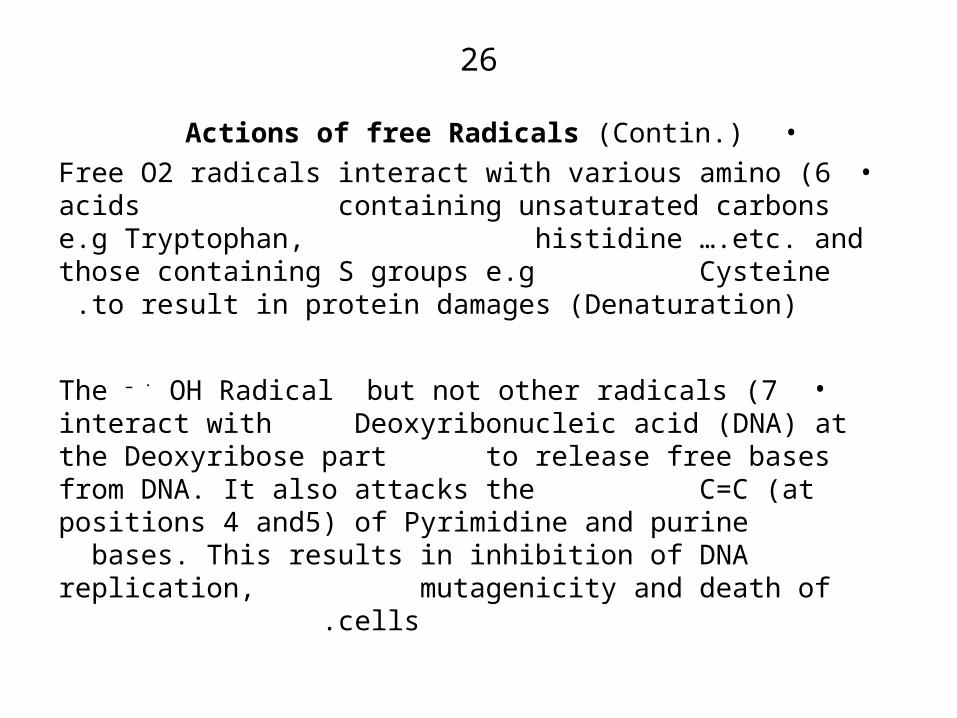
•Actions of free Radicals )Contin.(•6 )Free O2 radicals interact with various amino acids
containing unsaturated carbons e.g Tryptophan, histidine ….etc. and those containing S groups e.g
Cysteine to result in protein damages )Denaturation( .
•7 )The – . OH Radical but not other radicals interact with Deoxyribonucleic acid )DNA( at the Deoxyribose part to release free bases from DNA. It also attacks the C=C )at positions 4 and5( of Pyrimidine and purine bases. This results in inhibition of DNA replication,
mutagenicity and death of cells .

27•Anti – Oxidants
• 1 )These are substances which prevent the damaging actions of free radicals. They may be enzymes, sequestering agents or scavenging substances.
•2 )Enzymatic Anti-oxidants act intracellularly and convert the reactive radicals to less harmful products.
•Examples are )i( Superoxide dismutase )S.D.(
• O2. - S.D. H2O2
• (ii )Catalase
• H2O2 H2O + O2
• (iii )Glutathione peroxidase
• H2O2 + Glutathione H2O + oxidized glutathione
• disulphide. •
28
•Anti – Oxidants )Contin.(
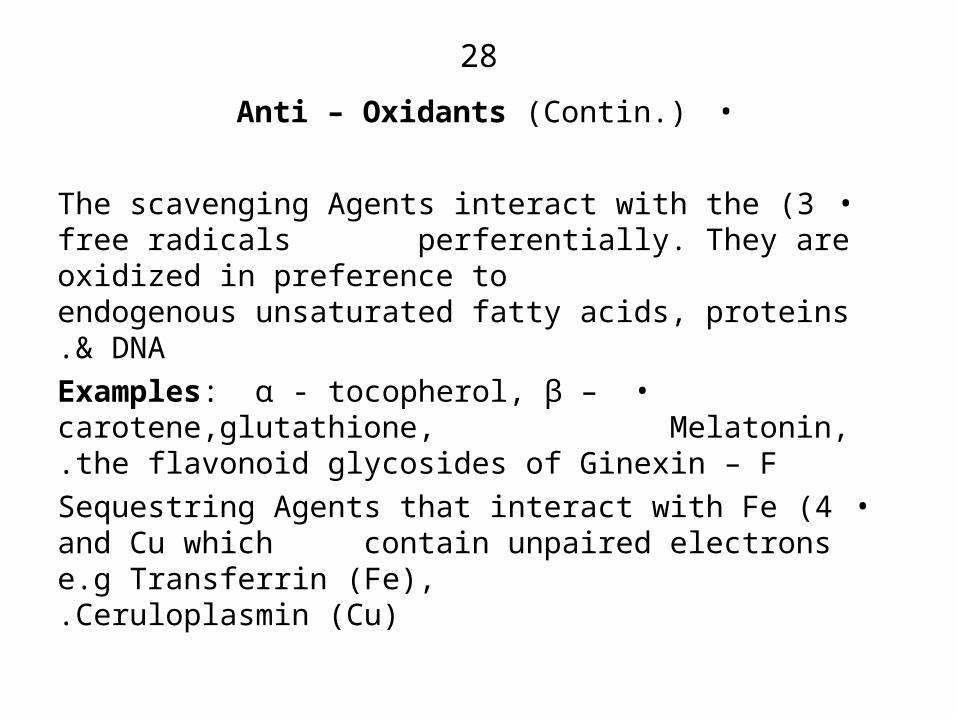
•3 )The scavenging Agents interact with the free radicals perferentially. They are oxidized in preference to endogenous unsaturated fatty acids, proteins & DNA.
•Examples: ⍺ - tocopherol, β – carotene,glutathione, Melatonin, the flavonoid glycosides of Ginexin – F.
•4 )Sequestring Agents that interact with Fe and Cu which contain unpaired electrons e.g Transferrin )Fe), Ceruloplasmin )Cu).
29
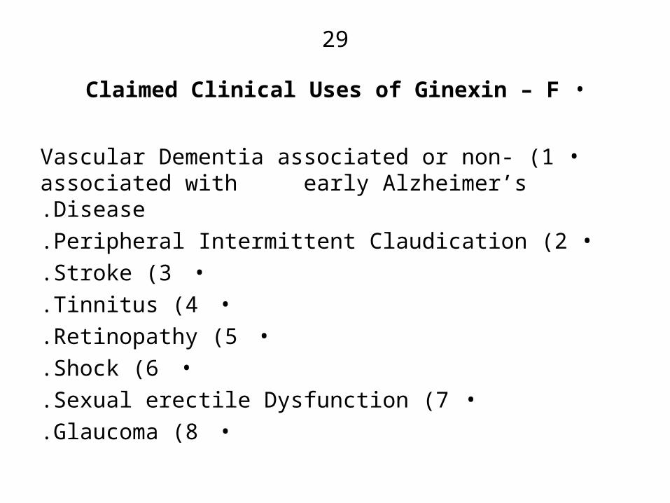
•Claimed Clinical Uses of Ginexin – F
•1 )Vascular Dementia associated or non-associated with early Alzheimer’s Disease.
•2 )Peripheral Intermittent Claudication.•3 )Stroke.
•4 )Tinnitus.•5 )Retinopathy.
•6 )Shock.•7 )Sexual erectile Dysfunction.
•8 )Glaucoma.
30
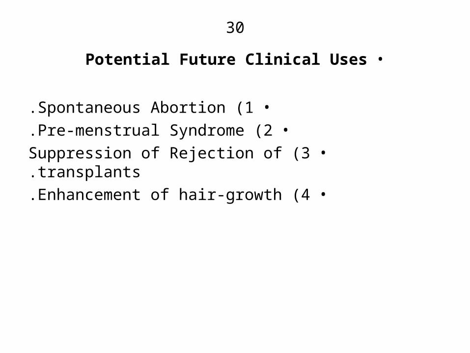
•Potential Future Clinical Uses
•1 )Spontaneous Abortion.•2 )Pre-menstrual Syndrome.
•3 )Suppression of Rejection of transplants.•4 )Enhancement of hair-growth.
31
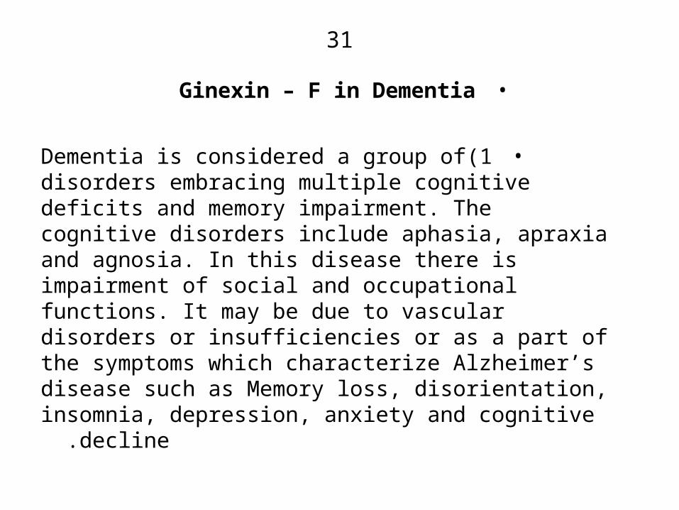
•Ginexin – F in Dementia
•1)Dementia is considered a group of disorders embracing multiple cognitive deficits and memory impairment. The cognitive disorders include aphasia, apraxia and agnosia. In this disease there is impairment of social and occupational functions. It may be due to vascular disorders or insufficiencies or as a part of the symptoms which characterize Alzheimer’s disease such as Memory loss, disorientation, insomnia, depression,
anxiety and cognitive decline .
32
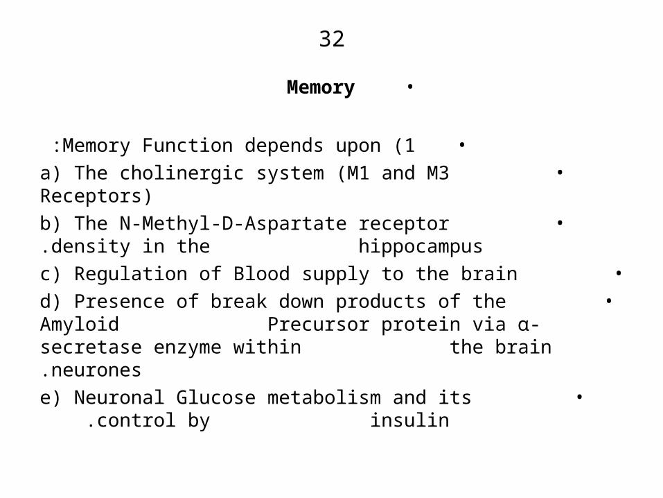
•Memory
•1 )Memory Function depends upon :• a( The cholinergic system )M1 and M3 Receptors(
• b( The N-Methyl-D-Aspartate receptor density in the hippocampus.
• c( Regulation of Blood supply to the brain• d( Presence of break down products of the Amyloid
Precursor protein via ⍺- secretase enzyme within the brain neurones.
• e) Neuronal Glucose metabolism and its control by insulin.
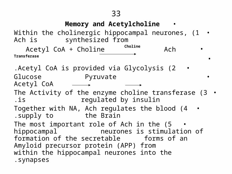
33•Memory and Acetylcholine
•1 )Within the cholinergic hippocampal neurones, Ach is synthesized from
• Acetyl CoA + Choline Choline
Ach •
Transferase
•2 )Acetyl CoA is provided via Glycolysis.• Glucose Pyruvate Acetyl CoA
•3 )The Activity of the enzyme choline transferase is regulated by insulin.
•4 )Together with NA, Ach regulates the blood supply to the Brain.
•5 )The most important role of Ach in the hippocampal neurones is stimulation of formation of the secretable forms of an Amyloid precursor protein )APP( from within the hippocampal neurones into the synapses.
34
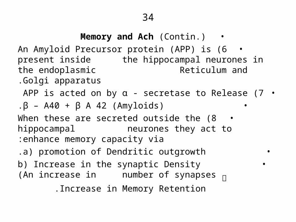
•Memory and Ach )Contin.(
•6 )An Amyloid Precursor protein )APP( is present inside the hippocampal neurones in the endoplasmic Reticulum and Golgi apparatus.
•7 )APP is acted on by ⍺ - secretase to Release • β – A40 + β A 42 )Amyloids).
•8 )When these are secreted outside the hippocampal neurones they act to enhance memory capacity via:
• a) promotion of Dendritic outgrowth.• b) Increase in the synaptic Density )An
increase in number of synapses ༜ Increase in
Memory Retention .
35
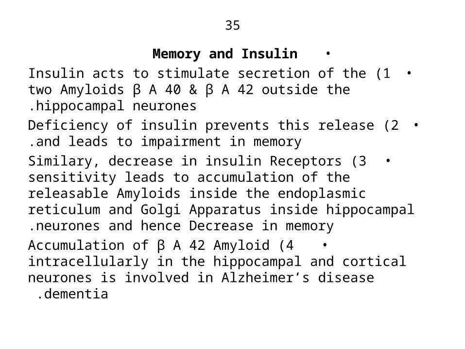
•Memory and Insulin•1 )Insulin acts to stimulate secretion of the two Amyloids
β A 40 & β A 42 outside the hippocampal neurones.•2 )Deficiency of insulin prevents this release and leads to
impairment in memory.•3 )Similary, decrease in insulin Receptors sensitivity
leads to accumulation of the releasable Amyloids inside the endoplasmic reticulum and Golgi Apparatus inside hippocampal neurones and hence Decrease in memory.
•4 )Accumulation of β A 42 Amyloid intracellularly in the hippocampal and cortical neurones is involved in
Alzheimer’s disease dementia .

36
– Thus, Disturbance and insufficiency of insulin or decrease in the sensitivity of its Receptor in both the hippocampus and the cortex leads to Disturbance in glucose metabolism )Glycolysis( with decrease in production of ATP and Acetyl CoA and hence: a( No stimulation of production of the Amyloids β A 40 and β A 42 via ⍺ secretase enzyme due to deficiency of Ach and
– b) No release of the Amyloids outside the neurones due to deficiency of insulin.
– ༜ Decrease in neuromal Activity at the
synapses leading to decrease in memory Dementia.

37
•Drugs in use for Dementia•1 )Levacecarnine
• 2.5 3 gm per day.• It enhances Ach synthesis.
•2 )Anti-oxidant Vitamin E + C.•3 )Cholinesterase Inhibitors:• a( Donepezil [Arisept]
• 5 10 mg / day• b( Rivastingmine [Exelon] Capsules.
• 1.5 mg 2x daily up to 12 mg / day• c( Galantamine [Reminyl]
• 24 mg / day• d( Huperzine A
• 50 100 µg 2 x daily.• This is an alkaloid obtained from Club Moss: Huperzia serrata
•Rate of success of Anti-Choline esterases• 45 50%
•4 )Memantine )NMDA Blocker( 5( Piracetam [Nootropil]
38
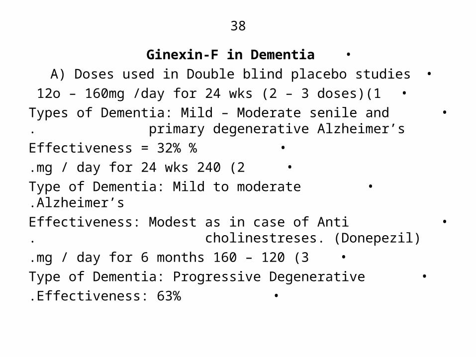
•Ginexin-F in Dementia•A( Doses used in Double blind placebo studies
•1)12o – 160mg /day for 24 wks )2 – 3 doses( • Types of Dementia: Mild – Moderate senile and
primary degenerative Alzheimer’s.• % Effectiveness = 32%•2 )240 mg / day for 24 wks.
• Type of Dementia: Mild to moderate Alzheimer’s.• Effectiveness: Modest as in case of Anti
cholinestreses. )Donepezil(.•3 )120 – 160 mg / day for 6 months.
• Type of Dementia: Progressive Degenerative• Effectiveness: 63%.

39
•Dementia )Contin.(
•4 )120 – 240 mg/day for 6 months• Type of Dementia: Multi-infarct dementia .
• Effectiveness: 27%.•5 )120mg/day for 24wks.
• Type of Dementia: Mild to Moderate.• Effectiveness: 52%
40
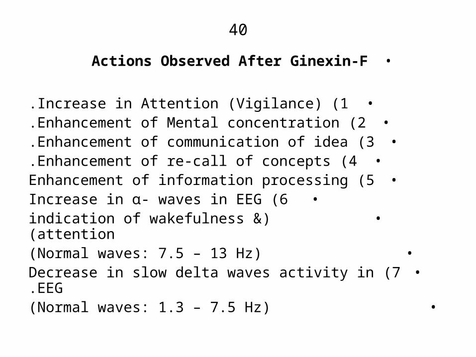
•Actions Observed After Ginexin-F
•1 )Increase in Attention )Vigilance(.•2 )Enhancement of Mental concentration.
•3 )Enhancement of communication of idea.•4 )Enhancement of re-call of concepts.
•5 )Enhancement of information processing•6 )Increase in ⍺- waves in EEG
•) indication of wakefulness & attention)•) Normal waves: 7.5 – 13 Hz)
•7 )Decrease in slow delta waves activity in EEG.•) Normal waves: 1.3 – 7.5 Hz)

41
•Probable Mechanisms of Action•1 )Enhancement of cerebal blood flow via a( direct action
b( Blockade of ⍺1 Receptors .• c) Antagonism of PAF-induced platelets
aggregation .• d) Release of [NO] which vasodilates blood
vessels and inhibits platelets aggregation.• e) Anti-oxidant effect against vasoconstricting
O2 radicals .
– f) Inhibition of cerebral oedema.
• The increase in blood supply ensures excellent supply of O2 and glucose and insulin resulting in elevation of ATP and creatine phosphate.
42
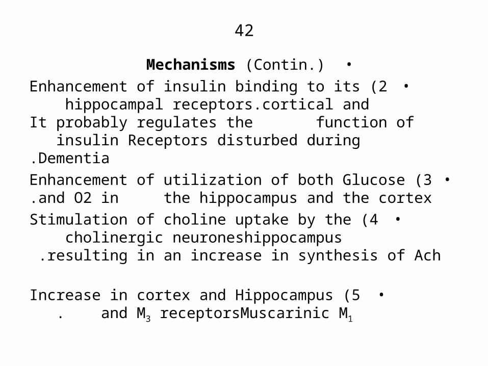
•Mechanisms )Contin.(•2 )Enhancement of insulin binding to its cortical and
hippocampal receptors. It probably regulates the function of insulin Receptors disturbed during
Dementia.•3 )Enhancement of utilization of both Glucose and O2 in
the hippocampus and the cortex.•4 )Stimulation of choline uptake by the hippocampus
cholinergic neurones resulting in an increase in synthesis of Ach.
•5 )Increase in cortex and Hippocampus Muscarinic M1 and M3 receptors .
43
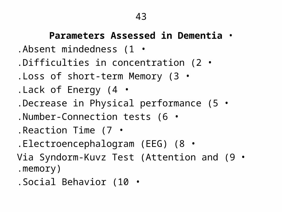
•Parameters Assessed in Dementia•1 )Absent mindedness.
•2 )Difficulties in concentration.•3 )Loss of short-term Memory.
•4 )Lack of Energy.•5 )Decrease in Physical performance.
•6 )Number-Connection tests.•7 )Reaction Time.
•8 )Electroencephalogram )EEG(.•9 )Via Syndorm-Kuvz Test )Attention and memory(.
•10 )Social Behavior.
44
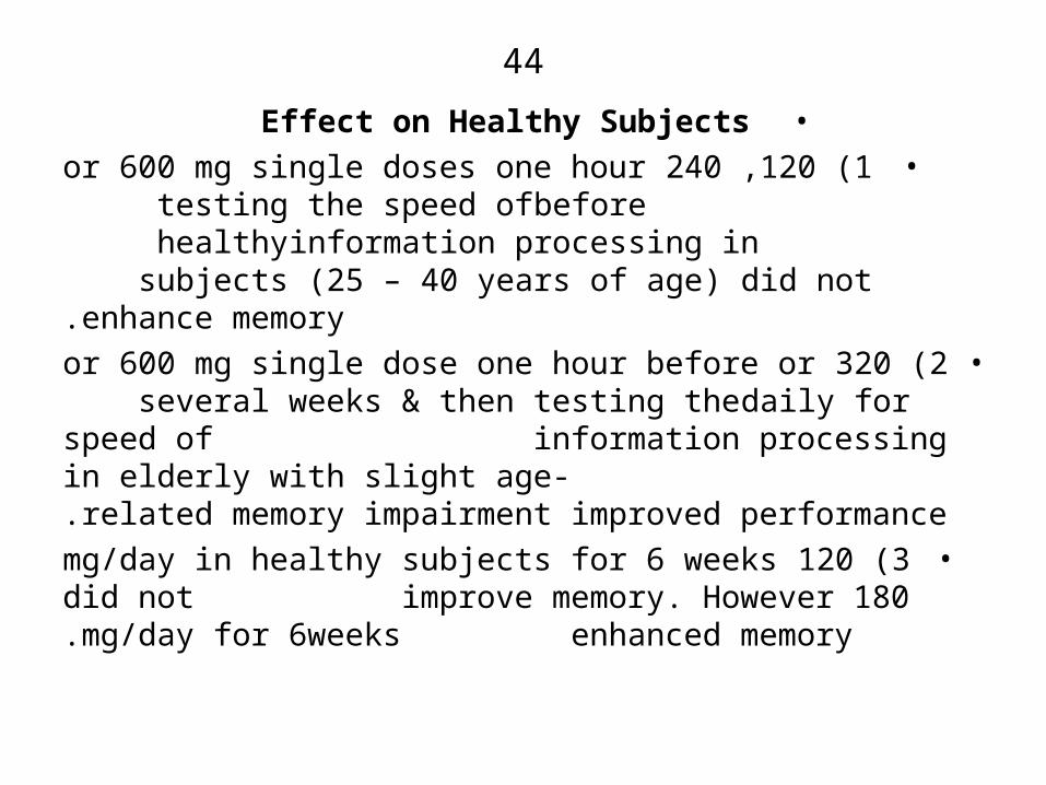
•Effect on Healthy Subjects•1 )120 ,240 or 600 mg single doses one hour before
testing the speed of information processing in healthy subjects )25 – 40 years of age( did not
enhance memory.•2 )320 or 600 mg single dose one hour before or daily for
several weeks & then testing the speed of information processing in elderly with slight age- related memory impairment improved performance.
•3 )120 mg/day in healthy subjects for 6 weeks did not improve memory. However 180 mg/day for 6weeks enhanced memory.

45
•General Notes Regarding Use in Dementia•1 )The effectiveness is Moderate. It depends upon the
severity of the disease.•2 )Also, moderate effectiveness )50%( in primary
Degenerative Alzheimer’s Dementia.•3 )Its rate of success )or efficiency( is similar to the
available anticholine esterases.•4 )Duration of treatment depends upon the condition of
the patient.
46
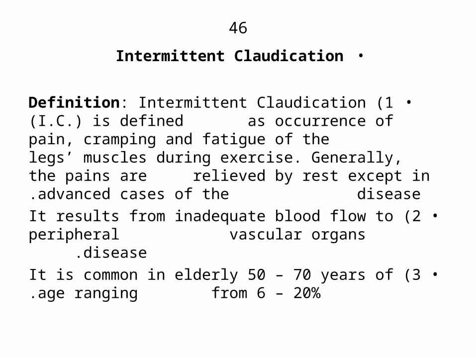
•Intermittent Claudication
•1 )Definition: Intermittent Claudication )I.C.( is defined as occurrence of pain, cramping and fatigue of the legs’ muscles during exercise. Generally, the pains are relieved by rest except in advanced cases of the disease.
•2 )It results from inadequate blood flow to peripheral vascular organs disease .
•3 )It is common in elderly 50 – 70 years of age ranging from 6 – 20%.

47
•I.C. )Contin.(
•4 )It is associated with atherosclerotic narrowing in iliac or femoral arteries or a distal leg artery.
•5 )Blood perfusion of the post stenotic tissues is highly reduced leading to local tissue hypoxia i.e ischaemia and Release of free Radicals, platelets aggregation and accumulation of lactic and pyruvic acids in tissues Pain.
•6 )Chronic Disease leads to Gangrene resulting in the possibility of limb amputation.
48
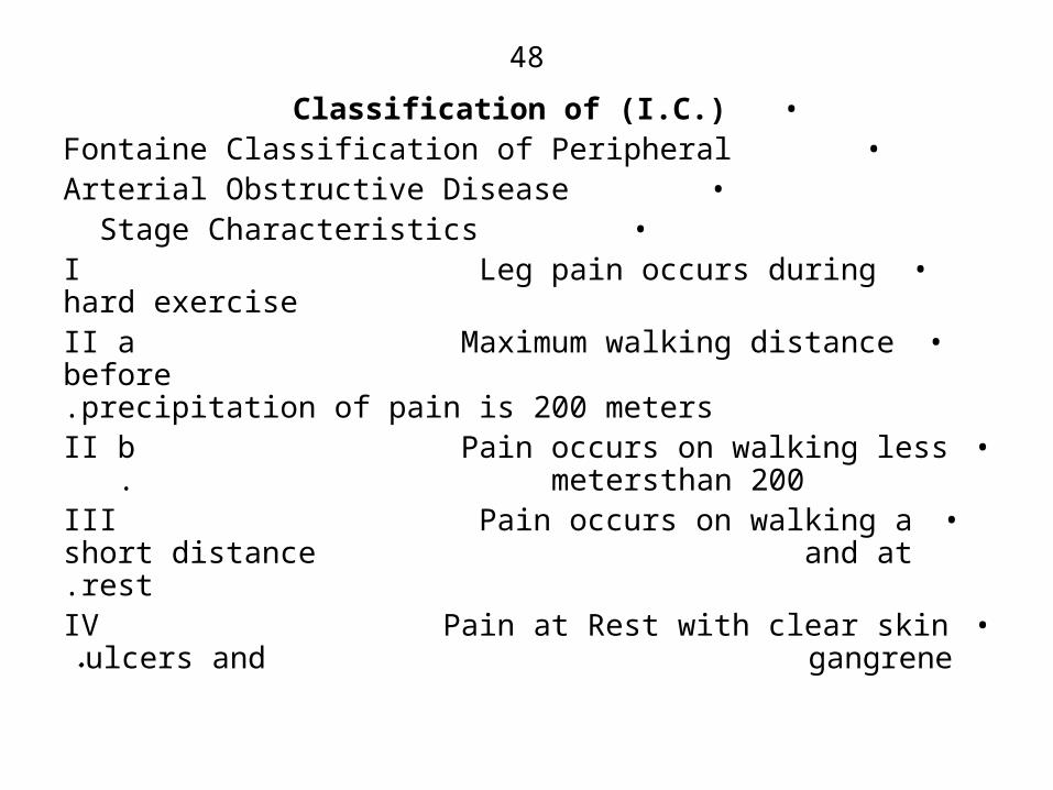
•Classification of )I.C.(• Fontaine Classification of Peripheral
• Arterial Obstructive Disease• Stage Characteristics
•I Leg pain occurs during hard exercise•II a Maximum walking distance before
precipitation of pain is 200 meters.•II b Pain occurs on walking less than 200
meters .•III Pain occurs on walking a short distance
and at rest.•IV Pain at Rest with clear skin ulcers and
gangrene.
49
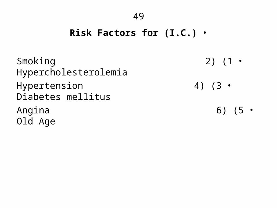
•Risk Factors for )I.C.(
•1 )Smoking 2( Hypercholesterolemia•3 )Hypertension 4( Diabetes mellitus
•5 )Angina 6( Old Age

50
•Strategy For Treatment•1 )Proper treatment of the associated diseases.
•2 )With holding of smoking.•3 )Drug treatment.
•Drugs Currently in use•1 )Pentoxifylline [Treatal] 400mg 3x daily
• a( Methylxanthine Derivative that inhibits cyclic AMP phosphdiesterase leading to elevation
of the level of cAMP .• b( It Blocks Adenosine receptors.
• c( The net action is inhibition of platelets aggregation and dilatation of blood vessels .
• d( Effectiveness = 39%.
51
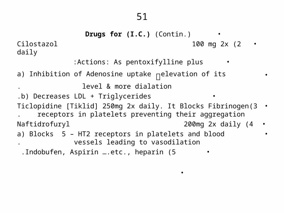
•Drugs for )I.C.( )Contin.(•2 )Cilostazol 100 mg 2x daily•Actions: As pentoxifylline plus:
• a( Inhibition of Adenosine uptake ༜elevation of its
level & more dialation.• b) Decreases LDL + Triglycerides.
•3)Ticlopidine [Tiklid] 250mg 2x daily. It Blocks Fibrinogen receptors in platelets preventing their aggregation.
•4 )Naftidrofuryl 200mg 2x daily• a) Blocks 5 – HT2 receptors in platelets and blood
vessels leading to vasodilation.•5 )Indobufen, Aspirin ….etc., heparin .
•

52
•Ginexin – F in )I.C.(•1 )Intake of 120 – 240mg/day prolonged the distance
walked by the patient without pain to an extent similar to pentoxifylline at a dose of 1.2gm/day. )Tread Mill Exercise(.
•2 )Duration of treatment: 24 weeks.
•3 )Mechanisms:
• 1 -Induction of Vasodilation via direct and indirect mechanisms via release of [NO] .
• 2 -Inhibition of platelets aggregation, induced by PAF by blocking its receptors and other aggregators via
NO – induced increase in cyclic GMP .
• 3 -Antagonism to PAF – induced ischemia resulting from accumulation of PMNL.
• 4 -Scavenging of free O2 Radicals that constrict blood vessels.
53
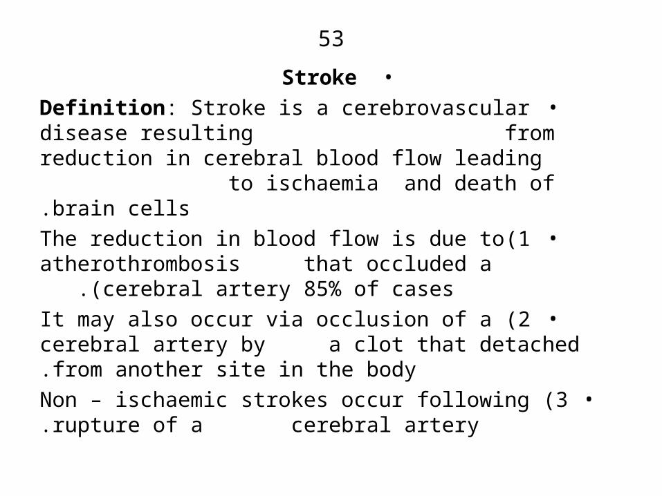
•Stroke•Definition: Stroke is a cerebrovascular disease resulting
from reduction in cerebral blood flow leading to ischaemia and death of brain cells.
•1)The reduction in blood flow is due to atherothrombosis that occluded a cerebral artery 85% of cases .)
•2 )It may also occur via occlusion of a cerebral artery by a clot that detached from another site in the body.
•3 )Non – ischaemic strokes occur following rupture of a cerebral artery.
54
•Stroke Complication
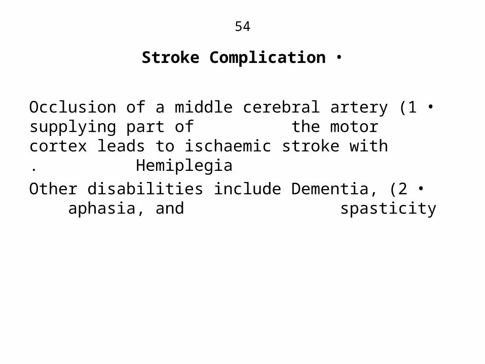
•1 )Occlusion of a middle cerebral artery supplying part of the motor cortex leads to ischaemic stroke with Hemiplegia.
•2 )Other disabilities include Dementia, aphasia, and spasticity
55
•Consequencies of Ischaemic Stroke
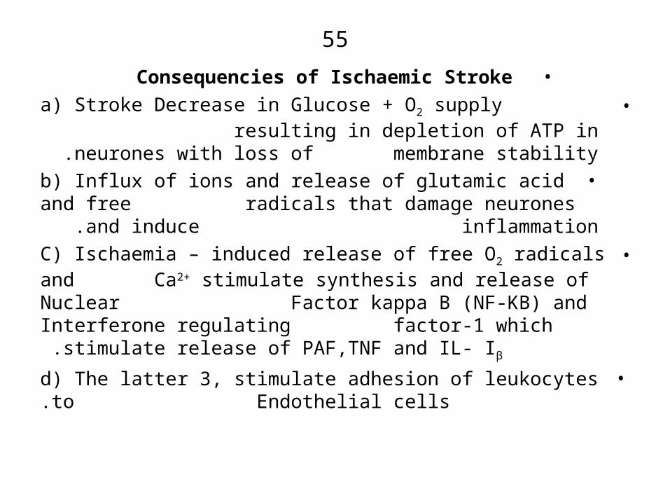
•a( Stroke Decrease in Glucose + O2 supply resulting in depletion of ATP in neurones with loss of
membrane stability .•b( Influx of ions and release of glutamic acid and free
radicals that damage neurones and induce inflammation .
•C( Ischaemia – induced release of free O2 radicals and Ca2+ stimulate synthesis and release of Nuclear Factor kappa B )NF-KB( and Interferone regulating
factor-1 which stimulate release of PAF,TNF and IL- Iβ .
•d( The latter 3, stimulate adhesion of leukocytes to Endothelial cells.
56
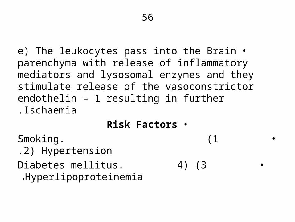
•e( The leukocytes pass into the Brain parenchyma with release of inflammatory mediators and lysosomal enzymes and they stimulate release of the vasoconstrictor endothelin – 1 resulting in further Ischaemia.
•Risk Factors• 1 )Smoking. 2( Hypertension.
• 3 )Diabetes mellitus. 4( Hyperlipoproteinemia.

57
•Treatment Strategy
•A( For Non – haemorrhagic ischaemic stroke:• 1 )I.V. Administration of a thrombolytic drug within 3
hours of occurrence.• 2 )Then administration of drugs to prevent
recurrencies i.e Anticoagulants.• 3 )Administer drugs to enhance stroke Recovery i.e
Administration of Neuroprotective Agents .

58
•Treatment: Thrombolytics•1)Recombinnant tissue Plasminogen Activator
• r t – PA [Alteplase]• It activates Plasminogen resulting in release of active
Plasmin that degrades the clot .•2 )Streptokinase [Kabikinase] 250000 i u i.v over 30
minutes then 100000 i u every hour for 24 – 72 hours.• It activates Plasminogen to release Plasmin.
•3 )Reteplase [Retavase].•4 )Others: Monteplase [Cleactor], Tenecteplase
[Metalyse]
59
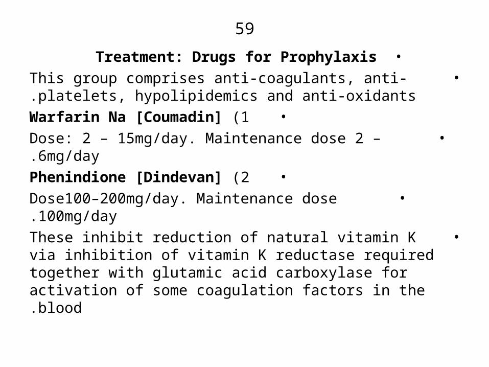
•Treatment: Drugs for Prophylaxis• This group comprises anti-coagulants, anti-platelets,
hypolipidemics and anti-oxidants.•1 )Warfarin Na [Coumadin]
• Dose: 2 – 15mg/day. Maintenance dose 2 – 6mg/day.•2 )Phenindione [Dindevan]
• Dose100–200mg/day. Maintenance dose 100mg/day.• These inhibit reduction of natural vitamin K via
inhibition of vitamin K reductase required together with glutamic acid carboxylase for activation of some coagulation factors in the blood.
60
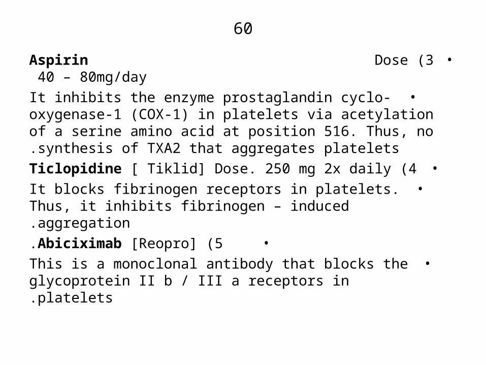
•3 )Aspirin Dose 40 – 80mg/day•It inhibits the enzyme prostaglandin cyclo-oxygenase-1
)COX-1( in platelets via acetylation of a serine amino acid at position 516. Thus, no synthesis of TXA2 that aggregates platelets.
•4 )Ticlopidine [ Tiklid] Dose. 250 mg 2x daily•It blocks fibrinogen receptors in platelets. Thus, it inhibits
fibrinogen – induced aggregation.•5 )Abiciximab [Reopro].
•This is a monoclonal antibody that blocks the glycoprotein II b / III a receptors in platelets.
61
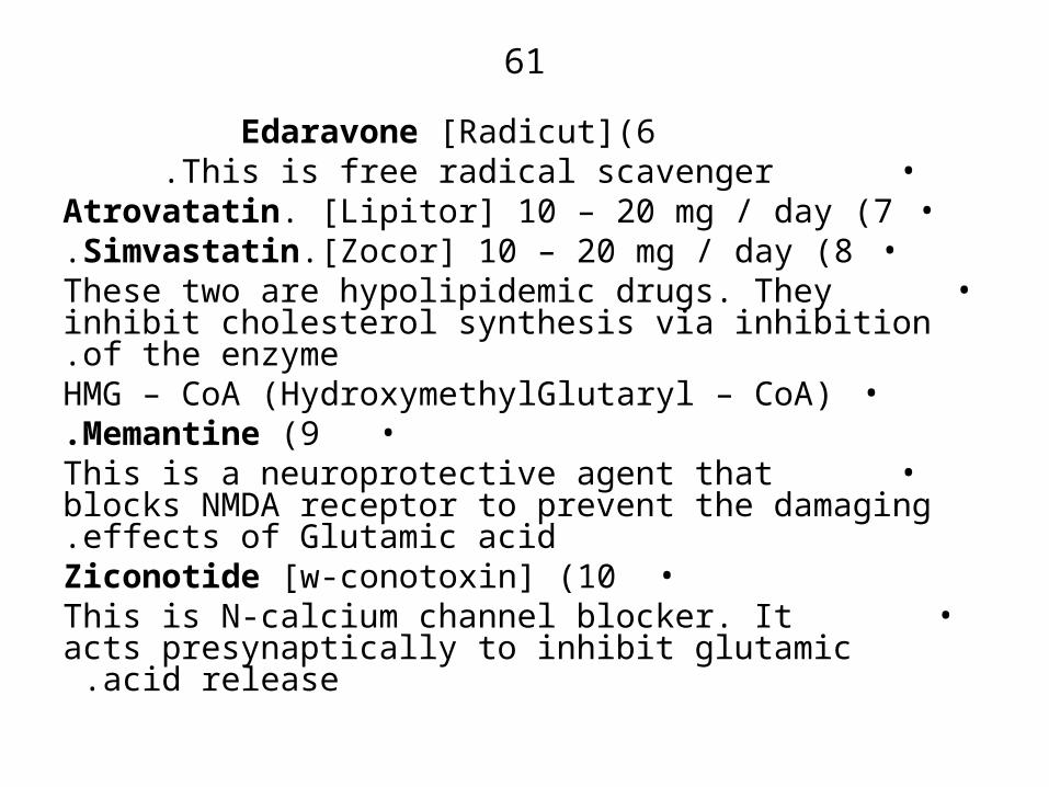
6)Edaravone [Radicut] • This is free radical scavenger .
•7 )Atrovatatin. [Lipitor] 10 – 20 mg / day•8 )Simvastatin.[Zocor] 10 – 20 mg / day.
• These two are hypolipidemic drugs. They inhibit cholesterol synthesis via inhibition of the enzyme.
•HMG – CoA )HydroxymethylGlutaryl – CoA(•9 )Memantine.
• This is a neuroprotective agent that blocks NMDA receptor to prevent the damaging effects of Glutamic acid.
•10 )Ziconotide [w-conotoxin]• This is N-calcium channel blocker. It acts
presynaptically to inhibit glutamic acid release .

62
•Enhancement of post-stroke recovery via use of:• 1 )Donepezil [Aricept]
• 2 )Rivastigmine [Exelon].• 3 )Galantamine [Reminyl
• These inhibit the enzyme ACh esterase leading to elevation of the level of ACh.
63
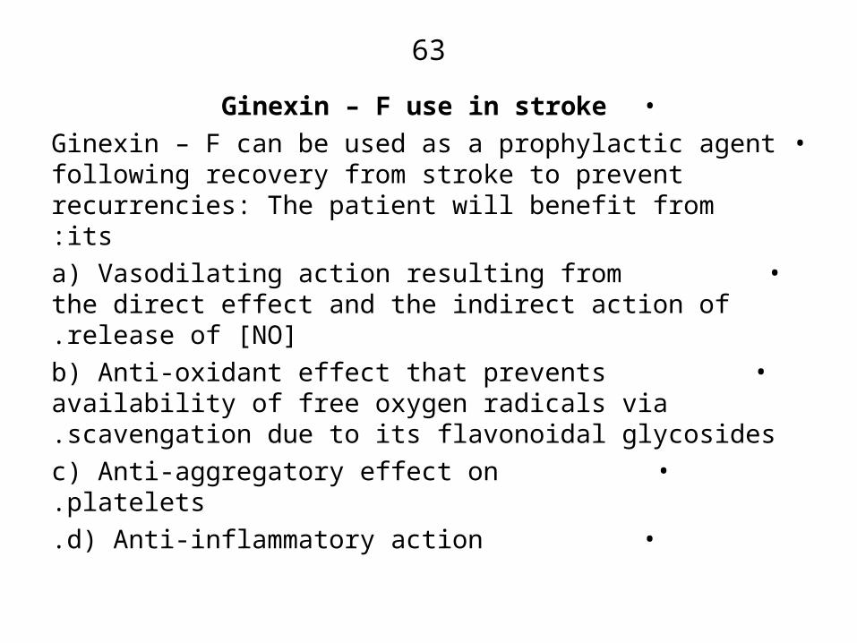
•Ginexin – F use in stroke•Ginexin – F can be used as a prophylactic agent
following recovery from stroke to prevent recurrencies: The patient will benefit from its:
• a( Vasodilating action resulting from the direct effect and the indirect action of release of [NO].
• b( Anti-oxidant effect that prevents availability of free oxygen radicals via scavengation due to its flavonoidal glycosides.
• c( Anti-aggregatory effect on platelets.• d( Anti-inflammatory action.

64
•Tinnitus•Definition: Tinnitus is an otological complain taking the
form of ringing in the ears. It affects about 10 – 12% of the population that are more than 65 years of age. In audiometric Normal subjects )normal hearers( the hearing level threshold is 20 – 25 dB )decibels( at frequencies ranging from 250 – 8000 Hz. The intensity of tinnitus is 10 – 44 dB at frequency of 3000 – 8000 Hz. The sounds are due to spontaneous vibrations of the
outer hair cells of the cochlea .

65
•Tinnitus )Contin.(• The sounds heard may be unilateral )one ear is
affected( or may be bilateral )both ears are affected(. The sounds heard may be ringing in 37% of patients, Buzzing in 11%, hissing in 8%, whistling in 7% and humming in 5% of the patients.
• Tinnitus may be objective meaning it is loud enough to be heard by the patient and his neighbors or it may be subjective heard by the patient only in most of the patients.
• Its effect is great in patients suffering from pain, depression or who are socially isolated and psychotic.
66
•Tinnitus )Contin.(• The clicking buzzing may indicate Palatal myoclonus
or contractions of the tensor tympani or stapelius muscle .
• The pulsatile sounds are also considered to be due to vibrations from turbulent blood flow that reaches the
cochlea .

67
•Parts Affected in Tinnitus•(Pathway)
•Damage or stimulation of the Kinocilia nerve attached to the Kinocilia cell in the inner ear sends impulses to the thalamus and then to the cerebral cortex. Tinnitus may result from an increase in the spontaneous firing rate of the auditory nerve as in the case of Salicylates. It may also occur following reduction in the activity of the auditory nerve inputs leading to disinhibition of the dorsal cochlear nucleus and an increase in spontaneous activity in the brain auditory system which is then expressed as tinnitus. e.g as in case of furosemide -induced tinnitus.

68
•Causes of Tinnitus
•1 )Poor blood perfusion of the inner ear due to vasoconstriction.
•2 )Rhythmic hyperactivity of the auditory reflex arc and impairment of the auditory neural pathway.
•3 )Dysfunction of the auditory nerve.•4 )Stimulation of the tympanic nervous plexus following
inflammation of the cavity of the middle ear )otitis media(•5 )Hypersensitivity of Tympani nerve.
•6 )Oedema of cortis’ organ.
69
•Causes of Tinnitus )Contin.(
•7 )Stenosis of the carotid artery.•8 )Atriovenous malformations.•9 )Tumors in the Jugular vein.
•10)Valvular heart disease. 11( High cardiac output•.12)Impacted cerumen. 13( Head injury •14)Vestibular schwannoma )Acuostic Neurema(
15)Meningitis 16( Cerebllum Tumors 17(Drugs.
18)Very loud noise. 19(Multiple sclerosis 20( Hypertension
21 )Anxiety. 22( Soft plate disorders .
70
•Drugs that cause tinnitus•1 )Salicylate and other non-selective COX enzyme
inhibitors.•2 )Aminoglycosides e.g Gentamycin; Kanamycin;
Streptomycin.•3 )Loop diuretics e.g Furosemide.
•4 )Vincristine.•5 )Cisplatin.
•6 )Quinine and Quinidine.•7 )Heavy metals poisoning.
71
•Treatment Strategy
•1)Assess tinnitus and identify its type using:• a( An audiometer.
• b( Determination of tone threshold.• c( Speech – Audiometry to determine hearing loss
i.e )Number and word comprehension(.• d( Monitoring of oto-coustic emissions to assess the
biomechanical performance of the outer hair cells.• e( Examine the head, oral cavity, cranial nerves the
5th , 7th and 8th .
72
•Treatment Strategy )Contin.(
•2 )Treat any causative disorder if possible.•3 )Withdraw any causative drug.
•4 )If the disease is due to exposure of loud sounds, protect the hearing process via use of ear-muff like devices or custom-molded devices that fit into external auditory meatus.
•5 )Administration of drugs noting that there is no 100% cure )up to 50 % Reduction(.

73
•Drugs Available for Treatment of Tinnitus
•A( Drugs that enhance blood flow to the cochlea :• 1 -The calcium channel Blockers e.g:
• a( Cinarizine [Cerepar] 25mg 3x daily• b( Flunarizine [Cibelium] 5 -10mg/day.
• c( Nicergoline [Semion] 5 – 15 mg / day 3x daily.• Efficacy : 20 – 30.%
• 2 -Anti-aggregatory and vasodilators e.g pentoxifylline• 3 -Direct vasodilator & PG releasers e.g Nicotinic
acid.
74•Treatment of Tinnitus )Contin.(
•B( Membrane Stabilizers:Carbamazepine [Tegretol]
Dose 50 – 100 mg / day It suppresses movement of Na+ and Ca2+ into the
cochlear nerve. Thus, it suppresses its activity to transmit impulses to the thalamus.
Effectiveness : 10 – 30 – 50.% C( Anxiolytics e.g:
Alprazolam [xanax] 0.5 mg 3x daily Lorazepam [Ativan] 1 – 2 mg / day.
These releive anxiety and suppress stress leading to emotional balance.
Effectiveness: 50.%

75
•D( Amitriptyline [Tryptisol]•50 – 150 mg / day
• Effectiveness: 10 – 30% •E( Non – Drug Treatment:
• a( A cupuncture which releases enkephalins and β- endorphins.
• Effectiveness: 5 – 30% • b( Electrical stimulation of cochlea via the external
canal of the ear.• Effectiveness: 10 – 50 %
76
•Treatment of Tinnitus )Contin.(
•F( New Drugs:• 1 )Memantine
• It blocks NMDA receptor of glutamic acid. It acts as a neuroprotector to the cochlear nerve.
• 2 )POU4F3
• This is an inner ear cell transcriptor factor that encodes nucleic acids. It stimulates regeneration
of damaged inner cells .•
77
•Ginexin – F and Tinnitus•1 )Ginexin – F when administered at a dose of 40mg 3x
daily for 12 weeks to patients with Tinnitus )subjective or idiopathic( resulted in a significant decrease in the severity of the disease in 57% of the patients.
•2 )The effectiveness of the drug is high in recently developing tinnitus.
•3 )Treatment of patients with 120mg/day for 2 months suppressed tinnitus by 60% whereas Nicergoline )a Vasodilator( 15mg/day for 2 months suppressed the symptoms by 40%.
•4 )In another study, administration of the extract 200mg/day )I.V.( in form of infusion followed by oral doses of 80mg 2x daily for 12 weeks decreased
significantly the subjective Tinnitus volume by-3.5 dB .

78•Mechanism of Action
• The beneficial action of Ginexin – F in suppressing the severity of tinnitus may have been exerted via Ginexin-F- induced.
• 1 )Vasodilation and enhancement of blood flow to the inner ears. The factors that are involved in this vasodilation include:
• a( Direct effect on blood vessels resulting in inhibition of release of intracellular Ca2+ with decrease in vasocontriction.
• b( Release of the vasodilators [NO] and PGI2 from the endothelium and smooth muscles of the blood vessels
• c( Antagonism of PAF- induced Platelets aggregation resulting in enhancement of the dilation.
• d( The antioxidant effect that prevents the availability of the vasoconstricting O2 free Radicals.
79
•Retinopathy•Definition: Retinopathy is a common ocular
microvascular Disease resulting from unwell controlled Diabetes mellitus and hypertension. Its ultimate Destination is Blindness.
•Signs : a( Generalized and focal retinal arteriolar narrowing.
• b( Atriovenous nicking.• c( Retinal haemorrhages.
• d( Swelling of the optic disk.•Other Causes: Inflammation and Endothelial dysfunction.
•
80
•Pathophysiology of Hypertension-induced Disease
•1)The high untreated blood pressure induces vasoconstriction and spasms resulting in narrowing of retinal arterioles.
•2 )This is followed by intimal thickness, hyperplasia of medial wall, hyaline degeneration and necrosis of retinal vessels and their endothelial cells.
•3 )Thus, Retinal Ischaemia results together with haemorrhages, hard exudates, occlusion of vein
branches and swelling of optic disc .
81
•Pathophysiology of Diabetes - induced Retinopathy• This is multifactorial since it involves polyol
accumulation, glycation, Oxidative stress and activation of protein kinases.
•a( Polyol Accumulation• 1 )The increased glucose in the blood is converted intra
cellularly into sorbitol via the enzyme Aldose reductase .
•2 )Sorbitol then exerts osmotic toxicity leading to damage of vascular cells, retina and the optic nerve .
82
•b( Protein Glycation• With the high level of glucose available some of it
interacts with proteins to form glycated proteins which stimulate free O2 radical production. These damage the neural cells.
•c( Oxidative Damage• Some of the high glucose undergo auto oxidation to
release free O2 Radicals.•d( Activation of Protein kinases
• An increase in the activity of the protein kinase-B2 is believed to stimulate production of matrix proteins such as collagen and fibronectin and enhances synthesis of endothelin resulting in thickening of the basement membrane and an increase in retinal vascular permeability leading to macular oedemas and decreases in the retinal blood flow.

83
•Growth Factors )GF(• In the process of Retinopathy some GF are believed
to be involved in the disease. These include:• a( vascular Endothelial GF which increases with
hypoxia, PAF release and retinal Ischaemia resulting in an increase in capillary permeability and formation of oedemas.
• b( The Transforming GF-β which is normally released by pericytes to inhibit endothelial proliferation and angiogenesis is found to be elevated during Retinopathy resulting in enhanced endothelial proliferation and angiogenesis.
84
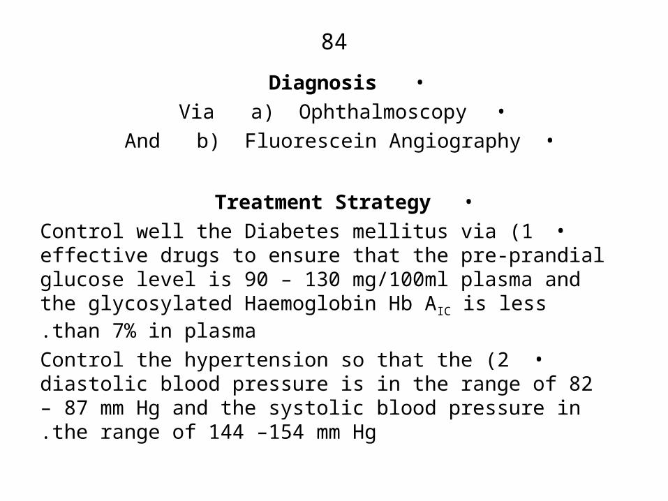
•Diagnosis•Via a( Ophthalmoscopy
•And b( Fluorescein Angiography
•Treatment Strategy•1 )Control well the Diabetes mellitus via effective drugs
to ensure that the pre-prandial glucose level is 90 – 130 mg/100ml plasma and the glycosylated Haemoglobin Hb AIC is less than 7% in plasma.
•2 )Control the hypertension so that the diastolic blood pressure is in the range of 82 – 87 mm Hg and the systolic blood pressure in the range of 144 –154 mm Hg.
85
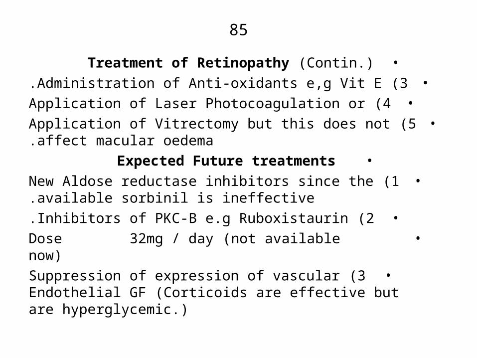
•Treatment of Retinopathy )Contin.(•3 )Administration of Anti-oxidants e,g Vit E.•4 )Application of Laser Photocoagulation or
•5 )Application of Vitrectomy but this does not affect macular oedema.
• Expected Future treatments•1 )New Aldose reductase inhibitors since the available
sorbinil is ineffective.•2 )Inhibitors of PKC-B e.g Ruboxistaurin.
• Dose 32mg / day )not available now(•3 )Suppression of expression of vascular Endothelial GF
)Corticoids are effective but are hyperglycemic.(

86
•Ginexin – F and Retinopathy•1 )Intake at a dose of 80mg/day for 6 months produced
significant improvements in patients with Diabetes- induced retinopathy.
•2 )Administration of a dose of 320mg /day for 3 months to diabetics with Retinopathy together with their normal diabetes treatment increased significantly some parameters that are decreased in the diabetics e.g it increased retinal blood flow rate. Furthermore it decreased some of those parameters that are increased such as: peroxidation of membranes, fibrinogen, blood viscosity, blood viscoelasticity and erythrocytes
rigidity .
87
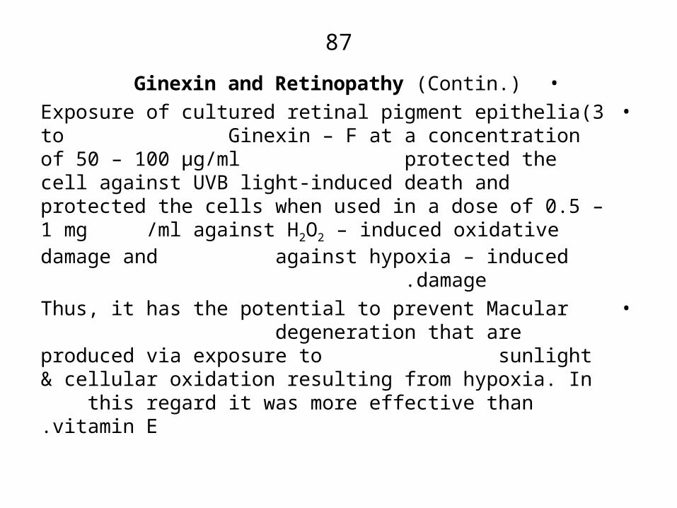
•Ginexin and Retinopathy )Contin.(•3)Exposure of cultured retinal pigment epithelia to
Ginexin – F at a concentration of 50 – 100 μg/ml protected the cell against UVB light-induced death and protected the cells when used in a dose of 0.5 – 1 mg /ml against H2O2 – induced oxidative damage and
against hypoxia – induced damage .• Thus, it has the potential to prevent Macular
degeneration that are produced via exposure to sunlight & cellular oxidation resulting from hypoxia. In this regard it was more effective than vitamin E.
88
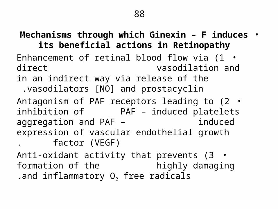
•Mechanisms through which Ginexin – F induces its beneficial actions in Retinopathy
•1 )Enhancement of retinal blood flow via direct vasodilation and in an indirect way via release of the
vasodilators [NO] and prostacyclin .•2 )Antagonism of PAF receptors leading to inhibition of
PAF – induced platelets aggregation and PAF – induced expression of vascular endothelial growth factor )VEGF(.
•3 )Anti-oxidant activity that prevents formation of the highly damaging and inflammatory O2 free radicals.
89
•Glaucoma•Definition: Glaucoma is an ocular disease caused by
sustained increase in intraocular pressure )I.O.P.(. This increase damages the optic nerve leading to loss of vision. The normal I.O.P. is 10 – 21 mm Hg but in glaucoma it increases above 30 mm Hg.
• Other factors that predispose to the disease include reduction in blood supply to the optic nerve, ocular trauma and migraine.
•Symptoms : In the first stage there are no clear symptoms except the high I.O.P. later symptoms include: Blurred vision ocular pain, redness, headache, nausea vomiting and seeing halos around light.
90
•Types of Glaucoma•a( Open-angle Glaucoma: this is the most common in
90% of the patients. Beside the high I.O.P. and using scanning laser imaging one can see optic-disk damage, enlargement of the optic-disk cup and haemorrhages.
•b( Closed angle Glaucoma : In this type there is an increase in the I.O.P. resulting from failure of the aqueous humor to flow through the pupil into the anterior chamber and hence No outflow through the trabercular system.
91
•Drugs Available for treatment of Glaucoma
•1)Timolol 0.25 – 0.5 % Eye Drops [Timpotic] 4x. It is non – selective β- blocker.
•2 )Betaxolol hydrochloride 0.25 & 0.5% Eye drops [Betopic] 4x daily .
• It is a selective β1- blocker .
• These Drugs decrease aqueous humor Synthesis .•3 )Pilocarpine hydrochloride 0.5 – 4% Eye drops
[Isoptocarpine] 4x daily.• It increases aqueous humor out flow from the anterior
chamber of the eye into the trabercular mesh work .
92
•4 )Acetazolamide Tablets )500mg( [Diamox] 500mg 2x daily. It decreases Aqueous humor synthesis.
•5 )Brinzolamide [Azopt] 1% Eye drops 3x. Both are carbonic anhydrase inhibitors.
•6 )Argon laser trabeculoplasty. Used to reduce the resistance of the trabercular mesh work to outflow of the aqueous humor.
•7 )Surgical trabeculectory .• An opening is created in the anterior chamber
angle to allow aqueous humer to flow from the anterior chamber below the conjunctiva .
93
•Ginexin – F use in Glaucoma•1 )Intake of 40 mg 3x daily for 4 weeks by Glaucoma
patients produced significant improvement in the visual field indices but did not decrease the I.O.P.
•2 Treatment of Glaucoma patients with 40 mg 3x for 2 days significantly increased the velocity of blood flow in artery by 23% but did not affect the I.O.P.
•3 )Treatment of rats before performing partial retinal degeneration using cautery decreased the loss of Retinal ganglion cells from 30% in control to 5% in treated animals

94
•Mechanisms of Action of Ginexin-F•The beneficial actions of Ginexin-F in Glaucoma may be
due to:• 1 -Its Vasodilatory action in retinal blood flow.
• 2 -Its platelets antiaggregatory effect that decreases blood viscosity and enhances its flow to the Retinal cells.
• 3 -Scavenging of free O2 Radicals that act to damage the retinal cells.
•Thus, in Glaucoma Ginexin-F can be used with other drugs to induce neuroprotective action.
95
•Shock• Shock occurs when the circulation of the arterial
blood is not adequate to perfuse various organs.•Types :
•1 )Hypovolemic: results from loss of blood via haemorrhages or loss of electrolytes and water as in case of severe burns, vomiting or diarrhoea.
•2 )Cardiogenic shock results from inadequate cardiac function.
•3 )Obstructive shock due to obstruction of the systemic or pulmonary circulation as in cases of massive pulmonary embolism.
•4 )Septic Shock: caused by severe infection with gram negative rods e.g E. coli and gram positive cocci e.g Staphylococcus.

96
5 )Neurogenic shock: results from spinal cord injury or fear mediated via reflex vagal stimulation .
•6 )Anaphylactic shock: due to antigen antibody reactions.
•Major Symptoms• 1 -Hypotension 2- Confusion
• 2 -Arrhythmias 4- Decrease in urine flow•Treatment
•1 )In case of haemorrhage infuse packed red cells with saline or physiological saline or lactated Ringer solution 0.5 – 2 liters .
•2 )Following correction of volume deficits administer Dopamine hydrochloride 200mg in 500ml NaCl injection at a rate of 1 -2 μg/kg/min. It elevates the blood flow.
•3 )Other symptomatic treatment as required.
97
•Ginexin –F and Shock• In patients with hypovolemic shock administration of
400mg I.V. infusion over 4 hours followed by 400mg orally/day for several days hastened recovery.
•Mechanism of Action:• The beneficial actions may be due to blockade of
PAF Receptors that mediate PAF actions when it is released in high quantities during shock.
98
•Other Potential Uses of Ginexin – F•1 )Treatment of spontaneous abortions.
•2 )Treatment of premenstrual Syndrome.•3 )Stimulation of Hair Growth.
•4 )Treatment of Raynaud’s Disease
99
•Side Effects of Ginexin – F
•Nausea, vomiting, diarrhoea, headache, dizziness, palpitations, flatulence, allergy.
•Precautions•1 )It enhances the actions of anticoagulants and anti-
Platelets .•2 )It inhibits Mono amine Oxidase enzyme.
•3 )No reports about its effects on pregnancy outcome.
100
• Finally, it should be recognized that any treatment that does not harm the patient it is worthwhile to consider it. A drug success rate may be 30 – 40% but may cure a specific patient.