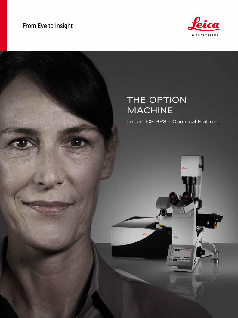
the option machine - leica microsystems tcs...a . b 5 the leica tcs sp8 is designed to meet all of...
TRANSCRIPT
Orde
r no.
: Eng
lish
???
???
∙ ??/
11/?
??/?
???
∙ Cop
yrig
ht ©
by
Leic
a M
icro
syst
ems
????
?,
Loca
tion,
Cou
ntry
, Yea
r. Su
bjec
t to
mod
ifica
tions
. LEI
CA a
nd th
e Le
ica
Logo
are
regi
ster
ed tr
adem
arks
of L
eica
Mic
rosy
stem
s IR
Gm
bH.
CONNECT
WITH US!
www.leica-microsystems.com
THE OPTION MACHINELeica TCS SP8 – Confocal Platform


3
“It is great if you can buy, with a small budget, a relatively standard microscope setup and if you need a special technique, you can up-grade it at a relatively low price.”
Bram van de Broek Netherlands Cancer Institute, NKI AvL Amsterdam
“I am very fond of the concept of modularity, because you don’t have to buy entirely new equipment every time you need additional capability.”
Jose Xavier Neto Laboratório Nacional de Biociências, Brasil
“When you start with a microscope configuration for your application, you don’t know how the research will develop. Especially in imaging facilities, we need to be flexible. And this flex-ibility is what I really like about the system.”
Astrid Schauss CECAD Imaging Facility, University of Cologne, Germany
CURRENT AND FUTURE APPLICATIONS
BY YOUR PARTNERS IN SCIENCE
4
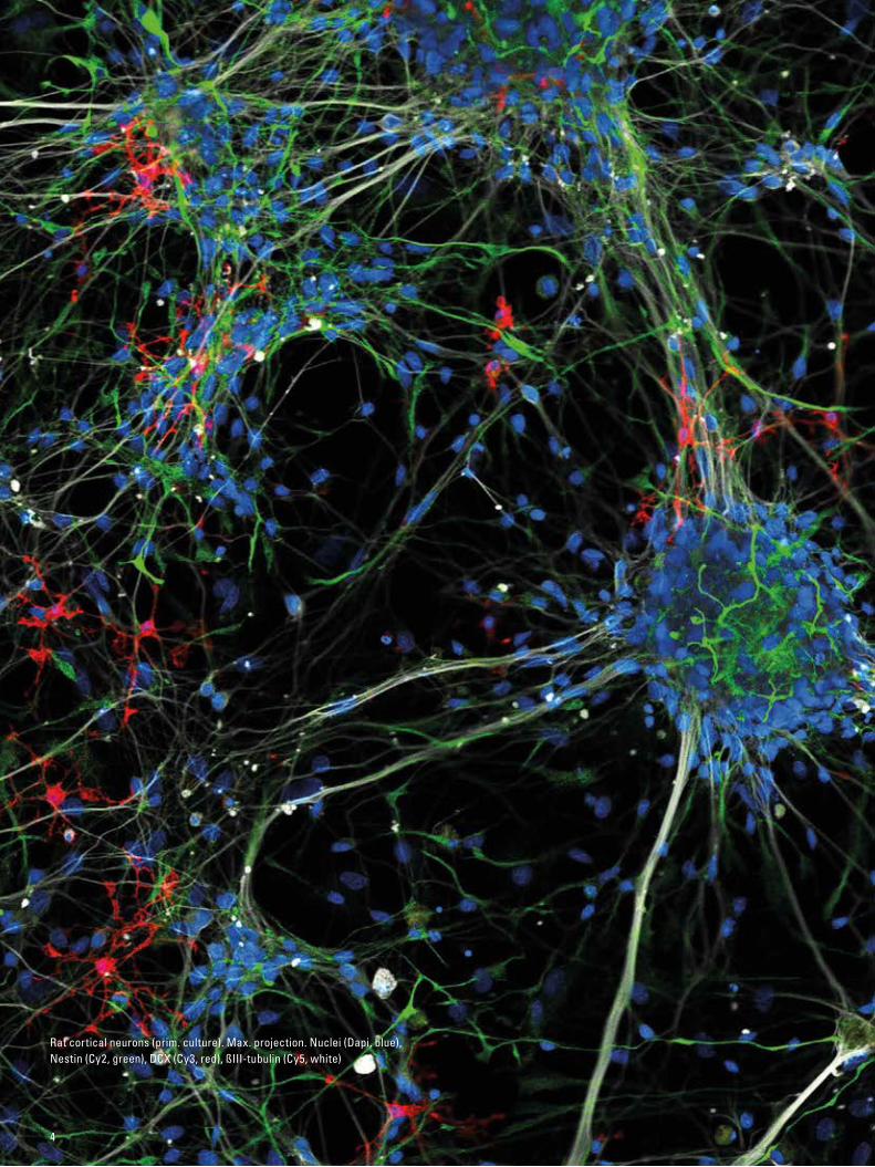
Rat cortical neurons (prim. culture). Max. projection. Nuclei (Dapi, blue), Nestin (Cy2, green), DCX (Cy3, red), ßIII-tubulin (Cy5, white)
A . B
5
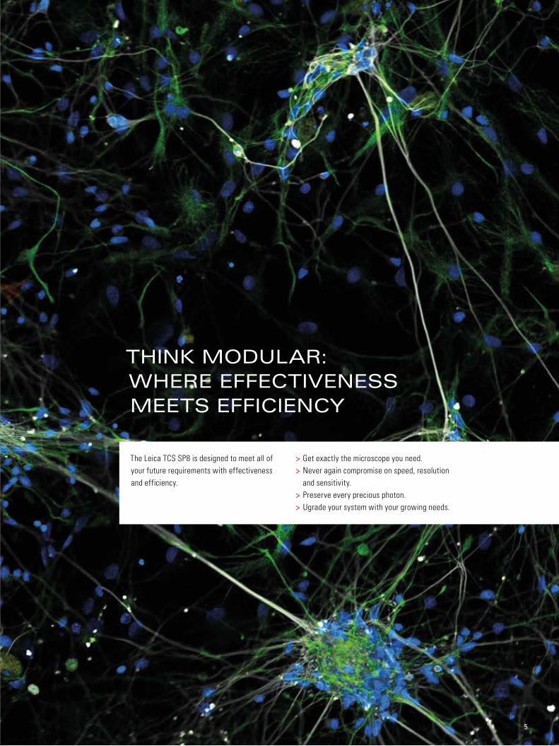
The Leica TCS SP8 is designed to meet all of your future requirements with effectiveness and efficiency.
> Get exactly the microscope you need. > Never again compromise on speed, resolution and sensitivity.
> Preserve every precious photon. > Ugrade your system with your growing needs.
THINK MODULAR: WHERE EFFECTIVENESS MEETS EFFICIENCY

6
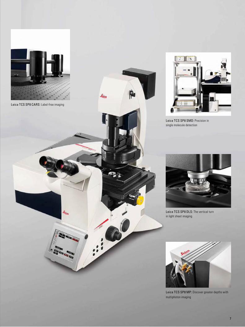
THE PLATFORM THAT IS READY FOR YOUR RESEARCH
GET WHAT YOU NEED NOW AND
UPGRADE WHENEVER NECESSARY.
Expand your possibilitiesDo you know which direction your research will take in the future? From live cell to quantitative imaging, from super-sensitivity to super-resolution, from multiphoton to light sheet imaging – with the Leica TCS SP8 you can always expand your possibilities.
Design your futureWe build on a modular concept: Tailor your microscope to your current needs and upgrade with additional functionalities at any time. Your investment in a Leica TCS SP8 will pay off – now and in the future.
Leica TCS SP8 X: Spectral freedom with the white light laser
Leica TCS SP8 STED 3X: Your next dimension in super-resolution
7
Leica TCS SP8 MP: Discover greater depths with multiphoton imaging
Leica TCS SP8 CARS: Label-free imaging
Leica TCS SP8 SMD: Precision in single molecule detection
Leica TCS SP8 DLS: The vertical turn in light sheet imaging

8
OPTIONS INSIDE
8
Mouse testis. Synaptonemal complex (Alexa 488, green), Nuclei (DAPI, blue), Protein X (Cy3, red). Courtesy of Yu Katsuyama and Noriko Osumi, Tohoku University, Miyagi, Japan.

9
Explore and analyzeSpend your time on your research while the Leica Application Suite X (LAS X) attends to microscope control. Its workflow-oriented design and intuitive software wizards guide you step by step through image acquisition, processing, and analysis. Tailor LAS X to your needs with additional software packages like 3D Visualization and Analysis, which allow you to understand the topology of your 3D image and quantify various aspects of intracellular structures. The examination and analysis of your experiments become extremely easy using the Leica TCS SP8 and LAS X.
Start thinking bigBreakthrough discoveries can happen when you are in the right place at the right time. Leica HCS A can speed up the process of discoveries through high content screening allowing you to standardize biological applications for rapid and reproducible results.
Computer Aided Microscopy (CAM) allows the detection of rare events using external image analysis software such as MatLab or the open-source ImageJ or CellProfiler software packages. Data is continuously streamed to external storage, and image analysis takes place simultaneously with data acquisition. Leica HCS A can respond to feedback from the analysis software, which simplifies large screening campaigns.
OPTIONS INSIDE
THE LAS X SOFTWARE PLATFORM CAN BE PERSONALIZED
TO YOUR WORK AND ASSISTS YOU IN PLANNING, EXECUTING,
AND ANALYZING YOUR EXPERIMENTS.
Without CAM (top): Important events may be missed.
With CAM (bottom): Time resolution is temporarily increased as needed. Process is studied in more detail and with better yield.
10
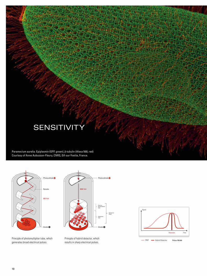
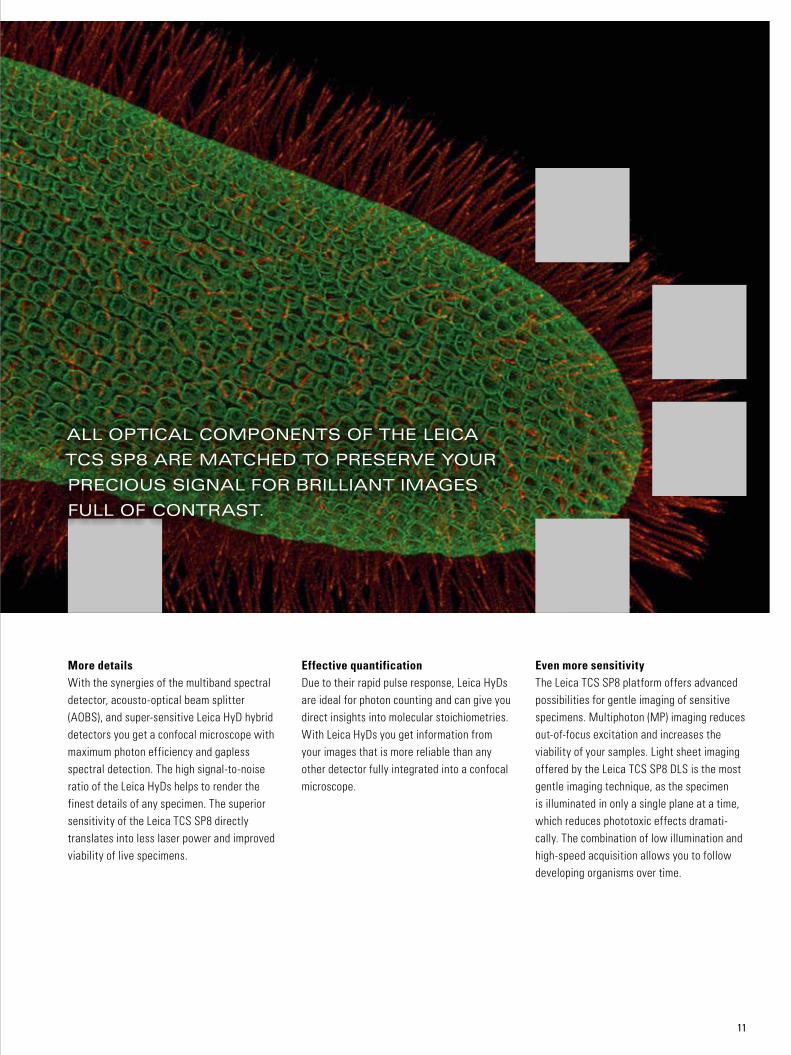
SENSITIVITY
Paramecium aurelia. Epiplasmin (GFP, green), β-tubulin (Alexa 568, red) Courtesy of Anne Aubusson-Fleury, CNRS, Gif-sur-Yvette, France.
Principle of photomultiplier tube, which generates broad electrical pulses.
Princple of hybrid detector, which results in sharp electrical pulses.
Photocathode
Anode
Dynode
Anode
800 Volt
ElectronBombardmentGain
AvalancheGain
AvalancheDiode
8000 Volt
Photocathode
Pulse Width
Pulse Width
11
More detailsWith the synergies of the multiband spectral detector, acousto-optical beam splitter (AOBS), and super-sensitive Leica HyD hybrid detectors you get a confocal microscope with maximum photon efficiency and gapless spectral detection. The high signal-to-noise ratio of the Leica HyDs helps to render the finest details of any specimen. The superior sensitivity of the Leica TCS SP8 directly translates into less laser power and improved viability of live specimens.
Even more sensitivityThe Leica TCS SP8 platform offers advanced possibilities for gentle imaging of sensitive specimens. Multiphoton (MP) imaging reduces out-of-focus excitation and increases the viability of your samples. Light sheet imaging offered by the Leica TCS SP8 DLS is the most gentle imaging technique, as the specimen is illuminated in only a single plane at a time, which reduces phototoxic effects dramati-cally. The combination of low illumination and high-speed acquisition allows you to follow developing organisms over time.
ALL OPTICAL COMPONENTS OF THE LEICA
TCS SP8 ARE MATCHED TO PRESERVE YOUR
PRECIOUS SIGNAL FOR BRILLIANT IMAGES
FULL OF CONTRAST.
Effective quantificationDue to their rapid pulse response, Leica HyDs are ideal for photon counting and can give you direct insights into molecular stoichiometries. With Leica HyDs you get information from your images that is more reliable than any other detector fully integrated into a confocal microscope.
12
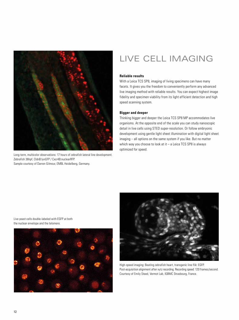
LIVE CELL IMAGING
Reliable resultsWith a Leica TCS SP8, imaging of living specimens can have many facets. It gives you the freedom to conveniently perform any advanced live imaging method with reliable results. You can expect highest image fidelity and specimen viability from its light efficient detection and high speed scanning system.
Bigger and deeperThinking bigger and deeper the Leica TCS SP8 MP accommodates live organisms. At the opposite end of the scale you can study nanoscopic detail in live cells using STED super-resolution. Or follow embryonic development using gentle light sheet illumination with digital light sheet imaging – all options on the same system if you like. But no matter which way you choose to look at it – a Leica TCS SP8 is always optimized for speed.
Live yeast cells double-labeled with EGFP at both the nuclear envelope and the telomere.
Long-term, multicolor observations: 17 hours of zebrafish lateral line development. Zebrafish 36hpf, CldnB:lynGFP / Cxcr4B:nuclearRFP. Sample courtesy of Darren Gilmour, EMBL Heidelberg, Germany.
High-speed imaging: Beating zebrafish heart, transgenic line flik: EGFP. Post-acquisition alignment after xytz recording. Recording speed: 120 frames/second. Courtesy of Emily Steed, Vermot Lab, IGBMC Strasbourg, France.
13
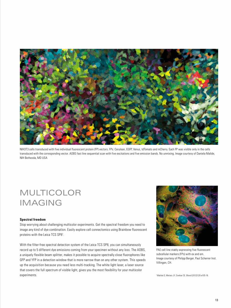
MULTICOLOR IMAGING
Spectral freedomStop worrying about challenging multicolor experiments. Get the spectral freedom you need to image any kind of dye combination. Easily explore cell connectomics using Brainbow fluorescent proteins with the Leica TCS SP81.
With the filter-free spectral detection system of the Leica TCS SP8, you can simultaneously record up to 5 different dye emissions coming from your specimen without any loss. The AOBS, a uniquely flexible beam splitter, makes it possible to acquire spectrally close fluorophores like GFP and YFP in a detection window that is more narrow than on any other system. This speeds up the acquisition because you need less multi-tracking. The white light laser, a laser source that covers the full spectrum of visible light, gives you the most flexibility for your multicolor experiments. 1 Malide D, Metais JY, Dunbar CE. Blood (2012)120:e105-16.
NIH3T3 cells transduced with five individual fluorescent protein (FP) vectors. FPs: Cerulean, EGFP, Venus, tdTomato and mCherry. Each FP was visible only in the cells transduced with the corresponding vector. AOBS fast line sequential scan with five excitations and five emission bands. No unmixing. Image courtesy of Daniela Malide, NIH Bethesda, MD USA
PAE cell line stably expressing five fluorescent subcellular markers (FPs) with ex and em. Image courtesy of Philipp Berger, Paul Scherrer Inst. Villingen, CH.
14
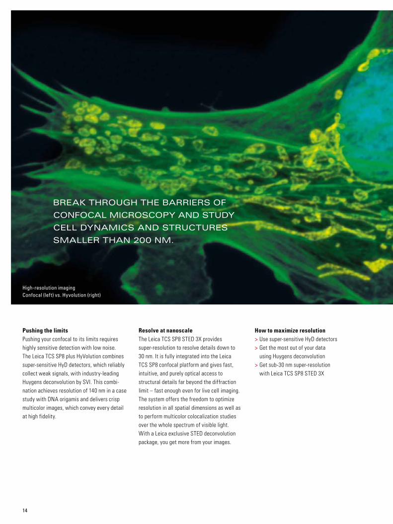
High-resolution imaging Confocal (left) vs. Hyvolution (right)
How to maximize resolution > Use super-sensitive HyD detectors > Get the most out of your data using Huygens deconvolution
> Get sub-30 nm super-resolution with Leica TCS SP8 STED 3X
BREAK THROUGH THE BARRIERS OF
CONFOCAL MICROSCOPY AND STUDY
CELL DYNAMICS AND STRUCTURES
SMALLER THAN 200 NM.
Pushing the limitsPushing your confocal to its limits requires highly sensitive detection with low noise. The Leica TCS SP8 plus HyVolution combines super-sensitive HyD detectors, which reliably collect weak signals, with industry-leading Huygens deconvolution by SVI. This combi - nation achieves resolution of 140 nm in a case study with DNA origamis and delivers crisp multicolor images, which convey every detail at high fidelity.
Resolve at nanoscaleThe Leica TCS SP8 STED 3X provides super-resolution to resolve details down to 30 nm. It is fully integrated into the Leica TCS SP8 confocal platform and gives fast, intuitive, and purely optical access to structural details far beyond the diffraction limit – fast enough even for live cell imaging. The system offers the freedom to optimize resolution in all spatial dimensions as well as to perform multicolor colocalization studies over the whole spectrum of visible light. With a Leica exclusive STED deconvolution package, you get more from your images.

15
RESOLUTION
High-resolution imaging with super-sensitive HyD detectors and Huygens deconvolution can resolve 140 nm in a case study with DNA origamis. Specimen: single molecule DNA-origamis with 140 nm defined spacing. (A) Confocal image. (B) HyVolution (C) Distance measured in B (green line). (D) Distribution of measured distances in nanometers.
Confocal AU 1.0
A
AU 0.6 +HyD and Deconvolution
Inte
nsity
freq
uenc
y
50.0100 200 300
Channel.001
140 nm
400 5000.00.000
0.061
0.123
0.184
0.246
0.307
0.369
0.430
0.492
0.553
0.615
0.676
0.738
0.799
0.861
0.922
0.984
5.0
10.0
15.0
20.0
25.0
30.0
35.0
40.0
45.0
100.0 150.0 200.0 250.0
[nm] Distance/nm
Inte
nsity
freq
uenc
y
50.0100 200 300
Channel.001
140 nm
400 5000.00.000
0.061
0.123
0.184
0.246
0.307
0.369
0.430
0.492
0.553
0.615
0.676
0.738
0.799
0.861
0.922
0.984
5.0
10.0
15.0
20.0
25.0
30.0
35.0
40.0
45.0
100.0 150.0 200.0 250.0
[nm] Distance/nmB C D

Tran
smis
sion
[%]
0300 400 500 600 700 800 900 1000
20
40
60
80
100
Wavelength [nm]
TISSUE CLEARING
Maximum depth, highest resolutionTransparent tissues help us to identify spatial arrange - ments and connectivity of cells and tissues. This is particularly helpful for identifying neuronal circuits. Clearing techniques provide maximum imaging depth and highest resolution when combined with the appropriate optics.
Reveal unexplored secretsWith the dedicated objectives for CLARITY and BABB treated specimens specifically designed by Leica Microsystems, you can reveal previously unexplored secrets. A motorized correction collar improves the resolution in deep tissue sections. Image whole organs at maximum imaging depth and resolution, and discover perfect CLARITY with single or multiphoton excitation.
300
Wavelength (nm)
HC FLUOTAR L25x/1.00 IMM motCORR VISIR
Tran
smis
sion
(%)
0
400 500 600 700 800 900
20
40
60
80
100
1000
Thy1-YFP adult mouse brain treated with CLARITY. Confocal imaging with excitation at 514 nm. Courtesy of K. Deisseroth and R. Tomer, Stanford University, Palo Alto, CA, USA.
16

STITCHING
From whole organisms to the highest level of detailThe Leica TCS SP8 offers the largest field of view in point scanning by using an X2Y scan mirror architecture. You can take images of whole animals or plants, often in one shot.
Mosaicking (stitching) is available for even larger specimens. Save time on stitching by covering the same area with a smaller number of images. Due to the homogenous field illumination provided by our scanner, your images are devoid of stitching artefacts. At the same time, every detail is preserved with high confocal resolution.
Coronar section of mouse nasal cavity. Mosaic compiled from 48 single images acquired using a high NA objective lens. (B) Close up from (A) indicating that high resolution is preserved at the whole field of view.
A
B
B
Large field – whole organism. Corona section of mouse nasal cavitiy.
17

18
CREATING THE OPTION MACHINEAN INTERVIEW WITH BILL HAY, SENIOR PROJECT MANAGER
OF THE SP8 DEVELOPMENT TEAM.
Why was the SP8 built on a platform principle?That was an answer to the customer require-ments, of course. If customers want to start with a confocal, but can’t afford the tricked-out system with all the bells and whistles, they can add new features year by year, just as their budget allows and science requires. When purchasing an SP8, our customers can look into the future and ask themselves what they are going to use it for and what they may need to add to it. We made an 'option' machine, so that the researcher has more options for the future.
So is it basically an expression of Ernst Leitz’s principle “with the user, for the user”?It does sum up very well what we’re doing. If we don’t do it for the user, there is no benefit to any of the things we do. If we’re not making a product that people need, we’re not helping to advance research and science. Therefore, we work with our customers to understand the science they are doing and how they’re doing it. Then we can incorporate certain features and technologies and anticipate some of the needs that will arise from the development of science. We learn from the user, so we help him again in return – it’s a symbiotic relationship.
How did you resolve the conflicting requirements of a confocal microscope, such as sensitivity, speed and resolution?First of all, efficiency is an area where we won’t compromise. Efficiency leads to sensitivity, so you don’t lose light. That’s the core principle of the SP8: efficient collection of light and
brightness. We improved optical coatings for the SP8 wherever possible with newer manu-facturing processes that help us to increase the optical efficiency. In the end the SP8 is designed to gather photons.
But the challenge is to accommodate all three: sensitivity, speed, and resolution. And where the requirements really conflict, you have to make sure that you have the flexibility to serve all customers across the spectrum. If they need the higher speed, at some point they have to accept compromise in resolution. But we have to be able to offer the highest resolution for the people who don’t need speed. You have to make sure you deliver the optimum combi-nation of all three.
What is the key to the SP8’s success?One of the major keys to its success, I would say, is the flexibility of the different options that we offer. But the SP8 also comes with an intel-ligent user interface, so that customers aren't overwhelmed by all the features and complexity. In the end, they shouldn’t even notice that we enable them to concentrate on their expe ri-ments, while we take care of controlling the system. In addition, we have a wide variety of tools and innovations – e.g., the white light laser, STED, and gating function – that are com bined in one system. And not to be for- gotten, our optics, efficiency, and image quality are second to none. It is extremely important to the user that they have the best possible image quality.
What does it mean to be a developer at Leica?It's never boring. We work in a field that is developing incredibly quickly. And with all the innovation that’s taking place, it’s a constant challenge to keep up and to keep ahead. The SP8 is certainly not the end of the road. We are always looking to improve on the things we already do, and to implement new features and methods that we don’t have yet. You never know what new developments might come along. One of the biggest challenges is to make a system that accommodates new develop-ments without having to change the platform. The light sheet module is a prime example of that. But that’s not the only thing that makes the work interesting. A whole team of gifted individuals comes together with a lot of brilliant ideas and innovations. This is the real basis for the SP8. I think that we have great people here with innovative ideas and a dedication to translate a vision into a real system for the user. We get to play with a machine like this and work every day on challenging new ideas. After all, it’s a pretty damn cool machine!
William C. Hay holds a Bachelor in Physics from Davidson College and a Master in Electrical Engineering from Duke University in North Caroline. He joined Leica Microsystems in 1989 in the semi-conductor applications department and switched to R&D project management in 1996. He was the project manager for the development of three generations of confocal microscopes: Leica TCS SP2, Leica TCS SP5 and Leica TCS SP8.

A . B
19
10/1
5 · O
rder
no.
: 159
3003
016
· © 2
015
by L
eica
Mic
rosy
stem
s CM
S Gm
bH.
Subj
ect t
o m
odifi
catio
ns. L
EICA
and
the
Leic
a Lo
go a
re re
gist
ered
trad
emar
ks o
f Lei
ca M
icro
syst
ems
IR G
mbH
www.leica-microsystems.comCONNECT WITH US!
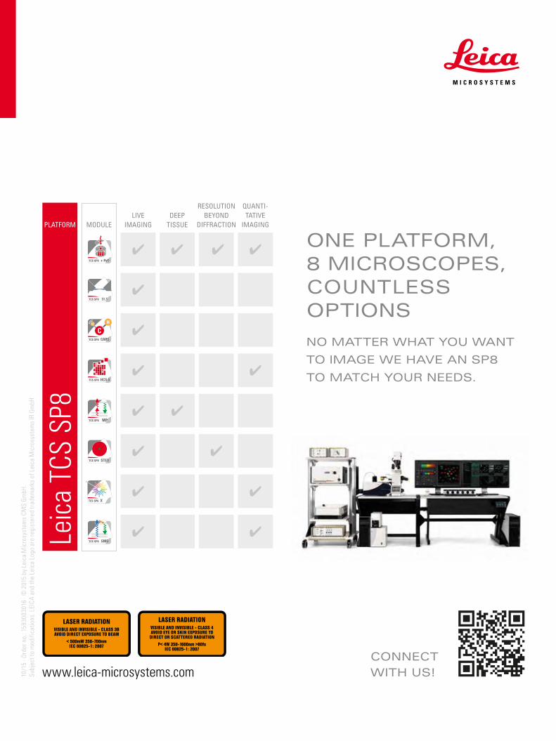
ONE PLATFORM, 8 MICROSCOPES, COUNTLESS OPTIONS
NO MATTER WHAT YOU WANT
TO IMAGE WE HAVE AN SP8
TO MATCH YOUR NEEDS.
PLATFORM MODULELIVE
IMAGINGDEEP
TISSUE
RESOLUTION BEYOND
DIFFRACTION
QUANTI-TATIVE
IMAGING
Leic
a TC
S SP
8