the ophthalmology anatomicalwith the position of the examiner's eye, and requires a total...
TRANSCRIPT

THE BRITISH JOURNAL OF OPHTHALMOLOGY
A NEW ANATOMICAL NOTATION OF THEVISUAL FIELD
BY
DR. M. URIBE TRONCOSONEW YORK
THE examination of the visual field deserves a more importantplace in the routine examination of the eyes than it now holds.Not only the beginner but the practising ophthalmologist as well,are deterred from a wider use of this method by the many com-plexities and loss of time its practical application entails. For thisreason the simplification and improvement of our present techniquewill do much in promoting a better understanding and largerapplication of this important diagnostic agency.One of the first technical difficulties the beginner has to deal
with, is the way of recording the visual field on the chart. Asmapped now the findings in the perimeter need to be invertedtwice. First comes the physiological inversion by which theimiage formed in the retina is projected to the opposite side of theeye. This requires the readings of the position of the test carrierto be made on the side of the perimeter opposite to the part of theretina under examination. The second inversion is madenecessary by the habit of considering the visual field in relationwith the position of the examiner's eye, and requires a totaltransfer of the findings for charting the right side of the patient'seyes to the right side of the observer's eye, the left side to, theleft, etc. This double inversion is the result of a faulty mentalhabit and is in complete violation of the anatomical ru,le whichrequires the subject to be considered always standing in frontof the observer.
In a previous paper(M) I have emphasized the confusion andmisunderstanding that the departure from this basal anatomicalrule has produced in all ophthalmological descriptions and theadvantages to be derived from the change to one standard positioninstead of two. The present method of recording the field ofvision was probably originated by the primitive use of the black-board and afterwards of the campimeter, in which the examinerplaces himself behind the patient. The limits of the field andscotomas are drawn on the blackboard in relation to the eyesof both, oonsidered in the same position on the space, and themapping, when necessary, is very simple, as it only requires asimilar drawing on a reduced scale., This method had theadvantages of enabling the observer, to visualize the conditionson whichi the' visual cone of the patient is projected into space,
280
on March 24, 2020 by guest. P
rotected by copyright.http://bjo.bm
j.com/
Br J O
phthalmol: first published as 10.1136/bjo.10.5.280 on 1 M
ay 1926. Dow
nloaded from

ANATOMICAL NOTATION OF THE VISUAL FIELD
and to refer all changes to the examiner's own right and left sides.With the advent of the perimeter, however, conditions for theexamination of the fields changed entirely. The observer had tostand in front of the patient for moving the test carriers, readingthe findings on the arc, and watching the central fixation of theeye. This change of position immediately required a readjust-ment of the mental picture of the visual cone of the patient, nowextending in front of the observer, the right side of the patientcorresponding to the left of the physician. The physiologicalinversion of the retinal images could be more clearly understoodand recorded on the arc of the perimeter in this position, thelarger temporal field corresponded to the larger extent of the nasalretina, and the restricted nasal field to the shorter area of thetemporal retina; the blind spot being about in the centre of thepercipient layer of this membrane (Elliot). But when the findingsin the arc were to be recorded on the chart, no change was madeto meet the new conditions, and the old schemes were used, withthe result that a second inversion became necessary to map thetemporal field A of the patient's eye (Fig. 1), at-the correspondingpoint B of the observer's eye. This second inversion althoughrelatively easy for a trained ophthalmologist, is difficult andrequires a considerable mental effort for the beginner. Thetransfer is much easier in the four cardinal points, viz.: temporal,nasal, upper and lower meridians, but for intermediate positions,300 or 600, 3000 or 3300, which need to be recorded at the sameinclination on the corresponding side of the observer's eye, thedifficulty increases a great deal, especially if, as frequentlyhappens, the graduation of the meridians in the card is differentfrom those in the perimeter.
In order to avoid the inevitable confusion and misunderstandingof the double inversion, the self-registering perimeters weredevised. In the simpler instruments the chart is carried by adisc which rotates with the arc, and the markings are made onthe side of the same colour, red or white, of the figures of thearc. Even with this perimeter, however, when the observer, forsaving time, tries to use both sides of the arc alternately, the sameembarrassment and confusion arises in regard to the place formarking and the inclination of the meridians. Only in the moreexpensive and complicated models are the records made auto-matically accurate.The difficulties of this faulty, irksome method of recording
the visual field are particularly evident when the student tries todetermine to which part -of the field a given defect or lesionin the retina corresponds. The mental habit of considering therelative positions of the papilla and the macula as we see themwith the ophthalnioscope, makes it difficult for the observer to
281
on March 24, 2020 by guest. P
rotected by copyright.http://bjo.bm
j.com/
Br J O
phthalmol: first published as 10.1136/bjo.10.5.280 on 1 M
ay 1926. Dow
nloaded from
282 THHE BRITISH JOURNAI OF OPHTHALAIOLOGY
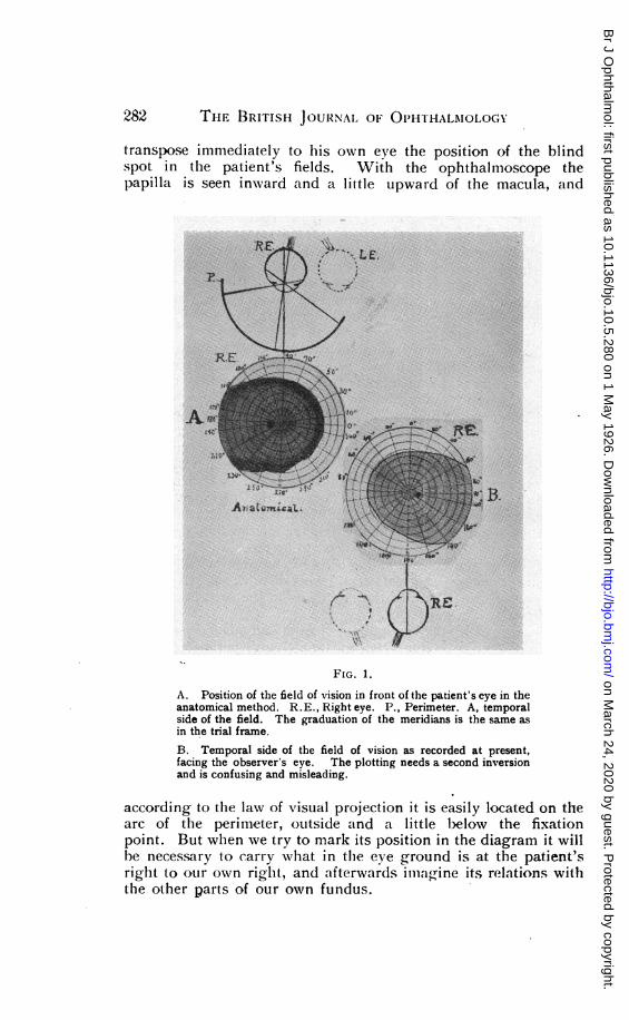
transpose immediately to his own eye the position of the blindspot in the patient's fields. With the ophthalmoscope thepapilla is seen inward and a little upward of the macula, and
Le.
Amf IF
up_ jjs:IN ti,AI,a t it 4
B.
FIG. 1.
A. Position of the field of vision in front of the patient's eye in theanatomical method. R. E., Right eye. P., Perimeter. A, temporalside of the field. The graduation of the meridians is the same asin the trial frame.B. Temporal side of the field of vision as recorded at present,facing the observer's eye. The plotting needs a second inversionand is confusing and misleading.
according to the law of visual projection it is easily located on thearc of the perimeter, outside and a little b}elow the fixationpoint. But wlhen we try to mark its position in the diagram it willhe necessary to carry what in the eve ground is at the patient'sright to our own righ-t, and afterwards inlagine its relations withthe other parts of our own fundus.
D 6 e
w
on March 24, 2020 by guest. P
rotected by copyright.http://bjo.bm
j.com/
Br J O
phthalmol: first published as 10.1136/bjo.10.5.280 on 1 M
ay 1926. Dow
nloaded from

ANATOMICAL NOTATION OF THE VISUAL FIELD
It is even more arduous to find the place which in the patient'sfield of vision will correspond with a characteristic change foundwith the ophthalmoscope. A thrombosis of the superior nasalartery of the right eye, for instance, will produce a sector-likedefect in the opposite side of the field, that is in the inferior
TzgtEr
FIG. 2.
Schematic representation of the optic pathways, fields of vision on bothsides as projected in the space in front of the observer, and records of thefields in the anatomical position.
temporal quadrant. With the perimeter this defect will beimmediately found in its proper place, but if the observer tries tovisualize the ophthalmoscopic lesion and then represent tohimself the defect produced in the field, he will have to transferthe-lesion first to his own eye, and the,n locate the defect in theopposite side according to the law of visual projection.
Probably on account of the early use of the campimeter and thefacility of representing the pathological changes in the patient, thedescription of diseases of the central nervous system has also been
283
on March 24, 2020 by guest. P
rotected by copyright.http://bjo.bm
j.com/
Br J O
phthalmol: first published as 10.1136/bjo.10.5.280 on 1 M
ay 1926. Dow
nloaded from
THE BRITISH JOURNAL OF OPHTHALMOLOGY
made considering the observer as standing behind the patient. Thewell-known diagrams of the central nervous system, the tracts, opticnerves and correlation of the two retinas are always representedin the unanatomical position; the only advantage of this is to beable to visualize the lesions as if they existed in the physician's ownorgans and designate the right and left sides by the same names.This has, however, numerous disadvantages. In hemianopsia,for instance, if the lesion is present in the right optical tract, theright halves of both retinas will be blind (Fig. 2). The patient,by the physiological inversion, sees only the right halves of allobjects and describes his symptoms in relation to his own body.In our clinical examination the patient is facing us and explainingwhich side of the objects he sees. With the perimeter we findthat the nasal field in the right eye and the temporal field in theleft are wanting; still following the custom, we ought to recordthe defect referring it to our own eyes, and make the diagnosisby changing sides and ascertaining which side of the objects ourhemianopic eye will see.Would it not be easier, following the anatomical rule, to
consider the visual cones of the patient projected into the spacein front of the observer, the right field in front of the right eye,the left before the left, and record the fieIds as they are found inthe arc of the perimeter, with only one inversion instead of two(Fig. 1, A, and Fig. 2)? The visual cones have their apices inthe nodal points of each eye and base in the infinite, but theobserver needs to consider only a section, corresponding eitherto the perimeter or the tangent screen.The same as with the perimeter, the findings in the Bjerrum
screen will be better understood and recorded in the anatomicalposition than by the old method. In fact it is difficult to under-stand how the plotting made in the graduated side of the curtainfacing the observrer, was not immediately recorded in a diagramexactly as it was found, instead of making the second inversionto the examiner's eye.No change in our instruments is necessary for the new method,
except that the campimeter should be made of cloth for thrustingpins and marking the limits and defects in the field on the sidefacing the operator.With the adoption of the new anatomical -field it would be
important to standardize the notation of the meridians in thecharts, which at the present time is very confusing. In somecharts 00 is placed above, 1800 below, and 900 on either side of thehorizontal meridian for both eves. In others for the right eye theOc is above, 900 at the left of the patient, 2700 to his right and1800 below. For the left eye 900 is at the right of the patient and
284
on March 24, 2020 by guest. P
rotected by copyright.http://bjo.bm
j.com/
Br J O
phthalmol: first published as 10.1136/bjo.10.5.280 on 1 M
ay 1926. Dow
nloaded from

ANATOMICAL NOTATION OF TIHE VISUAL FIELD
2700 to the left, the vertical meridian being the same as in theother eye.
In America the most accepted notation is 900 above, 2700below, and the 00 at the left side of the patient. In the anatomicalfield it will be desirable to adopt the last-mentioned notation,and represent the inclination of the meridians exactly in the sameway as the axis of the cylinders in the trial frame, that is the 00always at the left of each eye of the patient, 1800 to the right,900 above, and 2700 below (Fig. 1, A). In this way it will be easy
'4
/1S
FIG. 3.
Homonymous diplopia. The deviation of the left eye inward make's theimage of the candle fall at the inner side of the macula, M, and thisimage is projected outward, where a false image is seen at the left.
to find the inclination of every meridian and draw an accurat-ediagram even on a provisional chart, by comparison with thetrial frame.
I am well aware that this complete change in the m'apping of thevisual field will find a great opposition amongst ophthalmologists,who being trained in the double standard positilon, have acquiredthe habit of the mental process required to make the inversionquickly and without any mental -strain. They will find it un-necessary or even inadvisable to depart from their habits, but forthe beginner in o)phthalmology to be taulght in the anatomicalposition to which he has been accustomed, will mean much timesaved and much effort spared.The transition between the old and new method is very simple.
New charts may be printed with the same diagram they have at
285
on March 24, 2020 by guest. P
rotected by copyright.http://bjo.bm
j.com/
Br J O
phthalmol: first published as 10.1136/bjo.10.5.280 on 1 M
ay 1926. Dow
nloaded from

THE BRITISH JOURNAL OF OPHTHALMOLOGY
present, not exactly inverted, but as seen by translucency; thetemporal side of the chart in front of the temporal side of bothorbits of the patient. Even the old charts can be put to. service,simply changing sides, the right eye being used for the left andvice versa; but of course the graduation in the meridians willbecome inverted, which is not desirable.
Fixation Field.-The monocular and binocular fields of fixationwill be more easily understood and recorded in the anatomicalposition, than after the inversion necessary for transferring themto the position of the observer's eye.
In paralytic strabismus the examination of the binocular fieldof fixation with the tangent screen in the six cardinal directionswill detect the limitation of movement, the extent of the field forsingle and double vision, the kind of diplopia, and the separationof the two images, which can be plotted on the white side of thescreen and recorded on the anatomical card without any inversion.This will give the student a much better idea of the situation ofthe false image in space and its relations with the retina of thepatient, as he can visualize by the deviation of the optical axisthe new location of the image outside the macula and the directionin which the false image will be projected (Fig. 3).
It is a well-known fact that the chapter on abnormal motility ofthe eye is considered one of the most difficult to grasp by thestudent. The normal anatomy and physiological action of theextraocular muscles are taught with the cadaver or subject in thestandard anatomical position, but the abnormalities in the functionare considered not in the same way, the proper scientific method,but by reversing the findings to the observer's own eye, thuscreating a double system for the study and interpretation of thesecomplicated symiptoms with inevitable confusion and misunder-standing.
In a paper presented to the Ophthalmological Section of theAmerican Medical Association in May of this year, Dr. LutherC. Peter, of Philadelphia(2), endorses this anatomical method, andgives added reasons for the adoption of one universal standardinstead of two. The same author in a personal communication,states also his belief in the necessity of changing the recordingof the field of vision to the more scientific and accurate anatomicalposition.
REFERENCES1. M. Uribe Troncoso.-Anatomical versus Ophthalmological Nomenclature. Trans.
Internat. Con. Ojhthal., Washington, 1922.2. Luther C. Peter.-Anatomic Method of studying and recording the Motility of the
Eyes. Trans. Sec. Ophthal., Amer. Med. Assoc., May. 1925.
286
on March 24, 2020 by guest. P
rotected by copyright.http://bjo.bm
j.com/
Br J O
phthalmol: first published as 10.1136/bjo.10.5.280 on 1 M
ay 1926. Dow
nloaded from