the objective identification of dermatoscopic features using spectrophotometric intracutaneous...
TRANSCRIPT
The Objective Identification of Dermatoscopic Features using
Spectrophotometric Intracutaneous Analysis

Marc Moncrieff 1,4, Symon Cotton2, Ela Claridge3, Per Hall1
1 - Department of Plastic & Reconstructive Surgery, Addenbrooke’s Hospital, Cambridge, UK
2 - Astron Clinica, Cambridge, UK
3- Department of Computer Science, University of Birmingham, UK
4 - Department of Plastic & Reconstructive Surgery, West Norwich Hospital, Norwich, UK
SIA
• Technique developed in UK
• Analyses remitted light from the skin
• Produces ‘SIAgraphs’
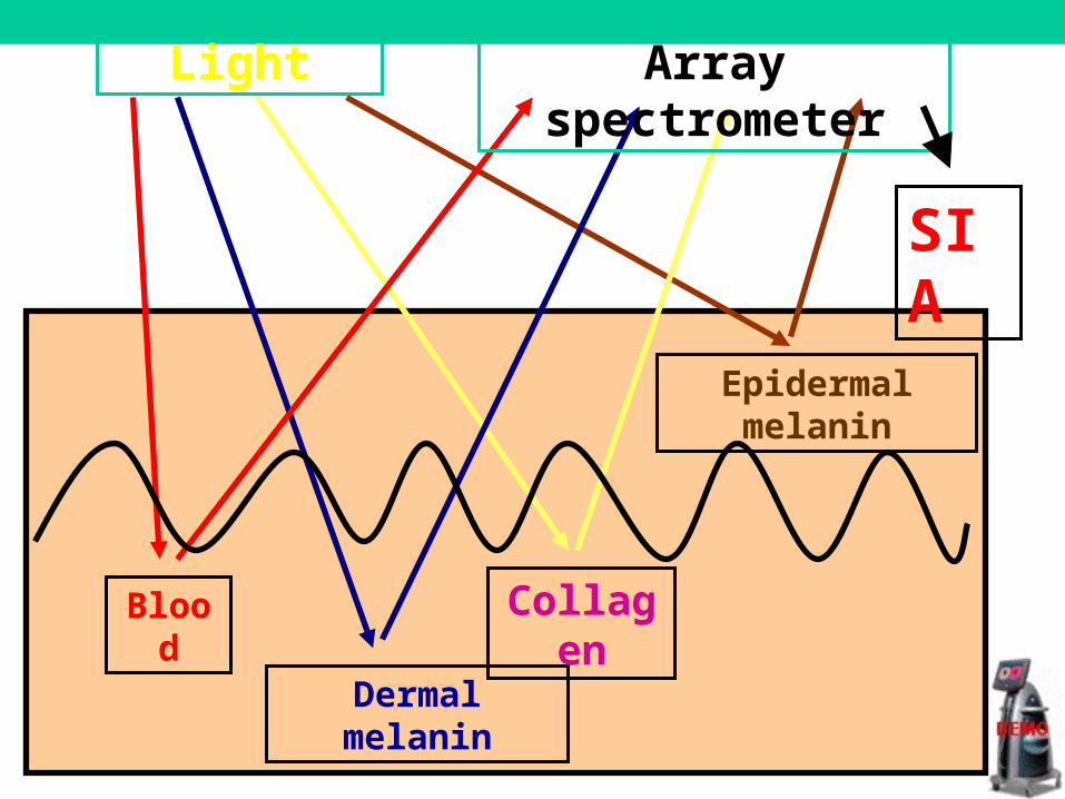
Epidermal melanin
CollagenBlood
Dermal melanin
Array spectrometerLight
SIA
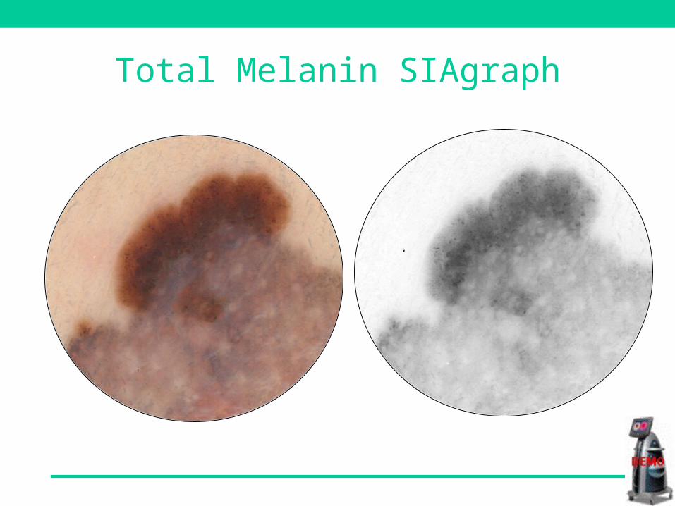
Total Melanin SIAgraph
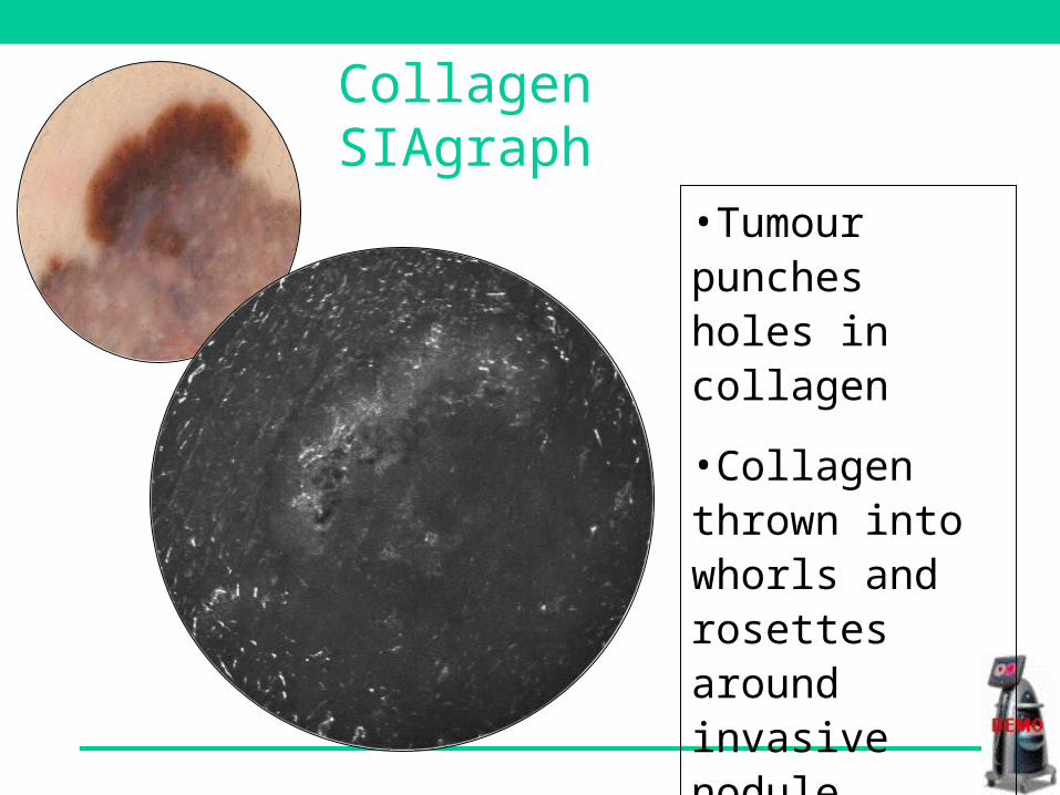
•Tumour punches holes in collagen
•Collagen thrown into whorls and rosettes around invasive nodule
•Thickening of collagen at invasive margin
Collagen SIAgraph
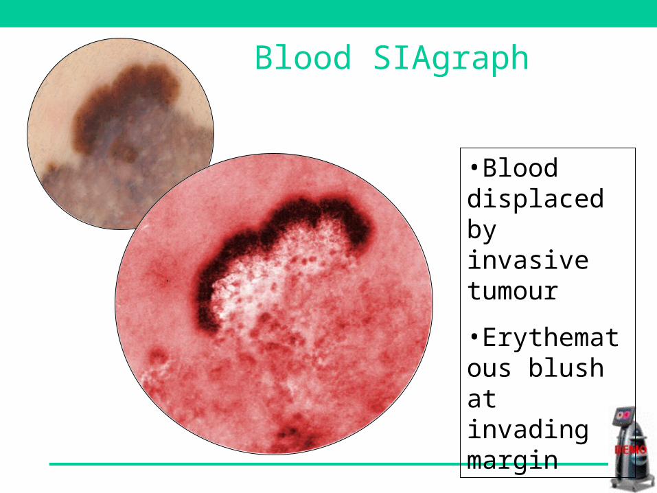
•Blood displaced by invasive tumour
•Erythematous blush at invading margin
Blood SIAgraph
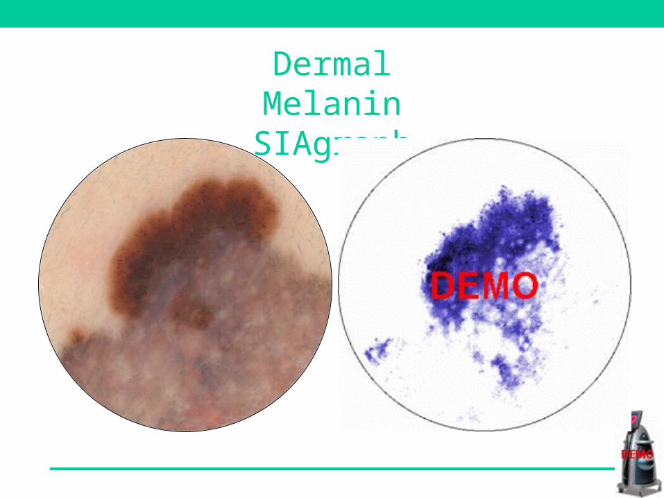
Dermal Melanin SIAgraph
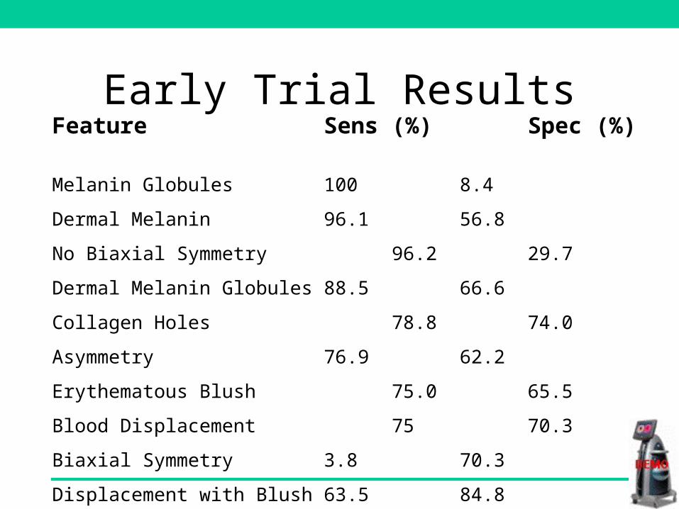
Early Trial ResultsFeature Sens (%) Spec (%)
Melanin Globules 100 8.4
Dermal Melanin 96.1 56.8
No Biaxial Symmetry 96.2 29.7
Dermal Melanin Globules 88.5 66.6
Collagen Holes 78.8 74.0
Asymmetry 76.9 62.2
Erythematous Blush 75.0 65.5
Blood Displacement 75 70.3
Biaxial Symmetry 3.8 70.3
Displacement with Blush 63.5 84.8
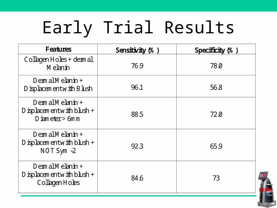
Early Trial ResultsFeatures Sensitivity (%) Specificity (%)
Collagen Holes + dermal Melanin 76.9 78.0
Dermal Melanin + Displacement with Blush 96.1 56.8
Dermal Melanin + Displacement with blush +
Diameter > 6mm 88.5 72.0
Dermal Melanin + Displacement with blush +
NOT Sym -2 92.3 65.9
Dermal Melanin + Displacement with blush +
Collagen Holes 84.6 73
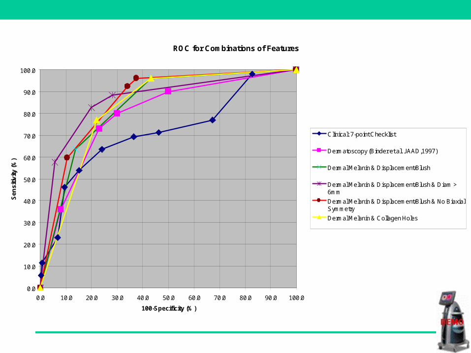
ROC for Combinations of Features
0.0
10.0
20.0
30.0
40.0
50.0
60.0
70.0
80.0
90.0
100.0
0.0 10.0 20.0 30.0 40.0 50.0 60.0 70.0 80.0 90.0 100.0
100-Specificity (%)
Sen
sitiv
ity (%
)
Clinical: 7-point Checklist
Dermatoscopy (Binder et al. JAAD,1997)
Dermal Melanin & Displacement/Blush
Dermal Melanin & Displacement/Blush & Diam >6mm
Dermal Melanin & Displacement/Blush & No BiaxialSymmetry
Dermal Melanin & Collagen Holes
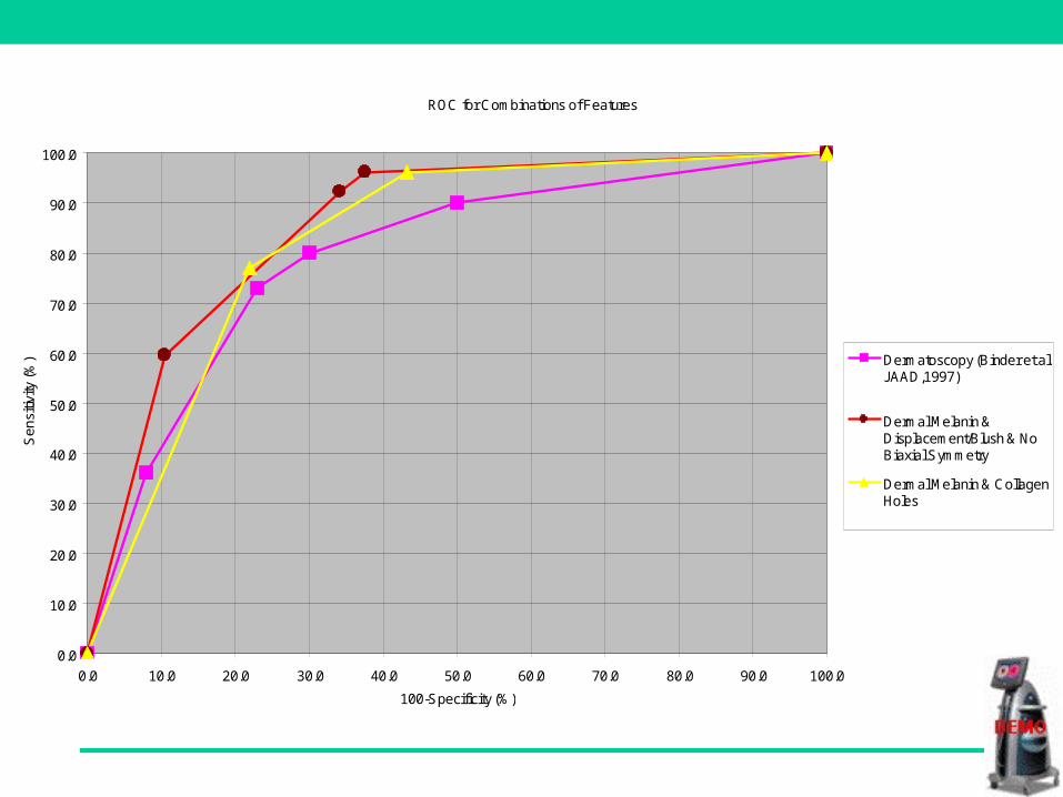
ROC for Combinations of Features
0.0
10.0
20.0
30.0
40.0
50.0
60.0
70.0
80.0
90.0
100.0
0.0 10.0 20.0 30.0 40.0 50.0 60.0 70.0 80.0 90.0 100.0
100-Specificity (%)
Sen
sitiv
ity (%
) Dermatoscopy (Binder et al.JAAD,1997)
Dermal Melanin &Displacement/Blush & NoBiaxial Symmetry
Dermal Melanin & CollagenHoles
Early Trial Results
• Very simple features identified
• As accurate as dermatoscopy
• Results do not include interpretation of
dermatoscopic features
Skin Surface Microscopy / Dermatoscopy
• A (useful) adjunct to clinical diagnosis
• Limitations
– training very limited and very necessary
– subjective findings
– subjective interpretation
Blue
Formed by:– Dermal melanin– dermal Hb– thinning of collagen– thickening of keratin
Hidden by:– epidermal melanin
– thrombosis
Bahmer et al (1990):
“Histopathologically this corresponds to superficial fibrosis with melanophages and / or pigmented malignant cells in the papillary dermis”
Bahmer et al (1990) J Am Acad Dermatol 23: pp. 1159-62
Blue - Grey (white) Veil
Menzies et al (1996):
“It is the single most significant surface microscopic finding of invasive melanoma”
Menzies et al (1996) An Atlas of Surface Microscopy of Pigmented Skin Lesions (p.18)
Blue - Grey (white) Veil
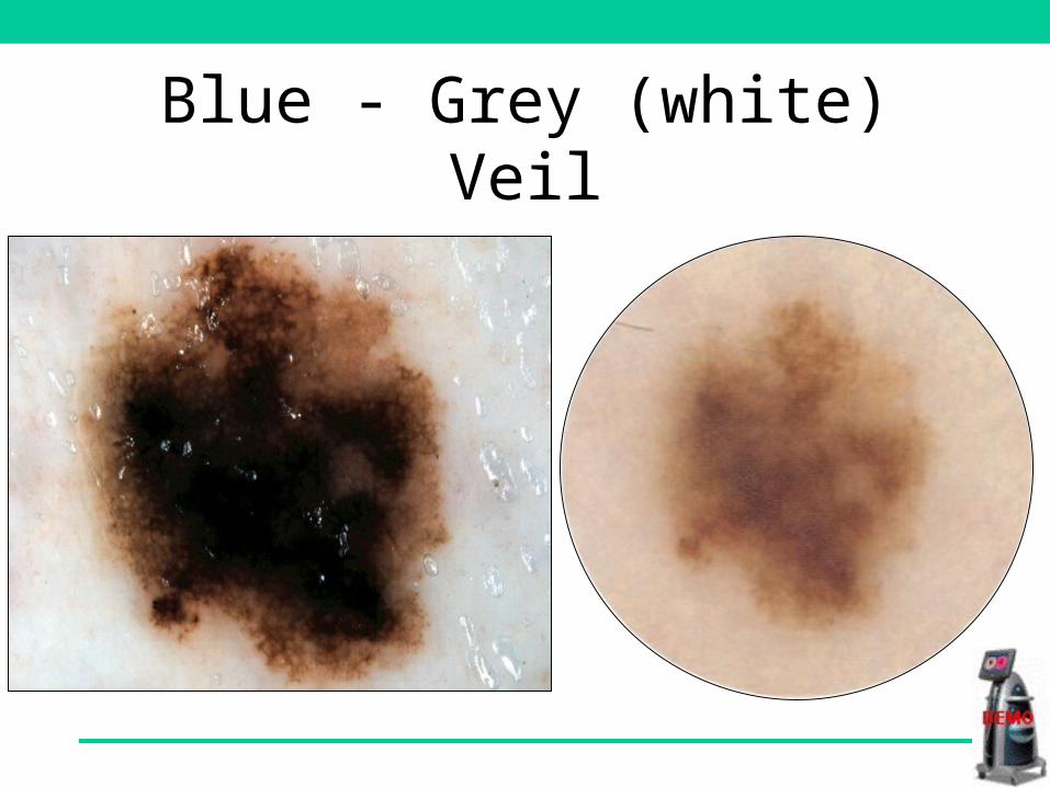
Blue - Grey (white) Veil
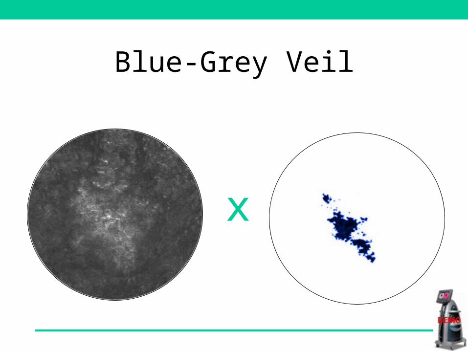
Blue-Grey Veil
x
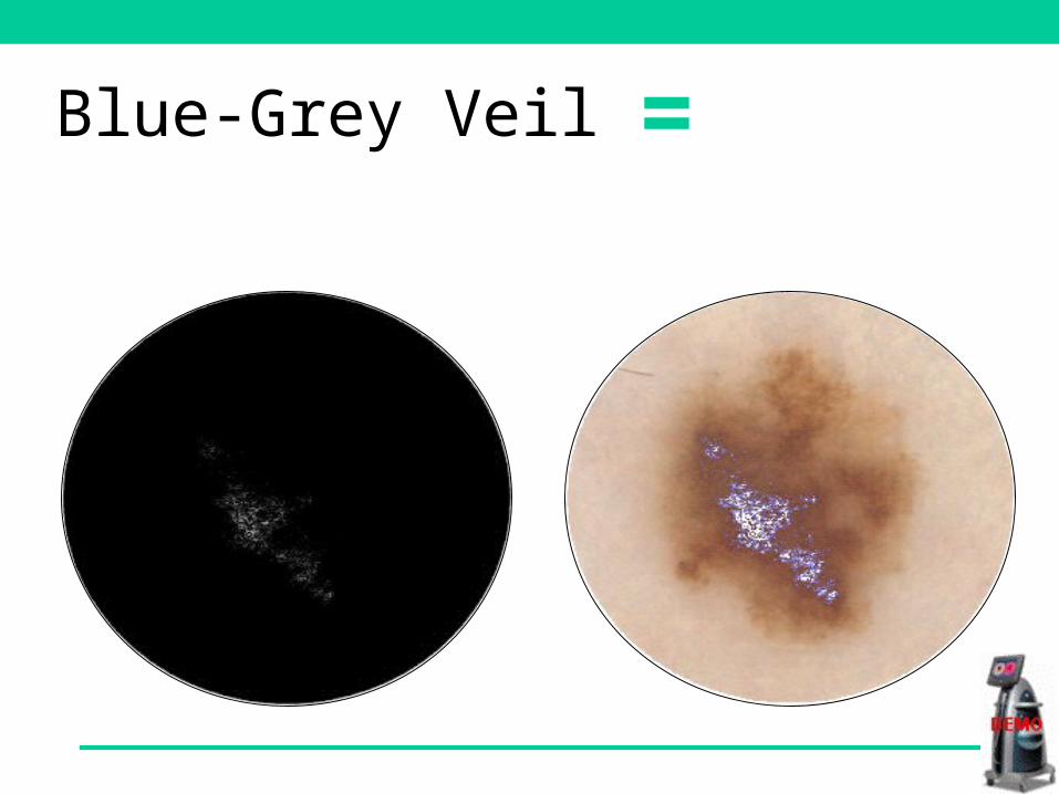
=Blue-Grey Veil
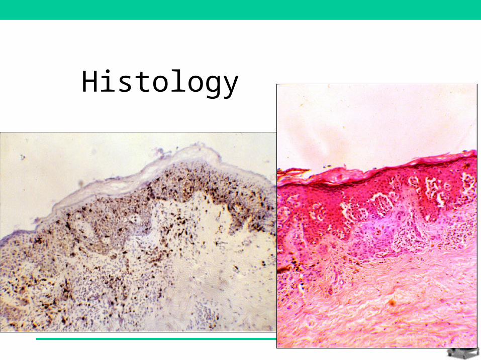
Histology
Summary
• Blue/grey veil detected by SIAscopy Sensitivity = 96% for invasive melanoma
• Allows objective assessment of surface microscopic features according to underlying histopathology
• The SIAscope is a diagnostic tool in its own right