the molecular defect underlying fucosidosis
TRANSCRIPT

8JMed Genet 1996;33:284-288
The molecular defect underlying canine
fucosidosis
Barbara J Skelly, David R Sargan, Michael E Herrtage, Bryan G Winchester
AbstractFucosidosis is a lysosomal storage diseasewhich affects humans and Englishspringer spaniel dogs. The disease is re-cessively inherited in both species and res-ults from a deficiency of the enzyme a-L-fucosidase. We have recendy cloned andsequenced the canine fucosidase gene(EMBL sequence admission numberX92448 (cDNA) and X92671-X92678 (in-dividual exonic data)). The gene spans12 kb and consists of eight exons. SSCPbased mutation analysis of affected an-imals was carried out on the coding regionof this gene both with exonic primers, andintronic primer pairs for each exon. A14 base pair deletion of the cDNA wasidentified at the 3' end of exon 1 in fu-cosidosis affected animals. Surprisingly,PCR based genomic cloning ofDNA fromthese animals showed an identical deletionin this DNA, ending at the start of intron1. This change causes a frameshift and, inconsequence, 25 novel codons are tran-scribed in exon 2 before the first of twoadjacent premature stop codons is en-countered.(JMed Genet 1996;33:284-288)
Key words: canine fucosidosis; oa-L-fucosidase; ly-sosomal storage disease; mutation detection.
The lysosomal storage disease fucosidosis hasbeen described in humans' and also in pedigreeEnglish springer spaniels in Australia and inthe United Kingdom.23 Affected dogs have aprofound deficiency (0-5% of the controlmean) of the enzyme oc-L-fucosidase whichcatalyses removal of fucose moieties from theoligosaccharide side chains of glycoproteinsand glycolipids. Pathology and clinical signsare the result of the lysosomal accumulation offucose containing glycoconjugates.' The cent-ral nervous system displays particular markedstorage abnormalities. Heterozygotes are with-out symptoms and possess fucosidase activitiesof approximately 50% of the control mean.The disease in the springer spaniel manifestsas a mainly neurological syndrome with mixedmotor and mental function deficits includingprogressive ataxia, proprioceptive difficulties,apparent blindness, change in temperament,dysphagia, dysphonia, and loss of learned be-haviour.5 As the disease progresses there isa rapid deterioration in body condition withmuscle wasting. Affected dogs die or areeuthanased by 3 to 4 years of age. The caninedisease has been likened to the delayed onsethuman form because dogs are clinically normal
until 18 months of age. Thus, disease sim-ilarities have led to the use of springer spanieldogs as models in bone marrow transplantationtrials.67Both the canine and the human diseases are
inherited as autosomal recessive conditions.Pedigree analysis of canine families suggeststhat a common British founder is present inall affected lines, including those in Australiawhich were derived from exported Britishbreeding stock. Testing for carrier status indogs has been conducted in the UK by analysisof enzyme activity.8 Heterozygotes should haveenzyme activities that are approximately 50%of the control mean. The division betweennormal and heterozygous dogs is, however,often indistinct and a molecular description ofthe disease causing mutation would be valuablein allowing definitive designations of genotypewithin the springer spaniel population.The cDNA for human oc-L-fucosidase
(FUCAl) has been cloned,9 and the genomicorganisation established.'0 In addition, mostfamilies with affected family members have hadthe molecular basis of the disease investigated.Early analyses described the obliteration of anEcoRI site in six patients from five fucosidosisfamilies" 12 and the deletion of the last twoexons at the 3' end of the gene.'3 As morepatients are analysed, it has become clear thatthere is considerable mutational variety withinthe patient groups studied.""'9 Most of themutations described involve single base sub-stitutions or small deletions or insertions. Onesplice site mutation has been identified.'" Noobvious mutation "hotspots" have so far beenidentified in human fucosidosis.We have recently cloned and sequenced the
canine gene for fucosidase (Skelly et al, sub-mitted for publication). The nucleotide se-quence data used in this paper will appear inthe EMBL, Genbank, and DDBJ NucleotideSequence Databases under the accession num-bers X92448 (cDNA sequence) and X9267 1-X92678 (sequences of exons 1-8). In this studywe have used SSCP and sequence analysis toinvestigate the mutation responsible for caninefucosidosis.
Materials and methodsThe dogs investigated in this work were frompedigree English springer spaniel breeding col-onies in the UK. Affected animals were iden-tified by enzymology as previously described8following referral to the Department of ClinicalVeterinary Medicine, University of Cambridge,or were identified from samples submitted dir-ectly by veterinarians.
Department ofClinical VeterinaryMedicine,University ofCambridge,Madingley Road,Cambridge CB3 OES,UKB J SkellyD R SarganM E Herrtage
Division ofBiochemistry andGenetics,Institute of ChildHealth,30 Guilford Street,London WClN 1EH,UKB G Winchester
Correspondence to:Dr Sargan.Received 20 October 1995Revised versionaccepted for publication11 December 1995
284
on April 3, 2022 by guest. P
rotected by copyright.http://jm
g.bmj.com
/J M
ed Genet: first published as 10.1136/jm
g.33.4.284 on 1 April 1996. D
ownloaded from
The molecular defect underlying canine fucosidosis
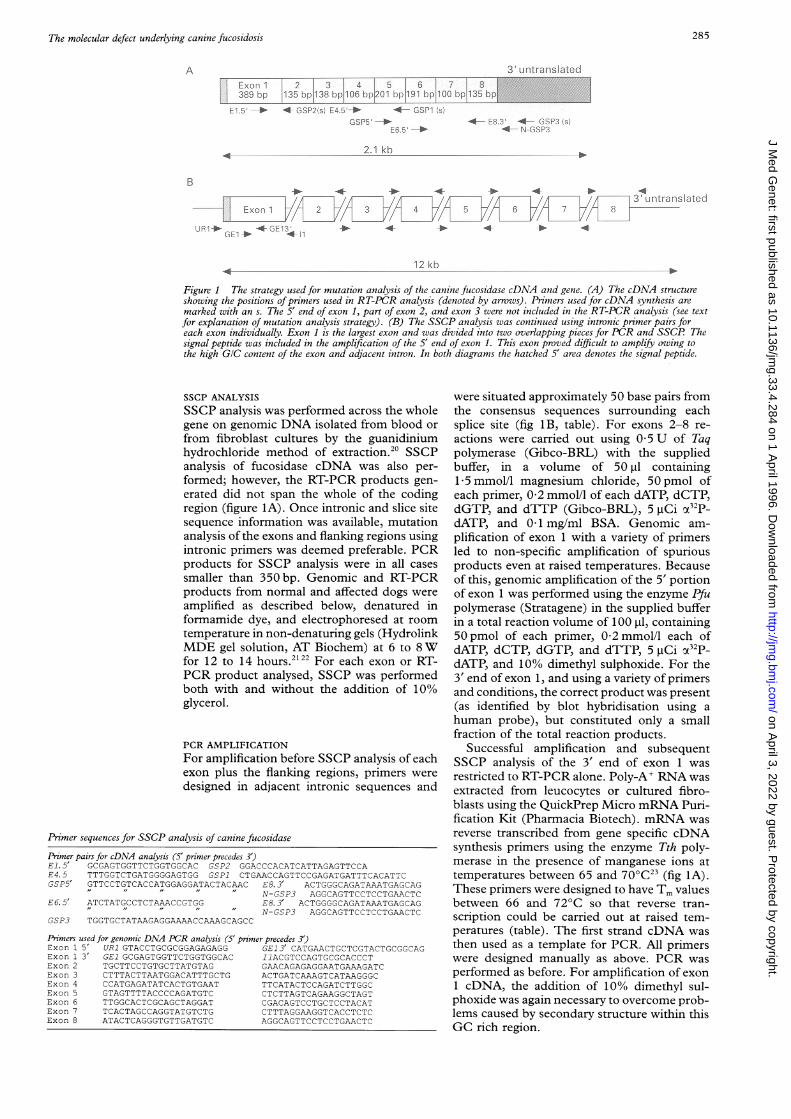
3' Ll ntra11slateciExon 1i 2 3 4 5 6 7 8
389 bsp ~135 bp 138 bpl106 bpji201 hp 191 bsp 100 bsp 135 bp
E .5 _ 4 GSP2:s. E4.5P_ 4 GSP1 s
E56 _
2 1 kb
4--E:. 41--GS'340-4.S-
B* * *
i/f 3 L tra islate diExon-i 3 6
UJ 1* GE-1 * "IE13*1 >
12 kb
Figure 1 The strategy used for mutation analysis of the canine fucosidase cDNA and gene. (A) The cDNA structureshowing the positions ofpnrmers used in RT-PCR analysis (denoted by arrows). Prinmers used for cDNA synthesis aremarked with an s. The 5' end of exon 1, part of exon 2, and exon 3 were not included in the RT-PCR analysis (see textfor explanation of mutation analysis strategy). (B) The SSCP analysis was continued using intronic primer pairs foreach exon individually. Exon 1 is the largest exon and was divided into two overlapping pieces for PCR and SSCP Thesignal peptide was included in the amplification of the 5' end of exon 1. This exon proved difficult to amplify owing tothe high GIC content of the exon and adjacent intron. In both diagrams the hatched 5' area denotes the signal peptide.
SSCP ANALYSIS
SSCP analysis was performed across the wholegene on genomic DNA isolated from blood or
from fibroblast cultures by the guanidiniumhydrochloride method of extraction.20 SSCPanalysis of fucosidase cDNA was also per-formed; however, the RT-PCR products gen-erated did not span the whole of the codingregion (figure IA). Once intronic and slice sitesequence information was available, mutationanalysis of the exons and flanking regions usingintronic primers was deemed preferable. PCRproducts for SSCP analysis were in all cases
smaller than 350 bp. Genomic and RT-PCRproducts from normal and affected dogs were
amplified as described below, denatured informamide dye, and electrophoresed at room
temperature in non-denaturing gels (HydrolinkMDE gel solution, AT Biochem) at 6 to 8Wfor 12 to 14 hours.2122 For each exon or RT-PCR product analysed, SSCP was performedboth with and without the addition of 10%glycerol.
PCR AMPLIFICATION
For amplification before SSCP analysis of eachexon plus the flanking regions, primers were
designed in adjacent intronic sequences and
Primer sequences for SSCP analysis of canine fucosidase
Primer pairs for cDNA analysis (5' primer precedes 3')El. 5' GCGAGTGGTTCTGGTGGCAC GSP2 GGACCCACATCATTAGAGTTCCAE4.5 TTTGGTCTGATGGGGAGTGG GSP1 CTGAACCAGTTCCGAGATGATTTCACATTCGSP5' GTTCCTGTCACCATGGAGGATACTACAAC E8. 3' ACTGGGCAGATAAATGAGCAG
N-GSP3 AGGCAGTTCCTCCTGAACTCE6. 5' ATCTATGCCTCTAAACCGTGG E8. 3' ACTGGGGCAGATAAATGAGCAG
I" 1 I" " N-GSP3 AGGCAGTTCCTCCTGAACTCGSP3 TGGTGCTATAAGAGGAAAACCAAAGCAGCC
Primers used for genomic DNA PCR analysis (5' primer precedes 3')Exon 1 5' UR1 GTACCTGCGCGGAGAGAGG GE13' CATGAACTGCTCGTACTGCGGCAGExon 1 3 GE1 GCGAGTGGTTCTGGTGGCAC I1ACGTCCAGTGCGCACCCTExon 2 TGCTTCCTGTGCTTATGTAG GAACAGAGAGGAATGAAAGATCExon 3 CTTTACTTAATGGACATTTGCTG ACTGATCAAAGTCATAAGGGCExon 4 CCATGAGATATCACTGTGAAT TTCATACTCCAGATCTTGGCExon 5 GTAGTTTTACCCCAGATGTC CTCTTAGTCAGAAGGCTAGTExon 6 TTGGCACTCGCAGCTAGGAT CGACAGTCCTGCTCCTACATExon 7 TCACTAGCCAGGTATGTCTG CTTTAGGAAGGTCACCTCTCExon 8 ATACTCAGGGTGTTGATGTC AGGCAGTTCCTCCTGAACTC
were situated approximately 50 base pairs fromthe consensus sequences surrounding eachsplice site (fig 1B, table). For exons 2-8 re-actions were carried out using 0 5 U of Taqpolymerase (Gibco-BRL) with the suppliedbuffer, in a volume of 50 pd containing15 mmol/l magnesium chloride, 50 pmol ofeach primer, 0-2 mmol/l of each dATP, dCTP,dGTP, and dTTP (Gibco-BRL), 5 ItCi (i32p_dATP, and 0.1 mg/ml BSA. Genomic am-
plification of exon 1 with a variety of primersled to non-specific amplification of spuriousproducts even at raised temperatures. Becauseof this, genomic amplification of the 5' portionof exon 1 was performed using the enzyme P:fupolymerase (Stratagene) in the supplied bufferin a total reaction volume of 100 ptl, containing50 pmol of each primer, 02 mmol/l each ofdATP, dCTP, dGTP, and dTTP, 5 pCi OCL32P-dATP, and 10% dimethyl sulphoxide. For the3' end of exon 1, and using a variety of primersand conditions, the correct product was present(as identified by blot hybridisation using a
human probe), but constituted only a smallfraction of the total reaction products.
Successful amplification and subsequentSSCP analysis of the 3' end of exon 1 was
restricted to RT-PCR alone. Poly-A' RNA was
extracted from leucocytes or cultured fibro-blasts using the QuickPrep Micro mRNA Puri-fication Kit (Pharmacia Biotech). mRNA was
reverse transcribed from gene specific cDNAsynthesis primers using the enzyme Tth poly-merase in the presence of manganese ions attemperatures between 65 and 700C23 (fig 1A).These primers were designed to have Tm valuesbetween 66 and 72°C so that reverse tran-scription could be carried out at raised tem-peratures (table). The first strand cDNA was
then used as a template for PCR. All primerswere designed manually as above. PCR was
performed as before. For amplification of exon1 cDNA, the addition of 10% dimethyl sul-phoxide was again necessary to overcome prob-lems caused by secondary structure within thisGC rich region.
A
285
on April 3, 2022 by guest. P
rotected by copyright.http://jm
g.bmj.com
/J M
ed Genet: first published as 10.1136/jm
g.33.4.284 on 1 April 1996. D
ownloaded from
Skelly, Sargan, Herrtage, Winchester
N..
.,0,,
.W_i.* O..
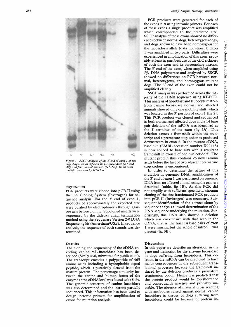
Figure 2 SSCP analysis of the 3' end of exon 1 of twodogs diagnosed as deficient in x-L-fucosidase (Al andA2) and four normal animals (Nl-N4). In all cases
amplification was by RT-PCR.
SEQUENCINGPCR products were cloned into pCR-II usingthe TA Cloning System (Invitrogen) for se-
quence analysis. For the 3' end of exon 1,products of approximately the expected sizewere purified by electrophoresis through agar-ose gels before cloning. Subcloned inserts weresequenced by the dideoxy chain terminationmethod using the Sequenase Version 2-0 DNASequencing kit (Amersham/USB). In sequenceanalysis, the sequence of both strands was de-termined.
ResultsThe cloning and sequencing of the cDNA en-
coding canine oc-L-fucosidase has been de-scribed (Skelly et al, submitted for publication).The transcript encodes a polypeptide of 465amino acids including a hydrophobic signalpeptide, which is putatively cleaved from themature protein. The percentage similarity be-tween the canine and human forms of theenzyme at the cDNA level was found to be 84%.The genomic structure of canine fucosidasewas also determined and the introns partiallysequenced. This information has been used to
design intronic primers for amplification ofexons for mutation analysis.
PCR products were generated for each ofthe exons 2-8 using intronic primers. For eachof these exons a single product was amplifiedwhich corresponded to the predicted size.SSCP analysis of these exons showed no differ-ences between normal dogs, heterozygous dogs,and dogs known to have been homozygous forthe fucosidosis allele (data not shown). Exon1 was amplified in two parts. Difficulties wereexperienced in amplification of this exon, prob-ably at least in part because of the G/C richnessof both the exon and its surrounding introns.The 5' end of the exon, when amplified usingPYu DNA polymerase and analysed by SSCP,showed no differences on PCR between nor-mal, heterozygous, and homozygous mutantdogs. The 3' end of the exon could not beamplified cleanly.SSCP analysis was performed across the ma-
jority of the cDNA sequence using RT-PCR.This analysis offibroblast and leucocyte mRNAfrom canine fucosidase normal and affectedanimals showed only one mobility shift, whichwas located in the 3' portion of exon 1 (fig 2).This PCR product was cloned and sequencedin both normal and affected dogs and a 14 basepair deletion of the mRNA was identified atthe 3' terminus of the exon (fig 3A). Thisdeletion causes a frameshift within the tran-script and a premature stop codon is produceddownstream in exon 2. In the mutant cDNA,base 393 (EMBL accession number X92448)is now spliced to base 408 with a resultantframeshift in exon 2 of one nucleotide 5'. Themutant protein thus contains 25 novel aminoacids before the first of two adjacent prematurestop codons is encountered.
In order to determine the nature of thismutation in genomic DNA, amplification ofthe 3' end of exon 1 was performed on genomicDNA from an affected animal using the primersdescribed (table, fig 1B). As this PCR didnot amplify with sufficient specificity, shotguncloning of the size fractionated PCR productsinto pCR-II (Invitrogen) was necessary. Sub-sequent identification of the correct clone bysequence analysis allowed determination of theDNA sequence underlying the mutation. Sur-prisingly, this DNA also showed a deletionwhich was coextensive with that seen in thecDNA; that is, the final 14 base pairs of exon1 were missing but the whole of intron 1 waspresent (fig 3B).
DiscussionIn this paper we describe an alteration in thegene and transcript for the enzyme fucosidasein dogs suffering from fucosidosis. This de-letion in the mRNA can be predicted to havemajor consequences in the subsequent trans-lational processes because the frameshift in-duced by the deletion produces a prematuretermination codon. Hence it is predicted thatthe protein product would be foreshortenedand consequently inactive and probably un-stable. The absence of material cross reactingwith antibodies raised against normal caninefucosidase in tissues of dogs suffering fromfucosidosis could be because of protein in-
286
on April 3, 2022 by guest. P
rotected by copyright.http://jm
g.bmj.com
/J M
ed Genet: first published as 10.1136/jm
g.33.4.284 on 1 April 1996. D
ownloaded from

The molecular defect underlying canine fucosidosis
Normal sequenceExon 1 Intron 1
Exon 1 Intron 1
T G C AFigure 3 (A) Sequence analysis of the 3' end of exon 1 and 5' end of exon 2 of canine fucosidase cDNA showing the 14 base pair cDNA deletion.(B) Comparison of mutant and normal sequences from genomic PCR products covering the 3' end of exon 1 and 5' end of intron 1, showing the 14 basepair DNA deletion.
stability or of the absence of antigenic epitopein the mutant protein."The mutation underlying the deletion in the
RNA is a coextensive deletion of the DNA.The splice donor site at the end of exon 1in normal dogs takes the form "CCG/GTGAGT..". This sequence, with a C at po-sition -2, is itself less close to a splice donorconsensus than the splice site created bythe deletion in the mutant gene "CAG/GTGAGT..". It is therefore unsurprising thatthe latter splice site is used efficiently.The description of the disease causing muta-
tion has important consequences for theEnglish springer spaniel breed. Until now, het-erozygotes have been identified on the basis ofa reduced enzyme activity.8 This method isunreliable and far from satisfactory when breed-ing programmes are being established. Thedescription of the mutation at the molecularlevel will allow a test to be designed which willidentify the mutant allele specifically, so thatheterozygous dogs can be identified easily andrapidly.
Fucosidosis in English springer spaniels pro-vides a useful animal model with which to studythe human disease. Dogs diagnosed bio-chemically as being homozygous for the disease-causing mutation have already been usedin trials to assess the efficacy of bone marrow
transplantation.72526 As a consequence of thiswork, bone marrow transplantation has beenattempted in a human patient.27 Now, withthe therapeutic emphasis turning towards thepossibility of gene replacement therapy, a fullermolecular understanding of the canine diseasemust be beneficial. Preliminary assessments ofgene transfer, using retroviral vectors, havebeen conducted in human and canine fuco-sidosis fibroblast cultures.28 The early iden-tification ofaffected animals and the knowledgeof the cDNA and genomic organisation of can-ine fucosidase could prove invaluable whenit comes to designing such gene replacementprotocols using the springer spaniel as a humandisease model.
We would like to thank Helen Cragg for her advice and tre-mendous encouragement in the earlier stages of this project,also David Entz who provided technical support. This workwas funded by the Wellcome Trust. BJS is the holder of aWellcome Clinical Research Studentship.
1 Durand P, Barrone C, Della Celia G. Fucosidosis. Pediatr1969;75:665-74.
2 Hartley WJ, Canfield PJ, Donnelly TM. A suspected newcanine storage disease. Acta Neuropathol 1982;56:225-32.
3 Littlewood JD, Herrtage ME, Palmer AC. Neuronal storagedisease in English springer spaniels. Vet Record 1983;112:86-7.
4 Abraham D, Blakemore WF, Dell A, et al. The enzymicdefect and storage products in canine fucosidosis. Biochem1984;221:25-33.
5 Herrtage ME. Canine fucosidosis. Vet Annual 1988;28:223-7.
A B
4*
I.
to40
00
aw.
*
041.
287
on April 3, 2022 by guest. P
rotected by copyright.http://jm
g.bmj.com
/J M
ed Genet: first published as 10.1136/jm
g.33.4.284 on 1 April 1996. D
ownloaded from

Skelly, Sargan, Herrtage, Winchester
6 Ferrara ML, Taylor RM, Stewart GJ. Age at marrow trans-plantation is critical for successful treatment of caninefucosidosis. Transplant Proc 1992;23:2282-3.
7 Taylor RM, Farrow BRH, Stewart GJ, Healy PJ, Tiver K.The clinical effects of lysosomal enzyme replacement bybone marrow transplantation after total lymphoid ir-radiation on neurologic disease in fucosidase deficientdogs. Transplant Proc 1988;20:89-93.
8 Barker CG, Herrtage ME, Shanahan F, Winchester BG.Fucosidosis in English springer spaniels: results of a trialscreening programme. 7 Small Anim Pract 1988;29:623-30.
9 Occhiodoro T, Beckman KR, Morris CP, Hopwood JJ.Human oa-L-fucosidase: complete coding sequence fromcDNA clones. Biochem Biophys Res Commun 1989;164:439-45.
10 Kretz KA, Cripe D, Carson GS, Fukushima H, O'Brien JS.Structure and sequence of the human oa-L-fucosidase geneand pseudogene. Genomics 1992;12:276-80.
11 Willems PJ, Darby JK, DiCioccio R, et al. Identification ofa mutation in the structural alpha-L-fucosidase gene infucosidosis. Am J7 Hum Genet 1988;43:756-63.
12 Kretz KA, Darby JK, Willems PJ, O'Brien JS. Char-acterisation of the EcoRI mutation in fucosidosis patients:a stop codon in the open reading frame. J Mol Neurosci1989;1:177-80.
13 Willems PJ, Gatti R, Darby JK, et al. Fucosidosis revisited:a study of 77 patients. Am J Med Genet 1991;38:111-31.
14 Williamson M, Cragg H, Grant J, et al. A 5' splice sitemutation in fucosidosis. J Med Genet 1993;30:218-23.
15 Seo HC, Willems PJ, Kretz KA, Martin BM, O'Brien JS.Fucosidosis: four new mutations and a polymorphism.Hum Mol Genet 1993;2:423-29.
16 Seo HC, Willems PJ, O'Brien JS. Six additional mutations infucosidosis: three nonsense mutations and three frameshiftmutations. Hum Mol Genet 1993;2:1205-8.
17 Seo HC, Kunze J, Willems PJ, et al. A single base deletionin a Turkish patient with fucosidosis. Hum Mutat 1994;3:407-8.
18 Seo HC, Yang M, Tonlorenz R, et al. A missense mutation(S63L) in a-L-fucosidase is responsible for fucosidosis inan Italian patient. Hum Mol Genet 1994;3:2065-6.
19 Seo HC, Heidemann PH, Lutz E, O'Brien JS. A nonsensemutation in two German patients with fucosidosis. HumMutat 1995;6: 184-5.
20 Jeanpierre M. A rapid method of purification of DNA fromblood. Nucleic Acids Res 1987;15:9611.
21 Orita M, Iwahana H, Kanazawa H, Hayashi K, SekiyaT. Detection of polymorphisms of human DNA by gelelectrophoresis as single strand conformation poly-morphisms. Proc Nad Acad Sci USA 1989;86:2766-70.
22 Orita M, Suzuki Y, Sekiya T, Hayashi K. A rapid andsensitive detection of point mutations and genetic poly-morphisms using the polymerase chain reaction. Genomics1989;5:874-9.
23 Myers TW, Gelfand DH. Reverse transcription and DNAamplification by a Thermus thermophilus DNA poly-merase. Biochemistry 1991;30:7661-6.
24 Barker C, Dell A, Rogers M, Alhadeff JA, Winchester BG.Canine oa-L-fucosidase in relation to the enzymic defectand storage products in canine fucosidosis. Biochem J1988;254:861-8.
25 Taylor RM, Farrow BRH, Stewart GJ. Correction ofenzymedeficiency by allogenic bone marrow transplantation fol-lowing total lymphoid irradiation in dogs with lysosomalstorage disease (fucosidosis). Transplant Proc 1986;18:326-9.
26 Taylor RM, Farrow BRH, Stewart GJ. Amelioration ofclinical disease following bone marrow transplantationin fucosidase-deficient dogs. Am J Med Genet 1992;42:628-32.
27 Vellodi A, Cragg H, Winchester B, et al. Allogeneic bonemarrow transplantation for fucosidosis. Bone MarrowTransplant 1995;15: 153-8.
28 Occhiodoro T, Hopwood JJ, Morris CP, Anson DS. Cor-rection of oa-L-fucosidase deficiency in fucosidosis fibro-blasts by retroviral vector-mediated gene transfer. HumGene Ther 1992;3:365-9.
288
on April 3, 2022 by guest. P
rotected by copyright.http://jm
g.bmj.com
/J M
ed Genet: first published as 10.1136/jm
g.33.4.284 on 1 April 1996. D
ownloaded from