the merican journal cancer - cancer...
TRANSCRIPT

THE MERICAN JOURNAL OF CANCER
A Continuation of The Journal of Cancer Research
VOLUME XXXIII JULY, 1938 NUMBER 3
CARCINOSARCOMA
OTTO SAPHIR, M.D., AND ALOYSIUS VASS, M.D.l
(From the Pathology Departmeizt of the Nelson Morris Institute, Michael Reese Haspitel, Chicago)
The histologic classification of malignant tumors is important not only from an academic (histogenetic) standpoint, but also from that of prognosis and treatment. Thus the application and dosage of irradiation are primarily and principally dependent upon histologic detail, as is also the grading of malignant tumors. While ordinarily it is fairly well within the power of the pathologist to classify a malignant tumor, occasional growths are encountered which, despite obvious malignant features, cannot, because of their peculiar histologic detail, easily be classed as the descendants of one particular cell type. These tumors present features which may lead to the belief that they are the result of atypical growth of more than one cell type.
All early pathologists considered their origin to be of dual nature. Thus, Virchow labeled them carcinosarcoma. He believed that within the primary car- cinomatous or sarcomatous elements, the stromal or epithelial portions respec- tively were subsequently or simultaneously stimulated to malignant growth. Herxheimer and others believed this occurrence to be more frequent in primary carcinomas. They held that a carcinoma could stimulate an excessive growth of the stroma; that when the stroma-proliferation reached the stage of malig- nancy, the resulting tumor would be a carcinosarcoma. Perhaps, also, the secondary malignancy might supersede the primary one.. This opinion was corroborated by Ehrlich and Apolant, and others, who observed that trans- planted mouse carcinomas changed their histologic appearance, first to car- cinosarcoma and eventually to sarcoma.
With the acceptance of the existence of these dual tumors, various at- tempts at classification were undertaken. Saltykow called tumors resembling carcinoma in some portions and sarcoma in others “ carcinoma sarcomatodes,” a term first employed by Herxheimer. Those growths which were the product
Interest in these tumors is as old as histopathology itself.
1 Aided by a grant from the A. B. Kuppenheimer Fund. 33 1

- --_ . .&-A FIG. 11. PRIMARY DUCT CARCINOMA OF BREAST. HEMATOXYLIN-EOSIN. X 85
FIGS. 12 AND 13. METASTASTS IN OVARY OF PRIMARY DUCT CARCIXOMA OF BREAST (FIG. 11) Note the round tumor cells. Hematoxylin-eosin. X 7 5 and X 160
FIGS. 14 AND 15. Two AREAS IN PRIMARY CARC~NOMA OF THYROID Note the spindle shape of the carcinoma cells in Fig. 15. Hematoxylin-eosin. X 75
FIG. 16. PRIMARY CARCINOMA OF ESOPHAGCS Note the presence of many connective-tissue cells which in some instances have been interpreted
as sarcoma cells. Hematoxylin-eosin. X 100
345

CARCINOSARCOMA 333
of carcinoma cells and recommended the use of the term “ pseudosarcomatous carcinoma ” or “ pseudocarcinosarcoma ” for carcinomas with sarcoma-like appearance. Semb, in describing tumors of the breast, mentioned the pos- sible misinterpretation of morphologically transformed carcinomas as sar- comas. Krompecher doubted the true dual nature of these tumors and conceived of them as originating from an embryonal cell, the “ transitional cell,” which supposedly possesses the potentialities of both epithelial and con- nective-tissue cells. Cohn stressed the fact that it is next to impossible to diagnose correctly immature tumors, and pointed out that scme of these, be- cause of the variability of tumor cells, may be interpreted as carcinosarcomas. Ewing’s point of view has been mentioned above.
Harvey and Hamilton, on the other hand, maintain that there is a double tumor which is a mixture of carcinoma and sarcoma and that this may be denominated carcinosarcoma. The sarcomatous development is probably an exaggeration of stroma reaction to invasion by carcinoma. They further point out that it is generally believed that the carcinoma cells exert a stimu- lating influence on the stroma cells, which in transplanted tumors in course of the transfers yield a tumor of fibroblastic origin. The current interpretation of the carcinosarcomatous structures in man and the lower animals, however, is, as these authors point out, a matter of much difficulty, and probably no single explanation will apply to all cases. The chief source may be the trans- formation of epithelial cells into spindle cells. These writers state, however, that there are tumors of both epidermic and glandular carcinoma type which may show, in part, aggregations of spindle-shaped epithelial cells, having much the appearance of a sarcoma, which may be denominated spurious carcinosarcoma or carcinoma sarcomatoides.
Kettle pointed out that, although it is possible that many of the carcino- sarcomas recorded are true mixed tumors, it is certain that others are merely examples of extreme polymorphism of carcinoma cells.
Willis stated that in human pathology no acceptable example of sarcoma- tous change in the stroma of a tumor has been described. All alleged in- stances of this occurrence are clearly traceable to the structural versatility of carcinomas as described by Kettle.
The discrepancy of opinion indicated by this brief review justifies a re-investigation of the tumors reported as carcinosarcomas.
In a previous study one of us (Saphir) has shown that in a series of care- fully studied tumors, principally epithelial, more than one type of apparently epithelial element was often present. In some tumors, particularly those re- ferred to as squamous-cell carcinomas with transitional-cell features, the latter often assumed spindle forms which upon cursory examination might easily be mistaken for sarcomatous elements. Only a large number of sections re- vealed the true nature of some of these tumors through observation of the change from transitional and basal cells to the spindle-shaped sarcoma-like elements. Upon superficial examination these tumors could have been in- terpreted as carcinosarcomas. Since a re-study of our very meager material on carcinosarcomas almost invariably revealed that these tumors could be interpreted as carcinomas of various types showing so-called morphologic variations, we decided to collect these tumors and to determine whether or

3 34 OTTO SAPHIR AND ALOYSIUS VASS
TABLE I: Location of 153 Carcinosarcomas Reported in the Literature -
Uterus. . . . . . . . . . . . . . . . . . . 36 Lung . . . . . . . . . . . . . . . . . . . . . . . . . . 7 Tube . . . . . . . . . . . . . . . . . . . . . . . . . . . . . . . . . 2 Kidney . . . . . . . . . . . . . . . . . . .
Breast . . . . . . . . . . . . . . . . . . . . . . . 32 Ureter . . . . . . . . . . . . . Thyroid gland, . . . . . . . . . . . . . . . . . . . . . . . . 1 7 Prostate. . . . . . . . . . . . . . . . . . . . . . . .
Esophagus 14 Middle e a r . . . . . . . . . . . . .
Gallbladder. . . . . . . . . . . . . . . . . . . . . . . . . . . . 2 Maxillary sinus. . . . . . . . . . . . .
Hypopharynx . . . . . . . . . . . . . . . . . . . . . . . . . . 1
. . . . . . . . . . . . . . . . . . . 7 Urinary bla . . . . . . . . . . . . . 1
. . . . . . . . . . . . 1 Sk in . . . . . . . . . . . . . . . . . . . . . . . . . . . . . 5
. . . . . . . . 1 Labium majus . . . . . . . . . . . . . . . . . . 1
. . . . . . . . . . . . . . . . . . . . . . . . . 1 Nasal cavity . . . . . . . . . . . . . . . . . . . . . . 1 Larynx. 6 Ciliary body. . . . . . . . . . . . . . . . . . . . 1
. . . . . . . . . . . . . . . . . . . . . . . 5 Salivary gland. . . . . . . . . . . . . . . . . . . .
. . . . . . . . . .
not they were single tumors. I t was also considered wise to re-study those instances of carcinosarcoma reported in the literature which were available to us, and to see if such an interpretation was indisputable.
In the following pages the carcinosarcomas reported in the literature are cited, our interpretation of these tumors is given, and similar tumors which have come to our observation are reported in view of the possibility of a diagnosis of carcinosarcoma. For the sake of coherence, these observations are given after the review of the literature on carcinosarcomas of individual organs or systems of organs.
REVIEW OF THE LITERATURE AND PERSONAL OBSERVATIONS
This review is by no means complete, but it covers most of the publica- tions in the American, English, French, German, and Italian literature. In the study of the reported tumors, we were concerned only with those which, although presenting apparently mixed characteristics, were obviously not teratomas. diagnoses were “ carcinosarcoma ” and, less frequently, “ carcinoma sarco- matodes.” Our study was based upon the descriptions and the illustrations accompanying the case reports. Our analysis concerned itself with the fol- lowing possibly complicating factors in the alteration of the fundamental his- tologic appearance of the tumor: (1) growth of a carcinoma into a benign tumor; ( 2 ) inclusion of benign epithelial cell formations within a malignant tumor of connective-tissue origin; ( 3 ) chronic productive inflammation in the vicinity of the tumor; (4) marked anaplasia; ( 5 ) morphologic variations in tumor cells, as described by one of us (Saphir); (6) history and histologic evidence of x-ray irradiation. In our interpretation of the fundamental char- acter of the tumor, the nature of the metastases was given due consideration.
The material for our investigation consisted of 153 reported carcinosar- comas, the distribution of which in various organs is presented in Table I.
Uterus: Table I1 lists the reported tumors and the nature of metastases of 36 uterine carcinosarcomas.’ In 19 instances pedunculated tumors pro- jected either from the endometrium into the uterine cavity or from the cervix
4 The terms most commonly used by the various authors in their
“*
*The report of Kubinyi of carcinosarcomas of the uterus, as quoted by Bczza, could not he verified.

CARCINOSAkCOMA 335
TABLE 11; Chrcinosarcomas of the Uterus
Date of Metastasis
Author Carcinoma Sarcoma Mixed No. Publicat ion - --
- - - 1 1893 Sehrt 2 1896 Iwanoff 3 1896 Niebergall - - - 4 1901 Fraen kel + 5 1904 Nebesky - 6 1904 Nebesky - - - 7 1908 Lindemann 8 1909 Forssner 9 1909 Forssner
- - -
- - - -
- - - - - - - - -
- - + 10 1909 Albrecht -
+ 11 1912 Stein - 12 1914 Saltykow - - 13 1914 Saltykow + 14 1915 Benthin - 15 1922 Klee 16 1922 Rrun 17 1922 Kleinsch mid t + 18 1922 Kleinschmidt - 19 1923 JaffC - - 20 1925 Frankl 21 1925 Frankl 22 1925 Frankl 23 1925 Frankl - 24 1925 Frankl 25 1925 Frankl
Frankl 26 1925 27 1925 Frankl 28 1926 Raltzer + 29 1928 30 1931 Leroux and Perrot - - 31 1932 Bosenberg - 32 1935 Daniel and Lazaresco - 33 1935 Harvey and Hamilton - 34 1935 Harvey and Hamilton - - 35 1936 Goldstine - 36 1936 Schwarz
- - - - -
- - - - - -
- - - -
- - - - - - - - - -
- - - - -
- - - - - - - - - - -
- - - Schiff mann -
- - - - - -
- - -
- - -
into the vagina. Microscopically they consisted of large spindle-shaped or elongated cells, similar to smooth muscle cells, among which many atypical anaplastic cells of varying size and multinucleated giant cells were also seen. Ulceration of the surface of the tumor was present in most instances, and in- flammatory changes were regularly evident within the stroma. The tissue around the base of the pedunculated mass was purely carcinomatous and was so interpreted by the author. In one case the pedunculated tumor did not consis; of spindle-shaped muscle cells, but of connective tissue enclosing uterine glands of somewhat irregular arrangement composed of cells with no anaplastic features. The stroma of this apparently polypoid growth con- tained a great number of inflammatory cells and many anaplastic tumor cells. Here, too, the tissue surrounding the tumor presented typical carcinomatous features and was so interpreted by the author.
In 8 instances parts of the tumors were distinctly carcinomatous. In other regions, however, chronic productive inflammatory changes were present.

FIG. 1, SQUAMOUS-CELL CARCINOMA O F UTERUS WlTH MANY LYMPHOCYTIC CELLS The tumor was irradiated. Tumors presenting similar pictures have been recorded in the
literature as carcinolymphosarcoma. Hematoxylin-eosin. X 60
FIG. 2. SQUAMOCS-CELL CARCINOMA O F CERVIX WITH TRANSITIONAL-CELL FEATURES Note the transitional cells simulating connective-tissue cells which surround islets of squamous
cells. Hematoxylin-eosin. X 50 [ L q e r t d cont. at foo l ul 9 . 3371
336

CARCINOSARCOMA 3 37
In these areas, besides typical carcinoma cell groups, there were tumor cells showing anaplasia with bizarre pleomorphism. These pleomorphic cells were interpreted by the authors as sarcoma cells. Tumors No. 20 and 23 belong- ing to this group were treated with x-ray irradiation and multiple curettages. In tumor No. 31 there were found, beneath and partly among the cells of a typical transitional-cell carcinoma of the cervix, many closely packed small round cells of uniform size and shape. These cells were interpreted by the author as those of a round-cell sarcoma.
In Case No. 11, a polypoid structure arising in the endometrium was present in an adenomyomatous uterus. Microscopically this polypoid struc- ture consisted of spindle cells enclosing endothelial-lined cavities containing blood; only a scanty stroma was present. Among the spindle cells were cells with large cytoplasms and irregularly outlined nuclei. Some of the cells con- tained multiple nuclei. Enclosed within the tumor tissue were irregularly formed glandular structures lined by partly stratified squamous cells differing somewhat in size and shape. Metastases occurring one year later did not contain these glandular structures.
Tumor No. 16 may be interpreted as a metastatic carcinoma within a myofibroma. The primary tumor was apparently a cystadenocarcinoma of an ovary which had been removed previously. Also in Kleinschmidt’s tumors it seems that a primary carcinoma had invaded a myofibroma. The metas- tasis in one of these was carcinomatous. Because of absence of sufficient description and illustrations case No. 1 cannot be discussed.
Nineteen tumors were characterized, as stated before, by a more or less pedunculated myomatous growth. The tissue surrounding the base of the myoma was in all instances purely carcinomatous, as admitted by the authors. I t is known that carcinoma cells invading connective or muscle tissue may become morphologically altered by the mechanical pressure of the invaded tissue. It seems, therefore, that this group of tumors should be considered as primary carcinomas invading myofibromas which may be undergoing various progressive and regressive changes, rather than that they should be interpreted as carcinosarcomas. That this interpretation is the more prob- able one for this group is demonstrated by the carcinomatous nature of the metastases in the few instances where these were present.
In the second large group of 8 cases, anaplastic tumors were encountered which showed in some parts distinct carcinomatous features, while in others they presented marked pleomorphism, which led to the diagnosis of carcino- sarcoma. I n all of these tumors the pleomorphism was present in areas of chronic productive inflammation. I n tumors No. 2 1 and 23 a long history corroborates the chronicity of the inflammatory process. I t seems that the pleomorphism of the tumor cells is more likely the result of the chronic in-
FIG. 3. ADENOCARCINOMA METASTASIS IN MYOFIBROMA Instances of this type were reported as carcinosarcoma.
Note the transitional type of cells in the left lower field.
Iron hematoxylin-eosin.
These are occasionally regarded as connective-tissue cells. Iron hematoxylin-eosin. X 110
FIGS, 5 AND 6 . Two INSTANCES OF PAPILLARY CYSTADENOCARCINOMA ARISING IN A MAMMARY DUCT
X 90
FIG. 4. PAPILLLFEROUS CYSTADENOCARCINOMA OF OVARY
Note the spindle shape of some of the tumor cells. Hematoxylin-eosin. X 210 and X 90
338 OTTO SAPHIR AND ALOYSIUS VASS
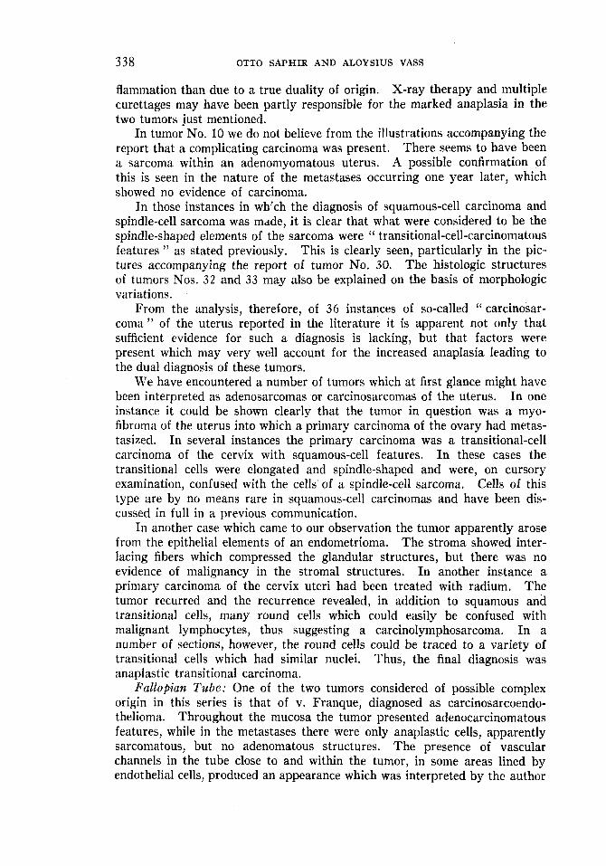
flammation than due to a true duality of origin. X-ray therapy and multiple curettages may have been partly responsible for the marked anaplasia in the two tumors just mentioned.
In tumor No, 10 we do not believe from the illustrations accompanying the report that a complicating carcinoma was present. There seems to have been a sarcoma within an adenomyomatous uterus. A possible confirmation of this is seen in the nature of the metastases occurring one year later, which showed no evidence of carcinoma.
In those instances in wh‘ch the diagnosis of squamous-cell carcinoma and spindle-cell sarcoma was mdde, it is clear that what were considered to be the spindle-shaped elements of the sarcoma were “ transitional-cell-carcinomatous features ” as stated previously. This is clearly seen, particularly in the pic- tures accompanying the report of tumor No. 30. The histologic structures of tumors Nos. 32 and 33 may also be explained on the basis of morphologic variations.
From the analysis, therefore, of 36 instances of so-called (‘ carcinosar- coma” of the uterus reported in the literature it is apparent not only that sufficient evidence for such a diagnosis is lacking, but that factors were present which may very well account for the increased anaplasia leading to the dual diagnosis of these tumors.
We have encountered a number of tumors which at first glance might have been interpreted as adenosarcomas or carcinosarcomas of the uterus. In one instance it could be shown clearly that the tumor in question was a myo- fibroma of the uterus into which a primary carcinoma of the ovary had metas- tasized. In several instances the primary carcinoma was a transitional-cell carcinoma of the cervix with squamous-cell features. I n these cases the transitional cells were elongated and spindle-shaped and were, on cursory examination, confused with the cells of a spindle-cell sarcoma. Cells of this type are by no means rare in squamous-cell carcinomas and have been dis- cussed in full in a previous communication.
In another case which came to our observation the tumor apparently arose from the epithelial elements of an endometrioma. The stroma showed inter- lacing fibers which compressed the glandular structures, but there was no evidence of malignancy in the stromal structures. In another instance a primary carcinoma of the cervix uteri had been treated with radium. The tumor recurred and the recurrence revealed, in addition to squamous and transitional cells, many round cells which could easily be confused with malignant lymphocytes, thus suggesting a carcinolymphosarcoma. In a number of sections, however, the round cells could be traced to a variety of transitional cells which had similar nuclei. Thus, the final diagnosis was anaplastic transitional carcinoma.
Pallopian Tube: One of the two tumors considered of possible complex origin in this series is that of v. Franque, diagnosed as carcinosarcoendo- thelioma. Throughout the mucosa the tumor presented adenocarcinomatous features, while in the metastases there were only anaplastic celIs, apparently sarcomatous, but no adenomatous structures. The presence of vascular channels in the tube close to and within the tumor, in some areas lined by endothelial cells, produced an appearance which was interpreted by the author

CARCINOSARCOMA 3 39
TABLE 111: Carcinosarcomas of Ovary *
Metastases Year of No. Publication Author Carcinoma Sarcoma Mixed
- ? 1 1901 Haacke - 2 1905 Lippmann - - 3 1913 Rothacker -
- +(?) 4 1916 Harbitz - 5 1916 Harbitz + 6 1922 Kleinschmidt 7 1929 Quin -.
+ - -
- - + - - -
* Vecchi’s report (Vecchi, G.: Arch. per le sc. med. 48: 135, 1926) was not available to US.
as endotheliomatous. The absence of adenomatous structures in metastases of primary adenocarcinomas is not rare and the presence of anaplastic cells in the metastases does not signify that the primary tumor contained sarcomatous elements. I t seems, therefore, assuming the validity of morphologic vari- ations of tumor cells, that this case was a primary adenocarcinoma of the fal- lopian tube with secondary anaplasia, rather than a simultaneous occurrence of tumors of different origin.
The second carcinosarcoma of the fallopian tube, in a fourteen-year-old girl, was reported by Motta. It was described as adenocarcinoma with pave- ment cells and spindle-cell sarcoma. The metastases were supposedly sar- comatous. The photomicrographs accompanying the report are of poor quality and do not show the presence of two tumors.
We have observed one instance of primary papilliferous adenocarcinoma in the tube, where the carcinoma cells assumed transitional forms which, as is pointed out later (pp. 343 and 356) resembled the cells of a spindle-cell sarcoma.
Ovary: In the literature reviewed there were 7 carcinosarcomas of the ovary (Table 111). Unfortunately only a few microscopic illustrations are available and the descriptions are not sufficiently ample to warrant detailed accounting. From the authors’ meager descriptions these tumors appear to have been mainly cystadenocarcinomas with closely packed groups of pleo- morphic and spindle-shaped cells growing between partly hyalinized connec- tive-tissue fibers. In our interpretation the latter areas represent carcinoma cells morphologically altered by growth into the restricted special environment of the fibrous wall of a fibrocystoma. The tumors described by Harbitz are in his own opinion questionable carcinosarcomas. One is seemingly a primary sarcoma and the other very likely a carcinoma with carcinomatous metastases.
There are a number of papilliferous cystadenocarcinomas in which the lining cells of the papillae are not cuboidal but either approach or actually are of transitional type. In these latter instances, when the transitional cells invade the core and the ovarian structures, they often assume spindle shapes, and the tumors may be confused with spindle-cell sarcomas and lead to the erroneous diagnosis of carcinosarcoma.
Breast: ‘ Thirty-two cases reported in the literature as carcinosarcoma of
A similar explanation may hold for Motta’s tumor.
According to Helwig, the first carcinosarcoma of the breast was reported by Kerbiriou and Danel. This report was not available to us.

3 40 OTTO SAPHIR AND ALOYSIUS VASS
TABLE IV: Carcinosarcomas of the Breast *
Metastases _______..- Year of Known dura-
No. Publication Author tion Of process Carcinoma Sarconm Mixed
1 2 3 4 5 6 7 8 9
10 11 12 13 14 1.5 16 17 18 19 20 21 22 23 24 25 26 21 28 29
1896 1906 1910 1910 1912 1912 1913 1913 1914 1915 1916 1920 1920 1922 1925 1925 1925 1928 1928 1931 1931 1932 1933 1933 1935 1935 1936 1936 1937
Uorsch Schlagenhaufer Coenen Orth Kettle Perrier Takano Waelle t Wehner HedrGn Harbitz (4 tumors) Kunsemuller Bergeret and Rotelho Jessup Kreibig Kreibig Kreibig Helwig Kuckens Sussi DeGaetani Stropeni Greenblatt Nowicki Harvey and Hamilton Harvey and Hamilton Pasternack and Wirth Mondor et al Sailer
* Not included are those tumors showing a large amount of cartilage or bone, which may Also not included is Melnichenko's report (Melnichenko, rather be interpreted as mixed tumors.
V. D.: Med. zhur. 5: 361, 1935), which was not available to us. t Tumor also shows some cartilage.
the breast are presented in Table IV." They are morphologically very similar. In 2 1 instances parts of the tumor revealed the characteristics of a carcinoma simplex arising from the ducts. In other regions, however, spindle and bizarre shaped anaplastic tumor cells were observed. The latter were found in areas which showed chronic productive inflammatory changes of variable extent. There were fat necrosis, granulation tissue with ( foreign-body) giant cells, marked fibrosis, calcification, and formation of osteoid tissue. One tumor consisted of a so-called adenoacanthoma which in some regions also revealed, within a very cellular stroma, seemingly anaplastic tumor cells with hyper- chromatic nuclei, some of which showed mitotic figures. In adjacent areas there was typical granulation tissue with foreign-body giant cells (fat ne- crosis). The entire tumor was enclosed in a firm, fibrous capsule. Tumor 16 consisted of bundles of elongated spindle-shaped cells throughout, among which were glandular structures lined by high cuboidal epithelial cells mani-
4 A tumor of the breast demonstrated by Schmincke is often referred to as carcinosarcoma. This tumor, however, is a sarcoma with benign epithelial structures which are constituents of an adenoma.

FIG. 7. PAPILLARY CYSTADENOCARCIXOMA OF BREAST, SAME AS FIG. 6.
FIG. 8. SPINDLE-SHAPED CARCINOMA CELLS OF A PRIMARY DUCT CARCINOMA OF THE BREAST
HEMATOXYLIN-EOSIN. x 180
Note the likeness of these cells to spindle cells of a sarcoma. Hematoxylin-eosin.
FIG. 9. DUCT CARCINOMA OF BREAST Note the transition from polygonal carcinoma cells to spindle cells. X 75
FIG. 10. SQUAMOUS-CELL CARCINOMA OF CERVIX WITH TRANSITIONAL-CELL FEATURES, SAME AS
X 100
Hematoxylin-eosin.
FIG. 2 .The spindle-shaped cells resemble sarcoma cells. Note the likeness to the spindle-shaped cells
of Figs. 6 t o 8. Hematoxylin-eosin. X 100
34 1

342 OTTO SAPHIR AND ALOYSIUS VASS
festing no gross anaplastic features. Recurrence of this tumor after excision consisted only of the spindle-shaped cells.
In other instances chronic productive inflammatory changes were present in the areas manifesting the “ sarcomatous ” features. In some instances where the history was available, this bore out the chronicity of the lesion. I t is possible that, as Ewing stated, the chronic inflammatory changes produce morphologic alterations of the carcinoma cells which to some degree may have been responsible for the diagnosis of sarcoma. As far as the tumor cells are concerned which are reproduced in the illustrations, they can be interpreted as carcinomatous in nature.
In tumor No. 2 7 , the questionable “ sarcomatous ” cells lie in a stroma which reveals chronic productive inflammatory changes. These cells, dis- tinctly anaplastic in appearance, are evidently carcinomatous. Tumor No. 16 was presented by the author in a discussion of carcinosarcoma, with the implication that the tumor in question was of that nature. Obviously, how- ever, it was an adenofibrosarcoma, perhaps a sarcoma arising in a fibro- adenoma.
Helwig’s case was interpreted as a carcinoma showing sarcomatoid meta- plasia. A similar tumor was found in the axillary lymph nodes and in recur- rent mammary nodules. From the pictures accompanying the report this tumor may perhaps be interpreted as a primary sarcoma of the breast. Kun- semuller’s tumor is also probably a simple sarcoma. That author stated that the nuclei of the sarcoma cells were similar to those cells which were inter- preted as carcinoma. Jessup reported before the New York Pathological Society a giant-cell sarcoma and a carcinoma in the same breast. Martland, in discussing this case, stated that he did not believe that this tumor was a carcinosarcoma but rather a simple carcinoma. Wood, however, held to the sarcomatous and carcinomatous nature of the tumor. Harbitz stressed the presence in his tumors of lymphocyte-like sarcoma cells, which were inter- preted as sarcomatous cells. From the description, however, it seems that these were rather anaplastic carcinoma cells. Harvey and Hamilton’s first case, and Bergeret and Botelho’s, seem to be primary carcinomas with a marked overgrowth of connective-tissue cells.
A large number of tumors reported, particularly Kuckens’, showed, in addition to the neoplasm, marked fibrotic changes, with or without the pres- ence of cysts. I t may be interesting at this time to mention Kuckens’ con- ception of the origin of carcinosarcoma of the breast. He assumed that there is a primary fibrosis of the breast. Either a primary carcinoma arises which secondarily causes the development of sarcoma of the fibrotic portions, or a sarcoma occurs primarily in the fibrotic breast which leads secondarily to the development of carcinoma. He postulated, however, that it might also be possible that a carcinoma or sarcoma of the breast occurs primarily and that the fibrosis is a secondary phenomenon, giving rise, in turn, to the sarcomatous or carcinomatous component respectively.
Kidner’s tumor is interesting but apparently does not belong to the car- cinosarcoma group. This author reported a primary carcinoma of the breast which was removed surgically. The recurrent tumor showed what was inter- preted as sarcoma. In this connection we may quote an instance which came

CARCINOSARCOMA 343
to our observation. A primary duct carcinoma was removed from a fifty-two- year-old woman, who died several years later. At autopsy several large masses were found in both ovaries, which histologically showed a large number of round cells, at first interpreted as sarcomatous in nature, until many sec- tions were examined and it was found that occasionally there was a tendency toward the arrangement of tumor cells in the form of plaques similar to those seen in primary duct carcinomas of the breast. This indicated that the ovarian neoplasm was a metastatic tumor of the Krukenberg type, the primary lesion being located in the breast.
Six out of nine metastases of these breast tumors showed carcinoma. In two instances (cases Nos. 12 and 23) the metastatic tumor consisted of inter- lacing bundles of malignant spindle cells which were considered sarcomatous because of a fine reticulum network as demonstrated by reticulum stain. Even with these special stains, however, it seems hazardous to base the dif- ferential diagnosis of carcinoma and sarcoma in these instances upon the presence or absence of reticulum fibers, in view of the evidence of marked chronic inflammatory changes with fibrosis. Though one of these tumors (case 23) may be considered from the accompanying photomicrographs as purely carcinomatous in nature, the possibility of a carcinosarcoma cannot be ruled out.
I t is interesting that in 2 1 out of the 29 breast tumors reported in the literature, the carcinomatous portions were duct carcinomas. We have ob- served a number of papilliferous duct carcinomas of the breast where, as in the tumors of the ovaries, the papillae were covered by transitional cells. These cells, when invading the breast aside from the ducts, assumed spindle- shaped forms and in some fields could not be differentiated from the cells of a transitional-cell carcinoma arising in the urinary bladder. As in these tu- mors, the malignant transitional cells of the papillary carcinomas of the breast are often thin with elongated spindle-shaped nuclei, and may easily be con- fused, therefore, with connective-tissue (sarcoma) cells. They are obviously malignant, as seen by the atypical mitotic figures and the relative anaplasia. But the fact that on the one hand typical adenocarcinomatous structures with papilliferous excrescences are present, and on the other hand cells which re- semble those seen in spindle-cell sarcomas, may lead to the erroneous diagnosis of carcinosarcoma.
I t thus seems that a number of the tumors reported in the literature as carcinosarcomas of the breast can be reconstructed as primary Carcinomas arising from the ducts, with papilliferous excrescences lined by epithelial cells of the transitional type which had invaded the adjacent breast tissue. These transitional cells have been interpreted as sarcoma cells.
One interesting tumor which at first was misdiagnosed may be described in more detail, since it seemed to have all the characteristics of a carcino- sarcoma of the breast, but finally was recognized as a papilliferous adenocar- cinoma arising in the ducts with secondary invasion of the breast. This tumor, clinically a benign lesion, removed from a young woman, was firm, grossly more or less well circumscribed, and revealed a grayish-pink, slightly opaque cut surface, interspersed with firm whitish strands and small minute cystic structures. Histologically there were many spindle-shaped cells with

344 OTTO SAPHIR AND ALOYSIUS VASS
TABLE V: Carcinosarcomas of Thyroid Gland * _II____
Date of No. Publication Author
Metastases Geographic ___I.-__
Location Carcinoma Sarcoma Mixed
1 2 3 4 5 6 7 8 9
10 11 1 2 13 14 15 16 17
1878 1898 1905 1907 1909 1909 1909 1909 1909 1913 1913 1916 1921 1929 1930 1930 1932
Kaufmann Kummer Salt ykow Herrenschmidt Nassetti Waechter Waech ter Waechter Waechter Simmonds Rassal & Rigaud Harbitz Giavotto Ziillner Oesterlin liolando Dal I’ozzo
Switzerland Switzerland Switzerland France Bologna Freiburg Freiburg Freiburg Freiburg Hamburg Toulouse Oslo Genoa Alps Great Lakes region Northern Italy Northern Italy
* Wells, and also Slye et al, have reported carcinosarcoma of the thyroid in animals. Milone’s report was not available.
hardly any recognizable cytoplasm and with oval nuclei which showed some variations in size and shape, and a number of atypical mitotic figures. These cells were arranged in rows and bands, and large groups of them were sepa- rated by scant connective tissue. In other fields a number of glandular structures were found which in part were compressed by the surrounding tumor cells and were lined by several layers of low cuboidal cells which showed no evidence of malignancy. The original diagnosis was carcinosarcoma of the breast. Only after many more sections had been studied carefully was the true nature of this tumor recognized, In serial sections a few obviously dilated glandular structures were noted with lining cells extending in finger- like projections into the lumen. In these regions the lining cells had assumed a spindle-shaped form, were apparently transitional in character and similar to those seen in the urinary bladder or in certain papilliferous cystadenocar- cinomas of the ovaries. At several points these cells extended into the sur- rounding parenchyma and stroma of the breast and formed large areas of spindle-shaped tumor cells, In the sections which were examined first, only these transitional tumor cells were seen surrounding some of the ducts, and they were interpreted as of connective-tissue origin. In short, what seemed to be a carcinosarcoma of the breast proved to be a primary carcinoma arising in the ducts. Tumor cells of the transitional type had secondarily invaded surrounding portions of the breast, giving the impression of sarcoma cells.
Thyroid Gland: Table V presents the 17 carcinosarcomas of the thyroid gland. I t is questionable whether Nassetti’s tumor (no. 5 ) belongs in this group, since it was apparently a primary carcinoma of the thyroid which was removed. The tumor recurred and the subsequent diagnosis was sarcoma. The others present more or less similar pictures. In some areas a typical carcinoma was encountered. In others, groups of anaplastic spindle-shaped or pleomorphic tumor cells with little intercellular substance were noted, In

- --_ . .&-A FIG. 11. PRIMARY DUCT CARCINOMA OF BREAST. HEMATOXYLIN-EOSIN. X 85
FIGS. 12 AND 13. METASTASTS IN OVARY OF PRIMARY DUCT CARCIXOMA OF BREAST (FIG. 11) Note the round tumor cells. Hematoxylin-eosin. X 7 5 and X 160
FIGS. 14 AND 15. Two AREAS IN PRIMARY CARC~NOMA OF THYROID Note the spindle shape of the carcinoma cells in Fig. 15. Hematoxylin-eosin. X 75
FIG. 16. PRIMARY CARCINOMA OF ESOPHAGCS Note the presence of many connective-tissue cells which in some instances have been interpreted
as sarcoma cells. Hematoxylin-eosin. X 100
345

346 OTTO SAPHIR AND ALOYSIUS VASS
all instances these cells were present in regions of thyroid tissue which showed marked secondary changes ranging from necrosis to fibrosis, calcification, and formation of cartilage and osteoid tissue. The anaplastic tumor cells in these areas were considered sarcomatous. Because the " sarcomatous " features were found principally in those regions of the thyroid gland which showed the degenerative changes, it seems that perhaps these changes secondarily altered the morphology of the tumor cells (Ewing). It may be of interest in this connection to point out that a number of these tumors are reported to have occurred in goiter regions and to have originated in long-standing goiters in elderly persons. Carcinosarcomas of the thyroid gland have also been re- ported as occurring spontaneously in the dog and rat.
The metastases of these reported tumors were considered definitely car- cinomatous in 2 instances and possibly sarcomatous in 1, definitely sarco- matous in 3 and questionable in 1. The diagnosis in the latter cases was based mainly on the spindle-shape of the cells. I t must be pointed out, how- ever, that, as Ewing emphasized, in the growth of carcinoma the relationship between epithelium and stroma may be such that the epithelial cells become elongated and of spindle form. Spindle-cell sarcoma of the thyroid, however, is a well known entity (Kaufmann).
We have encountered three tumors of the thyroid which were diagnosed as carcinoma simplex, but which have some earmarks of sarcoma, at least of those described in the older literature as round-cell sarcomas. Karsner states that it is difficult to 'distinguish between the large round-cell sarcoma and an epithelial tumor. I n our tumors there was a new formation of many round cells with hardly any recognizable cytoplasm. The nuclei were round or oval and densely stained, and there was no intercellular stroma. Because of the fact that in an occasional section it seemed that the tumor cells were de- rived from the lining cells of the very few recognizable follicles, because in many instances the nuclei were of outspoken vesicular type, and finally because of the absence of an intercellular stroma, it seems evident that these tumors were epithelial in nature.
Tongue: There is only one carcinosarcoma of the tongue in this series (Schwarz). This tumor was in its superficial areas a typical squamous-cell carcinoma. In its deeper layers were found, besides squamous cells, small groups of spindle-shaped and pleomorphic anaplastic tumor cells apparently inseparable from the stroma. The stroma in the deeper layers showed chronic inflammatory changes. The anaplastic pleomorphic cells in the deeper layers were considered by the author to be sarcomatous in nature. This tumor obviously must be classified as squamous-cell carcinoma with transitional-cell features (Saphir).
Pharynx: Harvey and Hamilton reported one instance of carcinosarcoma occurring in the hypopharynx. Though no definite objections against such a diagnosis can be stated, it seems very likely that this tumor may also be in- terpreted as a primary squamous-cell carcinoma with transitional-cell features,
Esophagus: Fourteen carcinosarcomas of the esophagus are shown in Table VI. Eight of these present essentially similar characteristics. Here, as in the tongue, the superficial layers were typical squamous-cell carcinomas, while in the deeper layers, besides the obvious carcinoma cell groups, smaller,

CARCINOSARCOMA 347
TABLE VI : Carcinosarcomas of Esophagus *
Year of No. Publication Author Metastases
1 1904 v. Hansemann 0 2 1908 Herxheimer 0 3 1912 Sokolbw 0 4 1918 Herxheimer 0 5 1921 Lang 0 6 1921 Lang 0 7 1928 Cilotti + 8 1929 Scarff 0 9 1930 Kahlstorf 0
10 1930 Kahlstorf 0 11 193 1 Carnevale-Ricci 0 12 1932 Kesch 0 13 1932 Bosenberg 0 14 1934 Blackburn 0
* Kobb’s report referred to as carcinosarcoma (Inaug. Dis. Frankfurt 1921) was not available to us.
diffusely infiltrating spindle-shaped and pleomorphic anaplastic tumor cells were noted. Histologic evidence of chronic productive inflammation was present in the areas of pleomorphism. The anaplastic and spindle-shaped cells in the deeper layers were considered sarcomatous.
In case No. 6 a pedunculated tumor had been previously removed sur- gically. This consisted microscopically of spindle cells and pleomorphic tumor cells which were interpreted as sarcomatous. Subsequent autopsy re- vealed at the base of the excised tumor diffusely infiltrating squamous cells with much anaplasia and also spindle-shaped tumor cells which were inter- preted as of a sarcomatous nature.
In a previous study, mentioned above, a number of tumors of the oral cavity and esophagus were reported which showed both features, squamous cells and transitional cells. The latter, in these locations as in other regions, were often spindle-shaped and could have been confused on superficial ex- amination with sarcoma cells. From our observations it seems that the spindle cells in the tumors described in the literature, which were interpreted as sarcoma cells, were more likely transitional epithelial cells. These tumors, reported as carcinosarcomas, with the possible exception of case 6 seem to us, therefore, to be carcinomas and may be designated as “ predominantly tran- sitional-cell carcinomas ” or as “ transitional-cell carcinomas with squamous- cell features.”
Herzog referred to an instance of “ apparent” carcinosarcoma of the esophagus. He pointed out the difficulties in differentiating carcinoma and sarcoma cells in tissues which show much newly formed connective tissue, Carnevale-Ricci’s case and also the tumor described by Cilotti seem to contain excellent examples of transitional epithelial cells simulating sarcoma cells. It should be mentioned, also, that Herzog implied that Herxheimer’s original carcinosarcoma of the esophagus may perhaps be interpreted as a squamous- cell carcinoma with transitional-cell features. I t is of interest to note that the question of the possible dual origin of tumors was first suggested in con-

348 OTTO SAPHIR AND ALOYSIUS VASS
TABLE VII: Carcinosarcomas of Stomach
Metastases - ~ _ _ _ _ ~ _ _ _ Year of No. Publication Author Site Carcinoma Sarcoma Mixed
1 1904 Queckenstedt Pylorus - - - 2 1908 I-indemann Anterior wall -I- 3 1923 lioesch Fundus + 4 1931 Gotting Pylorus - 5 1931 Schuback Diffuse +
- - - - - - - -
nection with these tumors of the esophagus. Thus Herxheimer first coined the expression, “ carcinoma sarcomatodes,” in reference to an esophageal tumor,
Stomach: Table VII lists 5 carcinosarcomas of the stomach. Tumors 2 , 3 , and 5 were in some regions typical adenocarcinomas. In others, however, diffusely infiltrating anaplastic (round, spindle-shaped, oval, and irregularly shaped) tumor cells were seen, which were considered sarcomatous. In these areas a particularly marked degree of chronic inflammatory change was evi- dent. In tumors l and 4 a pedunculated growth was found in the pyloric region. These tumors consisted mainly of spindle-shaped cells and fibrous tissue in which diffusely infiltrating anaplastic tumor cells were noted. At and surrounding the base the tumor was a typical adenocarcinoma. The pedunculated growths were considered by the authors sarcomatous. On re- viewing these last two instances critically, it seems that these pedunculated tumors were either fibromas or leiomyofibromas which were invaded sec- ondarily by a primary adenocarcinoma.
The tumors of the stomach and of the large intestines which in our ex- perience might easily be confused with carcinosarcomas are the carcinomas of the linitis plastica type. The minute glandular structures which are seen in these instances invading the entire wall are, on the one hand, indicative of carcinoma, On the other hand, the infiltration of the characteristic round or oval individual cells with barely any recognizable cytoplasm not in connection with glandular structures may suggest sarcoma. Though the presence of cells containing mucinous material with signet-shaped nuclei is pathognomonic of carcinoma, these may be absent in many fields. If the diffusely infiltrating cells with hyperchromatic nuclei and with no evidence of secretion are seen intermingled with the adenocarcinomatous structures, and if, as is so common in these instances, there is a large amount of newly formed connective tissue, then it might be conceivable that on superficial examination these fields would be confused with sarcomas and an erroneous diagnosis of carcinosarcoma be made.
Liver: Saltykow in his review of carcinosarcomas mentioned the case of Yamagiwa, called by the author a mixed, malignant tumor of embryonal origin, Similar tumors collected by us (those of Philipp, Hippel, Idzumi) also fall in this category and are obviously organoid, teratoid tumors and were so con- sidered by the authors.
The only carcinosarcoma available to us was that reported by Bosenberg. This tumor occurred in a cirrhotic liver which revealed in other portions a typical adenocarcinoma. In the more cirrhotic regions irregular spindle-
Tumors 2 , 3 , and 5 seem to belong in this group.
They therefore fall outside the discussion.

CARCINOSARCOMA 349
Fig. I 9.
FIG. 17 . PREDOMINANTLY SQUAMOUS-CELL CARCINOMA OF ESOPK.AGCS Note transitional type of tumor cells in addition to squamous cells. Iron hematoxylin-eosin.
x 1 2 0
FIG. 18. PRIMARY SQUAMOUS-CELL CARCINOMA OF PYRIPORM SINUS WITH TRANSITIONAL-CELL FEATURES
Note chronic inflammation with fibrosis and many connective-tissue cells.
FIGS. 1 9 AND 2 0 . LINITIS PLASTICA TYPE OF CARCINOMA OF THE STOMACH (SAME CASE) Note the characteristic signet-shaped cells in the right portion of Fig. 1 9 .
Similar instances are reported in the literature as carcinosarcoma. Iron hematoxylin-eosin. X 90
The field in Fig. 20 may suggest sarcoma. Iron hematoxylin-eosin. X 85 and 1 2 0
FIGS. 2 1 AND 22. PRIMARY CARCINOM4 OF LUNG (SAME CASE)
The field in Fig. 2 1 suggests squamous-cell carcinoma, that in Fig. 22 transitional-cell or oat- cell carcinoma. Iron hematoxylin-eosin. X 1 2 0
shaped and pleomorphic tumor cells were noted and considered by the author sarcomatous in nature. I t seems that this tumor is a simple adenocarcinoma with individual tumor cells compressed by the connective tissue of the cir- rhotic liver. Karsner has pointed out that the epithelial cells of a carcinoma of the liver may become elongated by connective-tissue overgrowth and con- fused with sarcoma cells.
350 OTTO SAPHIR AND ALOYSIUS VASS
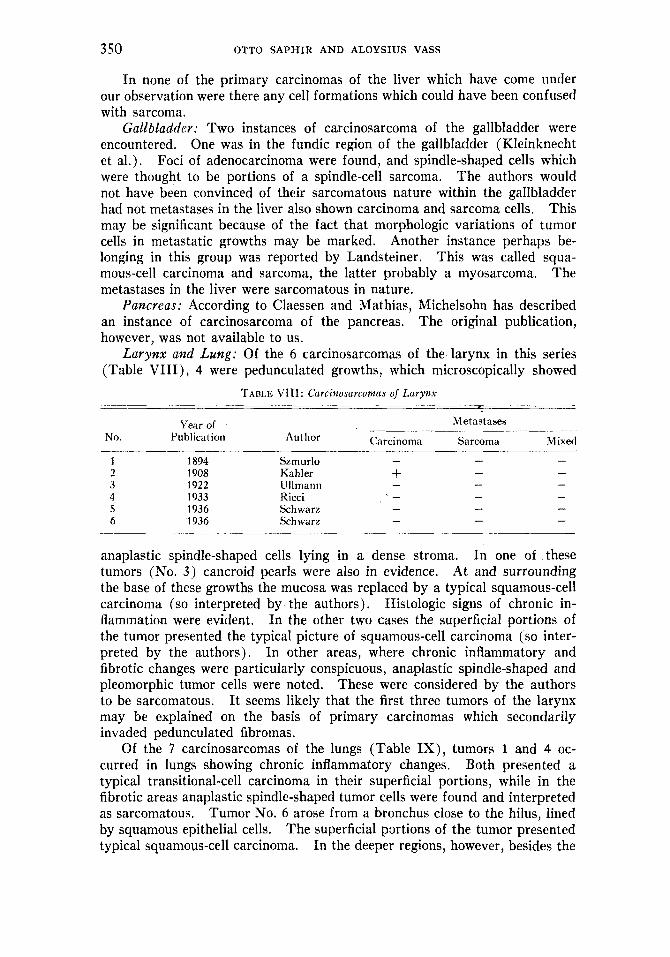
In none of the primary carcinomas of the liver which have come under our observation were there any cell formations which could have been confused with sarcoma.
Gallbladder: Two instances of carcinosarcoma of the gallbladder were encountered. One was in the fundic region of the gallbladder (Kleinknecht et al.). Foci of adenocarcinoma were found, and spindle-shaped cells which were thought to be portions of a spindle-cell sarcoma. The authors would not have been convinced of their sarcomatous nature within the gallbladder had not metastases in the liver also shown carcinoma and sarcoma cells. This may be significant because of the fact that morphologic variations of tumor cells in metastatic growths may be marked. Another instance perhaps be- longing in this group was reported by Landsteiner. This was called squa- mous-cell carcinoma and sarcoma, the latter probably a myosarcoma. The metastases in the liver were sarcomatous in nature.
Pancreas: According to Claessen and Mathias, Michelsohn has described an instance of carcinosarcoma of the pancreas. The original publication, however, was not available to us.
Larynx and Lung: Of the 6 carcinosarcomas of the larynx in this series (Table VIII) , 4 were pedunculated growths, which microscopically showed
TABLE VIII : Curcinosarcomas of Larynx ____ --____
Year of Metastases
N O . Publication Author Carcinoma Sarcoma Mixed - - - 1 1894 Szmurlo
2 1908 Kahler + 3 1922 Ullmann - 4 1933 Iticci 5 1936 Schwarz 6 1936 Schwarz
- - - -
- - - - - - - - -
anaplastic spindle-shaped cells lying in a dense stroma. In one of these tumors (No. 3 ) cancroid pearls were also in evidence. At and surrounding the base of these growths the mucosa was replaced by a typical squamous-cell carcinoma (so interpreted by the authors). Histologic signs of chronic in- flammation were evident. In the other two cases the superficial portions of the tumor presented the typical picture of squamous-cell carcinoma (so inter- preted by the authors). In other areas, where chronic inflammatory and fibrotic changes were particularly conspicuous, anaplastic spindle-shaped and pleomorphic tumor cells were noted. These were considered by the authors to be sarcomatous. I t seems likely that the first three tumors of the larynx may be explained on the basis of primary carcinomas which secondarily invaded pedunculated fibromas.
Of the 7 carcinosarcomas of the lungs (Table I X ) , tumors 1 and 4 oc- curred in lungs showing chronic inflammatory changes. Both presented a typical transitional-cell carcinoma in their superficial portions, while in the fibrotic areas anaplastic spindle-shaped tumor cells were found and interpreted as sarcomatous. Tumor No. 6 arose from a bronchus close to the hilus, lined by squamous epithelial cells. The superficial portions of the tumor presented typical squamous-cell carcinoma. In the deeper regions, however, besides the

CARCINOSARCOMA 351
TABLE IX: Carcinosarcomas of Lung
Year of No. Publicat ion
Metastases
Author Carcinoma Sarcoma Mixed ~
- + + +? -
1 1908 Kika + 2 1914 Saltykow + 3 1915 Frank + 4 1928 Selye - 5 1929 Ogawa - - - 6 1932 Bosen berg - - - 7 1933 Nowicki +
- - -
- -
typical carcinomatous features, elongated, spindle-shaped and pleomorphic cells were seen and were considered sarcomatous. Productive inflammatory changes were particularly evident here. Tumor 7 was partly a mucinous adenocarcinoma, while other portions showed spindle and pleomorphic cells. These were interpreted as sarcomatous. It may be that this tumor is a true carcinosarcoma, though the metastases were purely carcinomatous. Tumor 5 supposedly showed, in addition to carcinoma, a spindle-cell sarcoma. Though the original publication was not available, it seems evident from a study of an abstract of the case report that the spindle-cell sarcomatous fields were transitional or oat-cell carcinomatous portions. Tumor 3 showed an adeno- carcinoma and spindle-cell sarcoma. A mere glance at the photomicrographs accompanying the report, however, indicates that this tumor is a simple adeno- carcinoma with much fibrous connective tissue. Tumor 2 was interpreted as a large round-cell sarcoma with a few giant cells and squamous-cell carcinoma without keratinization. The accompanying photomicrographs of the sup- posedly carcinoma cells do not at all depict, or even resemble, carcinoma cells.
All the lung tumors, with the possible exception of tumor 7, when re- viewed critically, seem to be carcinomas. In both the larynx and the lungs, in addition to other types of carcinoma, transitional-cell carcinomas are relatively frequently encountered. In a previous communication it was shown that in these laryngeal carcinomas, in addition to the transitional cells, squamous-cells with keratinization are quite often found. I t seems clear that since transi- tional cells, as stated before, resemble connective-tissue cells, the finding of transitional and squamous cells may lead to the erroneous diagnosis of car- cinosarcoma. In the lungs are described tumors (Karsner and Saphir) often called oat-cell carcinomas, which, if not identical, are very similar to transi- tional-cell carcinomas. Tumors of the lungs have come to our observation in which predominantly transitional or oat-cells were found in some fields while in others squamous cells were seen. Because of the resemblance of these oat cells to malignant connective-tissue cells, an erroneous diagnosis of carcinosarcoma might be made. The metastases in the lung tumors 1 and 3 , considered in part sarcomatous, apparently consist of transitional cells.
Urinary Tract: Of the 8 tumors of the urinary tract (Table X) 4 arose in the urinary bladder, 3 in the prostate, and 1 in the ureter. The tumors of the urinary bladder were similar in histopathologic structure. Early in their history they were diagnosed as papilloma and carcinoma. Later, often after a history of many years, the tumors presented, besides the obvious carcino-
h

352 OTTO SAPHIR AND ALOYSIUS VASS
TABLE X: Carcinosarcomas of Urinary Tract __-- -
No. Year of
Publication
1910 1912 1931 1932 1932 1933 1934 1936
__ ~~~~~
Author __ 'Tanton Leuenberger Kenner Gabe Rijsen berg €'and Ehrhardt Schwarz
Site Carcinoma
Metastases ~ _ _ _ _ _ _ _ -
Sarcoma Mixed
Urinary bladder Urinary bladder Ureter Urinary bladder Urinary bladder Prostate Prostate Prostate
matous features, anaplastic spindle-shaped cells of varying size, with little intercellular substance, which cells were interpreted as sarcomatous. These tumors are apparently purely transitional-cell carcinomas, which are known to develop in the bladder the most bizarre anaplastic features. The long history, the early diagnosis of papilloma and carcinoma, the obvious chronic inflam- matory changes, and the lack of an intercellular fibrous network seem to in- dicate the probability of this interpretation. Nor do the so-called sarcomatous metastases reported by the authors seem characteristic of sarcoma.
In the last two " carcinosarcomas " of the prostate large grcups of dis- tinctly carcinomatous cells were evident. In addition, more diffusely infiltrat- ing single, irregular, anaplastic, spindle-shaped tumor cells were dispersed throughout whorls and bundles of large muscle fibers. There were also some multinucleated giant cells with centrally located nuclei. Chronic productive inflammatory changes were evident histologically. After a critical review of these tumors it seems probable that they are transitional-cell carcinomas of the urinary bladder which have invaded the prostate secondarily. The first tumor was described as a small-cell sarcoma and early adenocarcinoma. Ac- cording to the accompanying photomicrographs the " sarcomatous " portions may be interpreted as portions of a " carcinoma solidum '' (Kaufmann) . Kaufmann also states that transitions exist between diffusely infiltrating small- round-cell carcinomas (carcinoma solidum) and adenocarcinomas. This so- called carcinosarcoma of the prostate, therefore, seems to be similar to the type of carcinoma mentioned by Kaufmann.
This was a pe- dunculated growth arising in the middle third of the ureter and hanging down into the urinary bladder. The more distal portion of the tumor consisted of closely packed elongated, spindle cells and a myxomatous connective tissue with islets of cartilage. At and around the base a typical transitional-cell carcinoma was present. Islets of carcinoma cells (so interpreted by the author) also were noted in the proximal portion of the pedunculated mass. This tumor was interpreted by the author as a carcincsarcoma of embryonal origin. Although we cannot definitely deny the correctness of this diagnosis, we may interpret this t u m x as chondromyxofibrosarcoma, invaded secondarily by a transitional-cell carcinoma of the ureter. Thus may be explained the presence of carcinoma cells around the base of the tumor and in its proximal portions, and their absence in the distal portions of the pendulous mass.
There is also on record a carcinosarcoma of the ureter.

Fiq. 25.
Fisj.26. Fx. 23. PRIMARY CARCINOMA OF LUNG
Note the transitional type of tumor cells and more differentiated apparent squamous-cells. Iron hematoxylin-eosin. X 165
FIG. 24. PRIMARY CARCINOMA OF URINARY BLADDER Note the spindle-shaped transitional cells in the middle portion of the picture. Iron hema-
toxylin-eosin. X 225
FIG. 25. CARCINOMA OF URINARY BLADDER Note the resemblance of the tumor cells of transitional type to sarcoma cells. Iron hema-
toxylin-eosin. X 90
FIG. 26. SQUAMOUS-CELL CARCINOMA OF MOUTH WITH TRANSITIONAL-CELL FEATURES Note the infiltrating transitional cells which in some instances are interpreted as sarcoma cells.
Hematoxylin-eosin. X 75
353

3 54 OTTO SAPHIR AND ALOYSIUS VASS
Transitional-cell carcinoma of the urinary bladder is one of the malignant epithelial tumors which in many respects may resemble sarcoma. Thus, re- ported instances of sarcomas of the urinary bladder, such as studied by Jelm and others, probably are not sarcomas, but transitional-cell carcinomas. In the absence of papillary excrescences, when the fibrous cores from which the cells arise have disappeared, the cells often become more spindle-shaped and infiltrate the surrounding tissues in the form of clusters, or diffusely. Ewing speaks of spindle-cell carcinoma, to indicate the peculiar form of these cells. Carcinoma cells of this spindle shape are more commonly encountered in re- current tumors and in tumors invading the peritoneal surface or space of Retzius. When in some fields well preserved papillae are noted, with their cells still clearly recognizable as transitional epithelial cells, and in other fields transitional cells not arranged in the form of papillae, resembling sarcoma cells, infiltrate the mucosa and muscularis diffusely, a diagnosis of carcino- sarcoma may be reached. A number of tumors of this type came to our ob- servation and only careful studies by means of serial sections revealed that the cells first interpreted as spindle-cell sarcoma cells were transitional cells.
Gruber reported a tumor of the kidney which he called carcinosarcoma. This is the only instance on record of this type of tumor arising in the kidney which we have been able to find. I t seems clear, however, that the tumor in question belongs to the Wilms tumor group. Carpi, also, according to
.Claessen and Mathias, reported a carcinosarcoma of the kidney, but in the reference quoted, the report was not found.
Tumors in MisceZZaneous Locatiorts (Table XI) : Five carcinosarcomas of TABLE XI : Curcinosarcomas in Miscellaneous Locations
Year of No. Publication Author
Metastases ....
Site Carcinoma Sarcoma Mixed
1 1913 2 1923 3 1932 4 1932 5 1933 6 1928 7 1930 8 1921
9 1924- 10 . 1930 11 1932
Simmonds Ferrero Resenberg Hecker Bezza %lye Tj okonegoro Claessen and
Mathias Traina Rerger Morax and
Kerbrat
Skin Skin Skin Skin (lip) Skin (joint fistula) Middle ear Nasal cavity Salivary gland
Maxillary sinus Labium majus Ciliary body
the skin,G and one each of the middle ear, salivary gland, ciliary body, nasal cavity, maxillary sinus, and labium majus were found in the literature. Of those of the skin, the first showed superficially squamous-cell carcinoma features and in the deeper regions anaplastic, spindle-shaped cells, interpreted by the author as sarcomatous. This tumor developed in an area of der- matitis produced by x-rays. The second and third tumors were basal-cell
by Bczza, could not be verified. fi The report by Senger, referring to a sarcoma in close relation to a lupus carcinoma, as quoted

CARCINOSARCOMA 355
carcinomas in their superficial layers, while the deeper portions consisted of spindle-shaped cells in an abundant connective-tissue stroma with chronic inflammation. These portions were considered sarcomatous by the author. The fourth tumor was a metastatic growth of a previously excised unidentified tumor of the lip. Following excision of the primary tumor, irradiation was applied. An apparent metastatic growth in the neck consisted in part of typical malignant squamous cells, while in other portions there was an abundance of newly formed connective tissue with many spindle-shaped and pleomorphic cells, which were interpreted as sarcomatous. T o judge from the microscopic pictures, this tumor seems to be a simple squamous-cell car- cinoma. The fifth tumor apparently arose in a joint fistula. The microscopic picture may per- haps be interpreted as squamous-cell carcinoma with basal-cell features. The “ sarcomatous ” portions, however, seem to consist of non-malignant fibro- blastic cells.
The carcinosarcoma of the middle ear occurring in a woman sixty-three years of age, who had had otitis media with discharge since childhood, was a squamous-cell carcinoma, in some areas exhibiting anaplastic, spindle-cell features. The tumor of the nasal cavity can also be interpreted as a squa- mous-cell carcinoma with transitional-cell features.
In the study to which we have referred before (Saphir) a number of cancers of the skin were reported which undoubtedly are similar to these skin tumors and the tumor arising in the middle ear. I t was pointed out at that time that in these cases we were dealing not with two primary tumors but rather a single carcinoma with both squamous-cell and basal-cell features. The latter may occasionally be confused with malignant connective-tissue cells.
It con- sisted of epithelial and connective-tissue portions and resembled a malignant mixed tumor of salivary gland origin and was so interpreted by the author. Three months after excision and after x-ray therapy the patient died. The tumor had recurred and presented spindle-shaped and polymorphous, ana- plastic cells, which were interpreted by the author as sarcomatous. While this interpretation probably is correct, it seems that this tumor should not be classified as carcinosarcoma, in view of the fact that the original neoplasm was a mixed tumor. I t is possible, however, that the morphologic alterations might have been caused by the intervening irradiation.
In Traina’s tumor a number of giant cells were present, apparently of the type found in the epulis. This tumor is perhaps a carcinoma with in- flammatory changes resembling a giant-cell tumor. Berger’s tumor of the labium is interpreted by us as basal-cell carcinoma with squamous-cell fea- tures, severe chronic inflammatory changes, and much fibrosis. I t seems likely that basal cells compressed by the fibroblastic stroma were interpreted as sarcoma. Morax and Kerbrat’s tumor of the ciliary body was an outspoken melanotic tumor with spindle-shaped cells and epithelial-like structures which were interpreted as constituents of a melanotic carcinosarcoma. Such a com- bination, however, is often found in malignant melanotic tumors. A picture of a similar tumor is reproduced in a paper previously mentioned (Saphir).
This case is referred to as carcinosarcoma of the neck.
The salivary gland tumor was found in the submaxillary region.

356 OTTO SAPI-IIR AND ALOYSIUS VASS
DISCUSSION
A review of the available literature on carcinosarcomas and an analysis of tumors which have come to our observation which seemingly belong in thi: group reveal that most of the so-called carcinosarcomas are neither collision nor combination tumors but most commonly are primary carcinomas. The individual cells either show what has been called morphologic variation or have been distorted by chronic productive inflammatory changes within or close to the tumor. In some instances a primary sarcoma had invaded non- malignant epithelial structures forming inclusions which led to the erroneous diagnosis of carcinosarcoma. Occasionally a so-called carcinosarcoma could be interpreted as a primary carcinoma which had invaded a benign connective- tissue tumor.
In a study to which reference has been made (Saphir), it was brought out that various types of carcinomas may show distinct variations of the in- dividual cells or groups of cells to such an extent as to render their classifica- tion difficult. In many instances which had the appearance of squamous-cell carcinoma, islets of basal cells were found. Nests of transitional cells were present in squamous-cell carcinomas arising from mucous membranes, par- ticularly in the region of the oral cavity, esophagus, and cervix uteri. Vice versa, islets of squamous carcinoma cells were often present in carcinomas which at first appeared to be purely transitional-cell carcinomas. As stated previously, in some instances the transitional cells in these squamous-cell carcinomas were elongated and spindle-shaped, and could easily be confused with sarcoma cells. Thus, there are morphologic similarities between car- cinosarcomas as described in the literature and squamous-cell carcinomas with “ transitional-cell features.” The morphologic similarity of certain kinds of transitional cells of the bladder to spindle-cell sarcomas is well known and was discussed previously. I t was mentioned that Ewing speaks of “ spindle- cell ” carcinomas of the urinary bladder. In short, it seems obvious that a number of carcinosarcomas reported in the literature can be interpreted on the basis of such morphologic cellular variations. I t is of interest to note that Martin and Stewart emphasized that transitional-cell carcinomas, because of the fact that some of the tumor cells are spindle-shaped, are sometimes erroneously referred to as carcinosarcomas. They mentioned particularly tumors of the larynx, lungs, esophagus, bladder, and urethra,
A number of other reported carcinosarcomas are seemingly primary car- cinomas complicated by chronic productive inflammatory changes. Ewing pointed out that malignant tumors, when altered by inflammation, etc., tend to assume indifferent structures in which most of the original features are lost and from which it is usually hazardous to attempt to reach any conclusion regarding histogenesis. Inflammatory changes were most marked in those organs exhibiting cyclic changes, such as the uterus, breast and thyroid, and, conversely, those organs were more commonly the site of these tumors. I t seems quite plausible that connective-tissue proliferation with hyalinization, etc., resulting in compression of epithelial cells of tumors, may lead to mor- phologic changes in these cells and perhaps constitutes a factor in the faulty

CARCINOSARCOMA 3 57
diagnosis of these tumors. These changes are most evident in the thyroid gland and may explain some of the “ carcinosarcomas ” in this organ.
It is clear that tumors of organs which easily become infected, such as the oral cavity, cervix uteri, etc., also quite frequently show evidence of chronic inflammation. Still another factor causing chronic inflammatory changes of and around a primary tumor is x-ray irradiation, which was applied according to the history of some of the cases. I t seems evident that chronic inflam- matory changes with fibrosis, and also with the presence of many lymphocytes, may have suggested a connective-tissue tumor or occasionally a lymphosar- comatous element in primary carcinomas.
I t is, of course, true that chronic productive inflammatory changes are not necessarily confined to “ carcinosarcomas,” but are present in many malig- nant tumors. The inflammation seen in the latter, however, is more com- monly acute or chronic exudative in character rather than of a long-standing, chronic productive type, such as is almost invariably seen in “ carcinosar- comas.” This is borne out particularly in the linitis plastica type of car- cinoma, which was found to be the underlying malignancy in some of the reported instances of ‘‘ carcinosarcoma ” of the stomach.
In another group of tumors it seemed unquestionable that a carcinoma had invaded a previously existing benign connective-tissue tumor. The carcinoma was present in those organs in which connective-tissue tumors are not rare, particularly in the uterus and larynx, but also in the esophagus and stomach. Histologically the benign tumors were myomas, fibromyomas, and fibromas. Because of the occasional difficulties in differentiating malignant carcinoma cells from the cells of young benign connective-tissue tumors, and because of the presence of giant cells in some of these instances, the cells of the connec- tive-tissue tumors were interpreted as portions of a sarcoma. I t is known, however, that giant cells are not unusual in benign myofibromas. Also it is easily understood that because of the pressure of the fibers of the benign connective-tissue tumors, the carcinoma cells secondarily become altered, are compressed, and resemble sarcoma cells. Close inspection of the histologic pictures accompanying the case reports invariably lead us to believe in the carcinomatous nature of these cells.
In two instances, both of which presented adenomatous structures within spindle-cell sarcomas, the adenomatous structures were interpreted by the authors as blastomatous. These adenomatous structures were not found, however, in the recurrences, which were sarcomatous. From the accompany- ing illustrations it was not apparent that these adenomatous structures were malignant, whereas the connective-tissue tumor was obviously a sarcoma.
Thus, a study of the carcinosarcomas reported in the literature and our own pertinent experience indicate that many of these tumors are not carcino- sarcomas, but single malignant tumors, predominantly carcinomatous.
SUMMARY
One hundred and fif ty-three so-called carcinosarcomas reported in the literature are recorded. A critical review indicates that the carcinosar- comatous nature of these tumors is very questionable. Perhaps only three

358 OTTO SAPHIR AND ALOYSIUS VASS
or four of them may be designated as true carcinosarcomas. A number of our own observations are given on tumors which were at first thought to be carcinosarcomas but which, on more careful examination, were interpreted as primary carcinomas.
In evaluating the seemingly sarcomatous features of the reported carcino- sarcomas, the following complicating factors which play a r61e in the altera- tion of the fundamental histologic appearance of the tumors must be con- sidered: variations of carcinoma cells, some of which assume spindle shapes and may be interpreted as cells of a spindle-cell sarcoma, a factor particularly true of (‘ squamous-cell carcinomas with transitional features ”; marked ana- plasia of the carcinoma cells; chronic inflammation which either leads to morphologic changes of tumor cells, produces much connective tissue which may be regarded as part of a malignant connective-tissue tumor, or provokes a lymphocytic reaction sometimes taken as the lymphosarcoma component of some of these tumors; the invasion of a benign connective-tissue tumor by a carcinoma. Other instances of so-called carcinosarcomas are believed to be sarcomas which had invaded normal or metaplastic epithelial structures, the latter being interpreted as the “ carcinomatous ” elements.
REFERENCES
ALBRECHT, H.: Frankfurt. Ztschr. f . Path. 2: 191, 1909. ALBRECHT, H., AND HECHT, V.: Wien. klin. Wchnschr. 22: 1737, 1909. APOLANT, H.: Verhandl. d. deutsch. path. Gesellsch. 12: 3, 1908. ASADA, T.: Gann 16: 2 2 , 1922. BALTZER, H.: Virchows Arch, f . path. Anat. 259: 252, 1926. BASSAL, L., A N D RIGAUD: Arch. de mCd. expCr. 25: 491, 1913. BENTHIN, W.: Beitr. z. path. Anat. u. z. allg. Path. 60: 163, 1914-15. BERCER, L.: Bull, de 1’Assoc. franc. pour 1’Ctude du cancer 19: 292, 1930. BERCERET AND BOTELHO: Gynbc. et obst. 1: 139, 1920. BEZZA, P.: Tumori 7 : 601, 1933. BLACKBURN, G.: St. Bartholomew’s Hosp. J. 41: 138, 1934. Abst. in Am. J. Cancer 23:
BOSENBERG, M.: Ztschr. f . Krebsforsch. 36: 416, 1932. BRUN, A.: Centralbl. f . allg. Path. u. path. Anat. 33: 51, 1922. CARNEVALE-RICCI, F. : Oto-rho-laring. ital. 1 : 221, 1931. CARPI, U.: Gazz. med. ital. 61: 471, 1910. Quoted by Claessen and Mathias. CILOTTI, M.: Pathologica 20: 608, 1928. CLAESSEN, M., AND MATHIAS, E.: Beitr. z. klin. Chir. 123: 584, 1921. COENEN, H.: Beitr. z. klin. Chir. 68: 605, 1910. COHN, M.: Virchows Arch. f . path. Anat. 259: 30, 1926. DAL Pozzo, G.: I1 cancro 2: 217, 1931. Quoted from Abstract in Ztschr. f . Krebsforsch.
DAL Pozzo, G.: I1 cancro 3 : 156, 1932. DANIEL, G., AND LAZARESCO, S.: Rev. franq. de gynCc. et d’obst. 30: 883, 1935. DORSCH: Ueber Carcinom und Sarkom derselben Mamma, Inaug. Diss., Wiirzburg 1896.
EHRHARDT, W.: Centralbl. f . allg. Path. u. path. Anat. 59: 355, 1934. EHRLICH, P., AND APOLANT, H.: Centralbl. f . allg. Path. u. path. Anat. 17: 513, 1906. EHRLICH, P., AND APOLANT, H.: Berl. klin. Wchnschr. 44: 1399, 1907. EWINC, J.: Neoplastic Diseases, W. B. Saunders Co., Philadelphia, Ed. 3, 1928. EWINC, J.: Am. J. Path. 5: 99, 1929. FERRERO, V.: Arch. per le sc. med. 46: 285, 1923.
167, 1935.
36: 9, 1932.
Quoted by Coenen.

CARCINOSARCOMA 359
FORSSNER, H.: Arch. f . Gynak. 87: 445, 1909. FRAENKEL, E.: Monatschr. f . Geburtsh. u. Gynak. 14: 684, 1901. FRANK, A,: Schmidt’s Jahrb. d. ges. Med. 322: 149 (Erg. H.), 1915. FRANKL, 0.: Arch. f. Gynak. 124: 67, 1925. v. F R A N Q U ~ , 0.: Ztschr. f . Geburtsh, u. Gynak. 47: 211, 1902. GABE, J.: Brit. J. Urol. 4: 145, 1932. DE GAETANI, G.: Pathologica 23: 197, 1931. GIAVOTTO, G.: Pathologica 13: 95, 1921. GOTTING, P.: Frankfurt. Ztschr. f. Path. 41: 107, 1931. GOLDSTINE, M. T.: Am. J. Obst. & Gynec. 30: 143, 1935. GREENBLATT, R. B.: Am. J. Path. 9: 525, 1933. GRUBER, B.: Deutsche mil-arztl. Ztschr. 45: 404, 1916. HAACKE, R. : Ueber Geschwulstbildungen endothelialen Ursprungs in eincm Ovarialkystom,
v. HANSEMANN, D.: Berl. klin. Wchnschr. 2 7 : 353, 1890. v. HANSEMANN, D.: Ztschr. f . Krebsforsch. 1: 183, 1904. HARBITZ, F.: Beitr. z. path. Anat. u. z. allg. Path. 62: 503, 1916. HARVEY, W. F., AND HAMILTON, T. D.: Edinburgh M. J., 42: 337, 1935. HECKER, H.: Deutsche Ztschr. f . Chir. 237: 177, 1932. HEDREN, G.: Centralbl. f . allg. Path. u. path. Anat. 26: 265, 1915. HELWIG, F. C.: Arch. Path. 4: 162, 1927. HERRENSCHMIDT, A.: Bull. et mkm. SOC. anat. 9 (6th series): 300, 1907. HERXHEIMER, G.: Beitr. z. path. Anat. u. z. allg. Path. 44: 150, 1908. HERXHEIMER, G.: Centralbl. f . allg. Path. u. path. Anat. 29: 1, 1918. HERZOG, G.: Verhandl. d. deutsch. path. Gesellsch. 17: 346, 1914. HIPPEL, B.: Virchows Arch. f . path. Anat. 201: 326, 1910. IDZUMI, G.: Arch. f. klin. Chir. 100: 1181, 1913. IWANOFF, N. S.: Monatschr. f . Geburtsh. u. Gynak. 7: 295, 1898. JAFFE, R. H.: Surg. Gynec. & Obst. 37: 472, 1923. JELM, C. E.: J. Urol. 33: 599, 1935. JESSUP, D. S. D.: Proc. New York Path. SOC. 23: 21, 1923. KAHLER, 0.: Deutsche med. Wchnschr. 34: 644, 1908. KAHLSTORF, A,: Fortschr. a. d. Geb. der Kontgenstrahlen 42: 421, 1930. KARSNER, H. T.: Human Pathology, J. B. Lippincott Co., Philadelphia, 1926. KARSNER, H. L., AND SAPHIR, 0.: Am. J. Path. 6: 553, 1930. KAUFMANN, C.: Deutsche Ztschr. f . Chir. 11: 401, 1879. Quoted by Loeb. KAUFMANN, E. : Lehrbuch der speziellen pathologischen Anatomie, Walter de Gruyter & co., KERBIRIOU AND DANEL: J. d. sc. mkd. de Lille 1 : 175, 1897. KETTLE, E. H. : Lancet. 2 : 750, 1912. KETTLE, E. H.: Pathology of Tumors, H. K. Lewis & Co., London, Ed. 2, 1925. KIDNER, F. C.: Boston M. & S. J. 157: 836, 1908. KIKA, G.: Gann 2 : 84, 1908. KLEE, F.: Zentrlbl. f . Gynak. 46: 166, 1922. KLEINKNECHT, A., NESSMANN, V., AND OBERLING, CH.: Bull. Assoc., franc. p. l’ktude du
KLEINSCHMIDT, R.: Ztschr. f . Krebsforsch. 18: 126, 1922. KREIBIG, W.: Virchows Arch. f . path. Anat. 256: 649, 1925. KROMPECHER, E.: Beitr. z. path. Anat. u. z. allg. Path. 44: 88, 1908. V. KUBINYI, P.: Arch. f. Gynak. 97: 237, 1912. Quoted from Bezza. KUCKENS, H.: Beitr. z. path. Anat. u. z. allg. Path. 80: 116, 1928. KUMMER, E.: Rev. mCd. de la Suisse Rom. 18: 702, 1898. KUNSEMULLER, G. : Uber ein Karcinosarkom der Mamma, h u g . Diss., Breslau, 1920. LANDSTEINER, K.: Ztschr. f . klin. Med. 62: 427, 1907. LANG, F. J.: Virchows. Arch. f . path. Anat. 234: 485, 1921. LEROUX, R., AND PERROT, M.: Bull. Assoc. franq. p. 1. Ctude du cancer 20: 283, 1931. LEUENBERGER, S. G.: Deutsche Ztschr. f . Chir. 114: 1, 1912.
Inaug. Diss., Halle, 1901.
Berlin and Leipzig, 1931 (9-10 Aufl.). Quoted by Helwig.
cancer 20: 476, 1931.
Quoted by Loeb.

3 60 OTTO SAPHIR AND ALOYSIUS VASS
LEWIN, C.: Verhandl. d. deutsch. path. Gesellsch. 12: 50, 1908. LINDEMANN, A.: Ztschr. f . Krebsforsch. 6: 419, 1908. LIPPMANN, H.: Ztschr. f . Krebsforsch. 3: 293, 1905. LOEB, L.: Am. J. M. Sc. 125: 243, 1903. MARTIN, H. E., AND STEWART, F. W.: Am. J. Cancer 24: 273, 1935. MASON, R., AND WELLS, H. G.: J. Cancer Research 13: 207, 1929. MEYER, H.: Centralbl. f . allg. Path. u. path. Anat. 30: 291, 1919. MICHELSOHN : Ein Fall von primarem Sarcocarcinom des Pancreas, Inaug. Diss., Wiirzburg,
MILONE: Arch. per le sc. med. 48: 112, 1925-26. MONDOR, H., GAUTHIER-VILLARS, P., AND GOTTESMANN, H.: Ann. d’anat. path. 13: 783, 1936. MORAX AND KERBRAT: Bull. SOC. d’opht. de Paris, 1932, p. 186. Abst. in Am. J. Cancer 18:
MOTTA, G.: Ann. di ostet. 48: 611, 1926. NASSETTI, F.: Policlinico (sez. chir.) 16: 429, 1909. NEBESKY, 0.: Arch. f . Gynfk. 73: 653, 1904. NIEBERGALL, E.: Arch. f. Gynak. 50: 129, 1896. NOWICKI, W.: Virchows Arch. f . path. Anat. 289: 564, 1933. OESTERLIN, E. J.: Ann. Surg. 91: 610, 1930. OGAWA, K.: J, Orient, Med. 11: 133, 1929. Abst. in Ztschr. f . Krebsforsch. 31: 53, 1930. ORTH, J.: Charitk Ann. 34: 357, 1910. PAN& C.: Tumori 7: 244, 1933.
PERRIER, H.: Rev. mCd. de la Suisse Rom. 32: 447, 1.912. PHILIPP, P. W.: Jahrb. f . Kinderh. 18: 353, 1905. QUECKENSTEDT, H. : Ueber Karzinosarkome, Inaug. Diss., Leipzig, 1904.
QUIN, J. S.: Irish J. M. Sc., 1929, p. 579. RENNER, M. J.: Surg., Gynec. & Obst. 52: 793, 1931. RESCH, B.: Centralbl. f . allg. Path. u. path. Anat. 53: 275, 1932. RICCI, B.: Oto-rho-laring. ital. 3: 259, 1933. ROCA DE VIGALS, R.: Compt. rend. SOC. de biol. 110: 729, 1932. ROESCH, H.: Virchows. Arch. f . path. Anat. 245 : 9, 1923. ROLANDO, S.: Arch. ital. di chir. 2 5 : 502, 1930. ROTHACKER, A.: Ztschr. f . Krebsforsch. 12: 39, 1913. RUSSELL, B. R. G.: J. Path. & Bact. 14: 344, 1910. SALTYKOW, S.: Centralbl. f . allg. Path. u. path. Anat. 16: 547, 1905. SALTYKOW, S.: Verhandl. d. deutsch. path. Gesellsch. 17: 351, 1914, SAILER, S.: Am. J. Cancer 31: 183, 1937. SAPHIR, 0.: Morphologic Variations of Tumor Cells, To be published. SCARFF, G. R.: J. Laryng. & Otol. 44: 324, 1929. SCHIFFMANN, J.: Monatschr. f . Geburtsh. u. Gynak. 78: 287, 1928. SCHLAGENHAUFER: Centralbl. f . allg. Path. u. path. Anat. 17: 385, 1906. SCHMINCKE, A.: Monatschr. f . Geburtsh. u. Gynak. 39: 841, 1914. SCHONE, G.: Virchows. Arch. f . path. Anat. 195: 169, 1909. SCHUBACK, A.: Ztschr. f . Krebsforch. 33: 126, 1931. SCHWARZ, H.: Frankfurt. Ztschr. f . Path, 49: 247, 1936. SEHRT, E.: Beitr. z. Geburtsh. u. Gynak. 10: 43, 1905-06. SELYE, H.: Med. Klin. 24: 1197, 1928. SEMB, C.: Acta chir. Scandinav., vol. 64, Supp. X, 1928. SENGER: Berl. klin. Wchnschr. 48: 662, 1911. SHIMODA: Acta dermat. 9: 187, 1927. Quoted by Bezza. SIMMONDS, M.: Ztschr. f . Krebsforsch. 13: 307, 1913. SLYE, M., HOLMES, H. F., AND WELLS, H. G.: J. Cancer Res. 10: 175, 1926. SOKOLOW, A. N.: Centralbl. f . allg. Path. u. path. Anat. 23: 91, 1912. STEIN, A.: Monatschr. f. Geburtsh. u. Gynak. 36: 417, 1912. STROPENI, L.: I1 cancro 3: 236, 1932. Ahst. in Am. J. Cancer 20: 906, 1934.
1894. Quoted from Claessen and Mathias. Quoted from Pan&.
195, 1933.
PASTERNACK, J. G., AND WIRTH, J. E.: Am. J. Path. 12: 423, 1936.
Quoted by Claes- sen and Mathias.
Quoted by Dal Pozzo. Quoted by Bezza.

CARCINOS ARCOMA 361
SUSSI, L.: Policlinico (sez. chir.) 38: 514, 1931. SZMURLO: Medicyna, 29. Jahrgang, Warschau 1894. TAKANO, N.: Arch. f . klin. Chir. 103: 155, 1913. TANTON: Bull. et mCm. SOC. de chir. de Paris 36: 57, 1910. TJOKONEGORO, HADJIDHARMO: Nederl. Tijdschr. v. Geneesk. 74: 4792, 1930. TRAINA, S.: Tumori 11 : 36, 1924. ULLMANN, H.: Ztschr. f . Hals-, Nasen- u. Ohrenh. 1: 130, 1922. VIRCHOW, R. : Die krankhaften Geschwiilste, A. Hirschwald, Berlin, 1864-65, Vol. 2. WAECHTER, H. : Uber Carcino-Sarcome der Schildruse, Inaug. Diss. Freiburg i. Br. 1909. WAELLE, H.: Ueber das Carcinoma sarcomatodes der Mamma, Inaug. Diss. Zurich, 1913. WEHNER, E.: Frankfurt, Ztschr. f . Path. 16: 167, 1915. WELLS, H. G.: J. Path. & Bact. 7 : 357, 1901. WILLIS, R. A,: The Spread of Tumors in the Human Body, J. & A. Churchill, London, 1934. WOGLOM, W. H.: J. Cancer Res. 3: 47, 1918. WOGLOM, W. H.: Arch. Path. 2: 709, 1926. YAMAGIWA, IS.: Verhandl. der japon. path. Ges. I.
deutsch. path. Gesellsch. 1 7 : 351, 1914. ZOLLNER, F.: Deutsche Ztschr. f . Chir. 219: 321, 1929.
Quoted by Ullmann, H.
Quoted by Leuenberger, S. G.
Quoted by Saltykow in Verhandl. d.