the mechanism of action of penicillin
TRANSCRIPT

THE JOURNAL OF BIOLOGICAL CHEMISTRY Vol. 255, No. 9, lesue of May 10, pp. 3977-3986, 1980 Printed in U.S.A.
The Mechanism of Action of Penicillin PENICILLIN ACYLATES THE ACTIVE SITE OF BACILLUS STEAROTHERMOPHZLUS D-ALANINE CARBOXYPEPTIDASE.*
(Received for publication, August 23, 1979)
R. Rogers Yocum, James R. Rasmussen, and Jack L. Strominger From The Biological Laboratories, Harvard University, Cambridge, Massachusetts 02138
Penicillin kills susceptible bacteria by specifically inhibiting the transpeptidase that catalyzes the final step in cell wall biosynthesis, the cross-linking of pep- tidoglycan. It was hypothesized (Tipper, D., and Strom- inger, J. (1965) Proc. Natl. Acad. Sei. U. S. A. 64,1133- 1141) that 1) penicillin is a structural analog of the acyl- D-alanyl-D-alanine terminus of the pentapeptide side chains of nascent peptidoglycan, and that 2) penicillin, by virtue of its highly reactive /”ctam structure, ir- reversibly acylates the active site of the cell wall trans- peptidase. Although the cell wall transpeptidase has proven elusive, a closely related penicillin-sensitive cell wall enzyme, D-alanine carboxypeptidase, has been pu- rified from membranes of Bacillus stearothennophilus by penicillin affinity chromatography. By amino acid sequence analysis of 14C-labeled cyanogen bromide peptides generated and purified from this carboxypep- tidase covalently labeled with either [14C]penicillin G or the substrate, [‘4C]diacetyl-~-lysyl-~-alanyl-~-lac- tate, it was shown that the penicillin and substrate were both bound as esters to a serine at residue 36. Therefore, the second hypothesis stated above was proven to be correct for D-alanine carboxypeptidase.
Several new methods were developed in the course of this work, including 1) a rapid penicillin-binding assay, 2) use of hydroxylamine to protect peptides against carbamylation during ion exchange chroma- tography in concentrated urea solutions, and 3) gel filtration chromatography in 70% formic acid, a univer- sal solvent for peptides.
It has been known since 1965 that penicillin kills susceptible bacteria by inhibiting the transpeptidase that cross-links cell wall peptidoglycan (1, 2). Since that time, there has been considerable controversy in the literature as to the molecular details of this inhibition. Tipper and Strominger (2) hypoth- esized that 1) penicillin is a substrate or transition state analog of the acyl-D-Ala-D-Ala terminus of the pentapeptide side chain of uncross-linked peptidoglycan and that 2) the highly reactive p-lactam of the antibiotic irreversibly acylates the nucleophilic active site of cell wall transpeptidase. Based on biophysical and kinetic data, Ghuysen and co-workers have argued that penicillin might be an allosteric inhibitor of cell wall transpeptidase (3). Resolution of this controversy has been hampered by the fact that an enzyme which efficiently catalyzes cell w d transpeptidation in vitro has never been purified. Although bacterial membranes are capable of syn-
* This work was supported by a research grant from the National Science Foundation (PCM 78 24129). The costs of publication of this article were defrayed in part by the payment of page charges. This article must therefore be hereby marked “advertisement” in accord- ance with 18 U.S.C. Section 1734 solely to indicate this fact.
thesizing cross-linked peptidoglycan (4-6), solubilization of these membranes with detergents causes immediate loss of transpeptidase activity (6, 7).
The membranes of many species of bacteria contain one major and several minor proteins which bind penicillin cova- lently (7-9). Some of these proteins have been purified (7 , 8, 10-12), but none have been shown incontrovertibly to be the cell wall transpeptidase. Nakagawa et al. (13) claim to have isolated a penicillin-sensitive transpeptidase from Escherichia coli. However, 1 mol of this enzyme incorporates only 0.1 mol of substrate in 2 h, so it is not yet clear if this enzyme is the major peptidoglycan cross-linking enzyme. The major penicil- lin-binding proteins from the membranes of E. coli, Bacillus subtilis, and Bacillus stearothermophilus have been purified (10, 11, 14). In all three cases, these purified proteins specifi- cally catalyze the penicillin-sensitive hydrolysis COOH-ter- mind D-alanine from the peptide chain of cell wall-related substrates. Hence, these enzymes have been given the name D-alanine carboxypeptidase (CPase).’ Although the function of CPase in vivo has not been established, it has been sug- gested that it serves to limit cross-linking in cell walls by removing the terminal D-alanine which is essential for cross- linking. However, this suggestion was not supported by exper- iments designed to test it directly (15). It is also possible that CPase is actually the in vivo transpeptidase, but became “uncoupled” during purification (16). This notion is supported by the findings that CPases from several bacteria are capable of performing “unnatural” transpeptidation reactions when supplied with high enough concentrations of appropriate nu- cleophilic “acceptors” (3,17,18). In any case, the role of CPase and the identity of the transpeptidase require further inves- tigation.
Because CPases catalyze penicillin-sensitive reactions which utilize cell wall-related substrates in vitro and, because they can be purified in milligram amounts, they are the model system of choice for studying the mode of action of penicillin at the molecular level. It has been shown that the synthetic substrate, diacetyl-L-Lys-D-Ala-D-Ala (Ac2LAA) can be used to trap an acylenzyme intermediate for Staphylococcus au- reus CPase (18) and E. coli CPase (19). However, these two CPases are not available in amounts sufficient for protein- sequencing studies. Trapping of an acylenzyme intermediate of the more abundant B. subtilis CPase using [I4C]Ac2LAA could not be detected (20). Presumably, the deacylation step
The abbreviations used are: CPase, D-alanine carboxypeptidase; Ac~LAA, diacetyl-L-lysyl-D-alanyl-D-alanine; AczLALac, diacetyl-L- Iysyl-D-alanyl D-lactate; AczLA diacetyl-L-lysyl-D-alanine; t-Boc, t- butyloxycarbonyl; Bzl, benzyl; lac, lactate; Cbz, benzyloxycarbonyl; TPCK, tosylphenylmethyl chloroketone; CNBr, cyanogen bromide; PTH, phenylthiohydantoin derivative of amino acid A, absorbance; Vo, void volume; V,, salt volume; PhCHZ-SOz, phenylmethanesulfonyl fluoride; 6-APA, 6-aminopenicillanic acid.
3977

3978 B. Stearothermophilus Carboxypeptidase Active Site
of catalysis is rapid compared to the acylation step, so the steady state concentration of acylenzyme intermediate is low. It was subsequently shown that the ester analog, diacetyl-L- Lys-D-Ala-D-lactate was much more efficient in trapping an acylenzyme intermediate (20). This is presumably because the ester bond is more easily cleaved, resulting in a rate acceler- ation for acylation of the enzyme and a concomitant increase in the steady state concentration of acylenzyme intermediate.
In this study, ["C]Ac2LALac was used to trap an acylen- zyme intermediate of B. stearothermophilus CPase in nearly stoichiometric amounts. This allowed sequencing studies, which directly prove the hypothesis of Tipper and Strominger (Z), that penicillin acylates the active site of enzymes involved in cell wall biosynthesis. This paper provides the full details; a preliminary report has been published (21).
EXPERIMENTAL PROCEDURES
Cells-B. stearothermophilus ATCC 15952 was grown at 60°C in a rich medium (7). The glucose was autoclaved separately from the remainder of the ingredients to minimize caramelization. Cells were grown in a New Brunswick 250-liter fermentor at two-thirds maximum aeration to late-log phase (a Klett reading of 200), at which time aeration was raised to full, rapid chilling was started, and the cells were harvested. The cell paste was stored at -20°C. A typical yield was 5 g of cells/liter.
Membranes-In a Gilford Minimill cooled to 2"C, 1500 g of cells were pulverized with 750 ml of 120-pm acid-washed (to remove traces of heavy metals) glass beads (3M Co.) in 1500 ml of buffer containing 50 mM KPOa, 1 mM MgCL, 1 m~ dithiothreitol, pH 7.0. To minimize proteolyis, 400 mg of phenylmethane sulfonyl fluoride (Sigma) dis- solved in 20 ml of ethanol was added. Grinding was at full speed for a total of 30 min with intermittent reduction to one-third speed such that the mixture remained between 2°C and 8°C. All subsequent steps in membrane and enzyme purification were at 0" to 4°C unless otherwise stated. Glass beads were removed by suction frltration through Miracloth (Calbiochem). Whole cells and large cell wall fragments were removed from the filtrate by centrifugation for 10 min at 4,000 X g in a Sorvall GSA rotor. Membrane vesicles were pelleted by centrifuging the previous supernatant at 100,000 X g for 1 h. The membrane pellets were combined and resuspended in grinding buffer (see above) to give a total of 1 liter and homogenized in a Waring blendor. The membranes were stored at -70°C.
Purification of B. stearothermophilus CPase-CPase was purified from membrane vesicles of B. stearothermophilus by a modification of the covalent penicillin affinity chromatography method (14). A molecular spacer, 3-aminopropionic acid, was coupled to Sepharose 4B activated with 25 g of cyanogen bromide/100 ml of packed beads using the sodium carbonate-buffering method (22). A typical yield was 10 pmol of free carboxyl groups coupled/ml of beads as measured by titration. The spacer-coupled Sepharose was washed with IO volumes of 0.5 M NaCI, 0.1 M sodium acetate, pH 5.0, 10 volumes of water, and 2 volumes each of 25%, 50% 75% and 100% dioxane. The washed beads were suspended in 2 volumes of dioxane containing 0.3
TABLE I Purification of B. stearothermophilus ~ - u l u n i n e carboxypeptidase
Carboxypep-
alents* Stage of purification Protein" tidase equiv- Yield
mg Membranes' 25,100 241 Solubilized membrane su- 19,400 225
Cephalothin-treated su- 19,400 195 (100%)
6-APA column eluate 173 Soluble after dialysis 10 9
pernatant
pernatant d -
Precipitate after dialysis "E) 165 15:) 158 81% SP-SeDhadex eluate
" Measured by the Lowry assay (see "Experimental Procedures"). * Measured by ['4C]penicillin G-binding assay (see "Experimental
' From 1500 g of frozen cell paste. Procedures").
Not measurable due to presence of hydroxylamine.
1 2 3 HHH
Fraction Number FIG. 1. Purification of [''C]Ac&ALac. Crude ['4C]AczLALac
was fractionated on a column (1.5 X 100 cm) of Sephadex LH-20 in 0.1 M acetic acid. Fractions were 2.0 ml; 5 pl was assayed for radioac- tivity. Pool 1 contained No-['4C]acetyl-Lys-D-Ala-D-lactate, Pool 2 contained ['4C]Ac2LALac, and Pool 3 contained N'-acetyl-L-Lys-D- Ala-D-lactate.
M diisopropyl carbodiimide (Aldrich) and 0.2 M N-hydroxysuccinimide (Sigma) for 2 h at room temperature. After rinsing with 5 volumes of dioxane, the beads were suspended for 1 h at 0°C in 50 mM 6- aminopenicillanic acid (6-APA) and 0.1 M sodium pyrophosphate adjusted to pH 8.5 just before use. The 6-APA-Sepharose was then washed with 15 volumes of 50 mM KPO,, pH 7.0, and 15 volumes of 50 mM KPO,, pH 7.0, plus 1 M NaC1.
During the 1-h coupling reaction, CPase was solubilized from a thawed membrane suspension by addition of 4 M NaCl and 25% Triton X-100 to give final concentrations of 1.0 M NaCl and 5% Triton
48,000 X g for I h to remove insoluble material. Cephalothin (Lilly) X-100. This suspension was stirred 30 min at 0°C and centrifuged at
was added to the supernatant to give 5 pg/ml and the solution was stirred 30 min at room temperature. The washed 6-APA-Sepharose was then added to the solubilized crude CPase (1 ml of 6-APA- Sepharose/lO ml of solubilized membranes) and the mixture was swirled gently at 37°C for 30 min. The beads were then collected in a sintered glass funnel and washed with 10 volumes of 25 mM KPOo, 0.1% Triton X-100, 1 mM dithiothreitol, pH 7.0, and 10 volumes of 50 mM KPO,, 1 M NaCl, 0.1% Triton X-100, 1 m~ dithiothreitol, pH 7.0. CPase was eluted from the affkity resin with 3 volumes of 50 mM KP04, 1% Triton X-100,O.S M hydroxylamine-HC1, 1 mM dithiothre- itol, pH 7.0 over a period of 1% h. The eluted CPase was then dialyzed exhaustively against 10 mM NaP04, 1 mM dithiothreitol, 0.1% Triton X-100, pH 7.0, during which a variable amount of the CPase precipi- tated. The precipitate was removed by centrifugation at 48,000 X g for 1 h and the clear supernatant was concentrated on a column (1.2 X 40 cm) of SP-Sephadex equilibrated with the above dialysis buffer. CPase was eluted from SP-Sephadex by a step gradient of 50 mM NaP04, 1 mM ethylediaminetetraacetic acid, 1% Triton X-100, and 0.5 M sodium acetate, pH 7.0. The enzyme was monitored by a penicillin- binding assay described below. The CPase that had precipitated during dialysis was readily dissolved in the SP-Sephadex elution buffer and was fully active in binding penicillin. Pure CPase was stored at 4OC. The enzyme was greater than 95% pure: it gave one band at M, = 46,500 upon gel electrophoresis.
Synthesis of Ac2LALuc"Starting materials were obtained from the following sources: t-butyloxycarbonyl azide and N-hydroxysucci- nimide, Aldrich; [14C]acetic anhydride, Amersham; dibenzyloxycar- bonyl-x,-lysine, dicyclohexylcarbodiiide, and D-alanine (5 99.9% D isomer by enzymatic analysis (23)), Sigma. Elemental analyses were performed by Galbraith Laboratories, Knoxville, TN. Melting points were measured on a Buchi melting point apparatus. Optical rotation measurements were made on a Perkin Elmer model 141 polarimeter. High voltage electrophoresis employed Whatman No. 3MM paper and water/acetic acid/pyridine (1oOo:10:1) buffer, pH 3.5.
D-Lactic acid was prepared by fermentation (24) and converted to the lithium salt by titration to pH 7.0 with 1.4 N LiOH. The crude lithium-D-lactate was recrystallized from methanol/ethyl ether (1:3) to yield a colorless powder ([a12 = +14.1 (c 1.5, H@); literature value
B. Stearothermophilus Carboxypeptidase Active Site 3979 1
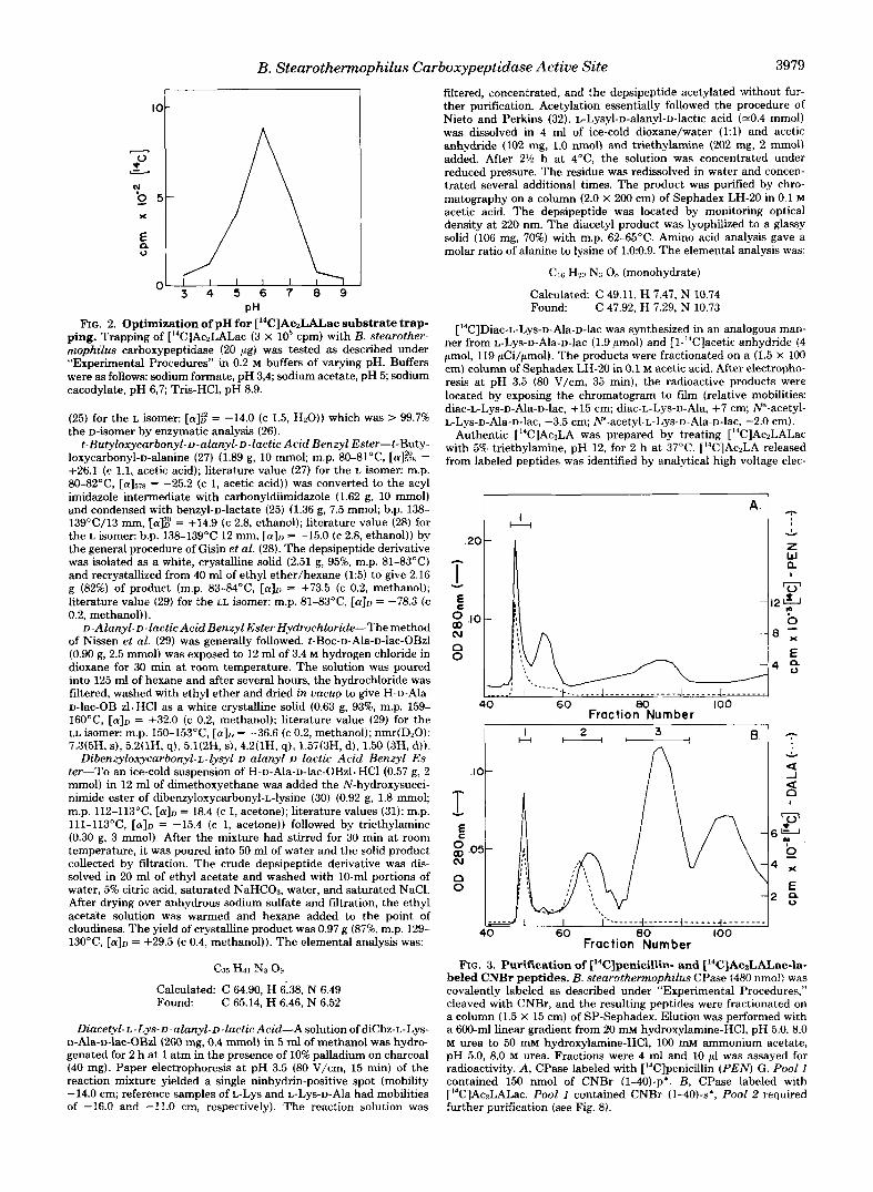
pH FIG. 2. Optimization of pH for ['4C]Ac~LAL.ac substrate trap-
ping. Trapping of ["C]AczLALac (3 X lo5 cpm) with B. stearother- mophilus carboxypeptidase (20 pg) was tested as described under "Experimental Procedures" in 0.2 M buffers of varying pH. Buffers were as follows: sodium formate, pH 3,4; sodium acetate, pH 5; sodium cacodylate, pH 6,7; Tris-HC1, pH 8.9.
(25) for the L isomer: [a]:: = -14.0 (c 1.5, H20)) which was > 99.7% the D-isomer by enzymatic analysis (26). t-Butyloxycarbonyl-D-alanyl-D-lactic Acid Benzyl Ester-t-Buty-
loxycarbonyl-D-alanine (27) (1.89 g, 10 mmol; m.p. 8O-8l0C, [a]% = +26.1 (c 1.1, acetic acid); literature value (27) for the L isomer: m.p. 80-82"C, [a1578 = -25.2 (c 1, acetic acid)) was converted to the acyl imidazole intermediate with carbonyldiimidazole (1.62 g, 10 mmol) and condensed with benzyl-D-lactate (25) (1.36 g, 7.5 mmol; b.p. 138- 139OC/13 mm, [a$' = +14.9 (c 2.8, ethanol); literature value (28) for the L isomer: b.p. 138-139°C 12 mm, [a], = -15.0 (c 2.8, ethanol)) by the general procedure of Gisin et al. (28). The depsipeptide derivative was isolated as a white, crystalline solid (2.51 g, 95%, m.p. 81-83°C) and recrystallized from 40 ml of ethyl ether/hexane (1:5) to give 2.16 g (82%) of product (m.p. 83-&1OC, [ a ] ~ = +73.5 (c 0.2, methanol); literature value (29) for the LL isomer: m.p. 81-83"C, [.ID = -78.3 (c 0.2, methanol)).
D-Alanyl-D-lactic Acid Benzyl Ester Hydrochloride-The method of Nissen et al. (29) was generally followed. t-Boc-D-Ala-D-lac-OBzl (0.90 g, 2.5 mmol) was exposed to 12 ml of 3.4 M hydrogen chloride in dioxane for 30 min at room temperature. The solution was poured into 125 ml of hexane and after several hours, the hydrochloride was filtered, washed with ethyl ether and dried in vacuo to give H-D-Ala- D-lac-OB zl.HC1 as a white crystalline solid (0.63 g, 93%, m.p. 159- 16OoC, [ a ] ~ = +32.0 (c 0.2, methanol); literature value (29) for the LL isomer: m.p. 150-153°C, [a], = -36.6 (c 0.2, methanol); nmr(D20): 7.3(5H, s), 5.2(1H, q), 5.1(2H, s) , 4.2(1H, q), 1.57(3H, d), 1.50 (3H, d)). Dibenzyloxycarbonyl-L-lysyl-D-alanyl- D-lactic Acid Benzyl Es-
ter-To an ice-cold suspension of H-D-Ala-D-lac-OBzl. HCl (0.57 g, 2 mmol) in 12 ml of dimethoxyethane was added the N-hydroxysucci- nimide ester of dibenzyloxycarbonyl-L-lysine (30) (0.92 g, 1.8 mmol; m.p. 112-113OC, [a]D = 18.4 (c 1, acetone); literature values (31): m.p. lll-l13"C, [@ID = -15.4 (c 1, acetone)) followed by triethylamine (0.30 g, 3 mmol). After the mixture had stirred for 30 min at room temperature, it was poured into 50 ml of water and the solid product collected by fdtration. The crude depsipeptide derivative was dis- solved in 20 ml of ethyl acetate and washed with 10-ml portions of water, 5% citric acid, saturated NaHC03, water, and saturated NaCl. After drying over anhydrous sodium sulfate and filtration, the ethyl acetate solution was warmed and hexane added to the point of cloudiness. The yield of crystalline product was 0.97 g (87%, m.p. 129- 130°C, [ a ] ~ = +29.5 (c 0.4, methanol)). The elemental analysis was:
C35 H41 NJ OY Calculated: C 64.90, H 6.38, N 6.49 Found: C 65.14, H 6.46, N 6.52
Diacetyl-L-Lys-D-alanyl-D-1acticAcid-Asolutionof diCbz-L-Lys- D-Ala-D-lac-OBd (260 mg, 0.4 mmol) in 5 d of methanol was hydro- genated for 2 h a t 1 atm in the presence of 10% palladium on charcoal (40 mg). Paper electrophoresis a t pH 3.5 (80 V/cm, 15 min) of the reaction mixture yielded a single ninhydrin-positive spot (mobility -14.0 cm; reference samples of L-LYS and L-Lys-D-Ala had mobilities of -16.0 and -11.0 cm, respectively). The reaction solution was
filtered, concentrated, and the depsipeptide acetylated without fur- ther purification. Acetylation essentially followed the procedure of Nieto and Perkins (32). L-LySyl-D-alanyl-D-laCtiC acid (a0.4 mmol) was dissolved in 4 ml of ice-cold dioxane/water (1:l) and acetic anhydride (102 mg, 1.0 nmol) and triethylamine (202 mg, 2 mmol) added. After 2% h at 4"C, the solution was concentrated under reduced pressure. The residue was redissolved in water and concen- trated several additional times. The product was purified by chro- matography on a column (2.0 X 200 cm) of Sephadex LH-20 in 0.1 M acetic acid. The depsipeptide was located by monitoring optical density a t 220 nm. The diacetyl product was lyophilized to a glassy solid (106 mg, 70%) with m.p. 6245°C. Amino acid analysis gave a molar ratio of alanine to lysine of 1.0:0.9. The elemental analysis was:
CM Hm Ns OR (monohydrate)
Calculated C 49.11, H 7.47, N 10.74 Found C 47.92, H 7.29, N 10.73
[14C]Diac-L-Lys-D-Ala-~-lac was synthesized in an analogous man- ner from L-Lys-D-Ala-D-lac (1.9 pmol) and [l-14C]acetic anhydride (4 pmol, 119 pCi/pmol). The products were fractionated on a (1.5 X 100 cm) column of Sephadex LH-20 in 0.1 M acetic acid. After electropho- resis at pH 3.5 (80 V/cm, 35 min), the radioactive products were located by exposing the chromatogram to film (relative mobilities: diac-L-Lys-D-Ala-D-lac, +15 cm; diac-L-Lys-D-Ala, +7 cm; N"-acetyl- L-Lys-D-Ala-D-lac, -3.5 cm; ~-acetyl-L-Lys-D-Ala-D-lac, -2.0 cm).
Authentic ["CIAczLA was prepared by treating [I4C]Ac2LALac with 5% triethylamine, pH 12, for 2 h at 37°C. [I4C]Ac2LA released from labeled peptides was identified by analytical high voltage elec-
Y
z W a I
'0'
? 0
- l 2 A
- 8 ; E
40 60 80 100 Fraction Number
I 2 H t"l-
3
1 0 W (u
E
n 0
FIG. 3. Purification of ['*C]penicillin- and ['4C]AczLALac-la- beled CNBr peptides. B. stearothermophibs CPase (480 nmol) was covalently labeled as described under "Experimental Procedures," cleaved with CNBr, and the resulting peptides were fractionated on a column (1.5 X 15 cm) of SP-Sephadex. Elution was performed with a 600-ml linear gradient from 20 mM hydroxylamine-HC1, pH 5.0, 8.0 M urea to 50 mM hydroxylamine-HCl, 100 mM ammonium acetate, pH 5.0, 8.0 M urea. Fractions were 4 ml and 10 pl was assayed for radioactivity. A, CPase labeled with [14C]penicillin (PEN G. Pool I contained 150 nmol of CNBr (1-40)-p*. B, CPase labeled with [I4C]Ac2LALac. Pool I contained CNBr (1-40)-s*; Pool 2 required further purification (see Fig. 8).

3980 B. Stearothermophilus Carboxypeptidase Active Site
TABLE I1 Amino acid compositions of ["C]penicilloyl and %substrate peptides from B. stearothermophilus CPase
Data are shown only for those residues present in excess of 0.1 mol/mol of peptide. The parentheses denote the numbers of residues found
40)-p* 40)-s* 40)-p' 40)-s' P* S* T;gf? Tryp(401
in the sequence. CNBr(1- CNBr(1- Staph(26- Staph(Z6- Pap(34-36)- Pap(34-36)-
Asx Thr Ser Glx Pro GlY Ala Val Met' Ile Leu Tyr Phe His LY s -4% Mol label/mol peptide
5.12 (5) 4.78 (5) 2.88 (3) 2.97 (3) 1.76 (2) 1.70 (2) 3.40 (3) 3.23 (3) 0.93 (1) 0.85 (1) 2.54 (2) 2.61 (2) 5.87 ( 6 ) 6.04 ( 6 ) 2.50 (2) 2.62 (2) 2.29 (2) 2.37 (2) 4.47 (5) 4.19 (5) 4.36 (4) 4.49 (4) 1.37 (1) 1.35 (1) 0.38 (0) 0.41 (0) 0.29 (0) 0.31 (0) 2.59 (3) 2.65 (3) 1.65 (1) 1.92 (1) 0.71 0.79
rnol/molpeptide
2.15 (2) 2.00 (2) 1.97 (2) 1.77 (2) 0.99 (1) 0.91 (1) 0.29 0.32
1.58 (1) 1.11 (1) 1.22 (1) 1.78 (1)" 1.16 (1) 1.03 (1) 2.18 (2) 2.34 (2)
1.22 (1) 1.26 (1) 2.01 (2) 2.09 (2)
0.12
1.93 (2) 2.99 (2)" 0.30 0.16 0.55 0.69
0.20
0.95 (1) 0.15
0.28
0.17
1.00 (I) 0.11
1.06 (1)
0.96
0.13
0.97 (1) 0.13
0.20 2.10 (1)"
1.00 (1)
0.94" 0.19 0.98
0.17 2.10 (2) 0.10 1.98 (2) 0.95 (1) 1.10 (1) 0.11 0.17
0.27 1.27 (1) 0.20 1.10 (1) 1.23 (1)
0.99 (1) 1.01 (1) 1.00
1.00 (1) 1.72 (2) 0.13 1.00 (1)
0.19
0.10 1.93 (2)
0.00 0.50 0.00
" One alanine and one lysine residue found in the composition but not found in the sequence are contributed by the covalently bound [I4C]diacetyl- L-Lys-D-alanyl moiety. ["CIPenicilloic acid gave no amino acids upon hydrolysis.
Determined as homoserine.
5 10 TIM @nsE: IIH GLU-SER-ALA-PRO-LEU-ASP-ILE-ARG-ALA-ASP-ALA-ALA- 2""""4"4-
5 10 15 20 2 - " " " " " - ) - w ~ ~ ~ ~ ~ ~ ~ ~ ~ a R ( 1 - N ) - P a : NH GLU-SER-ALA-PRO-LEU-ASP-ILE-ARG-ALA-ASP-ALA-ALA-ILE-LEU-VAL-ASP-ALA-GLN-THR-GLY-
25 30 35 a 40 LY~-ILE-LEU-TYR-GLU-LYS-ASN-ILE-ASP-THR-VAL-LEU-GLY-ILE-ALA-SER-NET-THR-LYS-MET +d4""" +
StaDh f26-40)-~* and -5 *
"""4-w"" I
Tryp (26-39)-p* and -s* I "'
* Pap (34-36)-p* and -s *
FIG. 4. Sequences of intact B. stearothemphilus carboxypeptidase and of the CNBr peptide containing the active site. Arrows represent residues sequenced directly by Edman degradation or by pancreatic carboxypeptidase A plus B digestion. The solid bars represent ["Clpenicillin- and 14C-substrate-labeled peptides which were isolated.
trophoresis (see above) along with the authentic compound. Penicillin- binding Assay-An assay was developed to quantitate
quickly proteins which covalently bind penicillin. The sample (whole membranes, solubilized membranes, or pure CPase), containing at most 1 mg of protein, was added to a reaction mixture to give a total volume of 100 p1 containing 1% Triton X-100, 50 mhf NaP04, 1 mM [I4C]penicillin G (52 to 56 Ci/mol, Amersham), pH 7.0. For assays of pure CPase, 4 mg/ml of Dextran T 70 (Pharmacia) was included as a carrier. After 5 min at 37"C, 0.9 ml of cold acetone was added to precipitate the [14C]penicilloyl-enzyme. The precipitates were col- lected on 25-mm Whatman GF/A glass fiber filters and washed twice with 5 ml of 50% ethanol, 0.1 M HC1 to remove unbound [14C]penicillin G. The filters were oven-dried and counted in toluene-Omnifluor (New England Nuclear). One microgram of CPase bound 1,600 cpm of [14C]penicillin G in this assay. Pretreatment of the sample with unlabeled penicillin G completely inhibited the binding; background was 60 cpm for membranes and 25 cpm for pure CPase. The assay was linear up to 1 mg of total protein, after which the filters became easily clogged.
['4C]AC2LALac, a modification of the above penicillin-binding assay Substrate-binding Assay-In order to optimize the trapping of
was used. The 10-4 assay mixture contained 1% Triton X-100,0.2 M buffer (buffering ion and pH was varied), and 20 pg of pure CPase. At
O"C, 2 pl of 1.25 mM [14C]Ac2LALac (119 Ci/mol) was added with mixing. Immediately, 100 p1 of 5% trichloroacetic acid was added. The resulting precipitate was collected, rinsed, and counted as described above for the penicillin-binding assay.
Preparative Binding of [14C]Penicillin G to CPase-A 2-fold molar excess of [I4C]penicillin G (52 to 56 Ci/mol) was incubated with CPase at 4 m g / d in SP-Sephadex elution buffer (see above) for 5 min at 37OC. Four volumes of cold acetone were added and the precipitated [14C]penicilloyl-enzyme was centrifuged at 2,000 X g for 10 min. This procedure gave 0.71 mol of ["Clpenicillin covalently bound/mol of CPase.
Preparative Trapping of [14C]Ac&ALac-CPase (4 mg/ml) was dialyzed exhaustively against 0.2 M sodium cacodylate, pH 6.0, 1% Triton X-100, 1 l ~ l ~ dithiothreitol. At O"C, [14C]Ac2LALac (2.6 Ci/ mol) was added to give a final concentration of 7 m. This was followed immediately by the addition of a cold solution of 100% (w/v) trichloroacetic acid to give 20% (w/v). The precipitated acyl-enzyme was centrifuged at 2,000 X g for 10 min and the pellet was washed once with 5% trichloroacetic acid and once with acetone. This proce- dure gave 0.85 mol of ["'C]Ac2LA covalently bound/mol of CPase.
Cyanogen Bromide Clea~age-[~~C]Penicilloyl- or ["C]Ac2LA-la- beled CPase was dissolved at 10 m g / d in 70% formic acid. Cyanogen bromide (CNBr) was added to give 100 mg/ml. The reaction mixture

B. Stearothermophilus Carboxypeptidase Active Site 3981
TABLE 111 Sequence data for A, 100 nmol of CNBr(l-40)-p8 and B, 50 nmol of
Staph(26-40)-p*. The yield of each PTH-derivative identified a t Step n, as well as the yield of the same amino acid a t steps n - 1
and n + 1, is given. Residue iden- Yield of identified residue at step
n - 1 n n + 1 Cycle (n) tified
A. 1 2 3 4 5 6 7 8 9
10 11 12 13 14 15 16 17 18 19 20 21 22 23 24 25 26 27 28 29 30 31
B. 1 2 3 4 5 6 7 8 9
10 11
Glu Ser Ala Pro Leu ASP Ile k g Ala ASP Ala Ala Ile
Val Leu
ASP Ala Gln Thr GlY LYS Ile Leu TYr Glu LYS Asn Ile ASP
Val LYS Asn Ile
Thr ASP
Val Leu GlY Ile Ala Ser
<1 3 2 6 5 9
<1 7 8
10 54 11 13 6 5
11
3 2
5 6 1 2
5 3
3
2 <I <1
1 3 1 2 2
<1
nmOl 71 30 80 37 46 50 51 26 37 38 54 63 49 41 33 27 30
23 12
22 24 16 16
-
-
- - 15 10
13 - - 38 16 18 29 25 11 18 31 12
3 3 7 7
10 11 19 28 11 13 63 34 18 19 11 15 20
9 7
13 13 7 6
9 7
11
3 2 2 3 4 2 4 7 2
PTH-derivatives of lysine, glutamine, and asparagine were iden- tified by gas chromatography and thin layer chromatography, but could not be reliably quantitated.
was flushed with Na, protected from light, and cleavage was allowed to proceed for 3 h a t 37"C, after which the volatiles were lyophilized.
Enzymatic Cleavages-Labeled peptides cleaved with TPCK- Trypsin (Worthington) in 0.2 M ammonium acetate, pH 8.2, S. aureus V8 protease (Miles) in 0.1 M ammonium acetate, pH 8.2, and papain (Worthington) in 0.1 M ammonium acetate, pH 5.5, 1 nm dithiothre- itol, 1 mu ethylenediamine tetraacetic acid. Enzyme concentrations were 10 to 50 pg/ml; peptide concentrations were 30 to 500 nmol/ml. All kest ions were for 1 h at 37°C and were terminated by addition of acetic acid to 10% (v/v).
Purification of Peptides-Labeled CNBr peptides were purified by ionexchange chromatography on SP-Sephadex C-25 in urea. The mixtures of CNBr peptides were dissolved in 8 M urea (Becton Dickinson, freshly deionized), 20 rims hydroxylamine-HCI, pH 5.0, and applied to a column (1.5 X 15 cm) of SP-Sephadex equilibrated with the same buffer. Peptides were eluted by a 600-ml linear gradient from the above buffer to 8 M urea, 50 mu hydroxylamine-HC1 and 0.1 M ammonium acetate, pH 5.0. Fractions containing label were pooled and dialyzed exhaustively against 1% formic acid in Spectropore 3 dialysis tubing (Spectrum Industries) which had a molecular weight cut-off of 3,500. When necessary, CNBr peptides were further purified
by gel filtration on a column (1.2 X 100 cm) of Bio-Gel P-10 (BioRad) in 70% formic acid.
Smaller peptides produced by enzymatic cleavage were purified by gel fitration on a column (0.9 X 170 cm) of Sephadex G-25 (superfine) in 0.1 M acetic acid or by ion exchange chromatography on a column (1 x 3 cm) of SP-Sephadex using a 300-ml linear gradient from 0.1 M acetic acid to 0.1 or 0.3 M ammonium acetate, pH 5.4.
Peptide Notation-Peptides are named by the method of cleavage (CNBr, cyanogen bromide; Staph, S. aureus V8 protease; Pap, papain; Tryp, trypsin) followed by the residue numbers contained in the peptide in parentheses. Peptides labeled with the [14C]penicilloyl moiety are signified by -p' and peptides labeled with the ['4C]Ac~LA substrate moiety are signified by -s*.
Sequencing-Polypeptides were sequenced by automated Edman degradation with a Beckman 890B (updated) Sequencer using a 0.1 M Quadrol program with combined benzene and ethyl acetate washes (33). Prior to application of each sample, 3 to 6 mg of Polybrene (Aldrich) was added to the cup in order to minimize extractive losses of short peptides (34 ) . Double coupling was used at the first step and double cleavage was used at prolines. Conversion to phenylthiohy- dantoins (PTH) was as described (35). Ethyl acetate-soluble PTH- derivatives were identified by thin layer chromatography on Cheng- Chin polyamide sheets (36) or by gas chromatography on 10% SP-400 (37). Leucine and isoleucine were resolved by converting the respec- tive PTH-derivatives to the trimethylsilyl derivative prior to gas chromatography and by back hydrolysis to free amino acids (38) . PTH-arginine was identified by back hydrolysis and amino acid analysis. PTH-norleucine was used as an internal standard for quan- titating yields of PTH-derivatives.
Because background and overlap gradually increased during se- quenator runs, residues were considered to be positively identified only if they rose at least 3-fold over background and fell to background in subsequent steps.
Miscellaneous-Protein was assayed by the Lowry method in the presence of 1% sodium dodecyl sulfate to eliminate interference from Triton X-100. When necessary, peptides were assayed by the fluores- camine reaction (39) after hydrolysis for 2 h at 110°C in 1 M NaOH in polypropylene tubes.
Amino acid analyses were obtained on a Beckman 121M instrument after 20 to 24 h hydrolysis in uucuo in 5.7 N HCI, with 1% phenol added to guard against oxidation. Some samples were oxidized with performic acid prior to hydrolysis for detection of cysteine (40).
Authentic [14C]penicilloic acid was generated by treating 200 p1 of 0.1 M ['4C]penicillin G with 1 unit of Bacillus cereus penicillinase (Sigma) for 2 h at 25°C in 50 mM NaP04, pH 7.0. ['4C]Penicilloic acid released from labeled peptides was identified by thin layer chroma- tography on Silica Gel F254 (Merck) along with the authentic com- pound using the solvent, water/acetic acid/n-butyl alcohol (1:2:4).
RESULTS
In a typical preparation, 1500 g of cells yielded 158 mg of pure CPase. Table I shows the yields and degree of purification at the various steps.
Fig. 1 shows the purification of ['4C]AczLALac (substrate) on Sephadex LH-20. The yield was 48% based on acetic anhydride. The purified compound gave one radioactive spot after high voltage electrophoresis at pH 3.5 under conditions which would detect >0.2% radioactive impurity. B. subtilis D-alanine carboxypeptidase (20), which is specific for the DD configuration, converted >98% of the depsipeptide to a single product which co-migrated with an authentic sample of [ 14C]Ac2LA.
The extent of covalent labeling of CPase with [14C]penicillin G showed little variation with time or pH (data not shown) because the reaction is fast and irreversible. However, the trapping of ['4C]Ac2LALac at the active site of CPase showed a marked dependence on substrate concentration (21), pH, and time of incubation. Trapping of [14C]AczLALac was shown to be maximal near pH 6.0 (Fig. 2), which coincides with the pH optimum for enzyme activity (12). Optimal trapping was obtained by immediately denaturing the transient complex at 0°C. In the 3 to 4 s required to add substrate and denaturant, 10 to 20% of the substrate is hydrolyzed to ['4C]AczLA under

3982 B. Stearothermophilus Carboxypeptidase Active Site
14 "S
1
50 70 90 110 Fraction Number
7
beled with ['4C]penicillin G or ['4C]AcJALac. CNBr(1-40)-p* FIG. 5. Purification of staphylococcal protease peptides la-
(150 nmol) or CNBr(l-40)-s* (90 nmol) was cleaved with staphylo- coccal protease (see "Experimental Procedures") and the resulting peptides were fractionated on a column (0.9 X 170 cm) of Sephadex
Fract ion Number
FIG. 6. Purification of tryptic peptides from Staph(26-40)- p*. Staph(26-40)-p* (40 nmol) was cleaved with trypsin (see "Exper- imental Procedures") and the resulting peptides were fractionated on a column (0.9 X 170 cm) of Sephadex G-25 in 0.1 M acetic acid. Fractions were 0.86 ml; 10 pl was assayed for radioactivity. Pool I contained 32 nmol of Tryp(26-39)-p*; Pool 2 contained 25 nmol of homoserine.
the conditions used for trapping (see "Experimental Proce- dures''). The procedure used for trapping B. subtilis acyl- CPase (20) of acidifying to pH 3 and precipitating with acetone gave only 0.1 mol of [14C]Ac2LA trapped/mol of B. stearo- thermophilus CPase.
In pilot experiments, it was shown that cleavage of CPase by a large excess of CNBr is complete in 3 h at 37°C. Thus, the usual 18 to 24-h cleavage (41) was avoided. CPase was labeled with ['4C]penicillin G and cleaved with CNBr. The resulting mixture of peptides was insoluble in aqueous buffers, so the mixture was fractioned by ion exchange chromatogra- phy in 8 M urea, which readily dissolved all of the peptides. Hydroxylamine-HC1 was included in all urea-containing buffers to scavenge cyanate which is constantly being pro- duced by hydrolysis of urea. Otherwise, extensive blocking of the NH2 termirlw of peptides occurred. The ['4C]penicilloyl peptide, CNBr(1-40)-1;*, was the first peptide to elute from SP-Sephadex (Fig. 3A) and was pure as judged by analytical isoelectric focusing (data not shown). The amino acid com-
1 1.0 - vo VS
L H 1 - I t I 1 I I I \
(u N
so 70 90 I IO I30 '."4 """" 4 "" """t""
Fraction Number
I- L
V &I
? 0 X
I
G-25 in 0.1 M acetic acid. Fractions were 0.86 ml; 10 pl was assayed for radioactivity. A, labeled with [14C]penicillin G. The indicated pool contained 90 nmol of Staph(26-40)-p*. B, labeled with ["C]- AczLALac. The indicated pool required further purification (see Fig. 91.
2
0 70 90 I IO
Fractlon Number ."
FIG. 7. Purification of papain peptides from Staph(26-40)- p* and Staph(26-40)-~*. Staph(2640bp' (15 nmol) or Staph(26- 40)-s* (15 nmol) was cleaved with papain (see "Experimental Proce- dures") and the resulting peptides were fractionated on a column (0.9 X 170 cm) of Sephadex G-25 in 0.1 M acetic acid. Fractions were 0.86 ml; 50 pl was assayed for radioactivity. A, labeled with [I4C]peniciuin G. The indicated pool contained 7 nmol of Pap(34-36)-p*. B, labeled with ['4C]Ac2LALac. The indicated pool contained 9 nmol of Pap(34- 36)-~*.

B. Stearothermophilus Carboxypeptidase Active Site 3983
Froctlon Number
FIG. 8. Further purification of CNBr(l-40)-s*. Pool 2 from Fig 2B was fractionated on a column (1.2 x 100 cm) of Bio-Gel P-10 in 70% formic acid. Fractions were 1.1 ml, 10 ~1 was assayed for radio- activity. Pool 1 contained 100 nmol of pure CNBr(l-40)-s’.
position of this peptide is given in Table II. Treatment of CNBr(l-40)-p* with 5% triethylamine, pH 12, for 3 h at 37°C caused the release of [r4C]penicilloic acid, identified by thin layer chromatography (see “Experimental Procedures”). CNBr(l-40)-p* (100 nmol) was sequenced through residue 29 in a single run on the sequenator (Fig. 4 and Table IIIA). The fist 12 residues of this sequence were identical with the first 12 residues of 50 nmol of uncleaved CPase (Fig. 4 and Ref. 44), demonstrating that CNBr(l-40)-p* was the NH*-terminal CNBr fragment of CPase.
The sequence of CNBr( 1-40)-p* was completed by isolating a staphylococcal protease peptide that overlapped the NHZ- terminal sequence. CNBr(l-40)-p* was cleaved with staphy- lococcal protease (see “Experimental Procedures”). The re- sulting mixture of peptides was fractionated on Sephadex G- 25 (Fig. 5A and “Experimental Procedures”) to give Staph(26- 40)-p*, which, incidentally, contained the [‘4C]penicilloyl la- bel. The composition (Table II) of Staph(26-40)-p* indicated the presence of 2 homoserine residues where only one was anticipated. In order to determine whether Staph(26-40)-p* contained cysteine, a sample was treated with 5% triethyla- mine to remove the penicilloyl moiety and then oxidized with perfonnic acid prior to amino acid analysis. No cysteine was present.
The frost 11 residues of Staph(26-40)-p* were determined by automated sequencing of 50 run01 of peptide (Fig. 4 and Table IIIB). No further residue could be identified, even though 4 residues remained. One of these residues was lysine and peptides containing lysine at or near the COOH terminus are known to wash out of the sequenator cup during the organic solvent rinses (42). Thus, the sequence of Staph(26- 40)-p* was attempted twice more, using 3-sulfophenylisothio- cyanate or 3,5-disulfonaphthalene isothiocyanate (Pierce) at the fust coupling step (42,43). But again, no residues could be identified beyond Step 11.
Cleavage of Staph(26-40)-p* with trypsin and fractionation on Sephadex G-25 (Fig. 6) yielded free homoserine (Tryp(40)) and a lCresidue, 14C-labeled peptide, Tryp(26-39)-p*, corre- sponding to the remainder of Staph(26-40)-p* (Table II). This implied that the penultimate residue of Staph(26-40)-p* was lysine. Digestion of Tryp(26-39)-p* with pancreatic carboxy- peptidases A plus B, as described (44), gave the sequence -(Ile, Ala, Ser, Hse)-Thr-Lys-COOH. Other investigators have noticed that Met-Thr peptide bonds are cleaved only very slowly by CNBr, but that the Met residue is nonetheless converted to homoserine upon acid hydrolysis (45). Thus, it was concluded that Staph(26-40)* ends with the sequence -Ser-Hse-Thr-Lys-Hse. It was also surmised that the Hse-Thr
peptide bond is resistant to Edman degradation. In order to pinpoint the location of the [‘?]penicilloyl label,
Staph(26-40)-p* was cleaved with papain and the resulting mixture of peptides was fractionated on Sephadex G-25 (Fig. 7A). A peptide, Pap(34-36)-p*, containing only isoleucine, alanine, and serine; and a stoichiometric amount of [‘“Cl- penicilloyl label, was isolated (Table II). This composition corresponds to residues 34 to 36 of CNBr (1-40)-p*. Since the only amino acid in this peptide with a chemically functional side chain is serine, it was concluded that the [‘4C]penicilloyl label was convalently bound to serine 36 as a serine ester.
CPase was covalently labeled with the [‘4C]Ac2LA moiety (see “Experimental Procedures.“). Cleavage of this acyl-en- zyme with CNBr and fractionation of peptides by ion exchange in urea (Fig. 3B and “Experimental Procedures”) gave two
123 4 HHH H
-7
-7 I 4- I
.L
3-
2- x
I-
o- . . - - - - - - -.
IO 20 30 40 50 O Fraction Number
FIG. 9. Further purification of Staph(26-40)-s*. Pooled mate- rial from Fig. 5B was applied to a column (1 x 3 cm) of SP-Sephadex in 0.1 M acetic acid. Peptides were eluted with a 300-ml linear gradient of 0.1 M acetic acid to 0.3 M ammonium acetate, pH 5.4. Fractions were 4 ml, 50 fi was assayed for radioactivity and 50 pl was assayed with fluorescamine (see “Experimental Procedures”). Pool 3 con- tained 35 nmol of pure Staph(26-40)-s*.
Fraction Number
FIG. 10. Separation of [“CIA&A from Pap(34-36). Pap(34- 36)-s* (5 nmol) was treated with base to hydrolyze the substrate moiety and the resulting mixture applied to a column (1 x 3 cm) of SP-Sephadex in 0.1 M acetic acid. Elution was performed with a 300- ml linear gradient of 0.1 M acetic acid to 0.1 M ammonium acetate, pH 5.4. Fractions were 4 ml; 50 al was assayed for radioactivity and 100 al was assayed with fluorescamine (see “Experimental Procedures”). Pool 1 contained 4 nmol of [“TIDALA and Pool 2 contained 3 nmol of Pap(34-36).

3984 B. Stearothermophilus Carboxypeptidase Active Site
radioactive peaks. These probably correspond to the homo- serine and homoserine lactone forms of the same peptide. The first component was pure as judged by analytical isoelectric focusing (data not shown). The second was impure, but was easily purified by gel fitration on Bio-Gel P-10 in 70% formic acid (Fig. 8). The composition of this peptide, CNBr(l-40)-s* (Table 11) was similar to that of CNBr(1-40)-p*. Cleavage of CNBr( 1-40)-s with Staphylococcal protease and fractionation of peptides on Sephadex G-25 (Fig. 5B) gave a pattern of I4C- label similar to that for the analogous chromatography where peptides were labeled with the [14C]penicilloyl moiety. How- ever, after Sephadex G-25 chromatography, some unlabeled material contaminated the [I4C]Ac2LA-labeled peptide. Thus, the labeled peptide was further purified by ion exchange chromatography on SP-Sephadex (Fig. 9 and ''Experimental Procedures"). The pure peptide, Staph(26-40)-s* had a com- position similar to that of Staph(26-40)-p* plus one alanine and one lysine derived from covalently attached [14C]Ac2LA (Table 11). Automated sequencing through residue 11 starting with 30 nmol of Staph(26-40)-s* gave a sequence identical with that of Staph(26-40)-p*. Cleavage of Staph(26-40)-~* with papain and fractionation of peptides on Sephadex G-25 (Fig. 7B) gave a pure peptide, Pap(34-36)-~*, the composition (Table 11) of which was consistent with a tripeptide containing a stoichiometric amount of ['4C]Ac2LA. To confim this, Pap(34-36)-s* was treated with 5% triethylamine (pH 12) for 2 h at 37°C to remove the substrate moiety and the resulting mixture was fractionated by ion-exchange on SP-Sephadex (Fig. 10). The flow-through peak contained ['4C]Ac2LA as shown by high voltage electrophoresis ("Experimental Pro- cedures") and the second peak contained the unlabeled tri- peptide Pap(34-36) (Table 11). Thus, it was concluded that [I4C]Ac2LA was covalently bound via an ester linkage to serine 36, the same residue labeled by [14C]penicilloyl.
DISCUSSION
Many of the methods described in this study are greatly improved over published methods and hence warrant some discussion.
Several different molecular spacers and two different peni- cillins were tested for affinity chromatographic purification of B. stearothermophilus CPase. Spacers of the general form X- (CH2),-COOH, for n = 0, 1, 2, 3, and 6, showed increasing nonspecific adsorption when 6-APA was used as the affinity ligand. However, the yield of CPase also increased with longer spacers. Therefore, 3-aminopropionic acid (n = 2) was chosen as the optimal spacer for maximum yield and minimum non- specific adsorption. Since penicillin G is a more potent inhib- itor of CPase than 6-APA,p-aminobenzyl penicillin was tested as a ligand. This ligand showed extremely high nonspecific adsorption and low yield and was not used in further experi- ments.
In the original method of penicillin affinity chromatography (14), 6-APA was attached via its 6-amino group to carboxyl groups on Sepharose beads directly with a water-soluble car- bodiimide. However, 6-AP'A contains a free carboxyl group as well as the free amino group, so one would expect 6-APA to polymerize under these conditions. Therefore, in this study, 6-APA was coupled to beads containing activated N-hydrox- ysuccinimide esters in the absence of carbodiimide, such that no polymerization of penicillin could take place. To synthesize the N-hydroxysuccinimide active ester, Cuatrecasas (46) rec- ommended dicyclohexyl carbodiimide. However, the reaction product of this reagent,, dicyclohexylurea, precipitates and is not readily removed from the Sepharose beads. Therefore, in this study, diisopropyl carbodiimide was used. The reaction
product from this coupling agent, diisopropyl urea, is soluble in dioxane and is readily rinsed from the Sepharose beads.
The method commonly used for measuring binding of [I4C]penicillin G to proteins (17, 19) involves a 16-h paper chromatography step and indicated that 1 mol of penicillin- binding protein bound only 0.04 to 0.28 mol of ['4C]penicillin G. However, by direct isolation of the ['4C]penicilloyl a CPase complex, it has been shown that the binding of [14C]penicillin G to CPase is nearly stoichiometric (44). The f i ter assay developed for this study is rapid (the entire assay takes about 1 h) and gives a more realistic 0.7 to 0.9 mol of ['4C]penicillin G bound/mol of CPase.
The purification of [14C]Ac2LALac by chromatography on Sephadex LH-20 was much improved over the published method which used preparative high voltage electrophoresis (20). The yield was increased from 35% to 48%, and the method was more convenient.
Penicillin covalently binds to CPase via an ester linkage which is relatively rapidly hydrolyzed in neutral or alkaline aqueous buffers. Thus, many of the standard techniques used in protein sequence studies such as long enzymatic digestions at pH 8 (35) and high resolution ion exchange chromatography at 55°C (47) had to be avoided. Therefore, short digestion times with high enzyme concentrations and chromatography at low pH were used throughout. The ['4C]penicilloyl and ['4C]Ac2LA labels were released quantitatively from peptides during the fist cycle of Edman degradation, most likely during the pH 9.5 coupling step. Therefore, the location of the labels had to be determined by isolating small labeled peptides.
The CNBr peptides of CPase were insoluble in aqueous buffers; hence, they had to be purified by ion exchange in 8 M urea. The labeled peptides bound to ion exchangers only a t very low ionic strength (less than 40 mM salt). Therefore, the standard technique of including 50 mM NH4' or Tris in concentrated urea solutions to protect peptides against NH2- terminal blockage by carbamylation (48,49) had to be avoided. It was found in pilot experiments (data not shown) that 20 mM hydroxylamine, presumably by virtue of its low pK of 6.7, was very effective in scavenging cyanate from urea solutions. Thus, ion exchange in urea could be performed at low ionic strength without NH2-terminal blockage.
Gel fitration for purification of large, insoluble peptides is commonly done in concentrated urea or guanidine solutions (50). In addition to the problems of carbamylation discussed above, these compounds are expensive in the highly purified form and they must be removed by dialysis afterwards. In the course of this study, it was found that gel fitration in 70% formic acid offers a good alternative. This is a good solvent for most peptides, including extremely hydrophobic ones, such as membrane-anchoring segments of proteins.2 Both Sepha- dexes and polyacrylamide (Bio-Gel P) gel filtration beads swell readily in 70% formic acid, but the Bio-Gel P series (up to P-30) give superior flow rates. Furthermore, chromatogra- phy on Bio-Gel P-10 in 70% formic acid gave much better resolution than published procedures using Sephadex G-50 in 88% formic acid (51) or Sephadex LH-60 in a mixture of 70% ethanol, 30% formic acid (52). Peptides can easily be located in 70% formic acid by absorbance at 280 nm or by the fluores- camine reaction (39). Finally, no dialysis is necessary.
This study has shown that both penicillin and a substrate can be bound covalently to B. stearothermophilus CPase in near-stoichiometric amounts. Both substrate and penicillin were bound via an ester linkage to serine 36. Thus, the prediction that penicillin acylates the active site of enzymes involved in cell waIl biosynthesis (2) has been proven correct.
R. R. Yocum and D. I. Waxman, unpublished observations.

B. Stearothermophilus Carboxypeptidase Active Site 3985
The hypothesis that penicillin is a transition state analog (2) could not be directly approached by this study and will probably have to await x-ray crystallographic analysis for confirmation.
A parallel study of the closely related CPase from B. subtilis (53) has shown that both penicillin and substrate acylate a serine residue in a region of that enzyme which is highly homologous to the active site of the B. stearothermophilus CPase. Homology between these two cell wall CPases and four p-lactamases is evident (53). In fact, it has recently been reported that P-bromopenicillanic acid, an active site-directed inhibitor of p-lactamases, binds covalently to serine 44 of B. cereus p-lactamase (54) in a region homologous to the active site of the two cell wall CPases, implying an evolutionary relationship between the two groups of enzymes. For a more complete discussion of these homologies, see the accompany- ing paper (53).
Since [14C]penicilloyl- and [14C]Ac2LA-labeled peptides were not recovered in quantitative yields at various steps of cleavage and purification (see “Results”), it was possible that either or both labels were covalently attached to additional sites on CPase, distinct from the site located in this study. However, no other labeled peptides were encountered. Fur- thermore, the finding of both labels a t a homologous site on B. subtilis CPase (53) supports the claim that the binding sites are specific and unique. Since entirely different methods of cleavage were used (CNBr and pepsin) to isolate labeled peptides from the two different bacilli, it is unlikely that base- catalyzed acyl migration occurred during preparation of the labeled peptides.
Bacterial D-alanine CPase can therefore be considered to be a serine protease with high specificity. Other well studied CPases include carboxypeptidases A and B from pancreas and carboxypeptidase Y from yeast. The pancreatic enzymes con- tain zinc at their active sites and probably have a mechanism distinct from that of the serine proteases (55, 56). CPase Y is presumed to be a serine exopeptidase because it is inhibited by phenylmethanesulfonyl fluoride, but not by sulfhydryl reagents (57). However, B. stearothermophilus CPase is not inhibited by PhCH2S02F; hence it is unique among the known CPases. The mechanism by which its active site serine is rendered nucleophilic requires further enzymological study. B. stearothermophilus CPase slowly but enzymatically re- leases and cleaves the [‘4C]penicilloyl moiety (17) to give [14C]phenacetylglycine (59; see also Ref. 60) and dimethyl- thiazoline carboxylate. Furthermore, evidence for a [I4C]phen- acetylglycyl-enzyme intermediate in a related Streptomyces CPase has been reported (61). Thus, the deacylation mecha- nism of D-alanine CPase is not a simple base-catalyzed hy- drolysis as is known for other peptidases such as chymotrypsin (62), but it is instead a complicated mechanism requiring further investigation.
It remains to be discovered whether the mode of action of penicillin demonstrated for CPase in this study can be gener- alized to the other penicillin-binding proteins of B. stearo- thermophilus and other species.
Acknowledgments-We would like to thank Doctors Robert Sauer and Harry Orr for many essential discussions.
REFERENCES
1. Wise, E. M., and Park, J. T. (1965) Proc. Natl. Acad. Sci. U. S.
2. Tipper, D. J., and Strominger, J. L. (1965) Proc. Natl. Acad. Sci.
3. Ghuysen, J.-M., Leyh-Bouille, M.. Frere, J.-M., Dusart, J., and
A . 54, 75-81
U. S. A. 54,1133-1141
Marquet, A. (1974) Ann. N . Y. Acad. Sci. 235, 236-268
4. Izaki, K., Matsuhashi, M., and Strominger, J . L. (1967) J. Biol.
5. Linnett, P. E., and Strominger, J . L. (1974) J. Biol. Chem. 249,
6. Mirelman, D., and Sharon, N. (1972) Biochem. Biophys. Res.
7. Blumberg, P. M., and Strominger, J . L. (1974) Bacteriol. Reu. 38,
8. Chase, H. A,, Reynolds, P. E., and Ward, J . B. (1978) Eur. J.
9. Spratt, B. G. (1977) Eur. J. Biochem. 72,341-352
Chem. 243,3180-3192
2489-2496
C O ~ J I U L ~ . 46, 1909-1917
291-335
Biochem. 88,275-285
10. Tamura, T., Imae, Y., and Strominger, J. L. (1976) J. Biol. Chem.
11. Yocum, R. R., Blurnberg, P. M., and Strominger, J . L. (1974) J.
12. Kleppe, G., and Strominger, J. L. (1979) J. Biol. Chem. 254,4856-
13. Nakagaki, J., Tarnaki, S., and Matsuhashi, M. (1979) Agric. Biol.
14. Blumberg, P. M., and Strominger, J. L. (1972) Proc. Natl. Acad.
15. Sharpe, A., Blumberg, P. M., and Strominger, J. L. (1974) J.
16. Lawrence, P. J., and Strominger, J . L. (1970) J . Biol. Chem. 245,
17. Blumberg, P. M., Yocum, R. R., Willoughby, E., and Strorninger,
18. Kozarich, J . W., and Strorninger, J . L. (1978) J. Biol. Chem. 253,
19. Curtis, S. J., and Strorninger, J . L. (1978) J. Biol. Chem. 253,
20. Rasmussen, J . R., and Strominger, J . L. (1978) Proc. Natl. Acad.
21. Yocum, R. R., Waxman, D. J., Rasmussen, J . R., and Strominger, J . L. (1979) Proc. Natl. Acad. Sci. IJ . S. A . 76, 2730-2734
22. March, S. C., Parikh, I., and Cuatrecasas, P. (1974) Anal. B i o ~ chem. 60, 149-152
23. Grassl, M. (1974) in Methods ofEnzymatic Analysis (Bergmeyer, H. U., ed) Vol. 4, pp. 1682-1685, Academic Press, New York
24. Brin, M. (1953) in Biochemical Preparation (Snell, E. E., ed) Vol. 3, p. 61, John Wiley and Sons, New York
25. Losse, G., and Bachmann, G. (1964) Chem. Ber. 97, 2671-2680 26. Noll, F. (1974) in Methods of Enzymatic Analysis (Bergrneyer,
H. U., ed) Vol. 3, pp. 1464-1468, Academic Press, New York 27. Schnabel, E. (1967) Justus Liebigs Ann. Chem. 702, 188-196 28. Gisin, B. F., Merrifield, R. B., and Tosteson, D. C. (1969) J . Am.
29. Nissen, D., Gilon, C., and Goodman, M. (1975) Mahromol. Chem.
30. Chladek, S., and Zemlicka, J. (1968) Collect. Czech. Chem. Com-
31. Nieto, M., Perkins, H. R., Leyh-Bouille, M., Frere, J.-M., and
32. Nieto, M., and Perkins, H. R. (1971) Biochem. J. 123, 789-803 33. Brauer, A. W., Margolies, M. N., and Haber, E. (1975) Biochen-
istry 14,3029-3035 34. Tarr, G. E., Beecher, J. F., Bell, M., and McKean, D. (1978) Anal.
Biochem. 84,622-627 35. Sauer, R. T., Niall, H. D., Hogan, M. L., Keutmann, H. T.,
O’Riordan, J. L H., and Potts, J . T., Jr. (1974) Biochemistry 13, 1994-1999
251,414-423
Biol. Chem. 249,4863-4871
4862
Chem. 43, 1379-1380
Sci. U. S. A . 68,2814-2817
Bacteriol. 117,926-927
3653-3659
J. L. (1974) J. Biol. Chem. 249, 6828-6835
1272-1278
2584-2588
Sci. U. S. A . 75,84-88
Chem. SOC. 91,2691-2695
1, 23-53
mun. 33,4299-4324
Ghuysen, J.-M. (1973) Biochem. J. 131, 163-171
36.
37. 38. 39.
40. 41. 42.
43.
44.
45.
Summers, M. R., Smythers, G. W., and Oroszlan, S. (1973) Anal.
Pisano, J . J. (1972) Methods Enzymol. 25, 27-43 Mendez, E., and Lai, C. Y. (1975) Anal. Biochem. 68,47-53 Udenfriend, S., Stein, S., Bohlen, P., Dairrnan, W., Leimgruber,
W., and Weigele, M. (1972) Science 178, 871-872 Hirs, C. H. W. (1967) Methods Enzymol. 11, 59-62 Gross, E. (1967) Methods Enzymol. 11, 238-255 Waterfield, M. D., and Bridgen, J. (1975) in Instrumentation in
Amino Acid Sequence Analysis (Perham, R. N., ed) pp. 41-73, Academic Press, New York
Duwlet, F. E., and Gurd, F. R. N. (1976) Anal. Biochem. 76,530- 538
Biochem. 53,624-628
Waxman, D. J., and Strominger, J . L. (1979) J. Biol. Chem. 254, 4863-4875
Maley, G. F., Bellisario, R. L., Guarino, D. U., and Maley, F. (1979) J. Biol. Chem. 254, 1288-1295

3986 B. Stearothermophilus Carboxypeptidase Active Site
46. Cuatrecasas, P., and Parikh, I. (1972) Biochemistry 11,2291-2299 47. Robb, R. J., Terhorst, C., and Strominger, J. L. (1978) J. Biol.
48. Hirs, C. H. W. (1967) Methods Enzymol. 11, 199-204 49. Stone, D., and Smillie, L. B. (1978) J. Biol. Chem. 253,1137-1148 50. Terhorst, C., Parham P., Mann, D. L., and Strominger, J . L.
(1976) Proc. Natl. Acad. Sei. U. S. A. 73,910-914 51. Konigsberg, W., Weber, K., Notani, G., and Zinder, N. (1966) J.
Biol. Chem. 241,2579-2588 52. Gerber, G. F., Anderegg, R. J., Herlihy, W. C., Gray, C. P.,
Biemann, K., and Khorana, H. G. (1979) Proc. Natl. Acad. Sci.
53. Waxman, D. J., and Strominger, J . L. (19801 J. Biol. Chem. 255,
54. Knott-Hunziker, V., Waley, S. G., Orlek, B., and Sammes, P. G.
Chem. 253, 5319-5324
U. S. A . 76, 227-231
3964-3976
(1979) FEBS Lett. 99, 59-61
55. Hartsuck, J. A,, and Lipscomb, W. N. (1971) in The Enzymes (Boyer, P. D., ed) Vol. 111, pp. 1-57, Academic Press, New York
56. Folk, J. E. (1971) in The Enzymes (Boyer, P. D., ed) Vol. 111, pp. 57-79, Academic Press, New York
57. Kuhn, R. W., Walsh, K. A., and Neurath, H. (1974) Biochemistv
58. Deleted in proof. 59. Hammarstrom, S., and Strominger, J . i. (1975) Proc. Natl. Acad.
60. Frere, J.-M., Ghuysen, J.-M., Degelaen, J., Loffet, A., and Perkins,
61. Marquet, A,, Frere, J.-M., Ghuysen, J.-M., and Loffet, A. (1979)
62. Hess, G. P. (1971) in The Enzymes (Boyer, P. D., ed) Vol. 111, pp.
13,3871-3877
Sci. U. S. A. 72, 3463-3467
H. R. (1975) Nature 258, 168-170
Biochem. J. 177,909-916
213-248, Academic Press, New York