the isolation and characterisation of ruminal mycoplasmas and … · 2016. 5. 27. · mollicutes....
TRANSCRIPT

Copyright is owned by the Author of the thesis. Permission is given for a copy to be downloaded by an individual for the purpose of research and private study only. The thesis may not be reproduced elsewhere without the permission of the Author.

THE ISOLATION AND CHARACTERISATION OF RUMINAL MYCOPLASMAS
AND THEIR INTERACTIONS WITH RUMINAL CELLULOLYTIC
MICROORGANISMS
A Thesis Presented in Partial Fulfilment
of the Requirements for the Degree of
MASTER OF SCIENCE
in Microbiology
at Massey University
GRAHAM ERNEST NA YLOR
1998

ABSTRACT
Six rumina} mycoplasmas (RM l O, RM l l , RM 12, RM 1 3, RM 1 4 and RM 1 5 ) were
isolated from the ruminal digesta of Friesian cows using anaerobic techniques. The
isolates were characterised using a range of culture media and molecular techniques to
provide phenotypic data. Cellular and colonial morphology were elucidated using light
microscopy, scan ning electron microscopy and transmission electron microscopy. It
was very apparent the isolates fel l into two distinct groups. Isolates RM l O, RM l l and
RM 1 2 had a growth requirement for sterol, were strictly anaerobic, were able to lyse the
cell wall of the Gram-negative bacterium E. coli, were proteolytic, were non-motile, had
trilaminar cell membranes, had no distinguishable cell wall, were pleomorphic, were
able to pass through 0.45 �m filters, were Gram-negative and produced H2. Therefore,
they were classified as members of the genus Anaeroplasma, within the class
Mollicutes. In the absence of 1 6S rRNA sequence data, it was not possible to determine
the phylogeny of the isolates. Analysis of cellular proteins by SDS-PAGE demonstrated
differences among the three isolates. Random Amplified Polymorphic DNA (RAPD)
profiles showed RM l l and RM 1 2 were more closely related to each other than to
RM l O. Isolates RM 1 3, RM 1 4 and RM 1 5 had the same characteristics as isolates
RM l O, RM l l and RM 1 2 except they were not able to lyse E. coli cell walls, were not
proteolytic and did not produce H2. These phenotypic characteristics identified them as
Anaeroplasma abactoclasticum. Analysis of cellular proteins by SDS-PAGE showed
variation in high molecular weight bands which suggested RM 14 and RM 1 5 were more
closely related to each other than to RM 1 3 . Evidence based on RAPD profiles of DNA
confirmed these relationships.
The range of carbohydrates used for growth was small and varied among the isolates.
Antibiotics to which both groups were sensitive were those which inhibit protein
synthesis and included, chloramphenicol, lincomycin-HCI and tetracycline. All isolates
had an optimum growth pH in the range pH 6.0 to 6.8 and an optimum growth
temperature in the range 42°C to 45°C.

11
The population density of ruminal mycoplasmas in hay-fed Friesian cows was between
1 07- 1 08
g- 1 of ruminal digesta_ A similar population density was observed in grass-fed
Friesian cows. The population density of Asteroleplasma species in both sets of animals
was between 1 05- 1 0
6 g- 1 . Therefore, ruminal mycoplasmas represent between 0. 1 - 1 .0%
of the total bacterial population in the bovine rumen.
Experimental evidence showed, that when grown in coculture with rumina} cellulolytic
fungi, some isolates reduced the extent of cellulose digestion by the fungus as follows;
Caecomyces communis (80%), Neocallimastixfrontalis (60%) and Piromyces communis
(70% ). Inhibition of fungal cellulolysis was most marked when the fungi were grown in
coculture with An. abactoclasticum (isolates RM 1 3 , RM 1 4 and RM 1 5) . The isolates
were also examined in coculture with ruminal cellulolytic bacteria. Cellulolysis by
Ruminococcus species, in coculture with ruminal mycoplasmas, was inhibited by
30-70% when growing on paper. Cellulolysis by Fibrobacter succinogenes and
Clostridium chartatabidum was not inhibited, and may have been slightly stimulated.
The mechanisms for the observed effects are not known.

iii
ACKNOWLEDGEMENTS
It is a great pleasure to say thanks to so many friends for their help and kindness during
the preparation of this thesis.
To my supervisors, Dr George Ionas, Institute of Molecular BioSciences, Massey
University, Palmerston North and Dr Keith Joblin, Rumen Microbiology Unit,
AgResearch, Grasslands Research Centre, Palmerston North, for their advice,
encouragement and support throughout this research project. Special thanks to Keith for
allowing me to include his SEM images of isolate RM 1 3.
To Dr Graeme Attwood, Bev Breslin, Diana Burgess, Dr Graeme Jarvis, Dr Brieuc
Morvan and Kerri Reilly of the Rumen Microbiology Unit, AgResearch, Grasslands
Research Centre, Palmerston North, for their friendship, advice and help in all sorts of
ways. Special thanks to those whose proof-reading skills and sense of humour with
regard to my scribblings, provided light-relief from what, at times, had become a chore.
To Helen Little and Joanne Morris of the Information Technology Group, AgResearch,
Grasslands Research Centre, Palmerston North, for teaching me how to use a computer
more effectively. Special thanks to Helen for her help with the final assembly of the
thesis.
To Doug Hopcroft and Crunch Bennett of the Keith Williamson Electron Microscopy
Unit, HortResearch, Palmerston North, for their ever friendly advice and help with TEM
images of the rumina! mycoplasmas.
To Ann Ainscough, Barbara McPhee, Sarah Nation and Stephen Northover of the
Crown Research Institutes' Library, Palmerston North, for their help, friendship and for
finding those difficult to locate references.

IV
To my Mum and Dad for their love and encouragement, and who in 1 959 had the
foresight to bring our family to live in New Zealand, with all the benefits that has
provided.
To my wife Ann, for her love, support and patience; for help with proof reading and
typing and for providing ever-welcome breaks.
This has been a real team effort.
Thanks for all your help.

V
DEDICATION
The late CECIL WILLIAM LEA, BSc (NZ), was a teacher who truly loved and lived his
vocation. He was a remarkable man and even today, 33 years since I last talked with
him, I remember him and his teaching with affection.
Cecil Lea taught at Feilding Agricultural High School from 1 936 until 1 965 having
graduated Bachelor of Science from the University of New Zealand in the 1 920s. A
colleague once said of him, "He was a stem taskmaster with little patience for "loungers
and loafers, and those allergic to work!" Yet underneath that rather stem countenance
was fostered a kindness and sympathy for the keen student, for the industrious pupil,
and a real sense of duty towards those given into his charge."
It is a pleasure to dedicate this thesis to his memory.

THE MICROBE
The microbe is so very small
You cannot make him out at all ,
But many sanguine people hope
To see him through a microscope.
His jointed tongue that lies beneath
A hundred curious rows of teeth;
His seven tufted tails with lots
Of lovely pink and purple spots,
On each of which a pattern stands,
Composed of forty separate bands;
His eyebrows of a tender green;
All of these have never yet been seen
But scientists, who ought to know,
Assure us that it must be so . . . .
Oh! let us never, never doubt,
What nobody is sure about!
from
The Bad Child's Book of B easts
by
Hilaire Belloc ( 1 870- 1 953)
Vl
vii
CONTENTS
ABSTRACT ...................................................................................................................................................... i
ACKNOWLEDGEMENTS ................................................................................................................................ iii
DEDICATION .................................................................................................................................................. V
THE MICROBE .............................................................................................................................................. vi
CONTENTS ................................................................................................................. .................................. vii
LIST OF FIGURES ............................................................................................................................................ X
LIST OF TABLES ............................................................................................................................................ xi
ABBREVIATIONS .......................................................................................................................................... xii
I INTRODUCTION ............................................................................... ....................................................... I
2 LITERATURE REVIEW ............................................................................................................................. 5
2.1 Introduction ................................................................................................. . . . . ............... .............. 5
2.2 The Rumen Microbial Ecosystem ................................................................................................. 5
2.3 Fibrolytic Rumina) Microorganisms .............................................................................................. 6
2.3.1 Fibrolytic Rumina) Bacteria ...................................................... ............................................ 6
2.3.2 Fibrolytic Rumina] Fungi ...................................................................................................... 7
2.3.3 Rumina) Protozoa .................................................................................................................. 9
2.4 Rumina) Bacteriophages ................................................................ ......................................... ..... I 0
2.5 Rumina) Yeasts .......................................... ............................................................ ...................... I 0
2.6 The Class Mollicutes ................................................................................................................... I I
2.6.1 Genetics of the Class Mollicutes .......................................................................................... 13
2.6.2 Pathogenicity of Mycoplasma spp . ............................ .......................................................... 14
2.6.3 Ruminal Mycoplasmas ........................................................................................................ 15
2.6.4 Pathogenicity of Anaeroplasma spp . ................................................................................... 18
2.6.5 Metabolism of Anaeroplasma intermedium and Asteroleplasma anaerobium .................... 18
2.6.6 Non- Rumina) Anaerobic Mycoplasmas ............................................................................... l 9
3 MATERIALS AND METHODS ................................................................................................................. 21
3.1 Introduction ............... .................................................................................................................. 21
3.2 Isolation of Rumina) Mycoplasmas ............................................................................................. 22
3.2.1 Media for the Isolation and Purification of Ruminal Mycoplasmas .................................... 22
3.3 Characteristics of Rumina) Mycoplasmas ................................................................................... 23
3.3.1 Morphology of Rumina) Mycoplasma Cells and Colonies .................................................. 23
3.3.2 Filtration of Rumina) Mycoplasma Cells ............................................................................. 25
3.3.3 Relationship between Temperature and Growth of Rumina! Mycoplasmas ........................ 25
3.3.4 Effect of pH on Growth of Rumina! Mycoplasmas .......... ......... .................. ..... ................... 25
viii
3.3.5 Growth Substrates for the Rumina) Mycoplasma Isolates ................................................... 25
3.3.6 Analyses of Fermentation End-Products .............................................................................. 26
3.3.7 Analyses of Fermentation Gases .......................................................................................... 26
3.3.8 Antibiotic Sensitivity of Rumina) Mycoplasmas ................................................................. 27
3.3.9 Lysis of Bacteria by Rumina) Mycoplasmas ........................................................................ 28
3.3. 1 0 Proteolysis by Rumina) Mycoplasmas ............................................................................... 29
3.3. 1 1 Lysis of Fungal Cell-Walls by Rumina) Mycoplasmas ...................................................... 29
3.3. 12 Chitinase Activity of Rumina) Mycoplasmas .................................................................... 29
3.3. 13 Sterol Requirements of Ruminal Mycoplasmas ................................................................ 30
3.3. 1 4 RAPD Analysis of Ruminal Mycoplasma DNA .................................................................. 3 1
3.3. 1 5 PAGE Analysis of Rumina) Mycoplasma Proteins ............................................................. 34
3.3.15. 1 Extraction and Measurement of Mycoplasma Cellular Proteins .............................. 35
3.3. 1 5.2 Electrophoresis of Mycoplasma Cellular Proteins ................................................... 36
3.4 Coculture Studies . . . . . . . . . . . . . . . . . . .. . . . . . . . . .. . . .... . . . . . . . . . . . . . . . . . . . . . . . . . . . . . . . . . . . . . . . . . . . . . . .. . . . . . . . . . . . . . . .. . . . ..... . . . . . . . . . . . . 37
3.4. 1 Effect of Rumina) Mycoplasmas on Cellulolysis by Rumina) Bacteria ............................... 37
3.4.2 Effect of Rumina) Mycoplasmas on Cellulolysis by Rumina) Fungi ................................... 38
3.4.3 Effect of Rumina) Mycoplasmas on N. frontalis Growing on Cellobiose ............................ 39
3.4.4 SEM Studies of Rumina) Mycoplasma RM 13 in Coculture with N. frontalis . . . . . . . . . . . . . .. . . . . . . 39
3.5 Enumeration of Bovine Rumina) Mycoplasmas .......................................................................... 40
4 RESULTS AND DISCUSSION . .. . . ..... . . .. . . . . . . . . . . .. . . . . . ...... . . .. .. . . .. . .. . . . . . . . . . ... . .. . . . . . . . . . . . .. . . . . . . .. . . . . . . . . . . . . ....... . . . . . . . . . 4 1
4. 1 Isolation of Rumina) Mycoplasmas ............................................................................................ . 4 1
4.2 Characteristics of Rumina) mycoplasmas ................................................................................... . 4 1
4.2. 1 Morphology of Rumina) Mycoplasmas .............................................................................. . 42
4.2.2 Filtration of Rumina) Mycoplasma Cells ............................................................................ . 46
4.2.3 Relationship between Temperature and Growth of Rumina) Mycoplasmas ....................... . 47
4.2.4 Effect of pH on the Growth of Rumina) Mycoplasmas ........................................................ 52
4.2.5 Growth Substrates for the Rumina) Mycoplasma Isolates ................................................... 56
4.2.6 Analyses of Fermentation End-Products .............................................................................. 58
4.2.7 Antibiotic Sensitivity of Rumina) Mycoplasmas ................................................................. 59
4.2.8 Lysis of Bacteria by Rumina) Mycoplasmas ........................................................................ 6 1
4.2.9 Proteolysis by Rumina) Mycoplasmas ................................................................................. 63
4.2. 1 0 Lysis of Fungal Cell-Walls by Rumina) Mycoplasmas ........... ...... ...... . .. ..... ..... .. ... .. .. ... ...... 64 4.2. 1 1 Chitinase Activity of Ruminal Mycoplasmas .................................................................... 64 4.2. 12 Sterol Requirements of Rumina) Mycoplasmas ................................................................. 65
4.2. 13 Identification of the Ruminal Mycoplasma Isolates .......................................................... 66
4.2. 1 4 RAPD Analysis of Rumina) Mycoplasma DNA .................................................................. 67
4.2. 15 PAGE Analysis of Rumina) Mycoplasma Proteins .............................................................. 69
4.3 Coculture Studies ........................................................................................................................ 7 1
4.3. 1 Effect of Rumina) Mycoplasmas on Cellulolysis by Rumina) Bacteria ............................... 7 1

lX
4.3.2 Effect of Rumina) Mycoplasmas on Cellulolysis by Rumina) Fungi ............. .. ........ .. .......... 74
4.3.3 Effect of Rumina) Mycoplasmas on N. frontalis Growing on Cellobiose ............................ 77
4.3.4 SEM Studies of Rumina) Mycoplasma RM 13 in Coculture with N. frontalis ..................... 78
4.4 Enumeration of Bovine Rumina) Mycoplasmas .......................................................................... 79
5 CONCLUSIONS ..................................................... ................................................................................. 83
6 APPENDIX: MICROORGANISMS USED IN THIS STUDY ............................................................................ 87
6.1 Bacteria ................................................... ................... .... ....................... . .......... . . ..................... 87
6.2 Rumina) Fungi .......... .............................................................................................................. 87
7 BIBLIOGRAPHY ............................................................................... ..................................................... 88
X
LIST OF FIGURES
Fig 4.1 Subsurface (left) and surface (right) colonies of isolate RM I 0 . .......... ..... ....................... .. ........... .43
Fig 4.2 Scanning electron micrograph to demonstrate the pleomorphic nature of isolate RMI3. Note the
nodulated, doughnut and dumbbell forms of the cells (arrowed) ................................................ ..... .44
Fig 4.3 Transmission electron micrograph of isolate RM I 0. Note the trilaminar cell membrane
(arrowed), lack of a cell wall and unevenly stained cytoplasm . ....................................................... .45
Fig 4.4 Transmission electron micrograph of isolate RM 14 showing the unevenly stained cytoplasm and
the pleomorphic nature of the cells .................................................................................................... 46
Fig 4.5 Effect of temperature on growth of isolate RMIO . ........................................................................ 47
Fig 4.6 Effect of temperature on growth of isolate RM 11 . ....................................................................... .48
Fig 4. 7 Effect of temperature on growth of isolate RM 1 2 . ............................. ..... .... ................................. .49
Fig 4.8 Effect of temperature on growth of isolate RM 1 3 . ........................................................................ 49
Fig 4.9 Effect of temperature on growth of isolate RM 14 . ........................................................................ 50
Fig 4.10 Effect of temperature on growth of isolate RM 15 ....................................................................... 51
Fig 4.11 Effect of pH on growth of isolate RM 10 ..................................................................................... 53
Fig 4.12 Effect of pH on growth of isolate RM 1 1 ..................................................................................... 53
Fig 4.13 Effect of pH on growth of isolate RM 12 ..................................................................................... 54
Fig 4.14 Effect of pH on growth of isolate RM 13 . .................................................................................... 54
Fig 4.15 Effect of pH on growth of isolate RM 14 . .................................................................................... 55
Fig 4.16 Effect of pH on growth of isolate RM 15 . .......................... .................................................... ...... 55
Fig 4.17 RAPD profiles of DNA from isolates RMIO to RMI5. For lane contents see Table 4.5 ..... ...... 68
Fig 4.18 PAGE profiles of the proteins from isolates RMIO to RMI5 ..................................................... 70
Fig 4.19 Photograph to illustrate the effect on cellulolysis by rumina! fungi in the presence of a rumina!
mycoplasma. Tube I. N. frontalis+ RM 14. Tube 2. N. frontalis alone. Tube 3. Control. ........... 75
Fig 4.20 Scanning electron micrograph of isolate RM 13 in coculture with the fungus N. frontalis. RM 1 3
is attached evenly to the thallus tissue of the fungus (arrowed) and to the paper support. ................ 79
Fig 4.21 Total population density of rumina) mycoplasmas in 3 cows, per gram of rumina! digesta . ....... 80
Fig 4.22 Population density of Asteroleplasma species in 3 cows, per gram of rumina! digesta . .............. 80
Fig 4.23 Total population density of rumina) mycoplasmas in 3 cows, per gram of rumina) digesta,
determined 14 days after those shown in Fig 4.21.. .. ......................................................................... 81
Fig 4.24 Population density of Asteroleplasma species in 3 cows, per gram of rumina) digesta,
determined 14 days after those shown in Fig 4.22 ............................................................................. 81

xi
LIST OF TABLES
Table 2.1 Classification of rumina) fungi ........... . . . ..... . . . . . . . .. . . . . . . . . . . . . . . . . . . . . . . . . ......... . . . .................................... 9
Table 2.2 Taxonomy and characteristics of the class Mollicutes• . . . . . . . .... .................................................. 1 3
Table 2.3 Taxonomy of the rumina) mycoplasmas• . . . . . . . . . . . . . . . . . . . . . . . . . . ..................... . . . . . . . . . . ................. . . ...... . I?
Table 2.4 Type Strains and G+C mol% of Rumina! Mycoplasma DNA • .............. ..... . . . . . . . . . . . . . .................. 18
Table 3.1 Antibiotic stock solutions and final concentration of each antibiotic in media . . . . .. . . . . ........ . . . . . . . 27
Table 4.1 Substrate utilisation by type strains of rumina) mycoplasmas and the isolates from this study . 57
Table 4.2 Antibiotic sensitivities• of rumina) mycoplasmas ......... ......... . . . . . . . . . .................... . . . . . . . . . . . . . . . . . . . . . . . 59
Table 4.3 Lysis of autoclaved bacterial cells by ruminal mycoplasmas in agar culture ............................ 62
Table 4.4 Lysis of autoclaved bacterial cells by rumina! mycoplasmas in broth culture ......... ....... . .......... 63
Table 4.5 DNA and primer combinations for the RAPD analysis ............................................................. 68
Table 4.6 Protein concentration of cell lysates and lane-loading of the PAGE gel ................................... 70
Table 4.7 Cellulolysis and H2 production by ruminal bacteria growing on cellulose in the presence of
rumina) mycoplasmas . . . . . . . . . ....... . . . . . . . . . . ........... . . . . .. . . . .. . . . . . . . . . . . . . . . . .. . . . . . . . . . . . . . . .. . . . . . . . . . . . . . . . . . .... . . . . . . . . . . . . . . . . . 73
Table 4.8 Effect of rumina! mycoplasmas on cellulolysis by the fungus N. frontalis . . .. . .. . . ..... . . . . . . . . . . . .. . . . .. 75
Table 4.9 Effect of rumina) mycoplasmas on cellulolysis by the fungus P. communis . ...... . .. . . . . . . . . . . . . ....... 76
Table 4.10 Effect of rumina) mycoplasmas on cellulolysis by the fungus C. communis . .. . .. . . .. .. .. . . . ....... . . . 77
Table 4.11 Effect of rumina) mycoplasmas on the fungus N. frontalis growing on cellobiose ................. 78
ABBREVIATIONS
ABS
ATP
BCRFB
bp BSA
oc CBR
CbRFB CBS
CH4 cm
-2
cm
cm-3
C02
CRFA
C RFB
CsB
CtSA
DNA
EDTA
FCWA
g
g -I
GC
H2
HPLC
hr
ICSBSTM:
IU kbp
kDa
1
LDH
M
mA mg
min min-1 ml
rnl-1 mm
mM Mmix
anaerobic buffer solution
adenosine triphosphate
basal clarified rumen fluid broth
base pair
bovine serum albumin
degrees Celcius
Coomassie B lue Reagent
cellobiose rumen fluid broth
Coomassie B lue Stain
methane
centimetre
per square centimetre per cubic centimetre
carbon dioxide
clarified rumen fluid agar
clarified rumen fluid broth
cellulose broth
chitin starch agar
deoxyribonucleic acid
ethylenediaminetetra-acetic acid, disodium salt
fungal cell wall agar
gram
per gram
gas chromatograph
hydrogen
high pressure liquid chromatography
hour
International Committee on Systematic Bacteriology Subcommittee on the Taxonomy of Mollicutes, 1 995.
international unit
kilobase pairs
kiloDaltons
litre
lactic dehydrogenase
molar
milliampere
milligram
minute
per minute
millilitre
per millilitre
millimetre
millimolar
mastermix
Xll
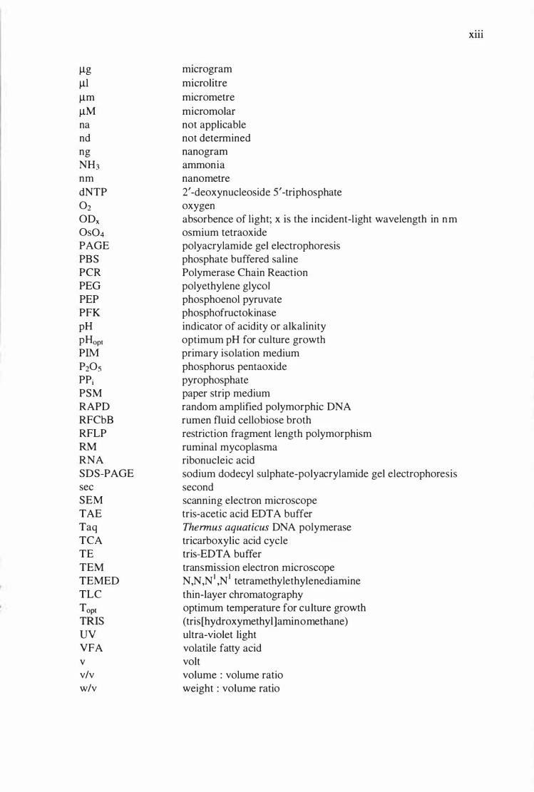
�g
�1
�m
�M
na
nd
ng
NH3
nm
dNTP 02 ODx Os04
PAGE
PBS
PCR
PEG
PEP
PFK
pH
pHopt PIM
P20s
ppi PSM
RAPD
RFCbB
RFLP
RM RNA
SDS-PAGE
sec
SEM
TAE
Taq
TCA
TE
TEM
TEMED
TLC
Topt TRIS
uv VFA
V v/v
w/v
micro gram
micro litre
micro metre
micro molar
not applicable
not determined
nanogram
ammonia
nanometre
2' -deoxynucleoside 5' -triphosphate
oxygen
absorbence of light; x is the incident-light wavelength in nm
osmium tetraoxide
polyacrylamide gel electrophoresis
phosphate buffered saline
Polymerase Chain Reaction
polyethylene glycol
phosphoenol pyruvate
phosphofructokinase
indicator of acidity or alkalinity
optimum pH for culture growth
primary isolation medium
phosphorus pentaoxide
pyrophosphate
paper strip medium
random amplified polymorphic DNA
rumen fluid cellobiose broth
restriction fragment length polymorphism
ruminal mycoplasma ribonucleic acid
sodium dodecyl sulphate-polyacrylamide gel electrophoresis
second
scanning electron microscope
tris-acetic acid EDT A buffer
Thermus aquaticus DNA polymerase
tricarboxylic acid cycle
tris-EDT A buffer
transmission electron microscope
N ,N ,N 1 ,N 1 tetramethy le thy lenediamine
thin-layer chromatography
optimum temperature for culture growth
( tris [hydroxymethy 1 ]aminomethane)
ultra-violet light
volatile fatty acid
volt
volume : volume ratio
weight : volume ratio
Xlll

1 INTRODUCTION
1
Ruminants are cloven-hoofed mammals which obtain food by browsing or grazing on
plants (Hungate, 1966). They are the earth' s dominant herbivores, due in part to the
evolution within this group of a mechanism utilising microorganisms to digest plant
components (Hungate, 1 975). Sheep, cattle, goats, deer and buffalo are all ruminants
(Hungate, 1966). There are 1 84 species of ruminating mammals, representing 74 genera
and 6 families within the kingdom Animalia. A feature common to most ruminants is a
four-chambered stomach comprising the reticulum, rumen, omasum and abomasum.
Exceptions to this general rule are found in the family Tragulidae (chevrotains and
mouse deer) where the stomach has just three chambers (Clarke, 1968). All ruminant
nutrition relies on the presence of the symbiotic intestinal microflora and microfauna.
Without this, ruminants would be incapable of digesting the plant materials which
constitute their diet (Hungate, 1966).
Fermentation of the digesta is carried out in the first two chambers (reticulum and
rumen) of the four-chambered stomach (Clarke, 1968). The rumen provides conditions
where moisture, pH, temperature, anaerobiosis and food supply are nearly constant.
These near constant conditions, lack of oxygen, and types of food, prevent many
microorganisms from growing within the rumen microbial ecosystem (Hungate, 1 960) .
Conversely, these same conditions allow microorganisms adapted to the rumen
environment, to proliferate to high numbers.
The temperature within the rumen is usually 39°C, although this may rise slightly in the
period after feeding when fermentation is maximal. However, the temperature will fall
with water intake (Dehority, 199 1 ; Theodorou et al, 1 992). The rumen pH usually lies
in the range 6.0 to 6.7 and is maintained by the secretion of an alkaline-buffered saliva
which neutralises the short-chain fatty acids produced during fermentation (Hungate,
1 988). The atmosphere within the rumen comprises 65% C02, 27% CH4, 7% N2; with
H2, H2S, and 02 being present in trace amounts (Hungate, 1966). Methane is formed in

2
the rumen by the reduction of C02 with H2, and not from volatile fatty acids (Hungate,
1 988).
S ignificant variation occurs in the numbers and types of microorganisms found within
the rumens of individual animals in a herd, and there are also marked differences in the
rumen populations of different ruminant species . This occurs because of host
specificity, dietary differences and protozoal predation on bacteria (Clarke, 1 968).
Some ciliate protozoa in the rumen prevent other protozoa becoming established by
predation and competition for food (Eadie, 1967). Deer maintain different ruminal
ciliate populations to those in sheep and cattle, in spite of ample opportunity for cross
infection (Clarke, 1968). Individuals of the same ruminant species also show gross
differences in ruminal ciliate protozoa populations even within a single flock or herd. It
is not uncommon to find several sheep in a flock without any holotrich protozoa, or
without some entodiniomorph protozoa which are present in all of the other members of
the flock (Clarke, 1968).
Swain et al ( 1 996), noted that animals on the same diet and penned together, allowing
micro flora and fauna to be exchanged, had differing bacteriophage (phage) populations.
This showed that the phage population of the rumen varies throughout the day, and also
that individual animals have unique phage populations. Not only did phage activity vary
between animals and groups of animals, but there were distinct diurnal variations in
animals fed once daily. Phage numbers fell shortly after feeding and rose to a peak
some 8- 1 0 hours later (Swain et al, 1996) .
In cattle fed once daily, ruminal bacteria population studies showed that regardless of
diet, the total bacterial numbers remained fairly constant throughout the day (Leedle et
al, 1982). The number of viable bacteria fell by 40% to 60% after feeding, but
increased to a maximum 1 6 hr after feeding. Soluble carbohydrate-utilising bacteria
predominated at all times (Leedle et al, 1 982).
Microbial digestion of plant tissues in the rumen produces volatile fatty acids (VF A),
principally acetate, propionate and butyrate, and these are used as energy sources by the
host. Propionate is a key metabolic intermediate, because it is the only VFA converted

3
into carbohydrate by the ruminant. Ruminant needs for carbohydrate are less than those
of non-ruminants, but carbohydrate is essential especially during periods of lactation.
Microbes from the rumen, when digested in the lower intestine, are a major source of
protein for the animal. In grazing ruminants, forage is ingested during approximately
one third of the 24 hr day. The rumen never empties, even during periods of starvation
(Hungate, 1 975).
At periodic intervals, the orifice between the reticulum and the omasum opens and
liquid and small particles of digesta flow into the omasum (Dehority, 199 1 ). In sheep,
the threshold size for digesta to pass from the reticulum to the omasum is between 1-2
mm, while in cattle the size is 2-4 mm (Ulyatt et al, 1 985). Larger plant fragments are
retained in the reticulo-rumen for further digestion. This process involves regurgitation
and re-chewing of digesta by the animal and further microbial degradation of
lignocellulose in the rumen (Dehority, 1 99 1 ) . In the omasum, fermentation acids and
bicarbonate are absorbed by the leaf-like layers of tissues between which the digesta
pass. Posterior to the omasum the ruminant alimentary tract is comparable to that of
most other animals except that the pancreatic juice contains an exceptionally high
concentration of ribonuclease; an adaptation to the abundant ribosomes in the bacteria to
be digested (Hungate, 1975).
The particular aspect of rumen microbiology which forms the basis of this thesis, is the
study of a group of microorganisms which have received little attention in the past. The
class Mollicutes comprises eight genera of bacteria, none of which possess a cell wall
(Robinson and Freundt, 1 987; Dybvig and Voelker, 1 996). Only two mollicute genera
are found in the rumen and these are loosely termed, ruminal mycoplasmas. The term is
misleading since no ruminal mycoplasma belongs to the genus Mycoplasma, but belong
to the genera, Anaeroplasma and Asteroleplasma (Robinson and Freundt, 1 987). Both
genera are obligately anaerobic, in contrast to the other 6 genera of mollicutes, which
are facultative anaerobes (Weisburg et al, 1 989) .
There are no studies which have investigated the ecological role of ruminal
mycoplasmas in ruminants. Likewise, no information is available as to how these
bacteria interact with other microbes, particularly the cellulolytic bacteria and

4
cellulolytic fungi within the rumen microbial ecosystem. Therefore, the contribution of
Anaeroplasma and Asteroleplasma species to the overall process of fibre digestion and
to ruminant nutrition is unknown. The data reported in this thesis will endeavour to
answer some of these questions.

2 LITERATURE REVIEW
2.1 INTRODUCTION
5
Much of the literature relating to the research topic of this thesis uses the term
"anaerobic mycoplasma" to describe those members of the class Mollicutes (bacteria
without cell walls) which can be isolated from the intestinal tract of ruminants.
Similarly it uses the terms "mollicutes" or "mycoplasma" to loosely describe any
member of the eight genera within the class Mollicutes. It is now felt a more precise
term for anaerobic mycoplasmas of rumen origin is "rumina! mycoplasma", and this
name will be used throughout this thesis. The descriptives, "mollicutes" and
"mycoplasma" will be used in their generic sense, unless specifically discussing the
class Mollicutes or the genus Mycoplasma, when the name will be italicised. To give an
overall picture, the review includes a brief description of the rumen microbial ecosystem
and describes a number of rumina! microorganisms. However only fibrolytic bacteria
and fungi which have been investigated in cocultural studies with the rumina!
mycoplasmas are discussed in any depth. The rumina! mycoplasmas are discussed in
detail later, in relation to their physiology, phylogeny and interactions with other
rumina! microorganisms.
2.2 THE RUMEN MICROBIAL ECOSYSTEM
The rumen microbial ecosystem is a diverse assemblage of interdependent and
interactive microorganisms (see reviews in Hobson and Stewart, 1997). Rumina!
microorganisms are found in the liquid phase of the digesta, associated with plant
tissues, and attached to the rumina! epithelium (Czerkawski and Cheng, 1988).
Microorganisms found within the rumen include bacteria, fungi, protozoa and
bacteriophage. They function to digest the host animal's fibrous diet and provide
nutrients to the host by way of fermentation end products. Ultimately their cells are
digested in the lower intestinal tract, and contribute an important source of protein for
the animal. The total number of anaerobic microorganisms in the rumen is between 1010
and 101 1 g- 1 (Hungate, 1966). Aerobic and facultative microorganisms have also been

6
isolated but are thought to be mainly transients (Stewart and Bryant, 1988). The
temperature of the ruminal digesta is fairly constant at 39°C, although this rises slightly
after feeding when the fermentation is at a maximum but falls with water intake
(Dehority, 1991; Theodorou et al, 1992). The rumen pH usually lies in the range 6.0 to
6.7, and is maintained by the introduction of alkaline-buffered saliva which neutralises
VFAs produced by the rumen fermentation (Hungate, 1988). The atmosphere in the
rumen comprises 65% C02, 27% CH4, 7% N2; with H2, H2S, and 02 being present in
trace amounts (Hungate, 1966).
2.3 FIBROL YTIC RUMINAL MICROORGANISMS
Ruminal microorganisms may be divided into two major groups; those directly involved
in plant tissue degradation, and those which use the end-products of these
microorganisms for growth. Fibrolytic microorganisms release sugars as a result of
polysaccharide digestion; the sugars are then utilised by non-fibrolytic microorganisms
to provide energy for cell growth. Fibrolytic and non-fibrolytic microorganisms produce
volatile fatty acids (VFAs), including acetate, propionate and butyrate which are used by
the host animal for energy. The capability for breaking down plant fibre in the rumen
rests with a relatively small number of bacterial, fungal and protozoal species.
2.3.1 Fibrolytic Rumina! Bacteria
Bacteria are the major group of microorganisms that degrade plant tissues in the rumen
(Akin and Benner, 1988). The plant cell-wall degrading bacteria include Butyrivibrio
fibrisolvens, Clostridium spp., Eubacterium cellulosolvens, Fibrobacter succinogenes,
Ruminococcus albus and Ruminococcus flavefaciens. F. succinogenes and
Ruminococcus spp. are generally regarded as the most active fibrolytic bacteria. F.
succinogenes degrades mainly cellulose, while some strains have the ability to degrade
storage polysaccharides such as starch, or structural polysaccharides such as pectin
(Stewart and Bryant, 1988). Access to plant material is generally gained via damaged
tissue or through stomata, while intact plant cuticles appear to be impermeable to either
mechanical penetration or enzymatic degradation (Chesson and Forsberg, 1988). Close
contact appears to be necessary for plant cell-wall degradation as many of the bacterial
enzyme systems involved are cell bound (Chesson and Forsberg, 1988; Dehority, 1991).

7
Cellulolytic bacteria are sensitive to low pH, so it is essential that fluctuations in rumen
pH are avoided if optimum fibre digestion is to be achieved (Wallace, 1992).
Non-fibrolytic bacteria in the rumen have indirect effects on plant tissue degradation by
utilising the intermediates and end products produced by plant cell-wall degrading
microbes. Selenomonas ruminantium for example, is incapable of fermenting cellulose
but will utilise the cellodextrins produced during the hydrolysis of cellulose by F.
succinogenes (Russell, 1985). Also succinate, produced by many plant cell-wall
degrading bacteria, never accumulates in the rumen. It is utilised by other bacteria such
as Selenomonas ruminantium to produce propionate (Scheifinger and Wolin, 1973)
which is essential to the ruminant because it is the only metabolic intermediate used in
gluconeogenesis (Wolin and Miller, 1988).
2.3.2 Fibrolytic Ruminal Fungi
Ruminal fungi have been the subject of extensive reviews recently. For references see
Mountfort and Orpin (1994) and Orpin and Joblin (1997). Ruminal fungi were first
observed when fungal rhizoids were seen attached to plant tissue in rumen contents
(Orpin, 1975). Previously it had been the practice to examine only the filtrate of rumen
contents; the solid fraction being discarded as largely unimportant. Flagellated
zoospores of chytridiomycete fungi had been observed earlier in the filtrate, but
incorrectly identified as protozoa. Braune (1913), reported by Hungate (1966), observed
what he described as flagellates in the rumens of calves and named them Callimastix
frontalis. These were almost certainly the zoospores of the ruminal fungus
Neocallimastixfrontalis, which were reclassified by Vavra and Joyon (1966) along with
flagellates from other environments into the genus Neocallimastix. Orpin (1975)
observed that " ... the vegetative stage of N. frontalis bears a strong morphological
resemblance to that of certain species of aquatic phycomycete fungi ... ".
Obligatory anaerobic fungi have been isolated from the rumen, omasum, abomasum,
small intestine, caecum, large intestine and faeces of cattle (Davies et al, 1993), and
from the alimentary tract of a wide variety of other animals (Theodorou et al, 1992).
These fungi do not possess mitochondria. The life-cycles of anaerobic fungi have three
parts; a motile zoospore, a vegetative thallus, and an aerotolerant survival stage. The

8
thallus carries a sporangia (fruiting body) from which zoospores are released at maturity
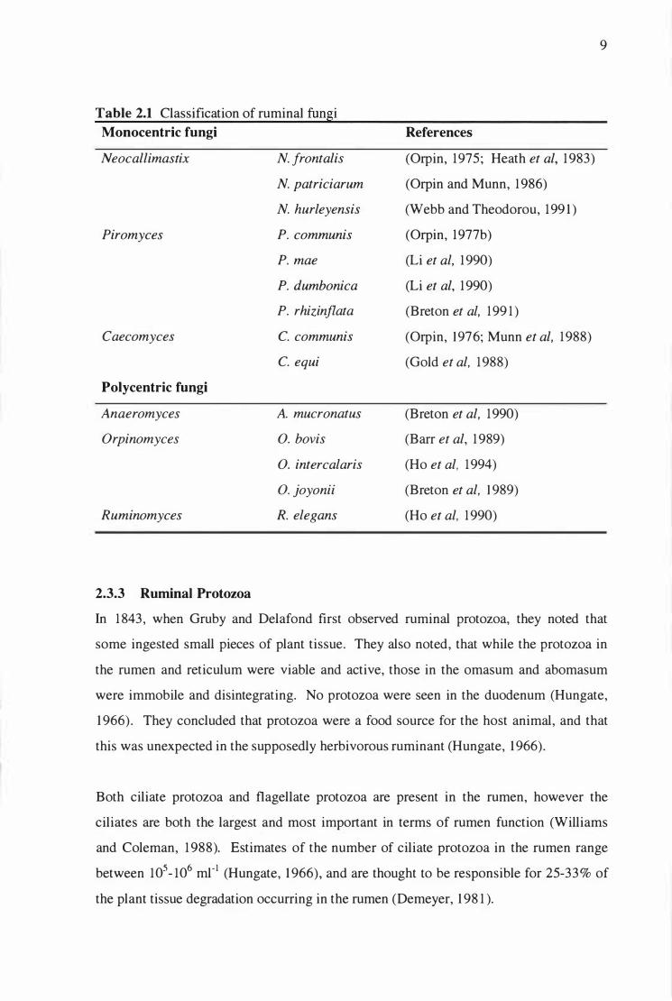
(Orpin and Joblin, 1997). The classification of the ruminal fungi is shown in Table 2.1.
Ruminal fungi are believed to play an important role in fibre digestion in the rumen
(Orpin, 1977(a); Bauchop, 1979), with the initial invasion of the plant material
occurring at sites of tissue damage (Bauchop, 1980), and at stomata (Orpin, 1977(a); Ho
et al, 1988). Following zoospore attachment and encystment, fungi develop highly
branched mycelia which grow inside plant fragments, where enzymes active against
structural polysaccharides are released. This allows the fungus access to fermentable
carbohydrates which are not immediately available to surface-acting ruminal bacteria. It
has been shown that fungi colonise fibrous plant tissue, and that lignin-containing
tissues are colonised preferentially (Bauchop, 1979; Grenet and Barry, 1988). The
evidence for this was the discovery that fungal numbers in vivo, as estimated by
sporangia counts, are higher in animals fed poor quality, fibrous diets. In contrast, fungi
were found to be absent from the rumen of sheep grazing soft, leafy diets, low in fibre
(Bauchop, 1979). The smaller populations of fungi found in the rumens of animals fed
soft leafy diets, may be due to the short residence time in the digestive tract, leading to
incomplete fungal life-cycles (Bauchop, 1989). Counts of 105 sporangia cm-2
were
found on the surface of lucerne (Medicago sativa) stems which had been suspended in
nylon bags in a sheep rumen (Bauchop, 1979). Estimates of fungal populations have
also been made on the basis of counts of zoos pores in rumen fluid (Joblin, 198 1 ).
Anaerobic fungi have been seen in the rumens of lambs 8- 10 days after birth (Fonty et
al, 1987). The presence of anaerobic fungi in saliva and faeces suggests these as
possible routes of transmission (Trinci et al, 1988). Spore-like structures have been
found in the rumen (Fonty and Joblin, 199 1), and the faeces of cattle (Davies et al,
1993). These resting spores are perhaps the way fungi survive outside the rumen, and
are possibly one route for transmission between animals (Fonty and Joblin, 199 1).
9
Table 2.1 Classification of rumina! funsi
Monocentric fungi References
Neocallimastix N. frontalis (Orpin, 1975; Heath et al, 1983)
N. patriciarum (Orpin and Munn, 1986)
N. hurleyensis (Webb and Theodorou, 1991)
Piromyces P. communis (Orpin, 1977b)
P. mae (Li et al, 1990)
P. dumbonica (Li et al, 1990)
P. rhizinflata (Breton et al, 199 1)
Caecomyces C. communis (Orpin, 1 976; Munn et al, 1988)
C. equi (Gold et al, 1988)
Polycentric fungi
Anaeromyces A. mucronatus (Breton et al, 1990)
Orpinomyces 0. bovis (Barr et al, 1989)
0. intercalaris (Ho et al, 1994)
0. joyonii (Breton et al, 1 989)
Ruminomyces R. elegans (Ho et al, 1990)
2.3.3 Rumina) Protozoa
In 1843, when Gruby and Delafond first observed ruminal protozoa, they noted that
some ingested small pieces of plant tissue. They also noted, that while the protozoa in
the rumen and reticulum were viable and active, those in the omasum and abomasum
were immobile and disintegrating. No protozoa were seen in the duodenum (Hungate,
1966). They concluded that protozoa were a food source for the host animal, and that
this was unexpected in the supposedly herbivorous ruminant (Hungate, 1966).
Both ciliate protozoa and flagellate protozoa are present in the rumen, however the
ciliates are both the largest and most important in terms of rumen function (Williams
and Coleman, 1988). Estimates of the number of ciliate protozoa in the rumen range
between 105- 106
ml- 1 (Hungate, 1966), and are thought to be responsible for 25-33% of
the plant tissue degradation occurring in the rumen (Demeyer, 198 1).

10
Two major groups of ciliate protozoa have been described; the entodiniomorphs, and the
holotrichs. More than 100 species of entodiniomorph protozoa have been found in the
rumen. All are strict anaerobes which feed mainly by engulfing particulate matter,
including other rumina! microorganisms (Williams and Coleman, 1988). The
entodiniomorphs lack the abundant cilia which cover the surface of holotrichs, but have
evolved specialised bands of syncilia (fused ciliary bands) which function both for
movement and food ingestion (Hungate, 1966).
Rumina! holotrich protozoa possess cilia over the entire body surface, each inserted
singly and not fused with others, except in the region of the mouth (Hungate, 1966).
There are two genera of holotrich protozoa found in the rumen; /sotricha and
Dasytricha. These are large microorganisms, with /. intestinalis recorded in the range
(97 - 13 1 J..lm x 68-87 J..Lm) (Hun gate, 1966). Holotrich protozoa are mainly involved in
the utilisation of non-structural carbohydrates and soluble sugars, and have only limited
ability to degrade plant structural polysaccharides (Demeyer, 1981 ).
2.4 RUMINAL BACTERIOPHAGES
Bacteriophage (phage) are an integral part of the rumen microbiota of sheep and cattle,
occurring in large numbers and in great diversity (Klieve, 199 1 ). Phage are obligate
pathogens of bacteria and their presence leads to the eventual lysis of the bacterial host.
Little is known about the effects of phage attack upon rumina! bacteria, or their effect on
nutrient cycling within the rumen. The size of the population suggests that they play a
significant role in bacterial lysis and the subsequent reduction in the efficiency of feed
utilisation (Swain et al, 1996). The number of phage found in rumina! fluid from sheep
and cattle lies in the range 2 x 10⁷ to 1 x 10⁸
ml- 1 (Klieve and Bauchop, 1988).
2.5 RUMINAL YEASTS
Clarke and di Menna (196 1) isolated yeasts from the rumens of cattle in New Zealand,
which they believed were true rumina! inhabitants. The yeasts were isolated using
aerobic techniques, grew at 39°C, and were not isolated from samples of the feed
material. Representatives of the Candida, Trichosporon and Rhodotorula genera were
found.

11
The yeast most commonly isolated from the rumens of cows in Denmark was Candida
krusei which, like the less commonly isolated Torulopsis pintolopesii and
Kluyveromyces bulgaricus, could reproduce under anaerobic conditions in vitro (Lund,
1974).
Lund ( 1980), reported the isolation of yeasts from ruminal digesta of the Musk Oxen
(Ovibos moschatus) in Greenland. The isolates most commonly found were Candida
spp. and Cryptococcus spp.. The ruminal digesta of these animals contained
predominantly woody plant parts of the Salix and Betula genera, in sharp contrast to the
grass and clover mix found in most New Zealand pasture-fed animals. The yeasts which
Lund ( 1980) described were isolated using aerobic techniques and many of those found
in the rumen of the musk oxen were also isolated from samples of peat, soil and algae in
a similar geographic location.
2.6 THE CLASS MOLL/CUTES
Members of the class Mollicutes are small, free-living procaryotes that pass through
0.45 Jlm filters, have genomes with a very low G+C content, lack cell walls and have
unusual nutritional needs (Weisburg et al, 1989; Dybvig and Voelker, 1996). Many are
pleomorphic, ranging from spherical, coccoid, cocco-bacilliary, ring and dumb-bell
forms, to short and long branching filaments. The coccoid forms are approximately 0.3
Jlm in diameter, while the filamentous forms can be more than 100 Jlm long (Boatman,
1979). The mollicute genome is believed to be the smallest of any free-living cell, and
because of this biosynthetic capacity is limited. More than 100 different species have
been isolated from humans, animals, plants and insects.
The class Mollicutes contains eight genera (Acholeplasma, Anaeroplasma,
Asteroleplasma, Entomoplasma, Mesoplasma, Mycoplasma, Spiroplasma and
Ureaplasma), (see Table 2.2) with classification based primarily on differences in
morphology, genome size, and some nutritional features (Robinson and Freundt, 1987).
The class is phylogenetically quite broad (Weisburg et al, 1989; Dybvig and Voelker,
1996). The two anaerobic genera within the class Mollicutes, Asteroleplasma and
Anaeroplasma, are only distantly related (Gundersen et al, 1994).
12
Acholeplasma spp. and Asteroleplasma anaerobium are able to grow in the absence of
pre-formed sterols (Weisburg et al, 1989), while more recently, Toth et al (1994)
reported a new genus, Mesoplasma, which also grow in the absence of sterol.
Mesoplasma species, of which there are 12, are pathogens of plants and insects. All
other mollicute genera require an exogenous supply of sterols for growth (Weisburg et
al, 1989). Mycoplasma, Ureaplasma and Spiroplasma spp. are unable to synthesize
fatty acids and these must be provided by the host. Ureaplasma spp. require exogenous
urea for growth, and for this reason are often associated with infections of the
genitourinary tract. M. gallisepticum, requires pre-formed phospholipids for growth
(Razin, 1992). By becoming dependent on host-derived nutrients, a considerable
amount of genetic information is saved (Razin, 1992).
The nutritional dependence of some members of the ruminal mycoplasmas on
exogenous sterol is a trait that has arisen several times during their evolution (Robinson
and Freundt, 1987). Nutritional requirements such as the need for sterol probably reflect
a loss of genes, but since different gene functions could be lost and result in similar
dependencies, care must be taken when using nutritional requirements as indicators of
biosystematic groupings (Toth et al, 1994 ) . It has been suggested, that during the
evolution of mycoplasmas from Gram-positive bacteria, the loss of cell walls, together
with many biosynthetic systems, has necessitated the adoption of a parasitic mode of
survival (Woese, 1987; Razin, 1992). All known mycoplasmas are parasites of humans,
other vertebrates, plants or arthropods (Razin, 1992). Mycoplasmas have complex
nutritional requirements and depend on the host for many amino acids, fatty acids,
sterols, and vitamins, (Razin, 1978; Rodwell, 1983; Razin, 1992). Because of this many
are not readily culturable.
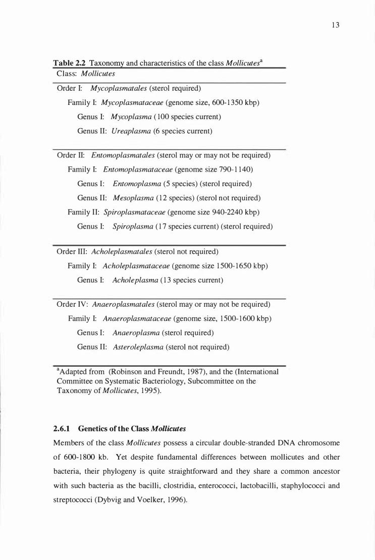
Table 2.2 Taxonomy and characteristics of the class Mollicutesa
Class: Mollicutes
Order 1: Mycoplasmatales (sterol required)
Family 1: Mycoplasmataceae (genome size, 600- 1 350 kbp)
Genus 1: Mycoplasma ( 100 species current)
Genus 11: Ureaplasma (6 species current)
Order 11: Entomoplasmatales (sterol may or may not be required)
Family 1: Entomoplasmataceae (genome size 790- 1 140)
Genus 1 : Entomoplasma (5 species) (sterol required)
Genus 11: Mesoplasma ( 1 2 species) (sterol not required)
Family 11: Spiroplasmataceae (genome size 940-2240 kbp)
Genus 1: Spiroplasma ( 1 7 species current) (sterol required)
Order Ill: Acholeplasmatales (sterol not required)
Family 1: Acholeplasmataceae (genome size 1 500- 1 650 kbp)
Genus 1: Acholeplasma ( 1 3 species current)
Order IV: Anaeroplasmatales (sterol may or may not be required)
Family 1: Anaeroplasmataceae (genome size, 1 500- 1 600 kbp)
Genus 1: Anaeroplasma (sterol required)
Genus 11: Asteroleplasma (sterol not required)
a Adapted from (Robinson and Freundt, 1987), and the (International Committee on Systematic Bacteriology, Subcommittee on the Taxonomy of Mollicutes, 1 995).
2.6.1 Genetics of the Class Mollicutes
1 3
Members of the class Mollicutes possess a circular double-stranded DNA chromosome
of 600- 1 800 kb. Yet despite fundamental differences between mollicutes and other
bacteria, their phylogeny is quite straightforward and they share a common ancestor
with such bacteria as the bacilli, clostridia, enterococci, lactobacilli, staphylococci and
streptococci (Dybvig and Voelker, 1996).

14
Characteristically, mycoplasmal tRNAs have fewer modified nucleosides, reducing the
need for modification enzymes and consequently, genomic information. Mycoplasma
and Ureaplasma species have only one DNA polymerase, while Acholeplasma and
Spiroplasma spp. (which have larger genomes of 1600- 1700 kb ), possess three distinct
DNA polymerases, in common with E. coli and other eubacteria (Razin, 1992).
Analyses of mollicute 16S rRNA sequences indicates a relationship to Gram-positive
bacteria whose DNA has a low percentage of guanine and cytosine (% G + C)
(Weisburg et al, 1989).
Members of the orders Mycoplasmatales and Entomoplasmatales use the UGA codon to
encode tryptophan at a frequency 10 times greater than UGG. The unconventional use
of UGA as a tryptophan codon is not shared by all mycoplasmas. Acholeplasma
laidlawii uses the universal genetic code and has a single tRNA species with an anti
codon CCA that can only translate the normal tryptophan codon UGG (Tanaka et al,
1989). This supports the idea that the change in the UGA assignment from a stop codon
to a tryptophan codon occurred after separation of the spiroplasma and mycoplasma
branches from the acholeplasma branches of the phylogenetic tree (Rogers et al, 1985).
The International Committee on Systematic Bacteriology Subcommittee on the
Taxonomy of Mollicutes (ICSBSTM) reported in 1995, that only UGG encodes
tryptophan in members of the orders Acholeplasmatales and Anaeroplasmatales. In the
case of anaeroplasmas, little is known about their molecular biology, but since they are
closely related to acholeplasmas, it is probable they also use the universal genetic code.
The genetic code used by Asteroleplasma anaerobium is unknown (Dybvig and
Voelker, 1996).
2.6.2 Pathogenicity of Mycoplasma spp.
Many members of the genus Mycoplasma cause disease in animals and humans, and are
usually host specific. Two species known to cause disease in ruminants are M. bovis
(calf pneumonia, mastitis and arthritis) and M. agalactiae (contagious agalactia in goats
and sheep) (Pettersson et al, 1996). Infections by pathogenic mycoplasmas are rarely of
the fulminant type, but are often chronic in nature. The mechanisms of mycoplasma
pathogenicity are largely unknown (Razin, 1992).

15
Mycoplasmas infecting humans and animals are mostly surface parasites, colonising the
epithelial linings of the respiratory and genitourinary tracts. Potent toxins have not been
associated with mycoplasmas. M. arthritidis, which has been implicated in arthritis,
produces a potent mitogen; a 27-kDa protein molecule (MAM) which has been defined
as one of the so-called superantigens. Superantigens, which are produced by a variety of
microbial agents, activate T cells by a unique pathway leading to T cell modification
and the induction of autoimmunity. It has been proposed that MAM participates in
chronic joint inflammation in rats and mice infected by M. arthritidis, not only by
activating T cells with the resulting liberation of inflammatory lymphokines, but also by
suppressing host defences. The contributions of infection and autoimmunity in the
chronic phase of the disease are unknown (Cole and Atkins, 1991). Mycoplasmal
infections are often chronic in nature, and it is unclear as to how these wall-less, rather
fragile microorganisms resist the immune defence mechanisms of their host. Some
pathogenic mycoplasmas are thought to undergo high-frequency phenotypic switching
involving variable antigens, which enables them to evade host defences (Razin, 1992).
2.6.3 Ruminal Mycoplasmas
Little is known about the group of microorganisms loosely termed "rumina!
mycoplasmas." They are classified within two genera, Anaeroplasma and
Asteroleplasma and are found within the rumen microbial ecosystem. Studies of
rumina! mycoplasmas in axenic culture, and in coculture with rumina! cellulolytic
bacteria and fungi form the central theme of this thesis and the research reported herein.
An obligately anaerobic microorganism from the rumen of cattle which lysed bacterial
cells was described by Hungate in 1966. The microorganism, which lacked a cell wall,
was subsequently characterised as a rumina! mycoplasma (Robinson and Hungate,
1973) and classified as Acholeplasma bactoclasticum. Later this classification was
reviewed and the microorganism reclassified into a new genus and species,
Anaeroplasma bactoclasticum, on the basis that it was an obligate anaerobe and
required sterols for growth (Robinson and Allison, 1975). A feature of the mycoplasma
was the production of an extracellular enzyme, which under anaerobic but not aerobic
conditions, degraded the peptidoglycan layer in the cell walls of Gram-negative bacteria
16
such as E. coli, causing lysis. Ruminal contents of both cattle and sheep were
subsequently found to contain similar mycoplasmas, while other mycoplasmas were
found which were abacteriolytic. The abacteriolytic mycoplasmas (107 - 108i1) were
present in higher numbers than the bacteriolytic type (105 - 107 g- 1 ) (Robinson et al,
1975). Neither of these types of mycoplasma were found in the caecal contents of
hamsters, horses, pigs, rabbits or turkeys (Robinson et al, 1975). The abacteriolytic
rumina! mycoplasmas comprise two types; the sterol-requiring Anaeroplasma
abactoclasticum, and the sterol non-requiring, Asteroleplasma anaerobium (Robinson
and Freundt, 1987).
Within the genus Anaeroplasma, only one species, An. bactoclasticum was originally
characterised as being able to lyse the cell-walls of Gram-negative bacteria (Robinson
and Allison, 1975). However, later work by Robinson and Rhoades ( 1977) using gel
immunodiffusion precipitation tests, showed that within the species An. bactoclasticum
three serovars could be distinguished. In contrast An. abactoclasticum was represented
by only one serovar. Gel diffusion precipitation tests also showed that rumina}
mycoplasmas, which were later to be placed into the genus Asteroleplasma, did not
cross-react with antisera from any of the other groups of rumina} mycoplasma under
test. Step hens et al ( 1985) described five distinct groups of ruminal mycoplasma based
on eH] DNA-DNA homology data; four Anaeroplasma spp. and one Asteroleplasma
sp., although not all had been named at that time. These groups were sufficiently
diverse for Robinson and Freundt ( 1987) to propose that ruminal mycoplasmas,
previously classified as An. bactoclasticum, be reclassified into three separate species -
An. bactoclasticum, and two new species, An. varium, and An. intermedium. All three
species were capable of lysing the cell walls of Gram-negative bacteria. Based on
distance matrix data, An. bactoclasticum and An varium were more closely related to
each other, than they were to An. intermedium (Weisburg et al, 1989). A fourth species,
An abactoclasticum was more distantly related (Weisburg et al, 1989). The four
Anaeroplasma spp., and As. anaerobium constitute the five groups described by
Stephens et al ( 1985). Asteroleplasma species are phylogenetically distant from all
Anaeroplasma species (Gundersen et al., 1994; Seemuller et al, 1994).

17
The polar lipids of Anaeroplasma spp. contain plasmalogens (alk-1-enyl glyceryl ethers)
which are found in anaerobic bacteria but not aerobic bacteria. A bacteriolytic strain
7LA (An. intermedium) contained about 50% of the amount of glycolipids present in the
non-bacteriolytic strain 6-1 (An. abactoclasticum) (Langworthy et al, 1975).
The rumina! mycoplasmas (Table 2.3) currently are classified according to the
recommendations of Robinson and Freundt ( 1987) who concluded, "Because of the
unique metabolic properties of the obligately anaerobic mycoplasmas, dependence on
strict anaerobiosis should outweigh the sterol requirement as the major taxonomic
factor. Therefore, we propose to classify the genera Anaeroplasma and Asteroleplasma
in a new family, Anaeroplasmataceae. Since this new family is not referable to either of
the two established orders of the Mollicutes, we propose the assignment of
Anaeroplasmataceae to a new order Anaeroplasmatales, as order ill of the class
Mollicutes." This classification now has been slightly changed following
recommendations of the ICSBSTM, (see Table 2.2); the rumina! mycoplasmas move
from Order Ill to Order IV.
Table 2.3 Taxonomy of the rumina! mycoplasmas a
Order IV: Anaeroplasmatales (sterol may or may not be required)
Family I :
Genus I :
Anaeroplasmataceae (genome size, 1,000 megadaltons)
Anaeroplasma (sterol required)
Species 1: An. abactoclasticum
Species 2 : An. bactoclasticum
Species 3: An. varium
Species 4: An. intermedium
Genus II : Asteroleplasma (sterol not required)
Species 1 : As. anaerobium
a Adapted from Robinson and Freundt (1987).
Details of the type strains of the rumina! mycoplasmas and the percentage G+C of their
DNA are shown in Table 2.4

Table 2.4 Type Strains and G+C mol% of Rumina! Mycoplasma DNAa
Ruminal Mycoplasma Type Strain G+C (mol% )
Anaeroplasma abactoclasticum (6-1=ATCC 27879) 29.3
Anaeroplasma bactoclasticum (JR=ATCC 271 12) 33.7
Anaeroplasma intermedium (7LA=ATCC 43166) 32.5
Anaeroplasma varium (A-2=ATCC 43 167) 33.4
Asteroleplasma anaerobium ( 161 =A TCC 27880) 40.2
a(Stephens et al, 1985; Robinson and Freundt, 1987)
18
It can be seen from Table 2.4 that the %G+C for An. bactoclasticum, An. intermedium
and An. varium are very similar, and the %G+C of An. abactoclasticum is somewhat
lower. The %G+C of Asteroleplasma anaerobium is somewhat higher than all
Anaeroplasma species to which they are phylogenetically only distantly related
(Robinson and Freundt, 1987; Weisburg et al, 1989; Toth et al, 1994). These groupings
are supported by phenotypic evidence, principally because An. abactoclasticum is the
only species within the genus Anaeroplasma not able to lyse the cell walls of Gram
negative bacteria and being non-proteolytic (Robinson and Freundt, 1987).
Asteroleplasma anaerobium is unique among the rumina! mycoplasmas in being able to
grow in the absence of sterols (Robinson and Freundt, 1987).
2.6.4 Pathogenicity of Anaeroplasma spp.
Robinson and Hungate (1973) and Robinson and Allison (1975) reported that they
observed no deaths in chicken embryos inoculated with active cultures of Acholeplasma
(syn. Anaeroplasma) bactoclasticum. Likewise, Truscott (198 1) reported no diseases of
cattle which could be attributed to the presence of Anaeroplasma spp. among their
rumina! microflora.
2.6.5 Metabolism of Anaeroplasma intermedium and Asteroleplasma anaerobium
Cell-free extracts of An. intermedium 5LA and As. anaerobium 161 T were examined for
enzymes of the Embden-Meyerhof-Parnas (EMP) pathway leading from glucose to the
triose phosphates and from 3-phosphoglyceric acid to pyruvate. It was shown that An.

19
intermedium 5LA possessed all eight enzymes of the EMP pathway whereas As.
anaerobium lacked hexokinase activity (Petzel et al, 1990). The phosphofructokinase
(PFK) of As. anaerobium was ATP-dependent in common with most bacteria and
Mycoplasma spp., but the PFK of An. intermedium was PPi -dependent, in common with
Acholeplasma laidlawii and the propionibacteria (Petzel et al, 1990). The lactic
dehydrogenase of both microorganisms was activated by fructose 1 ,6-diphosphate. An.
intermedium 5LA and As. anaerobium 16 1 T can therefore be distinguished from each
other by their enzyme activities. The occurrence of 3 uncommon PPi-dependent kinases
among the rumina! mycoplasmas may also be useful in determining their phylogenetic
relationship to other mycoplasmas, and to the Gram-positive bacteria (Petzel et al,
1990).
2.6.6 Non-Ruminal Anaerobic Mycoplasmas
Anaerobic mycoplasmas, which did not require sterol and were abacteriolytic, were
isolated from the intestinal tract of pigs (Binder and Kirchhoff, 1988). Like other
mycoplasmas, the surface colonies were 'fried egg' in appearance, growth was not
inhibited by penicillin and they were bounded by a trilaminar membrane without a
distinguishable cell wall (Binder and Kirchhoff, 1988). Resistance to digitonin and
growth in the absence of cholesterol, placed them into the genus Asteroleplasma. These
isolates from the pig were not thought to be Asteroleplasma anaerobium , but probably
belonged to a new species since they were serologically distinct from anaerobic
mycoplasmas previously isolated from the pig, or from the gut of ruminants (Binder and
Kirchhoff, 1988). However, they commented that . . . "the primary cultivation of these
mollicutes was possible only under strict anaerobic conditions .. " but then .. "with
increasing passage the organisms became less sensitive to oxygen." (Binder and
Kirchhoff, 1988). Since the fundamental requirement for mollicutes to be classified
within the genus Asteroleplasma is an absolute need for anaerobic growth conditions,
their second admission suggests that this isolate was perhaps a member of the
facultative, sterol non-requiring genus Acholeplasma, rather than the obligately
anaerobic, sterol non-requiring genus Asteroleplasma within the class Mollicutes.
Two species of obligately anaerobic mycoplasma were the predominant microbes in a
methanogenic, glucose-limited, enrichment culture obtained from a sewage sludge

20
digester (Rose and Pirt, 198 1 ) . In pure culture, one of the anaerobic mycoplasmas,
tentatively named Anaeroplasma sp. strain London, fermented glucose primarily to
butyric acid, H2, and C02 while the other produced CH4 from H2 and C02 and was
named Methanoplasma elizabethii. Both were classified within the family
Mycoplasmataceae on the basis of colonial and cellular morphology, ability to pass
through 0.45 J.lm filters, and resistance to penicillin (Rose and Pirt, 198 1). Neither
microorganism appears in the current literature on the classification of the anaerobic
mycoplasmas.
3 MATERIALS AND METHODS
3.1 INTRODUCTION
21
All chemicals were of analytical grade and obtained from British Drug Houses, UK,
unless stated otherwise. All antibiotics were supplied by Sigma Chemicals, USA. All
dehydrated microbiological media was supplied by Difco Laboratories, USA. Other
chemicals or reagents are as acknowledged in the text.
Unless stated otherwise, all media were prepared under stringent anaerobic conditions
using the procedure of Hungate ( 1969), utilising a gas phase of 100% 02-free C02. All
components of the medium, except the reducing agent L-cysteine-HCl, were boiled to
remove dissolved 02 and then saturated with 02-free C02 while cooling to 45cc (agar),
or in ice-water (broth), when the L-cysteine-HCl was added. Agar media were
dispensed 5 rnl per tube and broth media 10 ml per tube in Hungate culture tubes
(Bellco Glass, Inc., NJ, USA), under 100% 02-free C02 and sterilised by autoclaving at
12 1 cc for 20 min. All media had a final pH of 6.4-6.6 unless stated otherwise.
Clarified rumen fluid was prepared when rumina! digesta was removed from a Friesian
cow via the rumen fistula, strained through several layers of cheesecloth to remove large
particulate matter, and clarified by centrifugation at 16,000 x g for 15 min at 4 cc.
Clarified rumen fluid was stored at -2occ until required.
Mineral solution 1 consisted of: KH2P04 (6.0 g), NaCl ( 12.0 g), (NH4)2S04 (12.0 g),
CaCh 2H20 (1.58 g), MgS04·7H20 (2.5g) and distilled water to 1,000 rnl.
Mineral solution 2 consisted of: K2HP04 ( 6.0 g) and distilled water to 1 ,000 rnl.
Salt solution A consisted of: KH2P04 (3.0 g), NaCI (6.0 g), (N�)2S04 (1.5 g),
CaCh 2H20 (0.79 g), MgS04·?H20 ( 1 .2 g) and distilled water to 1,000 rnl.
Salt solution B consisted of: K2HP04 (6.0 g) and distilled water to 1,000 rnl.
3.2 ISOLATION OF RUMINAL MYCOPLASMAS
22
All of the ruminal mycoplasma cultures isolated in this study were obtained from the
ruminal digesta of non-lactating, hay-fed Friesian cows at AgResearch, Grasslands
Research Centre, Palmerston North. The ruminal digesta were removed via the rumen
fistula and immediately processed in the laboratory.
3.2.1 Media for the Isolation and Purification of Ruminal Mycoplasmas
Primary Isolation Medium (PIM) consisted of: clarified rumen fluid ( 40.0 ml), mineral
solution 1 (3.75 ml), mineral solution 2 (3.75 ml), distilled water (52.5 ml), glucose
(0.05 g), cellobiose (0.05 g), starch (0.05 g), tryptone (0.2 g), yeast extract (0. 1 g),
resazurin (0.1 mg), autoclaved E. coli cells (0.5% w/v), Na2C03 (0.4 g), benzylpenicillin
(Na+ salt, 1 x 10⁵ IU), L-cysteine-HCl (50 mg) and agar ( 1.5 g).
Clarified Rumen Fluid Agar (CRFA) and Broth (CRFB) consisted of: clarified rumen
fluid (40.0 ml), mineral solution 1 (3.75 ml), mineral solution 2 (3.75 ml), distilled
water (52.5 ml), glucose (0.2 g), cellobiose (0.2 g), starch (0.2 g), tryptone (0.2 g), yeast
extract (0.1 g), resazurin (0.1 mg), Na2C03 (0.4 g), L cysteine-HCl (50 mg) and agar
(1.4 g in CRFA).
The E.coli used in PIM and used also to supplement CRFB for the bacterial cell-wall
lysis studies, was grown aerobically in Brain Heart Infusion Broth (3.7% w/v), for 24 hr
at 39°C. Cells were pelleted by centrifugation ( 15,000 x g for 10 min), resuspended and
washed once in phosphate-buffered saline, autoclaved at 12 1 oc for 20 min, cooled to
room temperature, recentrifuged and stored as a pellet at -20°C until required.
Ruminal mycoplasmas were isolated from agar roll tubes using the method of Hungate
(1969) and the E. coli-containing PIM agar medium of Robinson et al ( 1975). The
medium was kept at 45°C in a water bath prior to inoculation with ruminal digesta.
Sample dilution was performed using hypodermic syringes (1 ml) and needles (21 gauge
x 2.5 cm) which were flushed with 02-free C02 prior to each transfer. Particulate plant
material in the inoculum often blocked the hypodermic needle during the first transfer,
so a reversed sterile 1 ml glass pipette was used for this dilution. In this case the septum

23
was removed from the first tube of each dilution series to allow the inoculum to be
added from the pipette, the septum was replaced and this initial transfer serially diluted
to 10- 10• Following inoculation, the molten agar tubes were rolled under cold water by
hand to solidify the agar in a thin film over the entire internal surface of the tube. All
tubes were incubated vertically at 39°C.
After 4 days incubation, discrete colonies could be seen in the roll tubes at dilutions of
10-5 and greater. Well separated colonies in the 10-7 dilution tubes were picked from the
agar using a Pasteur pipette with a tip bent to 90°. By gentle suction using a mouth
tube, the colony was drawn into the pipette from where it was transferred to CRFB
medium supplemented with benzylpenicillin (800 IU mr 1) and streptomycin sulphate
(80 !J.g ml- 1). The change from PIM medium was necessary because although PIM was
a suitable medium to isolate rumina! mycoplasmas in agar, turbidity due to the E.coli
component of the medium made it unsuitable for OD600 growth measurements. The two
media were essentially the same but with differing carbohydrate concentrations. After
24 hr incubation the CRFB cultures were used to inoculate fresh rol l tubes of CRFA
medium and a further dilution series prepared. These were again incubated for 4 days,
when colony picking was repeated into a further series of CRFA medium, devoid of
antibiotics. The culture tubes were rolled as before and incubated at 39°C. This
procedure was repeated twice more to ensure pure cultures were obtained.
3.3 CHARACTERISTICS OF RUMINAL MYCOPLASMAS
All isolates obtained during the current study were characterised using the anaerobic
procedures of Hungate ( 1969) and the tests outlined below.
3.3.1 Morphology of Ruminal Mycoplasma Cells and Colonies
The colonial morphology of isolates growing on CRF A medium was established by
examination of 72 hr cultures using a 20 x dissecting microscope (Olympus Optical Co.
Ltd., Japan). Morphology and motility of individual mycoplasma cells growing in
CRFB medium was determined by examination of wet mounts using a V an ox AHBT3
microscope (Olympus Optical Co. Ltd., Japan). Wet mounts of each isolate were
prepared under cover slips and examined using phase contrast microscopy and by
Nomarski Differential Interference Microscopy at final magnifications of 400 x (dry)
24
and 1,000 x (oil). Dried and heat-fixed films of cells, stained using Hucker' s
modification o f the Gram stain (Hucker, 1922), were examined at a final magnification
of 1,000 x (oil). Details of CRFA and CRFB medium are given in section 3.2.1
Isolates RM 10 and RM 14 were examined by transmission electron microscopy (TEM)
to determine cell morphology and ultrastructure. Cells were fixed in a mixture of 3%
(v/v) glutaraldehyde and 2% (v/v) formaldehyde in 0. 1 M phosphate buffer pH 7.2, then
left for 2 hr at room temperature, mixed with a 20% (w/v) solution of BSA in water and
centrifuged to produce a pellet. The pellet was sliced, washed twice in 0.1 M phosphate
buffer pH 7 .2. and given a secondary fix in 1% Os04 in the same buffer for 30 min at
room temperature. The fixed pellet was washed twice more in buffer, dehydrated by
passage through a graded acetone series, infiltrated in a 50/50 mixture of Procure 812
epoxy resin with acetone and left on a stirrer overnight. Fresh resin ( 1 00%) was used to
infiltrate the samples for a further 8 hr, after which the samples were embedded in
silicone rubber moulds and left to cure at 60°C for 48 hours. Then 1 �m sections were
cut and stained with Toluidine Blue and observed using light microscopy to locate the
specimens. The block was trimmed to produce EM sections of 90- 100 nm thickness.
The sections were cut using a diamond knife then stained with uranyl acetate and lead
citrate. The sections were observed using a Philips 20 1C Transmission Electron
Microscope (The Philips Company, Eindhoven, The Netherlands).
Scanning Electron Microscope (SEM) studies of one isolate (RM 13) were carried out to
gain an understanding of the gross morphology of the cells and to assess if ruminal
mycoplasmas played any part in the hydrolysis of cellulose. The fixation and
dehydration of the samples was as for TEM, and critical point drying was done with
liquid C02. Specimens were mounted on aluminium stubs, sputter-coated with gold and
examined using a Cambridge Model 250 Mkiii Scanning Electron Microscope
(Cambridge Instruments, UK).

3.3.2 Filtration of Ruminal Mycoplasma Cells
25
Cultures of ruminal mycoplasmas grown for 24 hr in CRFB medium were filtered
through "Supor Acrodisc 32", 0.2 J...lm syringe filters (Gelman Sciences, MI, USA) and
"Minisart NML" 0.45J...1m syringe filters (Sartorius AG, Germany). Following filtration,
0.5 ml of filtrate was directly injected into pre-warmed CRFB medium at 39°C.
Cultures were incubated vertically and unshaken at 39°C for 24-48 hr, and growth
assessed by measurement at OD600 using a Spectronic 20D spectrophotometer. Details
of CRFB medium are given in section 3.2. 1.
3.3.3 Relationship between Temperature and Growth of Rumina) Mycoplasmas
Determination of the relationship between temperature and the growth of mycoplasma
isolates was carried out in CRFB medium using an Arrhenius Block (Elliot, 1963) to
provide temperatures in the range 20°C to 65°C. The actual temperatures of incubation
were 20, 25, 3 1, 34, 36.5, 39.5, 42, 45, 47.5, 52, 58, and 65°C. All culture tubes were
incubated in a vertical position and not shaken. Growth was assessed at intervals over a
24 hr period by measurement of OD600 made against a medium blank.
3.3.4 Effect of pH on Growth of Ruminal Mycoplasmas
The effect of pH on the growth of rumina! mycoplasmas was determined in CRFB
medium where the pH had been adjusted by adding 2M HCI or 2M NaOH, prior to
dispensing the medium into Hungate culture tubes. The pH was monitored after
autoclaving using a PHM62 pH meter (Radiometer A/S, Denmark). Media at nine pH
values between pH 5.0 and pH 8.75 were tested. An inoculum of 0.2 rnl of each ruminal
mycoplasma grown in CRFB medium, was injected into the test-media and the cultures
incubated vertically and unshaken at 39°C. Growth was assessed after 10 hr by changes
in OD600. All measurements were made against a medium blank. Details of CRFB
medium are given in section 3.2. 1.
3.3.5 Growth Substrates for the Ruminal Mycoplasma Isolates
The range of substrates capable of supporting growth of rumina! mycoplasma isolates
was determined in Basal Centrifuged Rumen Fluid Broth (BCRFB) containing one of
the following: L( +) arabinose, D( +) cellobiose, cellulose, D(-) fructose, D( +) galactose,
26
D( +) glucose, glycerol, lactose, D-maltose, D( +) mannose, D( +) raffinose, D-salicin,
starch, sucrose or D( +) xylose to a final concentration of 0.1 %. BCRFB consisted of:
clarified rumen fluid (40.0 ml), mineral solution 1 (3.75 ml), mineral solution 2 (3.75
ml), distilled water (52.5 ml), tryptone (0.2 g), yeast extract (0. 1 g), resazurin (0. 1 mg),
Na2C03 (0.4 g) and L-cysteine-HCI (50 mg). The substrates were made up as stock
solutions (2% w/v) in Anaerobic Buffer Solution (ABS) and sterilised by autoclaving at
l 15°C for 10 min. ABS consisted of: salt solution A (17 ml), salt solution B ( 17 ml),
distilled water (66 ml), NaHC03 (0.5 g), resazurin (0. 1 mg) and L-cysteine-HCI (50 mg)
was prepared by the same method used for anaerobic media (see section 3.1). Culture
media (9.5 ml) was supplemented with each substrate to a final concentration of 0. 1%
(w/v) by the addition of 0.5 ml of substrate stock solution. All media were inoculated
with 0. 1 m1 of isolate grown to mid-log phase in CRFB medium (OD600 of 0.1) and the
cultures incubated 16 hr at 39°C. Growth was assessed by OD600 readings made against
a medium blank. Growth was measured in cellulose cultures, by analysis of the
microbial protein concentration, using the method of Bradford ( 1976). The pH of all
media was monitored after growth.
3.3.6 Analyses of Fermentation End-Products
Fermentation products of the rumina} mycoplasmas were identified qualitatively by
HPLC (Bio-Rad Laboratories, USA), using a modified method of Joblin et al ( 1990).
Cultures were grown for 24 hr in Cellobiose Rumen Fluid Broth (CbRFB) medium
which consisted of: clarified rumen fluid (30 ml), salt solution A ( 17 ml), salt solution B
( 17 ml), distilled water (36 ml), cellobiose (0.2 g), NaHC03 (0.5 g), resazurin (0. 1 mg)
and L-cysteine-HCI (50 mg). Analyses were carried out isocratically using two Bio-Rad
Aminex HPX-87H (300 mm x 7.8 mm) ion exclusion columns in series. The mobile
phase, 5 mM H2S04, was maintained at a constant flow rate of 0.6 m1 min- 1 • A column
temperature of 65°C was maintained throughout. Fermentation products were detected
by UV absorption at 210 nm, and a qualitative assessment of end products made by
comparison with standards of known retention time.
3.3.7 Analyses of Fermentation Gases
Headspace analyses of cultures were performed using the method of Wallace and Joblin
(1985). The H2 concentration in the culture headspace was determined using an

27
Aerograph 660 Gas Chromatograph (GC) (Varian Instruments, USA) fitted with a
thermal conductivity detector. Gases were separated on a Porapak Q column at ambient
temperature, using 02-free N2 as the carrier gas at a flow rate of 1 0 ml min- 1 • Following
incubation, all cultures were equilibrated to room temperature and the fermentation gas
volume measured by displacement of a well-lubricated 1 0 ml glass syringe plunger. A
0.3 ml sample of headspace gas was injected onto the GC, previously calibrated using
pure H2• Peak heights were compared with those of known volumes of pure H2 and
these were used to determine H2 production.
3.3.8 Antibiotic Sensitivity of Ruminal Mycoplasmas
The ruminal mycoplasma isolates were tested for their antibiotic sensitivities using a
method similar to that of Robinson et al ( 1 975) but with additional antibiotics included.
Studies were carried out in CRFB medium. Details of the antibiotics tested are given in
Table 3 . 1 . Stock antibiotic solution (0. 1 ml) was added to 9.9 ml of the growth
medium. All media were inoculated with 0.3 ml of the isolates grown to an 00600 of
0. 1 in CRFB medium. Cultures were incubated vertically and unshaken at 39°C and
growth assessed after 24 hr by measurement of 00600 in a Spectronic 200
Spectrophotometer (Milton Roy Co., NY, USA). Details of ABS are given in section
3 .3 .5 .
Table 3.1 Antibiotic stock solutions and final concentration of each antibiotic in media.
Antibiotic Stock Solution Solvent Final Concentration
Bacitracin 1 00 mg ml- AB Sa 1 .0 mg mr1
Benzy 1 penicillin 2 mg ml- 1 ABS 20 �g ml- 1
Chloramphenicol 2 mg ml- 1 ABS 20 �g ml- 1
D-Cycloserine 500 �g ml- 1 ABS 5 .0 �g ml- 1
Digitoninb 12 mg ml- 1 ABS 60 �g ml- 1
Lincomycin-HCl 2 mg ml- 1 ABS 20 �g ml- 1
Streptomycin S04 2+ 2 mg ml- 1 ABS 20 �g ml- 1
Tetracyclinec 2 mg ml- 1 methanol/ ABS 20 �g ml- 1
Thallium ( 1) acetate 200 �g ml- 1 ABS 2.0 �g ml- 1
aAnaerobic Buffer Solution (ABS).60nly 50% pure by TLC; cDissolved in 1 ml of methanol and made to 1 0 ml with ABS.

28
3.3.9 Lysis of Bacteria by Ruminal Mycoplasmas
The capability of each isolate to lyse bacterial cell walls was examined using two
methods.
Serial dilutions from 10- 1 to 10- 10
of each isolate in CRFA medium (incorporating 0.5%
w/v killed E. coli XL1 cells) (see section 3.2. 1 ), were used to examine the ability of the
isolates to lyse cell walls of the Gram-negative bacterium E .coli. After incubation at
39°C for 7 days, the roll tubes were examined for clearing zones in the E. coli
suspension surrounding the mycoplasma colonies.
Autoclaved ruminal Gram-negative bacteria were also incorporated into CRFA medium
(0.5% w/v). The Gram-negative ruminal bacteria tested were Butyrivibrio fibrisolvens
H 17C, Lachnospira multiparus D32, Prevotella ruminicola ruminicola A TCC 19 189
and Succinovibrio dextrinosolvens 24. The Gram-positive ruminal bacterium
Streptococcus bovis JB 1 was included as a negative control. Details of CRFA medium
are given in section 3.2. 1.
Autoclaved E. coli XL1 and autoclaved ruminal bacteria were added to 24 hr broth
cultures of ruminal mycoplasmas (OD600 of 0. 1) in CRFB medium to a final OD6oo of
0.25. The cultures were incubated for a further 96 hr, mixed by inversion and the OD600
measured to determine bacterial cell lysis . All readings of OD600 were taken against a
medium blank. The rurninal bacteria tested were Butyrivibrio fibrisolvens H 17C,
Lachnospira multiparus ATCC 19207, Megasphaera elsdenii T8 1, Prevotella
ruminicola ruminicola ATCC 19 189, Selenomonas ruminantium ruminantium HD4 and
Succinovibrio dextrinosolvens 24. Tubes of media, with autoclaved bacteria but without
rurninal mycoplasmas, were used as controls and the Gram-positive ruminal bacterium
Streptococcus bovis JB 1 was included as a negative control. Details of CRFB medium
are given in section 3.2. 1 .

29
3.3.10 Proteolysis by Ruminal Mycoplasmas
A modification of the CRFB medium of Robinson et al ( 1975), containing agar ( 1.4%
w/v), casein harnmersten (0.2% w/v) and starch (0. 1% w/v) was used to detect
proteolysis by rumina! mycoplasmas. Cultures of each isolate (OD600 of 0. 1 ) in CRFB
medium, were serially diluted through the agar medium to obtain separate colonies.
Following inoculation by syringe, the culture tubes were rolled under a stream of cold
water until the agar solidified. After incubation at 39°C for 4 days, the casein
suspension was examined for clearing zones surrounding the mycoplasma colonies,
indicating casein hydrolysis. Details of CRFB media are given in section 3.2. 1.
3.3.1 1 Lysis of Fungal Cell-Walls by Rumina) Mycoplasmas
Large scale cocultures of the fungus Neocallimastix frontalis and the methanogen
Methanobrevibacter smithii were grown in RFCbB medium to provide fungal cell-wall
material for inclusion in Fungal Cell Wall Agar (FCWA) medium. Fungal growth was
more luxuriant in the presence of M. smithii. RFCbB medium consisted of : salt solution
A (340 rnl), salt solution B (340 rnl), distilled water (720 rnl), clarified rumen fluid (600
rnl), cellobiose ( 1 0 g), NaHC03 ( 1 0 g), resazurin (2.0 mg) and L-cysteine-HCl ( 1.0 g).
After 4 days growth, the fungal tissue was harvested by low speed centrifugation,
washed in sterile distilled water and ball-milled for 7 days at 4°C as a 5% w/v
suspension. The suspension was incorporated into FCWA medium which consisted of:
clarified rumen fluid (40.0 rnl), mineral solution 1 (3.75 rnl), mineral solution 2 (3.75
ml), distilled water (42.5 rnl), ball-milled fungal cell wall suspension ( 10 rnl), tryptone
(0.2 g), yeast extract (0. 1 g), resazurin (0. 1 mg), Na2C03 (0.4 g), L-cysteine-HCl (50
mg) and agar ( 1.4g). Each isolate (0.2 rnl) was serially diluted ( 10- 1 to 1 0- 10) by syringe
in FCW A medium and the agar rolled within the tubes. After 2 1 days incubation at
39°C, the cultures were examined for any appearance of clearing zones in the fungal cell
wall suspension surrounding the mycoplasma colonies.
3.3.12 Chitinase Activity of Ruminal Mycoplasmas
Dilution tubes of Chitin Starch Agar (CtSA) and CtSA lacking added starch were
prepared in roll tubes. The media were inoculated with 0.2 rnl of each mycoplasma
isolate grown to an OD600 of 0. 1 in CRFB medium. The cultures were serially diluted
30
( 1 0- 1 to 1 0- 10) by the transfer of 0.5 ml of inoculum. CtSA consisted of: clarified rumen
fluid (40.0 ml), mineral solution 1 (3.75 ml), mineral solution 2 (3.75 ml), distilled
water (50.5 ml), 3.33% chitin suspension (2 ml), soluble starch (50 mg), tryptone (0.2
g), yeast extract (0.1 g), resazurin (0. 1 mg), Na2C03 (0.4 g), L-cysteine-HCl (50 mg)
and agar ( 1.5 g). To prepare the chitin suspension, 10 g of chitin (Sigma C-7 170) was
ball-milled in 300 ml of distilled water for 4 days at 4°C, then dialysed against 3
changes of distilled water over 3 days at 4 oc to remove soluble sugars and salts. The
cultures were incubated for 2 1 days at 39°C and examined using a dissecting
microscope to detect zones of clearing in the chitin surrounding the rumina}
mycoplasma colonies. Details of CRFB medium are given in section 3.2. 1.
3.3.13 Sterol Requirements of Ruminal Mycoplasmas
The sterol requirements of rumina! mycoplasmas were determined using the non-rumen
fluid, Modified Medium 10 (MM 10) of Robinson et al (1975), a modification of
Medium 10 of Caldwell and Bryant, ( 1 966). MM 10 consisted of: mineral solution 1
(3.75 ml), mineral solution 2 (3.75 ml), distilled water (92.2 ml), soluble starch (0.2 g),
glucose (0.2 g), cellobiose (0.2 g), tryptone (0.2 g), yeast extract (0.05 g), volatile fatty
acid (VFA) solution (0.3 1 ml), resazurin (0. 1 mg), Na2C03 (0.4 g) and L-cysteine-HCI
(50 mg). The VFA solution consisted of: acetic acid ( 17.0 ml), propionic acid (6.0 ml),
n-butyric acid (4.0 ml), iso-butyric acid ( 1.0 ml), n-valeric acid (1.0 ml), iso-valeric acid
( 1.0 ml) and DL-a-methylbutyric acid ( 1.0 ml). The VFA solution was stored at -20°C.
To establish the sterol requirements of the isolates, MM 10 was supplemented with E.
coli 055:B5 lipopolysaccharide (LPS) B (Boivin) (Difco) and cholesterol to final
concentrations of 0.25 mg ml-1 and 20 J..lg ml- 1 respectively (Robinson and Allison,
1975). The cholesterol stock solution contained: cholesterol (20 mg) dissolved in 1 ml
of ethanol and diluted to 10 ml with Anaerobic Buffer Solution (ABS). This was added
to the MM 1 0 prior to autoclaving. The serum bottle containing the E. coli LPS ( 100
mg) was gassed with 02-free C02 before ABS ( 1 0 ml) was added by injection. The LPS
solution was sterilised by passage through a 0.45J..lm membrane filter before addition to
the sterile MM l O. Ruminal mycoplasmas were grown in CRFB medium to mid-log
phase, centrifuged and washed in ABS, recentrifuged and resuspended in ABS to an
OD600 of 0. 1. The cells were washed to prevent carry-over of sterol-like material from
the rumen fluid in CRFB medium. MM10 and MM lO with added cholesterol and/or

31
lipopolysaccharide were inoculated with 0.2 ml of the washed mycoplasmas. The extent
of growth stimulation in each of the supplemented media was determined by
measurement of the OD600 after 24 hr incubation at 39°C and confirmed by cellular
protein measurements using the method of Bradford ( 1976). Details of CRFB medium
are given in section 3.2. 1 . Details of ABS are given in section 3.3.5.
To reduce the level of sterol contamination in MM l O, a sterol-free anaerobic solution of
yeast extract (2%) was prepared. This solution consisted of: yeast extract (2 g) in salt
solution A ( 1 7 ml), salt solution B ( 1 7 ml), distilled water (66 ml) and NaHC03 (0.5 g)
and was extracted five times with diethyl ether in a separating funnel to remove lipids.
Following extraction, the aqueous solution was evacuated briefly to remove residual
ether, boiled to remove dissolved 02 and cooled under 02-free C02, in an ice-water
bath. 50 mg of L-cysteine-HCl was added, the solution dispensed in 10 ml volumes and
sterilised by autoclaving at 121 oc for 20 min. To further lower sterol contamination of
the medium, a sterol-free anaerobic solution of tryptone (2% w/v) was similarly
prepared for inclusion into MM l O. These solutions were incorporated into MM l O in
place of their solid equivalents and the volume of water in the medium adjusted
accordingly .
3.3.14 RAPD Analysis of Ruminal Mycoplasma DNA
Ruminal mycoplasmas were grown in CRFB medium overnight, to a final OD600
between 0.1 and 0.12, and the cells pelleted in a Sorvall RC2B Refrigerated Centrifuge
fitted with a Sorvall SS34 head (15,000 x g for 15 min at 4°C). Uninoculated medium
was treated in the same way to provide a control for the RAPD analysis. Pelleted cells
were washed twice by resuspending in TE buffer, centrifuged (15,000 x g for 15 min at
4°C) and then resuspended in TE buffer to a cell density of approximately 108
ml- 1 • The
TE buffer consisted of 10 mM Tris-HCI and 1 mM EDTA (pH 8.0) in distilled water.
Details of CRFB medium are given in section 3 .2.1.
DNA was extracted from the pelleted cells using DNAzol reagent (Gibco BRL, NY,
USA). Approximately 3 x 10⁷ cells (determined by Thoma cell count) of each isolate
were transferred to a sterile 1.5 ml microcentrifuge tube, 1.0 ml of DNAzol reagent was
added and the cells and reagent mixed by gentle pipetting, which allowed the cells to

32
lyse and the DNA to be released. Shearing effects on the DNA were minimised by the
use of wide mouthed pipette tips. Samples were centrifuged at 10,000 x g for 10 min at
4 oc to remove insoluble cell fragments, RNA and polysaccharides from the
homogenate. The resulting viscous fluid was transferred to a fresh 1.5 ml
microcentrifuge tube. To each 1.0 ml of DNAzol/ cell homogenate, 0.5 ml of 99- 100%
ethanol was added. The solutions were mixed by inversion and stored 3 min at room
temperature. DNA appeared as a cloudy precipitate. This was pelleted by
centrifugation at 10,000 x g for 2 min at 4°C and the DNA washed with 1.0 ml of 70%
ethanol. The wash solution was carefully removed by decanting, and the pellet dried in
vacuo at room temperature. DNA samples were rehydrated in 200 J.ll of sterile TE
buffer and allowed to redissolve overnight at 4°C. An equal volume of
phenol/chloroform/iso-amyl alcohol (25:24: 1, v/v/v) was added to each sample and
mixed gently. Samples were centrifuged 10,000 x g for 2 min at 4 oc. The aqueous
(top) phase was removed for further treatment, taking care to leave the proteinacious
interface behind. The phenol/chloroform/iso-amyl alcohol treatment was repeated until
the interface was clear. The top layer was again retained and an equal volume of
chloroform iso-amyl alcohol added, the samples were mixed and recentrifuged. To the
aqueous phase (top layer), 1/10 volume 3M NaOAc pH 5.2 and 1.0 ml of 99- lOO%
ethanol were added. All samples were left at -20°C overnight to allow the DNA to
precipitate. The samples were centrifuged for 20 min at 10,000 x g at 4 oc to pellet the
DNA, the supernatant was removed, the DNA rinsed with 70% ethanol, air dried in
vacuo and resuspended in 200 J.ll of sterile TE buffer.
The purity and concentration of the DNA was estimated by measurement of the OD260
and OD280 using a SpectraMAX 250 microtitre plate reader (Molecular Devices, CA,
USA).
RAPD methodology was used to demonstrate polymorphisms in DNA of the rumina!
mycoplasmas using the method of Gardiner et al (1996). Random primers of 10
nucleotides each (10mers) were employed which had the following nucleotide
sequences: OPAA04 (5'-AGGACTGCTC-3'), OPAA06 (5'-GTGGGTGCCA-3') and
OPAA09 (5'-AGATGGGCAG-3') (Operon Technologies Inc., CA, USA).

33
A mastermix (MMix) of reactants was made to reduce pipetting errors and to provide
consistency. All reagents were stored on ice. The MMix consisted of: Taq l Ox buffer
(48 �1), 50 mM Mg2+ ( 1 4 �1), 5 mM dNTP ( 1 9 �1), Taq DNA polymerase (6 �1) (all
Gibco BRL, NY, USA), 1 6% formamide (48 �1) and sterile distilled water (249 �1) .
The MMix was divided into 3 equal volumes of 1 28 �1, and 16 11 1 of primer solution (2
�), added to give a total volume of 1 44 �1. Each Polymerase Chain Reaction (PCR)
comprised 1 8 �1 of this solution, and 2 �1 of rumina! mycoplasma DNA (3-4 ng) added
as the final reactant.
PCR was performed using a Corbett FfS-320 Thermal Sequencer (Corbett Research,
Australia), with a melting temperature of 94°C, an annealing temperature of 36°C and
an elongation temperature of 72°C. The thermal sequencer was programmed to provide
the following conditions: 94/4; [94/ 1 , 361 1 , 72/2] x 40; 721 10; 4/ 1 CC I min). The final
step held the PCR products at 4 oc.
PCR products were separated on a 2% agarose gel comprising: 1 % NuSeive agarose
(FMC BioProducts, ME, USA) and 1 % Ultrapure agarose, (Gibco BRL, NY, USA) in
T AE buffer. T AE Buffer consisted of: Trizma base ( 48.4 g), glacial acetic acid ( 1 1 .42
ml), 0.5 M EDT A (pH 8.0) (20 ml) and distilled water to 1 ,000 ml. The Loading Buffer
consisted of: bromophenol blue (0.25 g), xylene cyanol FF (0.25 g), glycerol (30 ml) and
distilled water to 1 00 ml. The solutions were stored at room temperature. Loading
buffer (4 �1) was added to each PCR product prior to loading 1 8 �1 of product/buffer
onto the gel. A 1 00 bp ladder (Pharmacia Biotech, Sweden) was included for PCR
product size-comparison purposes. PCR products were electrophoresed at 1 00 volts
(about 1 50 mA) for 3 .00 hr using a Model 1 000/500 Power Supply (Bio-Rad
Laboratories, CA, USA).
Following electrophoresis, gels were stained in an aqueous solution of ethidium bromide
(5 �g ml- 1 ) for 1 0 min, followed by a wash in cold tap water for 1 hr. PCR products
were visualised using a UV transilluminator (Spectronics Corporation, NY, USA) and
photographed with Polaroid 667 (ISO 3000) black and white film (Polaroid, UK).

34
3.3.15 PAGE Analysis of Ruminal Mycoplasma Proteins
The method used to electrophoretically separate proteins according to their molecular
size was that of Laemmli ( 1 970). A polyacrylamide gel solution was polymerized
between glass plates and placed between upper (anode) and lower (cathode) reservoirs
of Tris-Glycine Reservoir Buffer (pH 8 .3) . The polyacrylamide gel comprised two
parts; a lower running gel, overlaid by the stacking gel .
The running gel consisted of the following: Lower Tris Buffer (5 .0 rnl), Running Gel
Solution (6.7 rnl), distilled water (8.3 rnl), N�2+ persulphate solution (0. 1 ml) and
TEMED (0.0 1 rnl). The solutions were mixed thoroughly after each addition to ensure
even polymerization. The gel solution was poured between the glass plates of the
electrophoresis apparatus, and when the desired gel length (approximately 1 1 cm) was
attained, disti lled water was carefully pipetted on to the top of the mixture. The water
had two functions; it allowed any bubbles on the gel surface to escape leaving a flat
surface and provided anaerobic conditions, essential for complete gel polymerization.
Polymerization was complete when an interface appeared between the gel and the water.
The stacking gel was composed of the following: Upper Tris buffer (2.5 rnl), Stacking
Gel Solution ( 1 .5 ml), distilled water (6.0 ml), NH/+ persulphate solution (0.05 ml) and
TEMED (0.0 1 rnl). The distilled water overlaying the polymerized running gel was
decanted and the surface of the gel rinsed with 1 -2 m1 of freshly prepared, but
unpolymerised stacking gel solution. More stacking gel solution was added to fill the
remaining space between the glass plates i .e . approximately 5 ml . A perspex comb used
to form the sample wells was inserted into the stacking gel between the glass plates and
the stacking gel solution left to polymerise for a further 5- 1 0 min.
The solutions required to make the running and stacking gels were as follows: N� 2+ persulphate solution which consisted of: N�2+ persulphate (0. 1 g) and distilled water to
1 .0 ml. This solution was freshly made for each polyacrylamide gel . Lower Tris Buffer
which consisted of: Trizma Base ( 18 . 1 7 g), 1 0% solution of SDS in distilled water (4.0
ml) and distilled water to 1 00 ml. The Trizma Base was added to 70 rnl of distilled
water; the pH adjusted with 1 2M HCl to pH 8 .8 ; the SDS solution added and the final
volume made to 1 00 m1 with distilled water. Running Gel Solution which consisted of:

35
acrylamide (30 g), methylene-bis-acrylamide (0.5 g) and distilled water to 1 00 ml. The
solution was filtered through a single layer of Whatman No 1 filter paper prior to
storage at 4°C. Stacking Gel Solution which consisted of: acrylamide (30 g) dissolved
in 70 ml of distilled water which was allowed to return to room temperature before
methylene-bis-acrylamide ( 1 .6 g) was added, and the solution made to 1 00 ml. The
solution was filtered through a single layer of Whatman No 1 filter paper prior to
storage at 4°C. Upper Tris Buffer which consisted of: Trizma Base (6.06 g), 1 0%
solution of SDS in distilled water (4.0 ml) and distilled water to 1 00 ml. The Trizma
Base was added to 70 ml of the distilled water; adjusted with 1 2M HCl to pH 6.8 ; the
SDS solution was added and the volume made to 100 ml with distilled water. TEMED
is the acronym for N,N,N1 ,N1 tetramethylethylenediamine.
Tris-Glycine Reservoir Buffer consisted of: Trizma Base (6.07 g), glycine (28.8 g) and
SDS (2.0 g) in distilled water. The pH was adjusted to pH 8.3, prior to the volume
being made to 2,000 ml. The solution was stored at room temperature.
3.3.15.1 Extraction and Measurement of Mycoplasma Cellular Proteins
A batch culture of each rumina! mycoplasma was grown in 300 ml of CRFB medium for
24 hr at 39°C, then centrifuged at 1 5 ,500 x g for 1 0 min at 4°C. The cell pellet was
washed twice in 0. 1 5M phosphate buffered saline (PBS), recentrifuged and finally
resuspended in 300 �1 of PBS. An aliquot (0. 1 ml) of rumina! mycoplasma cells was
diluted 3 fold and 1 0 fold in 0.2 M NaOH and heated at 1 00°C for 3 min to release
cellular proteins. The protein solutions were cooled to room temperature and further
diluted 1 0 x in 0.2M NaOH to give 30 x and 1 00 x dilutions. A sub-sample (0. 1 ml)
from each of the four dilutions was mixed with 5 ml of Coomassie Blue Reagent (CBR)
and transferred to spectrophotometer tubes. The OD595 was measured and the protein
concentration determined from the standard curve.
The protein standard curve was prepared using a modification of the method of Bradford
( 1976). Bovine serum albumin (BSA) standards consisting of 0 to 1 00 �g in a total
volume of 0. 1 ml were prepared in 0.2 M NaOH and heated to 1 00°C for 3 min. The
BSA solutions were cooled to room temperature and 5 ml of Coomassie Blue Reagent
(CBR) added to each sample. The tubes were covered with Parafilm to prevent

36
contamination with other protein then mixed by inversion. A sub-sample of this
solution w as transferred to spectrophotometer tubes, or alternatively to microtitre plates
and the OD595 measured. A standard curve for the determination of protein
concentration was constructed.
The solutions required for this section comprised: Phosphate Buffered Saline (PBS)
which consisted of: NaCl (8.5 g), Na2HP04 (2.7 g), NaH2P04 (0.39 g) and distilled
water to 1 ,000 ml. The final pH was 7.2. Coomassie Blue Reagent (CBR), which
consisted of: Brilliant Blue G-250 (0. 1 g), 95% ethanol (50 ml), 85% (w/v) phosphoric
acid ( 1 00 ml) and distilled water to 1 ,000 ml . The Brilliant Blue G-250 dye was
dissolved in the mixture of ethanol and phosphoric acid and diluted to 1 ,000 ml with
disti lled water. The reagent was filtered through two layers of Whatman No 1 filter
paper and stored in an amber-glass bottle at room temperature.
3.3.15.2 Electrophoresis of Mycoplasma Cellular Proteins
Aliquots of SDS-sample buffer (25 f.ll) , and bromophenol blue tracking dye ( 1 2 .5 f.ll),
were added to 0. 1 ml of disrupted ruminal mycoplasma cells and heated to 1 oooc for 3
min to solublise cellular protein. The solutions were cooled to room temperature,
centrifuged at 4,000 x g for 1 5 min and then loaded into wells on the gel . 80 f.lg of
protein, solublised in Bromophenol-Blue tracking dye, was added to each well .
Glycerol 8% (v/v) present in the tracking dye ensured that samples settled to the bottom
of each well . The volumes of lysate required are reported in the results and were
different for each isolate. The position and identity of each lane was marked on the
glass plate using a felt pen.
The solutions required for this section were: SDS-Sample Buffer which consisted of: �mercaptoethanol ( 10 ml), SDS (6.0 g), Upper Tris Buffer (25 ml) and distil led water to
1 00 ml. Bromophenol Blue Tracking Dye which consisted of: bromophenol blue (0.25
g), glycerol ( 40 ml) and distilled water to 50 ml.
The protein lysates, in 50 f.!l increments, were loaded onto the stacking gel and
electrophoresed at 1 5 mA for 60 min. The gel was run with the anode at the origin.
During this time the wells were topped up with protein solution until the total protein

37
added to each lane reached 80 f..Lg. When the tracking dye migrated to the stacking
geVrunning gel i nterface, the current was decreased to 1 0 mA. It was maintained at this
value until the tracking dye was about 0.5 cm from the bottom edge of the running gel
(approximately 4.5 hr).
Following electrophoresis, the glass plates were carefully separated and a reference
mark made on the gel by removing the lower left hand corner. The gel was placed into
the staining bath containing the Coomassie Blue Stain (CBS) and left to stain overnight.
The gel was removed from the CBS solution and destained in 200 ml of 1 0% (v/v)
acetic acid while being gently agitated on a rocking table. The acetic acid solution was
replaced several times over the next 24 hr until the gel background was transparent,
leaving only the protein bands stained blue. Finally the gel was washed in water to
protect the optical surfaces of the imaging system from acetic acid fumes. Gel details
were recorded using an Alpha Innotech Gel Doe Imaging System (Alpha Innotech
Corporation).
Coomassie Blue Stain (CBS) consisted of: Bri l liant Blue R-250 (0.4 g), isopropanol
(250 ml) glacial acetic acid ( 100 ml) and distilled water to 1 ,000 ml. The stain was
filtered through two layers of Whatman No 1 filter paper before being stored at room
temperature.
3.4 COCUL TURE STUDIES
3.4.1 Effect of Rumina) Mycoplasmas on Cellulolysis by Rumina) Bacteria
To determine if rumina! mycoplasmas interacted with cellulolytic bacteria, cocultures of
each bacterium with each ruminal mycoplasma isolate were established in Paper Strip
Medium (PSM). PSM consisted of: salt solution A ( 1 7 ml), salt solution B ( 1 7 ml),
clarified rumen fluid (30 ml), distilled water (36 ml), NaHC03 (0.5 g), resazurin (0. 1
mg) and L-cysteine-HCl (50 mg). 80 mg of Whatman No. 1 filter paper (cellulose) in 1
cm strips was added as sole carbon source. Bacterial cultures in PSM, without added
ruminal mycoplasmas were used as controls. All of the cellulolytic bacteria were

38
obtained from the Rumen Microbiology Unit Culture Collection, AgResearch,
Grasslands Research Centre, Palmerston North.
The bacterial isolates used in this study were; Clostridium chartatabidum DSM 5482,
Fibrobacter succinogenes S85, Ruminococcus albus RA 7 and Ruminococcus
jlavefaciens FD 1
An inoculum of 0.2 rnl of each bacterium, grown in Cellulose Broth (CsB) medium, was
inoculated into pre-warmed PSM containing 80 mg of Whatman No. 1 filter paper per
tube, and left 24 hr for the bacteria to establish. Then 0.2 ml of each rumina!
mycoplasma isolate, grown overnight in CRFB medium, was inoculated into the
bacterial cultures in PSM. All cocultures were gently mixed and incubated vertically
and unshaken at 39°C for 7 days. The degree of interaction between the bacteria and the
rumina! mycoplasmas was assessed by measuring H2 production and by re-weighing the
washed and dried paper residue following fermentation. CsB medium consisted of: salt
solution A ( 1 7 ml), salt solution B ( 1 7 rnl), distilled water (2 1 ml), clarified rumen fluid
(30 ml), 2% cellulose suspension ( 1 5 ml), NaHC03 (0.5 g), resazurin (0. 1 mg) and L
cysteine-HCl (50 mg). The 2% cellulose suspension used in CsB medium was prepared
by ball-milling 1 2 g of Whatman No. 1 filter paper in 600 rnl of distilled water for 96 hr
at 4 oc. The suspension was dialysed at 4 oc against several changes of distilled water
for 72 hr to remove soluble sugars.
3.4.2 Effect of Ruminal Mycoplasmas on Cellulolysis by Ruminal Fungi
To determine if rumina! mycoplasmas interact with rumina! fungi, cocultures of each
fungus with each rumina! mycoplasma were established in PSM. These cocultures, with
pure fungal cultures in PSM as controls, would allow the extent of rumina! mycoplasma
inhibition of the fungus to be assessed. All rumina! fungi were obtained from the
Rumen Microbiology Unit Culture Collection, AgResearch, Grasslands Research
Centre, Palmerston North and were maintained on Sisal Broth (SB) medium, prior to the
coculture studies. SB medium consisted of: salt solution A ( 1 7 rnl), salt solution B ( 1 7
rnl), clarified rumen fluid (30 rnl), distilled water (36 rnl), NaHC03 (0.5 g), resazurin
(0. 1 mg) and L-cysteine-HCl (50 mg). A 2 cm piece of sisal twine (Agave sisalone L)
was added as sole carbon source. Detail s of PSM are given in section 3 .4. 1 .

The fungal cultures used in this study were; Caecomyces communis strain B7,
Neocallimastix frontalis strain Rold, and Piromyces communis strain B 1 9.
39
Zoospores of each fungus were inoculated from SB cultures (3 days old) into triplicate
tubes of PSM, pre-warmed to 39°C and the cultures incubated for 24 hr to allow fungal
zoospores to encyst and commence fungal growth. 0.2 ml of each CRFB-grown rumina!
mycoplasma (OD6oo of 0. 1 ) , was inoculated into the triplicate fungal cultures in PSM.
All tubes were gently mixed by inversion and incubated vertically and unshaken at
39°C. After 4 days, the cultures were removed from the incubator and cooled to room
temperature. Hydrogen in the head-space was determined using a GC. The final pH of
the culture was measured as quickly as possible to avoid any change associated with loss
of C02 to the atmosphere. The residual paper in each tube was washed twice with
distilled water, dried in vacuo over phosphorus pentaoxide (P205) and reweighed.
3.4.3 Effect of Ruminal Mycoplasmas on N. frontalis Growing on Cellobiose.
The rumina! fungus N. frontalis and rumina! mycoplasmas were grown in cocultures on
Rumen Fluid Cellobiose Broth (RFCbB) medium for 4 days at 39°C. RFCbB medium
consisted of: salt solution A ( 1 7 ml), salt solution B ( 1 7 ml), distilled water (36 ml),
clarified rumen fluid (30 ml), cellobiose (0.5 g), NaHC03 (0.5 g), resazurin (0. 1 mg)
and L-cysteine-HCl (50 mg). When fermentation was complete, the culture head-spaces
were analysed for H2 and the final culture pH recorded.
3.4.4 SEM Studies of Ruminal Mycoplasma RM13 in Coculture with N. frontalis
SEM studies were carried out to examine the morphology of isolate RM 1 3 growing in
coculture with the rumina! fungus N. frontalis. The methods employed to prepare the
samples are given in section 3.3 . 1

3.5 ENUMERATION OF BOVINE RUMINAL MYCOPLASMAS
40
Ruminal mycoplasmas were enumerated on two occasions, 1 4 days apart, in the rumens
of three non-lactating, Friesian cows being fed a diet of meadow hay ad libitum.
Enumerations of total populations of rumina! mycoplasmas were carried out using serial
dilutions of ruminal digesta from 1 0- 1 to 1 0- 1 0 in CRFA roll tubes containing
benzylpenicillin ( 1 ,000 IU mr1 ) . For these studies a new syringe was used for each
transfer throughout the dilution series, to prevent inoculum carry-over. A second series
of C RFA roll tubes with benzylpenicillin ( 1 ,000 IU mr1 ) and digitonin (60 flg mr 1 ) was
inoculated over the same dilution range. Digitonin inhibits the growth of the sterol
requiring Anaeroplasma species, but not the sterol non-requiring Asteroleplasma
anaerobium. Details of CRFA medium are given in section 3 .2. 1 .

4 RESULTS AND DISCUSSION
4.1 ISOLATION OF RUMINAL MYCOPLASMAS
4 1
Ruminal mycoplasmas were isolated from the rurninal digesta of non-lactating, rumen
fistulated, hay-fed Friesian cows using the method of Robinson et al ( 1975). The PIM
agar used for the isolations contained suspended, autoclaved E. coli cells (0.5% wet wgt.
/v) which were important in distinguishing rurninal mycoplasmas which lysed Gram
negative bacterial cell walls from those which did not (Robinson et al, 1 975) . After
incubation at 39°C for 4 days, colonies of both lytic and non-lytic mycoplasmas were
observed up to the 1 0·7 dilution. The E. coli suspension gave the agar a slight opacity
and lytic colonies were distinguished by zones of lysis in the E. coli suspension. Wel l
separated colonies of both types were picked from the anaerobic agar. Colony selection
followed by broth culture and further colony isolation was repeated twice more to obtain
pure cultures for further study. Culture purity was confirmed by Gram staining of heat
fixed cells and by phase contrast observation of wet mounts. These showed no evidence
of culture contamination. Using this approach, eight pure isolates were obtained but
two subsequently failed to grow during sub-culturing. The six isolates, RM l O, RM 1 1 ,
RM 1 2, RM 1 3 , RM 1 4 and RM 1 5 were utilised in the present study.
4.2 CHARACTERISTICS OF RUMINAL MYCOPLASMAS
Anaerobic isolation and culture procedures were shown to be essential for the successful
growth and study of all isolates. No rurninal mycoplasma grew in media deliberately
exposed to 02. Efforts were made to obtain cultures of known ruminal mycoplasmas for
comparison purposes. However, type cultures of rurninal mycoplasmas are no longer
available from the American Type Culture Collection (ATCC) or the culture collection
at the Deutsche Sarnmlung von Mikroorganismen und Zellkulturen (DSM) Germany.
The type cultures originally deposited with the A TCC were Anaeroplasma
abactoclasticum (ATCC 27879), An. bactoclasticum (ATCC 27 1 1 2), An. intermedium
(ATCC 43 1 66), An. varium (ATCC 43 1 67) and Asteroleplasma anaerobium (ATCC
27880).

42
Lyophilised type cultures of rumina! mycoplasmas were obtained from Dr Mark
Rasmussen, United States Department of Agriculture, but these proved to be non-viable.
4.2.1 Morphology of Ruminal Mycoplasmas
After four days incubation at 39°C, sub-surface colonies of all isolates growing in
CRFA medium were golden to fawn in colour, often multilobed, and approximately 0.6-
0.8 mm in diameter (Fig 4. 1 ). Surface colonies were approximately 1 .0- 1 .5 mm in
diameter, more lightly-coloured than sub-surface colonies, had umbonate elevation and
were circular with an undulate edge (Fig 4. 1 ) . Surface colonies of rumina!
mycoplasmas are often described in the literature as having a "fried-egg" appearance,
and this description applies to rumina!, as well as non-rumina! mycoplasmas from a
number of genera (Robinson and Freundt, 1 987). In the present study, the colonies of
all six isolates were similar and had a "fried-egg" appearance.
Phase contrast microscopy examination of the six isolates grown in CRFB medium
showed that all had similar morphological features and therefore were not
distinguishable using this procedure. Cells from 1 0 hr cultures of all isolates were
coccoid, non-motile and between 0.5- 1 .5 J..lm in diameter. Older cells ( 48-72 hr) had a
variety of pleomorphic forms including dumb-bell shaped cells ( 1 -2 J..lm) and cells with
small spherical surface-projections (Fig 4.2). All cultures ( 10 hr, mid to late log phase)
gave a Gram-negative reaction when stained. In the absence of a cell wall, the cells
retain the last stain used. These observations are in agreement with those of Robinson
and Hungate ( 1 973), and of Robinson et al ( 1 975) who described the morphology of
Anaeroplasma isolates. There is no definitive description in the literature of the cellular
or colonial morphology of members of the genus Asteroleplasma, but it seems likely
that these would be similar to Anaeroplasma species. Nomarski Differential
Interference Microscopy (NDIM) is a technique which gives cells a three-dimensional
appearance, because one side of the specimen appears lighter than the other as if light
was falling on it and casting shadows (Rawlins, 1 992). The method often gives surface
and intracellular detail not observable using phase contrast microscopy. In the present
study, the six mycoplasmas were observed using NDIM. This showed cellular detail

43
already seen by phase contrast microscopy, but revealed nothing new about the cellular
morphology of rumina! mycoplasmas.
Fig 4 .1 Subsurface (left) and surface (right) colonies of isolate RM l O
Following multiple sub-culture, i n the absence of antibiotics over a 3 0 month period,
there was no evidence of any isolate reverting to a cell-walled bacterial form, which
confirmed the isolates as members of the class Mollicutes and not bacterial L-forms.
L-forms of bacteria are generally regarded as protoplasmic elements not having a
defined shape and lacking the rigid component of a cell wal l . The loss of shape can be
correlated with the loss of peptidoglycan constituents in the cell wall (Mandelstam and
McQuillen, 1 973) .
Scanning electron microscope (SEM) studies of isolate RM 1 3 were carried out to
increase our knowledge of the morphology of rumina! mycoplasmas. A variety of cell
morphologies were seen and cells like bi-concave discs, some similar to grains of wheat
with longitudinal grooves, and others shaped like dumb-bells were observed. Some

44
cells appeared to be budding (Fig 4.2). There are no previous SEM studies of ruminal
mycoplasmas reported in the literature.
Fig 4.2 Scanning electron micrograph to demonstrate the pleomorphic nature of isolate
RM 1 3. Note the nodulated, doughnut and dumbbell forms of the cells (arrowed)
Transmission electron microscope (TEM) studies were conducted on thin sections of
two of the isolates RM l O (Fig 4.3) and RM 14 (Fig 4.4). The cells were devoid of
recognisable internal features, were pleomorphic, bounded by a trilaminar cell
membrane, and had no distinguishable cell wall. These observations are in agreement
with those of Robinson and Hungate ( 1 973), Robinson et al ( 1 975) and Kudo et al
( 1 990). Robinson and Allison ( 1 975) were able to demonstrate nuclear material and a
cluster of ribosomes in a TEM section ( 1 20,000 x) of an Anaeroplasma species.

O.StJm .
Fig 4.3 Transmission electron micrograph of isolate RM l O. Note the trilaminar cell
membrane (arrowed), lack of a cell wall and unevenly stained cytoplasm.
45
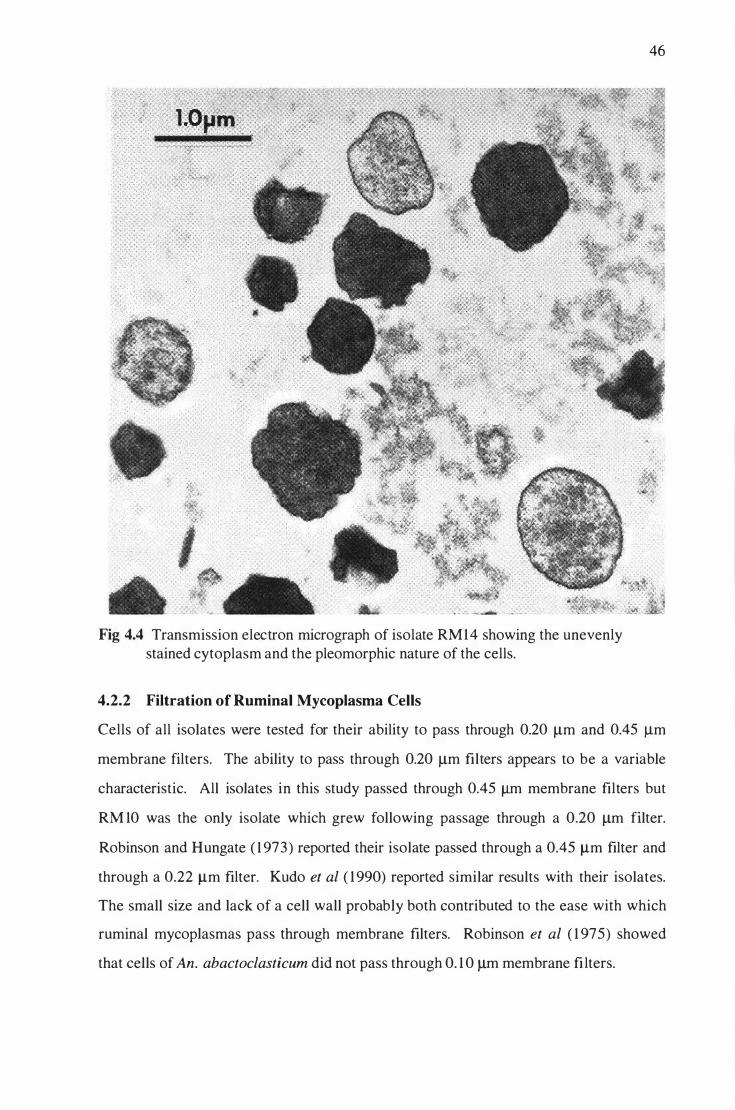
Fig 4.4 Transmission electron micrograph of isolate RM 14 showing the unevenly
stained cytoplasm and the pleomorphic nature of the cells.
4.2.2 Filtration of Ruminal Mycoplasma Cells
46
Cells of all isolates were tested for their ability to pass through 0.20 J..lm and 0.45 f.Lm
membrane filters. The ability to pass through 0.20 J..lm filters appears to be a variable
characteristic. All isolates in this study passed through 0.45 J..Lm membrane filters but
RM 10 was the only isolate which grew following passage through a 0.20 J..lm filter.
Robinson and Hungate ( 1 973) reported their isolate passed through a 0.45 f.Lm filter and
through a 0.22 f.Lm filter. Kudo et al ( 1 990) reported similar results with their isolates.
The small size and lack of a cell wall probably both contributed to the ease with which
rumina! mycoplasmas pass through membrane filters. Robinson et al ( 1 975) showed
that cells of An. abactoclasticum did not pass through 0. 1 0 J..lm membrane fi lters.

47
4.2.3 Relationship between Temperature and Growth of Ruminal Mycoplasmas
The optimum temperature for growth (T opt) of isolates was determined from their
growth in CRFB medium. Growth was measured at seven different temperatures over a
24 hr period by reading culture OD600 against a medium blank. The temperatures at
which growth was measured were 3 1 , 34, 36.5, 39.5, 42, 45 and 47.5°C. For the
purposes of the following discussion, the optimum growth temperature was considered
to be the temperature at which growth increased most rapidly in the shortest time after
culture inoculation.
Results for each of the rumina} mycoplasma isolates are given in Figs 4.5 to 4. 1 0.
0.25
0.2 � � � "" Q 2- 0. 1 5 ., u c 0: ..c
0.1 ... 0 "' ..c < 0.05
0 0 2 3 4 5
Time (hr) 6
Fig 4.5 Effect of temperature on growth of isolate RM 10.
7 8 9
For isolate RM 1 0, the growth curves are given in Fig 4.5. These show that the optimum
temperature for the growth of this i solate was 42°C. From Fig 4.5 it can be seen that
cel ls grown at 42°C and 45°C lysed rapidly after reaching maximum growth, as shown
by decreasing culture density. Cell lysis increased as the incubation temperature
increased. It must be remembered that these growth studies were done in batch culture
and do not, in all respects, parallel what would happen in the rumen where the
fermentation is closer to being continuous in nature.

48
0.3
0.25
g .., 0.2 Q 0
--- 31C --- 34C
'-' � --- 36.5C (.I = 0.15 = --- 39.5C
.c .. --- 42C 0 Vl .c --- 45C < 0.1 --- 47.5C
0.05
0 0 2 3 4 5 6 7 8 9 10 1 1 12
Time (hr)
Fig 4.6 Effect of temperature on growth of isolate RM 1 1 .
The seven temperature growth curves for isolate RM 1 1 are shown in Fig 4.6. These
show that the optimum temperature for the growth of this isolate was 42°C. At 45°C
and 42°C, cells began to lyse after 8 .5 hr growth. At temperatures below the optimum,
growth was slower and cells appeared less prone to lysis. The results in Fig 4.6 clearly
show that the lysis of isolate RM 1 1 was temperature dependent rather than growth rate
dependent. Although growth rates were similar at 45°C, 42°C and 39.5°C, RM l l lysed
readi ly at 45°C and 42°C but not at 39.5°C, the temperature nearest that found in the
rumen . There was no growth at 3 1 oc or 4 7 .5° C.
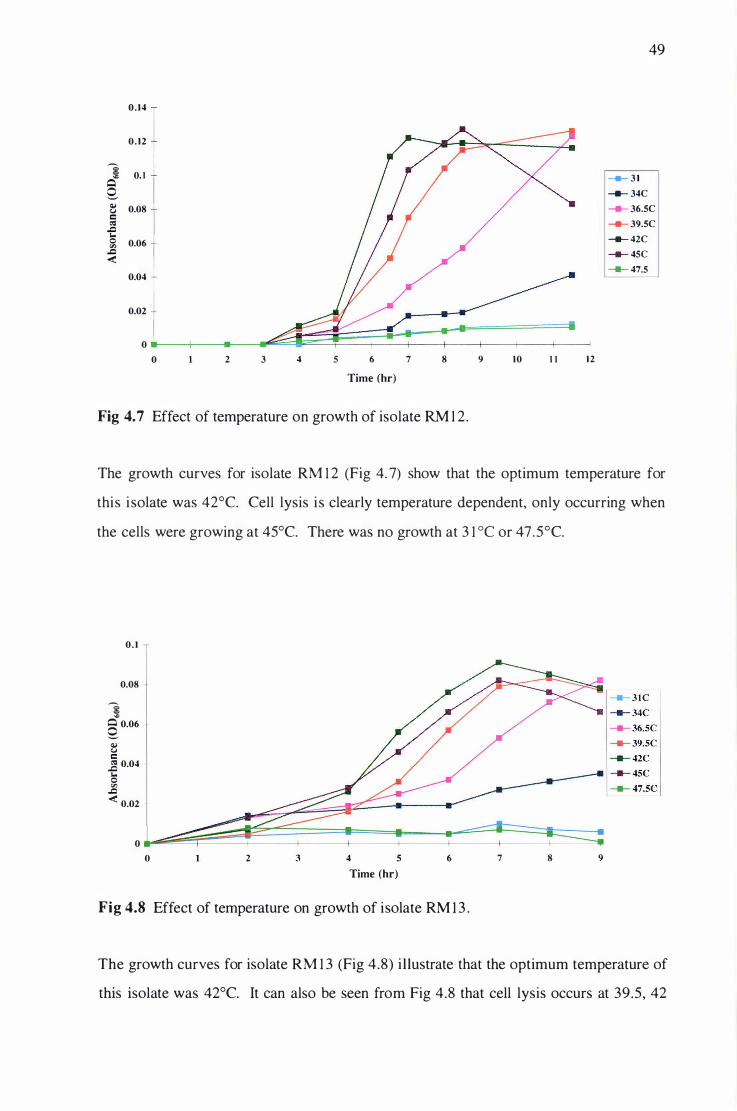
49
0.14
0.12
g 0.1 "" Q ----- 31
Q --- 34C � 0.08 u --- 36.5C c: ..s ,Q --- 39.5C ... 0 0.06 "' --- 42C
,Q �
--- 45C
----- 47.5 0.04
0.02
0
0 2 3 4 5 6 7 8 9 10 1 1 12
Time (hr)
Fig 4. 7 Effect of temperature on growth of isolate RM 1 2.
The growth curves for isolate RM 1 2 (Fig 4. 7) show that the optimum temperature for
this i solate was 42°C. Cell lysis i s clearly temperature dependent, only occurring when
the cells were growing at 45°C. There was no growth at 3 1 oc or 4 7 .5° C.
0.1
0.08
g "" � 0.06
� u c: _g 0.04 ... 0 "' ,Q � 0.02
0 0 2 4 5
Time (hr) 6
Fig 4.8 Effect of temperature on growth of isolate RM 1 3 .
7 8 9
The growth curves for isolate RM 1 3 (Fig 4.8) illustrate that the optimum temperature of
this isolate was 42°C. It can also be seen from Fig 4.8 that cell lysis occurs at 39.5, 42

50
and 45°C, but that the onset of cell lysis was most rapid at 42°C and 45°C. There was
no measurable growth at 3 1 oc or 47.5°C.
0. 1 4
0. 12
1 Q 0. 1
s � 0.08 = ell .Q 2; 0.06 "' .Q
< 0.04
0.02
0 0 2 3 4 5
Time (hr) 6
Fig 4.9 Effect of temperature on growth of isolate RM 1 4.
7
......... 3 1C
......... 34C
------- ......... 36.5C --- 39.5C -- 42C
--- 45C
--.---- -- 47.5C
8 9
The effect of temperature on the growth of mycoplasma RM 1 4 is shown in Fig 4.9. The
data clearly shows that the optimum temperature for this isolate was 42°C. The growth
rates were similar at 42°C and 45°C, but cell lysis appeared to be occurring more rapidly
at 45°C. The temperature of the rumen is 38°C-40°C (Hobson, 1 997). In general, for
cells of isolate RM 1 4 growing at temperatures close to that of the rumen, there was no
evidence of cell lysis. No measurable growth was seen at 3 1 oc or 47 .5°C.
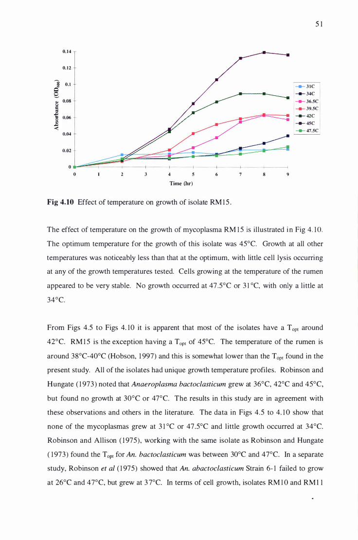
5 1
0.14
0.12
g 0.1 .., Q -- 31C Q, -- 34C Q.j 0.08 -- 36.SC u = � -- 39.SC
..c .. 0.06 0 -- 42C "' ..c -- 4SC
< 0.04 -- 47.SC
O.o2
0 0 2 3 4 5 6 7 8 9
Time (hr)
Fig 4.10 Effect of temperature on growth of isolate RM 1 5 .
The effect of temperature on the growth of mycoplasma RM 1 5 is illustrated i n Fig 4. 1 0.
The optimum temperature for the growth of this isolate was 45°C. Growth at all other
temperatures was noticeably less than that at the optimum, with little cell lysis occurring
at any of the growth temperatures tested. Cells growing at the temperature of the rumen
appeared to be very stable. No growth occurred at 47 .5°C or 3 1 °C, with only a little at
34°C.
From Figs 4.5 to Figs 4. 10 it i s apparent that most of the isolates have a Topt around
42°C. RM 1 5 is the exception having a Topt of 45°C. The temperature of the rumen is
around 38°C-40°C (Hobson, 1 997) and this is somewhat lower than the T opt found in the
present study. All of the isolates had unique growth temperature profiles. Robinson and
Hungate ( 1 973) noted that Anaeroplasma bactoclasticum grew at 36°C, 42°C and 45°C,
but found no growth at 30°C or 47°C. The results in this study are in agreement with
these observations and others in the literature. The data in Figs 4.5 to 4. 1 0 show that
none of the mycoplasmas grew at 3 1 °C or 47.5°C and l ittle growth occurred at 34°C.
Robinson and Allison ( 1 975) , working with the same isolate as Robinson and Hungate
( 1 973) found the Topt for An. bactoclasticum was between 30°C and 47°C. In a separate
study, Robinson et al ( 1 975) showed that An. abactoclasticum Strain 6- 1 failed to grow
at 26°C and 47°C, but grew at 3 7°C. In terms of cell growth, isolates RM l O and RM 1 1

52
grew to a maximum 00600 between 0.2 and 0 .3 , whereas all other isolates failed to grow
to an OD600 greater than 0. 1 4. This suggests two things; that some isolates were better
adapted to CRFB medium or some isolates were lysing faster than their cells were
dividing. All i solates became more prone to lysis as the incubation temperature
increased. Also, all isolates had a broader temperature growth range below their
optimum than above it. A comparison of the effects of temperature on growth for the
six isolates shows that the growth curves for isolates RM 1 3, RM 1 4 and RM 1 5 are
similar to each other but very different to those of RM 10, RM 1 1 and RM 1 2. This may
indicate that isolates RM 1 3, RM 1 4 and RM 1 5 are related species.
4.2.4 Effect of pH on the Growth of Rumina) Mycoplasmas
The six isolates were each inoculated into nine CRFB-based media adjusted to a final
pH of 5 .0 to 8 .75 and growth measured after 1 0 hr incubation. 39°C was chosen as the
growth temperature for these experiments because, for most isolates, little cell lysis
occurred at this temperature. Uninoculated medium was used as the reference for
growth measurements.
The effects of pH of culture media on the growth of isolates RM l 0, RM 1 1 and RM 1 2
are shown in Figs 4. 1 1 , 4. 1 2 and 4. 1 3 respectively. RM 1 0, RM 1 l and RM 1 2 grew in
the pH range 5 .5 to 7.2 with a pHopt of 6.5 for RM l O and RM 1 1 but with a pHopt closer
to pH 6.0 for RM 1 2. The pH growth profiles of RM 10 and RM 1 1 were similar but
differed significantly from that of RM 1 2. The optical densities reached by RM 1 0 and
RM 1 1 at their pHopr were considerably greater than that reached by RM 1 2.
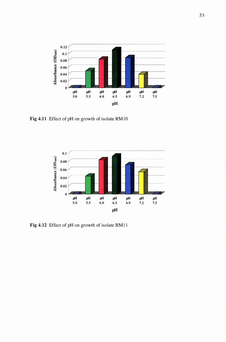
53
0.12 ,.-.. � 0.1
Q 0 0.08 ._, � � 0.06 c (IS .J:l 0.04 r.. 0 "' .J:l
< 0.02
0
pH pH pH pH pH pH pH 5.0 5.5 6.0 6.5 6.9 7.2 7.5
pH
Fig 4.1 1 Effect of pH on growth of isolate RM 10
0.1
'§ 0.08 Q �
0.06 � � c (IS 0.04 .J:l r.. 0 "' 0.02 .J:l
< 0
pH pH pH pH pH pH pH 5.0 5.5 6.0 6.5 6.9 7.2 7.5
pH
Fig 4.12 Effect of pH on growth of isolate RM 1 1

54
0.08
� Q 0.06 0 '-' � � 0.04 c � .t:l. &.. 0 0.02 rll .t:l.
< 0
pH pH pH pH pH pH pH 5.0 5.5 6.0 6.5 6.9 7.2 7.5
pH
Fig 4.13 Effect of pH on growth of isolate RM 1 2
Figs 4. 1 4 to 4. 1 6 show the effects of culture pH on the growth of isolates RM 1 3, RM 14
and RM 1 5 respectively. Isolate RM 1 3 grew within the range pH 5 .5 to 7.9 whereas
isolates RM 1 4 and RM 1 5 grew over the wider range of pH 5.0 to 7.9. Isolates RM 1 3,
RM 1 4 and RM 1 5 all had a pHopt of 6.5 to 6.9, and all grew to a similar culture density at
this pH. The similar responses to culture pH for isolates RM 1 3 , RM 1 4 and RM 1 5
suggest that they all may be similar. A comparison of these pH profiles with those in
Figs 4. 1 1 to 4. 1 3 indicates that they are markedly different from isolates RM 1 0, RM 1 1
and RM 1 2.
0.08 ,.-.,
� Q 0 0.06 '-' � � 0.04 c � .t:l. &.. 0 O.o2 rll .t:l.
< 0
pH pH pH pH pH pH pH pH 5.0 5.5 6.0 6.5 6.9 7.2 7.5 7.9
pH
Fig 4.14 Effect of pH on growth of isolate RM 1 3 .

55
0 . 1 ,.-.. § 0.08 Q
0 ._., 0.06 Q,l C.l = � 0.04 .c r.. 0 "' 0.02 .c
< 0
pH pH pH pH pH pH pH pH 5.0 5.5 6.0 6.5 6.9 7.2 7 .5 7.9
pH
Fig 4.15 Effect of pH on growth of isolate RM 14.
0.08
i Q 0.06 0 ._., Q,l C.l 0.04 = � .c r.. 0 0.02 "' .c
< 0
pH pH pH pH pH pH pH pH 5.0 5.5 6.0 6.5 6.9 7.2 7.5 7.9
pH
Fig 4.16 Effect of pH on growth of isolate RM 1 5 .
Based on the growth response to culture pH, there appeared to be two groups of rumina!
mycoplasma in this study; one consisting of isolates RM 1 0, RM 1 1 and RM 1 2 (Figs 4. 1 1
to 4. 1 3) and the other of isolates RM 1 3, RM 1 4 and RM 1 5 (Figs 4. 1 4 to 4. 1 6). The
differences between the two groups were particularly evident in media at higher pH with
isolates RM 1 3, RM 1 4 and RM 1 5 all showing measurable growth at pH 7 .5 and 7 .9 in
contrast to isolates RM 1 0, RM 1 1 and RM 1 2 which did not grow at these pH values.
None of the isolates grew at pH 8.75 . The pH of the rumen is usually in the range of pH
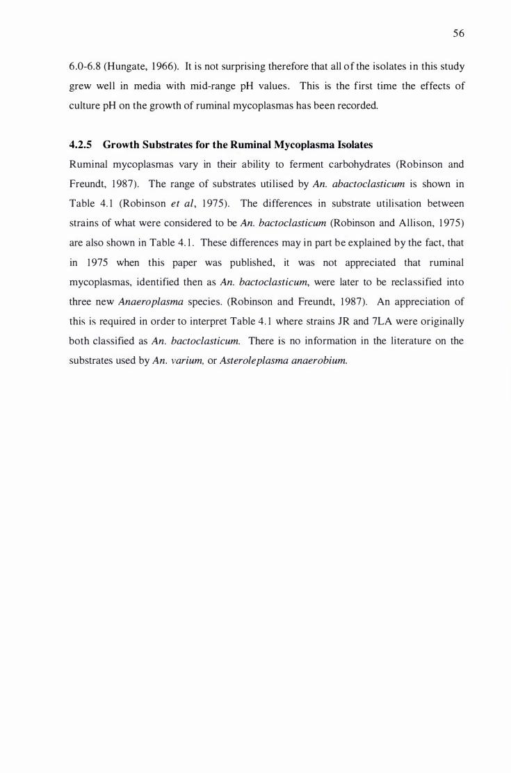
56
6 .0-6.8 (Hungate, 1 966). It is not surprising therefore that all of the isolates in this study
grew well in media with mid-range pH values . This is the first time the effects of
culture pH on the growth of rumina! mycoplasmas has been recorded.
4.2.5 Growth Substrates for the Ruminal Mycoplasma Isolates
Rumina! mycoplasmas vary in their ability to ferment carbohydrates (Robinson and
Freundt, 1 987). The range of substrates uti lised by An. abactoclasticum is shown in
Table 4. 1 (Robinson et al, 1 975). The differences in substrate utilisation between
strains of what were considered to be An. bactoclasticum (Robinson and Allison, 1 975)
are also shown in Table 4. 1 . These differences may in part be explained by the fact, that
in 1 975 when this paper was published, it was not appreciated that rumina!
mycoplasmas, identified then as An. bactoclasticum, were later to be reclassified into
three new Anaeroplasma species. (Robinson and Freundt, 1 987). An appreciation of
this is required in order to interpret Table 4. 1 where strains JR and 7LA were originally
both classified as An. bactoclasticum. There is no information in the literature on the
substrates used by An. varium, or Asteroleplasma anaerobium.
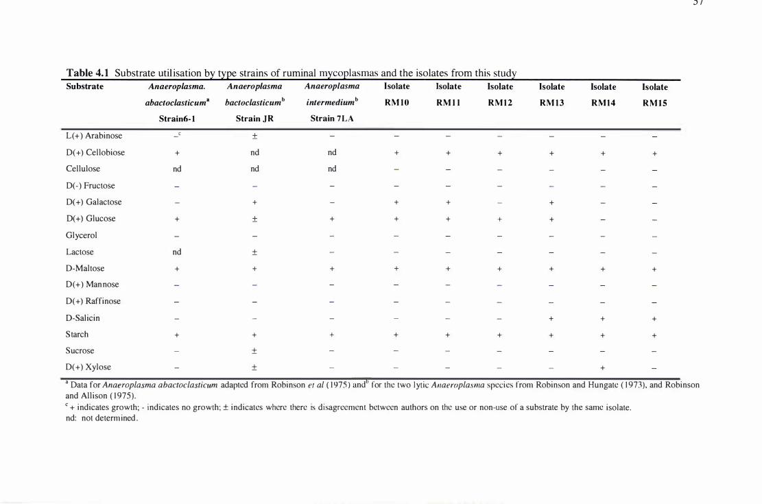
J !
Table 4. 1 Substrate util isation b� t�Ee strains of rumina! m�coElasmas and the isolates from this stud� Substrate Anaeroplasma. Anaeroplasma Anaeroplasma Isolate Isolate Isolate Isolate Isolate Isolate
abactoclasticum8 bactoclasticumb intermediumb RM lO RM l l RM12 RM 13 RM14 RMIS Strain6-l Strain JR Strain 7LA
L( +) Arabinose -c ±
D( +) Cellobiose + nd nd + + + + + +
Cellulose nd nd nd
D(- ) Fructose
D( +) Galactose - + - + + - +
D(+) Glucose + ± + + + + +
Glycerol
Lactose nd ±
D-Maltose + + + + + + + + +
D( +) Man nose
D( +) Raffinose
D-Salicin - - - - - - + + +
Starch + + + + + + + + +
Sucrose - ±
D(+) Xylose - ± - - - - - +
• Data for Anaeroplasma abactoclasticum adapted from Robinson et al ( 1975 ) andb for the two lytic Anaeroplasma species from Robinson and Hungate ( 1973), and Robinson and Allison ( 1975). c + indicates growth; - indicates no growth; ± indicates where there is disagreement between authors on the use or non-use of a substrate by the same isolate. nd: not determined.

5 8
The range of substrates used by isolates i n the present study i s shown i n Table 4. 1 .
Cellobiose, maltose and starch were utilised by all isolates. Arabinose, cellulose,
fructose, glycerol, lactose, mannose, raffinose, and sucrose were not metabolised by any
isolate. RM 1 4 was the only isolate to utilise xylose. Isolates RM 1 3, RM 1 4 and RM 1 5
utilised salicin, while isolates RM l O, RM l l and RM 1 2 did not. The use of sal icin
could provide a useful selection medium for this group of isolates. It can also be seen
from Table 4. 1 , that the substrates used by isolates RM l O, RM l l , RM 1 2, RM 1 3, RM 1 4
and RM 1 5 are the same as those used by the type strains ; the use of salicin by isolates
RM 1 3 , RM 1 4 and RM 1 5 being an exception.
4.2.6 Analyses of Fermentation End-Products
The fermentation products of rumina! mycoplasmas grown in Cellobiose Rumen Fluid
Broth (CbRFB) medium were identified by HPLC comparison. End-products were
qualitatively assessed by UV detection at a wavelength of 2 10 nm by comparison with
standards of known concentration and retention time. Software problems and the failure
of the refractive index detector made quantitative analysis difficult. Because the
refractive index detector fai led, it was not possible to analyse culture supematants for
ethanol, or trace the disappearance of cellobiose from the cultures. Hydrogen was
analysed by gas chromatography. Although small changes in some fermentation end
products were difficult to assess because of the relatively high background levels of
VFAs in the culture medium, it was found that the major end-products produced by
isolates RM l O, RM l l and RM 1 2 were formate, acetate, lactate, and H2. The major end
products produced by isolates RM 1 3, RM 14 and RM 1 5 were formate, acetate and
lactate. These isolates did not produce H2. The results agree with those of Robinson
and Hungate ( 1 973) who found that Acholeplasma (syn. Anaeroplasma) bactoclasticum
produced formate, acetate, lactate, H2 and C02 following growth on galactose. They
noted that because of a high VFA background in their medium, more accurate results
were achieved using 14C-galactose as the growth substrate and radiochemical methods to
assay end-products. Robinson et al ( 1 975) employed 1 4C-starch to examine the end
products of An. abactoclasticum and found formate, acetate and lactate as the major
products. Small amounts of 14C were detected also in succinate, C02 and several
unidentified products.

4.2. 7 Antibiotic Sensitivity of Ruminal Mycoplasmas
59
The susceptibility of isolates to antibiotics was assessed after 24 hr growth in CRFB
medium which contained the antibiotics listed in Table 4.2 at concentrations described
in section 3 .3 .8 .
Table 4.2 Antibiotic sensitivities3 of rumina! mi:CO£lasmas
Compound RMlO RMl l RM12 RM13 RM1 4 RM15
Bacitracin + +
Benzylpenicillin + + + + + +
Chloramphenicol
D-Cycloserine + + + + + +
Digitonin
Lincomycin-HCl
Streptomycin S04 2- + + + + + +
Tetracycline
Thallium (I) acetate + + + + + +
a Antibiotics were assessed as inhibitory if the 00600 of the culture at 24 hr was < 0.03
and are shown as - in Table 4.2. Cultures in which antibiotics showed no inhibition had 00600 values at 24 hr > 0.075 and were recorded as + in Table 4.2.
It was observed that in general , antibiotics which inhibited protein synthesis were the
most effective inhibitors of rumina! mycoplasmas. Chloramphenicol, digitonin,
lincomycin-HCl and tetracycline inhibited all isolates. Chloramphenicol binds to the
ribosomal 50S subunit inhibiting protein synthesis (Mandelstam and McQuillen, 1 973).
Lincomycin-HCl inhibits protein synthesis in Gram positive bacteria, anaerobes and
mycoplasmas, but is generally ineffective against Gram negative aerobic bacteria (Mims
et al, 1 993). Tetracycline binds to a protein of the ribosomal 30S subunit and inhibits
ribosomal translocation in both Gram-positive and Gram-negative microorganisms
(Mandelstam and McQuillen, 1 973). Digitonin is a naturally occurring steroid glycoside
active in the disruption of plasma membranes (Elias et al, 1 977). Robinson and Allison
( 1 975) showed that the ruminal mycoplasma, An. bactoclasticum was sensitive to
digitonin. Robinson et al ( 1 975) found that Anaeroplasma species were inhibited by
digitonin (20 J..lg mr 1 ), whereas the sterol non-requiring strains of rurninal mycoplasma

60
(now reclassified as Asteroleplasma anaerobium) were not inhibited by 200 j.lg ml- 1
digitonin. All of the rumina! mycoplasmas in this study were sensitive to digitonin (60
j.lg mr 1 ) and thus may be members of the genus Anaeroplasma. There are no previous
studies in the l iterature on the sensitivities of rumina! mycoplasmas to l incomycin-HCl
or to chloramphenicol . In this study, bacitracin was found to inhibit four of the six
rumina! mycoplasmas tested (Table 4.2). Bacitracin inhibits bacterial cell wall synthesis
by interfering with murein production (Mims et al, 1 993) and is also an inhibitor of
proteases (van Noort et al, 1 99 1 ) . The rumina! mycoplasma, An. abactoclasticum, has
been reported to be sensitive to bacitracin, streptomycin and D-cycloserine (Robinson et
al, 1 975). In the present study, there was no effect on the growth of any isolate by
benzylpenicillin or 0-cycloserine. Both antibiotics are involved in the inhibition of
bacterial cell wall synthesis (Mims et al, 1 993) and as such would not be expected to
inhibit the growth of rumina! mycoplasmas. Robinson and Hungate ( 1973) reported that
Acholesplasma (syn. Anaeroplasma) bactoclasticum was not inhibited by 5000 IU
benzylpenicillin mr 1' but was sensitive to 20 j.lg mr 1 terramycin (tetracycline) .
Although most species within the class Mollicutes are susceptible to the tetracyclines,
lateral transfer of tetracycline resistance genes from walled bacteria (streptococci ) to
mollicutes has been shown to occur in isolates from the human genital tract (Roberts,
1 990) . There was no inhibition by streptomycin sulphate or thallium (I) acetate of any
of the isolates in this study. Streptomycin binds to ribosomal components inhibiting
protein synthesis by causing mis-reading of m-RNA (Mandelstam and McQuillen,
1973). The negative results for thallium (I) acetate were unexpected; Robinson et al
( 1 975) and Robinson and Allison ( 1 975) found that al l of their isolates were sensitive to
thallium (D acetate. In contrast, Robinson and Hungate ( 1 973) noted that their isolate
was uninhibited by thallium en acetate, but in a personal communication to them,
Allison and Robinson suggested that . . . "the thallium may have been precipitated by the
sulfide used as a reducing agent." In this study the medium did not contain added
sulphide, but sulphide in the rumen fluid component in the medium, or the L-cysteine
HCl used as a reducing agent, may have had a similar effect.
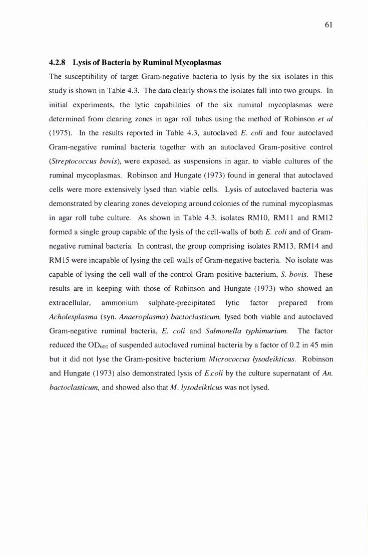
4.2.8 Lysis of Bacteria by Ruminal Mycoplasmas
6 1
The susceptibility of target Gram-negative bacteria to lysis by the six isolates i n this
study is shown in Table 4.3. The data clearly shows the isolates fall into two groups. In
initial experiments, the lytic capabilities of the six rumina! mycoplasmas were
determined from clearing zones in agar roll tubes using the method of Robinson et al
( 1 975). In the results reported in Table 4.3, autoclaved E. coli and four autoclaved
Gram-negative rumina! bacteria together with an autoclaved Gram-positive control
(Streptococcus bovis), were exposed, as suspensions in agar, to viable cultures of the
rumina! mycoplasmas. Robinson and Hungate ( 1 973) found in general that autoclaved
cells were more extensively lysed than viable cells. Lysis of autoclaved bacteria was
demonstrated by clearing zones developing around colonies of the rumina! mycoplasmas
in agar roll tube culture. As shown in Table 4.3, isolates RM l O, RM l l and RM 1 2
formed a single group capable of the lysis of the cell-walls of both E. coli and of Gram
negative rumina! bacteria. In contrast, the group comprising isolates RM 1 3 , RM 1 4 and
RM 1 5 were incapable of lysing the cell walls of Gram-negative bacteria. No isolate was
capable of lysing the cell wall of the control Gram-positive bacterium, S. bovis. These
results are in keeping with those of Robinson and Hungate ( 1 973) who showed an
extracellular, ammomum sulphate-precipitated lytic factor prepared from
Acholesplasma (syn. Anaeroplasma) bactoclasticum, lysed both viable and autoclaved
Gram-negative rumina! bacteria, E. coli and Salmonella typhimurium. The factor
reduced the 00600 of suspended autoclaved rumina) bacteria by a factor of 0.2 in 45 min
but it did not lyse the Gram-positive bacterium Micrococcus lysodeikticus. Robinson
and Hungate ( 1 973) also demonstrated lysis of E.coli by the culture supernatant of An.
bactoclasticum, and showed also that M. lysodeikticus was not lysed.
62
Table 4.3 Ll:sis of autoclaved bacterial cells bl: rumina! ml:coElasmas in agar culture
Ruminal Mycoplasma
Bacterium RMlO RMl l RM12 RM13 RM14 RM15
B. fibrisolvens Hl 7C +a + +
L. multiparus ATCC 1 9207 + + +
P. ruminicola A TCC 1 9189 + + +
S. bovis JBJ
Sue. dextrinosolvens 24 + + +
E. coli XLI + + +
a +, lysis by mycoplasma; -, no lysis by mycoplasma. For full genus and species names
of the bacteria see Chapter 6: "Microorganisms used in this study."
Lysis of bacteria by rumina] mycoplasmas was also tested in broth culture (Table 4.4).
This was found to be an easier method for assessment of the lytic properties of rumina]
mycoplasmas because it was faster and less labour intensive. The method had the
advantage that only one tube of each test bacterium I mycoplasma combination was
required, rather than the six or seven required for dilution series using the roll tube
method. Lysis was determined by measurement of culture OD600 after 96 hr incubation
at 39°C. The results in Table 4.4 show that E. coli and six Gram-negative rumina]
bacteria, exposed to the six rumina] mycoplasmas in broth culture, were lysed by
isolates RM l O, RM l l and RM 1 2, but not lysed by isolates RM 1 3, RM 1 4 and RM 1 5 .
The data in Table 4.4 also show that the Gram-negative ruminal bacteria Megasphaera
elsdenii and Selenomonas ruminantium, not tested using the agar method, were also
lysed by isolates RM lO, RM l l and RM 1 2. The Gram-positive cell wall of S. bovis was
resistant to lysis by all isolates. Examination of the data in Tables 4.3 and 4.4
confirmed that both methods used to study cell wall lysis gave identical results.

63
Table 4.4 L�sis of autoclaved bacterial cells b� rumina! m�coElasmas in broth culture
Ruminal Mycoplasma
Bacterium RMlO RMl l RM12 RM13 RM14 RMlS
B. fibrisolvens H1 7C +a + +
L. multiparus ATCC 1 9207 + + +
M. elsdenii T81 + + +
P. ruminicola ATCC 19189 + + +
Sel. ruminantium HD4 + + +
S. bovis JBJ
Sue. dextrinosolvens 24 + + +
E. coli XLI + + +
a + , lysis by mycoplasma; -, no lysis by mycoplasma. For full genus and species names
of the bacteria see Chapter 6: "Microorganisms used in this study."
One of the principal characteristics which divides species within the genus
Anaeroplasma is the ability (or inability) of isolates to lyse Gram-negative bacterial cell
walls (Robinson and Freundt, 1 987). The results of both agar and broth lysis assays
showed that only bacteria with Gram-negative cell walls were lysed by the rumina!
mycoplasmas. When rumina! mycoplasmas were first characterised, all of those which
lysed Gram-negative bacterial cell walls were classified as Anaeroplasma
bactoclasticum. However further analysis, using a variety of tests, indicated that the
rumina! mycoplasmas capable of the lysis of Gram-negative cell walls were a
heterogenous assemblage of species which were later reclassified as either An.
bactoclasticum, An. intermedium or An. varium (Stephens et al, 1985 ; Robinson and
Freundt, 1 987).
4.2.9 Proteolysis by Ruminal Mycoplasmas
The ability of the six rumina! mycoplasmas to hydrolyse casein (a high molecular
weight phosphoprotein) were determined, after 4 days incubation at 39°C, from clearing
zones in agar roll tubes using a modified method of Robinson and Hungate ( 1 973). The
data clearly showed the isolates fell into two groups. Isolates RM l O, RM 1 1 and RM 1 2
produced clearing zones i n the casein suspension, while isolates RM 1 3 , RM 14 and

64
RM 1 5 did not. These results are in agreement with those of Robinson and Hungate
( 1 973) whose isolate was proteolytic and Robinson et al ( 1 975) whose isolate was not.
4.2.10 Lysis of Fungal Cell-Walls by Ruminal Mycoplasmas
To test the hypothesis that the inhibition of rumina! fungi by rumina! mycoplasmas was
a consequence of fungal cell wall lysis, required a medium which would support the
growth of mycoplasmas and also demonstrate the utilisation of the fungal cell wall as a
carbon source. The simplest way was to use the development of clearing zones around
mycoplasma colonies growing on agar roll tubes which contained finely ground fungal
cell wall as the growth substrate. A major problem was grinding the fungal material
fine enough to give an even suspension in an agar medium. Fungal cell walls have a
leather-like consistency and in spite of prolonged ball-milling, no really suitable method
was found which produced a homogenous product. A fungal cell-wall medium
(FCW A) was prepared but this did not produce a truly homogenous agar and under the
dissecting microscope, FCW A had a granular appearance. The agar was inoculated with
the rumina] mycoplasmas (0.2 ml) (00600 of 0. 1 ), and incubated at 39°C for 2 1 days.
Although the non-uniformity of the agar may have masked hydrolysis of the fungal cell
wall , no evidence of clearing zones around any mycoplasma colony was seen. It seems
that the inhibition of ruminal fungi by rumina] mycoplasmas is therefore unlikely to
involve the degradation of the fungal cell wall .
4.2.1 1 Chitinase Activity of Ruminal Mycoplasmas
Chitin, a polymer of chitobiose and N-acetylglucosamine, is a major component of the
cell wall of ruminal fungi (Kopecny et al, 1 996). The chitinolytic bacterium
Clostridium tertium ChK5 has been shown to inhibit cellulolysis by three genera of
ruminal fungi (Kopecny et al, 1 996). Also, chitinases from Streptomyces griseus inhibit
the cellulases of the fungus N. frontalis possibly by disrupting the association between
the cellulase and components of the fungal cell wall (Wilson and Wood, 1 992). It was
not known if ruminal mycoplasmas produce a chitinase, but one possible mechanism
whereby rumina} mycoplasmas inhibit ruminal fungi may be via the hydrolysis of chitin
in the fungal cell wall (Joblin, pers. comm., l 996) .
65
Cultures of ruminal mycoplasma isolates inoculated into Chitin Starch Agar produced
no clearing zones in the chitin after 2 1 days incubation at 39°C, although colonies were
clearly visible. A similar experiment with no starch supplementation, produced poor
mycoplasma growth and again no clearing zones were seen in the chitin ; the
mycoplasmas were probably growing on low levels of an unknown substrate carried
over in the rumen fluid component of the medium. This result confirms the FCW A
findings (see section 4.2. 1 0) . Since no lysis of either fungal cell wall or pure chitin was
demonstrated, it is probable the inhibition of rumina! fungi by rumina! mycoplasmas
was not as a consequence of a mycoplasmal chitinase. With limited degradative
capabilities and a dependence on host-derived enzymes, it would perhaps have been
surprising had rumina! mycoplasmas been capable of hydrolysing chitin.
4.2.1 2 Sterol Requirements of Ruminal Mycoplasmas
The sterol requirement of rumina! mycoplasmas are second only to the requirement for
strictly anaerobic growth conditions when identification is being considered. Rumina]
mycoplasmas which require sterol for growth belong to the genus Anaeroplasma, while
those which are sterol non-requiring belong in the genus Asteroleplasma. To determine
their sterol requirements, all of the isolates in this study were inoculated into medium
MM 1 0, supplemented with E. coli l ipopolysaccharide (LPS) (Boivin) and cholesterol
using the method described by Robinson et al ( 1 975) . Isolates RM 1 0, RM l l and RM 1 2
failed to grow in MM 1 0, either alone, when the medium was supplemented as above or
when further supplemented with additional vitamins. The lack of growth of RM 1 0,
RM 1 1 and RM 1 2 in MM lO suggested an unidentified growth factor was missing from
MM l O or the supplements. Isolates RM 1 3, RM 1 4 and RM 1 5 al l grew in MM 10 and
were all stimulated by LPS and very strongly by LPS and cholesterol together. In a
separate experiment in CRFB medium, all six isolates were inhibited by low
concentrations of digitonin (60 �g rn1"1 ) . Robinson et al ( 1 975) stated, when
commenting on their isolates . . . "growth of the sterol requiring strains 6- 1 and 1 7 1
(Anaeroplasma species) were inhibited by digitonin (20 �g rnl"1 ) whereas growth of the
sterol non-requiring strain 1 6 1 was unaffected by 200 �g rnl-1 of digitonin and only
slightly by 500 �g ml- 1". Robinson et al ( 1 975) stated " Cholesterol requirement may
also be determined by the selective sensitivity of sterol-requiring mycoplasmas to
digitonin. Digitonin sensitivity seems to parallel the ability of ruminal mycoplasmas to

66
incorporate cholesterol ." The indirect evidence based on the sensitivity of the six
isolates to low concentrations of digitonin supported the identification of isolates RM 1 0,
RM 1 1 , RM 1 2, RM 1 3, RM 1 4 and RM 1 5 as members of the genus Anaeroplasma.
4.2.1 3 Identification of the Ruminal Mycoplasma Isolates
The characteristics of isolates RM 1 0, RM 1 1 and RM 1 2, placed them into the genus
Anaeroplasma. They were classified on the basis that they were strictly anaerobic, were
sensitive to low concentrations of digitonin (60 j..lg mr 1 ) which indicated a requirement
for sterols in growth media, had no cell wall, passed membrane filters with pore sizes of
0.45 j..tm, produced H2, were proteolytic and were able to lyse the cell walls of E. coli
(Robinson et al, 1 975; Robinson and Freundt, 1987). The evidence for the abil ity of
RM 10, RM 1 1 and RM 1 2 to lyse the cell walls of Gram-negative bacteria is given in
Table 4.3 and 4.4. The precise identity of the three i solates was not possible because
three Anaeroplasma species are capable of lysing the cell walls of Gram-negative
bacteria, namely, An. bactoclasticum, An. varium and An. intermedium (Robinson and
Freundt, 1987). The isolates may belong to any of these species, or to a new species.
1 6S rRNA sequence data would help confirm species identification (Weisburg et al,
1989; Kuske and Kirkpatrick, 1992).
The characteristics of isolates RM 1 3 , RM 14 and RM 1 5 place them into the genus
Anaeroplasma. The characteristics of these isolates were very similar to those of
isolates RM l O, RM 1 1 and RM 1 2 except they were unable to lyse the cell walls of
Gram-negative bacteria, were not proteolytic and did not produce H2. Additional
evidence for a requirement for exogenous sterol was shown by the strongly stimulated
growth seen in MM 1 0 supplemented with cholesterol and E. coli LPS. The isolates
were therefore classified as Anaeroplasma abactoclasticum; the only Anaeroplasma
species not able to lyse the cell walls of Gram-negative bacteria (Robinson et al, 1 975 ;
Robinson and Freundt, 1 987). Confirmation of this identity would have been possible
had 1 6S rRNA sequence data been available (Weisburg et al, 1989; Kuske and
Kirkpatrick, 1 992) .

67
4.2.14 RAPD Analysis of Rumina) Mycoplasma DNA
The RAPD method to examine the DNA of rumina! mycoplasmas for genomic
fingerprints was chosen because no prior knowledge of the DNA nucleotide sequence
was required, the method was faster than RFLP, no restriction enzyme digestion of
DNA was necessary and the process was relatively easy to perform. The RAPD method
was developed independently by Williams et al ( 1 990) and Welsh and McClel land
( 1 99 1 ). Will iams et al ( 1 990) used single arbitrary primers to demonstrate DNA
polymorphisms in DNA samples from humans, soybean, corn and Neurospora crassa.
Welsh and McClelland ( 1 99 1 ) demonstrated DNA polymorphisms in DNA from mice
using single random primers, but extended the methodology to show that primers used
in pairs would also generate unique banding patterns. The RAPD method has also been
successfully applied using DNA from procaryotes to demonstrate intraspecies
discrimination in Listeria monocytogenes (Czajka et al, 1 993), to distinguish among
strains of Xanthomonas campestris (Manulis et al, 1 994) and to identify conserved
regions in actinomycete genomes (Mehling et al, 1 995). In this study, random
oligonucleotide primers of 1 0 bases ( l Omers) with 60-70% G+C were used to amplify
the DNA of rumina! mycoplasma isolates using the Polymerase Chain Reaction (PCR).
The DNA and primer combinations as outlined in Table 4.5. DNA was extracted using
DNAzol reagent; the cells lysed readily and good yields of DNA were achieved using
the procedures recommended. The DNA was quantified spectrophotometrically using a
multiplate reader which measured OD260 and OD280. An OD260 of 1 is equal to
approximately 50 11g rnl- 1 of double-stranded DNA. The ratio between OD260 and OD280
gives an estimate of the purity of the nucleic acid. Pure preparations of DNA have a
OD26o/OD28o ratio of 1 .8 . RNA was also present in the extracts, as shown by agarose
gel electrophoresis. The RNA did not interfere with subsequent RAPD analysis which
was performed using the method of Gardiner et al ( 1 996). The details of the PCR cycle
times and temperatures are given in section 3.3 . 1 3 .

Table 4.5 DNA and primer combinations for the RAPD analysis
Lane DNA Primer Sequence 1 1 OObp ladder na 2 RM l O OPAA04 5' -AGGACTGCTC-3 ' 3 RM 1 1 OPAA04 4 RM 1 2 OPAA04 5 RM 1 3 OPAA04 6 RM 1 4 OPAA04 7 RM 1 5 OPAA04 8 1 OObp ladder na 9 RM l O OPAA06 5 ' -GTGGGTGCCA-3 '
1 0 RM 1 1 OPAA06 1 1 RM 1 2 OPAA06 1 2 RM 1 3 OPAA06 1 3 RM 1 4 OPAA06 1 4 RM 1 5 OPAA06 1 5 1 00bp ladder na 1 6 RM 1 0 OPAA09 5 ' -AGATGGGCAG-3' 1 7 RM 1 1 OPAA09 1 8 RM 1 2 OPAA09 1 9 RM 1 3 OPAA09 20 RM 1 4 OPAA09 2 1 RM 1 5 OPAA09 22 1 OObp ladder na
68
2 3 4 5 6 7 8 9 I 0 I I 1 2 13 14 15 16 1 7 1 8 1 9 20 2 I 22
Fig 4.17 RAPD profiles of DNA from isolates RM I O to RM 1 5. For lane contents see
Table 4.5

69
Fig 4 . 1 7 illustrates the PCR products of isolate-DNA, amplified using the RAPD
technique, after separation on a 2% agarose gel . Isolate RM l O produced unique banding
patterns with primers OPAA04 and OPAA06, but with primer OP AA09 gave similar
banding patterns to isolates RM l l and RM 1 2. Isolates RM l l and RM 1 2 produced
banding patterns which were the same for all three primers tested, which suggested that
these two isolates were identical. Isolate RM 1 3 had a unique banding profile with al l
the primers tested and was not similar to any other isolate. Isolates RM 1 4 and RM 1 5
gave the same banding patterns with all primers tested which suggested that these two
isolates were identical. Control reactions were run using distilled water in place of
DNA with no bands being produced. The results showed the RAPD procedure to be an
effective method for the demonstration of genomic fingerprints in rumina} mycoplasma
DNA.
4.2. 1 5 PAGE Analysis of Rumina) Mycoplasma Proteins
The method used to electrophoretically separate rumina} mycoplasma proteins according
to their molecular size was that of Laemmli ( 1970). When the mycoplasma cells were
lysed with SDS prior to protein analysis by the method of Bradford ( 1 976), the solutions
of RM I O, RM 1 1 , & RM 1 2 were clear, while those of RM 1 3, RM 14 & RM 1 5 were
cloudy. This suggested a compositional difference between the two groups of rumina!
mycoplasma. This observation may reflect that of Langworthy et al ( 1 975) who noted
"The bacteriolytic strain 7LA contained only about 50% of the amount of glycolipids
present in the non-bacteriolytic strain 6- 1 ". Strain 7LA is now classified as An.
intermedium, while strain 6- 1 is classified as An. abactoclasticum (Robinson and
Freundt, 1 987). The observation does not endow identity to the two groups of rumina!
mycoplasma, but may reflect differing glycolipid composition.
Details of the identity of the rumina! mycoplasma lysate in each lane of the gel, the
concentration, and volume required to give the necessary 80 �g of protein per lane, are
shown in Table 4.6.

70
Table 4.6 Protein concentration of cell lysates and lane-loading of the PAGE gel
Lane Isolate Protein Cone. of Lysate Cell Lysate per Lanea
(J.Lg mr1) (J.Ll)
RM I O 1 500 73.3
2 RM l l 1 860 59. 1
3 RM I 2 8 1 0 1 35 .8
4 RM I 3 900 1 22.2
5 RM 1 4 1 500 73.3
6 RM I 5 900 1 22.2
a Volume of lysate to give 80 IJ.g of protein per gel lane.
RM I O RM I I RM 1 2 RM 1 3 RM 1 4 RM 1 5
Fig 4 .18 PAGE profiles of the proteins from isolates RM I O to RM 1 5

7 1
The first impression from the gel was that there were two major groupings within the
rumina! mycoplasmas being studied (Fig 4. 1 8) . It was clear that isolates RM 1 0, RM 1 1
and RM 1 2 formed one distinct group while isolates RM 1 3, RM 1 4 and RM 1 5 formed
the other. Within each group of three isolates, there were minor differences in the
protein bands, sufficient to distinguish between the isolates. RM 1 1 and RM 1 2 both
possessed one strong, but different low molecular weight band, which RM 1 0 lacked.
The presence of a high molecular weight protein band in RM 1 3 , which was absent in
RM 1 4 and RM 1 5, suggested that RM 1 4 and RM 1 5 were more closely related to each
other than they were to RM 1 3 .
4.3 COCUL TURE STUDIES
Many studies have demonstrated interactions between rumina! microorganisms grown in
coculture. Studies have shown that when rumina! bacteria were cocultured with ruminal
fungi, the bacteria effected fungal xylanolysis in a variety of ways (Williams et al,
1 994) . When ruminal fungi were cocultured with the methanogen, Methanobrevibacter
smithii, it was shown that both the rate and extent of xylan utilisation by the fungi were
increased in the coculture (Joblin et al, 1990). There are no previous coculture studies
to show what effect rumina! mycoplasmas have on cellulolysis by rumina! bacteria.
Likewise no previous studies have been undertaken to show the effect of rumina!
mycoplasmas on cel lulolysis by rumina! fungi. However during fungal isolation we
have previously noted the eo-isolation of rumina! mycoplasmas, suggesting there may
be an association between these two groups of microorganisms.
4.3.1 Effect of Ruminal Mycoplasmas on Cellulolysis by Ruminal Bacteria
Cellulolytic ruminal bacteria and the rumina! mycoplasmas were cocultured to establish
if there was inhibition of bacterial cellulolysis and was it measurable. The cellulose
substrate, paper strip, was digested by the bacteria but not digested by the rumina!
mycoplasmas. Growth of the cellulolytic bacteria was monitored by disappearance of
the paper and production of H2. Hydrogen was produced both by the bacteria and
isolates RM l O, RM l l and RM 1 2. Isolates RM 1 3 , RM 1 4 and RM 1 5 did not produce
H2. Hydrogen production by the bacteria was not constitutive, but can be a useful

72
metabolite to follow as an indicator of growth. The results (Table 4.7) indicate that
cellulolysis was inhibited in both R. albus (30-60%) and R. jlavefaciens (50-80%) when
grown in coculture with the rumina! mycoplasmas, while C. chartatabidum and F.
succinogenes appeared to be slightly stimulated by the coculture. The H2 produced in
the coculture of F. succinogenes with isolates RM l O, RM l l and RM 1 2 is a reflection of
production by the mycoplasma alone, because F. succinogenes does not produce H2.
Similarly, an examination of the results (Table 4.7) show increased H2 in cocultures of
R. flavefaciens with RM l O, RM l l and RM 1 2 which probably reflect a stimulation of
mycoplasmal H2, rather than a stimulation of H2 production by the bacteria. This in turn
suggested that rumina! mycoplasmas were cross feeding on intermediates produced as a
result of the degradation of paper by the ruminococci . The H2 production results
suggest that the cocultures have little effect on the production of H2 by any of the
cellulolytic bacteria. Given that F. succinogenes is Gram-negative, it i s unclear why on
the basis of the demonstrated lysis of other Gram-negative bacteria by isolates RM l O,
RM l l and RM 1 2, that this bacterium was not also lysed and why cellulolysis was
uninhibited. Cellulolytic bacteria often produce extracellular slime when growing on
ball-milled cellulose and this may have protected F. succinogenes from lysis by these
rumina! mycoplasmas. The mechanism whereby R. albus and R. flavefaciens (both
Gram-positive) were inhibited is not known. The inhibition was substantial and may
have profound consequences for the rumen fermentation if the in vitro observations
were also shown to occur in vivo.

73
Table 4.7 Cellulolysis and H2 production by ruminal bacteria growing on cellulose in
the presence of ruminal mycoplasmas
Cellulose Used Culture mga % b H2 (ml)3
Clostridium chartatabidum 6.2 ± 0.4 1 00 2.02 ± 0. 1 8
+RM 1 0 6.3 ± 2.0 1 00 2.0 1 ± 0. 1 1
+RM 1 1 7.2 ± 0.5 1 20 2 .02 ± 0. 1 2
+RM 1 2 6.7 ± 1 .5 1 1 0 1 .96 ± 0. 1 2
+RM 1 3 7.0 ± 0.7 1 1 5 1 .99 ± 0.06
+RM 14 7. 1 ± 0.8 1 1 5 2. 1 0 ± 0.20
+RM 1 5 6.6 ± 0.5 1 05 2.0 1 ± 0. 1 5
Fibrobacter succinogenes 32. 1 ± 0.8 l OO 0
+RM 10 32.8 ± 1 .2 l OO 0.07 ± 0.02
+RM 1 1 34.2 ± 3 . 1 1 05 0.05 ± 0.03
+RM 1 2 34.3 ± 1 .3 1 05 0.03 ± 0.0 1
+RM 1 3 35. 1 ± 2.2 1 1 0 0
+RM 14 33 .5 ± 0.3 1 05 0
+RM 1 5 33. 1 ± 1 .8 1 05 0
Ruminococcus a/bus 1 1 .2 ± 0.8 1 00 2 .05 ± 0.08
+RM 1 0 6.5 ± 0.6 55 2 .03 ± 0.0 1
+RM 1 1 7 .4 ± 1 .0 65 1 .87 ± 0.07
+RM 1 2 6.9 ± 0.8 60 1 .96 ± 0.04
+RM 1 3 7 . 1 ± 3 .3 60 1 .89 ± 0.06
+RM 14 8.4 ± 2.0 70 1 .86 ± 0.04
+RM 1 5 4.4 ± 2 . 1 40 1 .82 ± 0.08
Ruminococcus flavefaciens 4.8 ± 0.7 1 00 0.09 ± 0.05
+RM 1 0 2.5 ± 1 .5 50 0.22 ± 0. 14
+RM 1 1 0.9 ± 1 . 1 20 0.28 ± 0.04
+RM 1 2 2.6 ± 1 .4 50 0.29 ± 0.02
+RM 1 3 2.6 ± 0.9 50 0. 1 0 ± 0.02
+RM 14 1 .8 ± 1 .4 35 0. 1 0 ± 0.02
+RM 1 5 1 .7 ± 0.7 30 0. 1 0 ± 0.03
a Data represents mean and standard deviation of triplicate cultures
b Cellulolysis by bacteria in axenic culture taken as 1 00%.

4.3.2 Effect of Ruminal Mycoplasmas on Cellulolysis by Ruminal Fungi
74
Rumina! fungi are recognized as producers of some of the most active cellulolytic
enzymes (Wood et al, 1 986). Fungal cellulolysis was inhibited by the action of
compounds produced by rumina! bacteria of the Ruminococcus and Butyrivibrio genera
(Stewart et al, 1 992; Joblin and Naylor, 1 994). Previous studies have shown that
rumina! mycoplasmas were often eo-isolated with rumina! fungi but the degree to which
they interacted was unknown (Joblin and Naylor, unpublished data) . To establish if
there was interaction, cellulolytic fungi and rumina! mycoplasmas were grown m
coculture and the effects on fungal cellulolysis monitored. Cocultures of each fungus
with each rumina! mycoplasma were established in PSM. These cocultures, with pure
fungal cultures on PSM as controls, were examined to give information on substrate
utilisation and H2 production. This allowed an assessment of the extent to which
rumina! mycoplasmas and fungi interact in coculture. The substrate, paper strip, was
digested by the fungi, but not by the rumina! mycoplasmas. Fungal growth was
monitored by disappearance of the paper (cellulose) and by production of H2. Fungal H2
production is constitutive. Mycoplasma isolates RM 1 0, RM 1 1 and RM 12 also produce
H2, while isolates RM 1 3 , RM 14 and RM 1 5 do not.
In a preliminary experiment to see if rumina! fungi were inhibited by rumina!
mycoplasmas, paper-containing-medium PSM, was inoculated with the fungus N.
frontalis alone and N. frontalis with mycoplasma RM 1 4. The results are shown in Fig
4. 1 9 . In the culture inoculated with N. frontalis alone (tube 2), the paper was almost
totally digested and the paper strip had collapsed completely. In contrast, the paper strip
in the culture containing the fungus and the rumina! mycoplasma RM 14 (tube 1 )
remained intact, similar to the paper in the uninoculated control (tube 3) . This
experiment formed the basis for further experiments involving rumina! fungi of the
Neocallimastix, Piromyces and Caecomyces genera cocultured with the six rumina!
mycoplasma isolates. The results are shown in Tables 4 .8 , 4.9 and 4. 1 0 respectively.
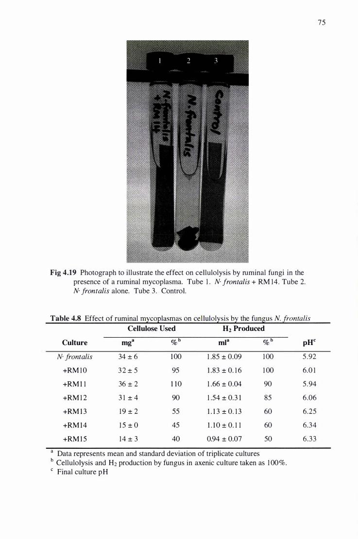
Fig 4.19 Photograph to illustrate the effect on cellulolysis by rumina! fungi in the
presence of a rumina! mycoplasma. Tube 1 . N. frontalis + RM 14. Tube 2.
N. frontalis alone. Tube 3 . Control.
Table 4.8 Effect of rumina! mycoplasmas on cellulolysis by the fungus N. frontalis
Cellulose Used H2 Produced
Culture mga % b mla % b pHc
N. frontalis 34 ± 6 1 00 1 .85 ± 0.09 1 00 5 .92
+RM 1 0 3 2 ± 5 95 1 .83 ± 0. 1 6 1 00 6.0 1
+RM 1 1 36 ± 2 1 10 1 .66 ± 0.04 90 5 .94
+RM 1 2 3 1 ± 4 90 1 .54 ± 0.3 1 85 6.06
+RM 1 3 19 ± 2 55 1 . 1 3 ± 0. 1 3 60 6.25
+RM 14 1 5 ± 0 45 1 . 1 0 ± 0. 1 1 60 6.34
+RM 1 5 14 ± 3 40 0.94 ± 0.07 50 6.33
a Data represents mean and standard deviation of triplicate cultures
b Cellulolysis and H2 production by fungus in axenic culture taken as 1 00%.
c Final culture pH
75

76
Not all of the isolates inhibited cellulolysis by N. frontalis (Table 4.8). Isolates RM l O,
RM l l and RM 1 2 did not inhibit N. frontalis to any extent, while isolates RM 1 3 , RM 1 4
and RM 1 5 inhibited the fungus between 45% and 60% based on cellulose degradation
data. The final culture pH also reflected the degree to which each fungus was inhibited
by the rumina! mycoplasmas. Fungal H2 production was also reduced when fungal
growth was inhibited.
Table 4.9 Effect of rumina! mycoplasmas on cellulolysis by the fungus P. communis
Cellulose Used H2 Produced
Culture mga % b ml3 % b pHc
P. communis 50 ± 3 1 00 0.6 1 ± 0.07 1 00 5 .89
+RM 10 45 ± 3 90 0.64 ± 0.04 1 05 5 .82
+RM 1 1 49 ± 2 1 00 0.6 1 ± 0.07 1 00 5.76
+RM 1 2 52 ± 2 1 05 0.57 ± 0.04 90 5 .74
+RM 1 3 1 9 ± 0 40 0.47 ± 0.0 1 80 6.25
+RM 14 1 5 ± 4 30 0.56 ± 0.06 90 6.35
+RM 1 5 1 4 ± 2 30 0.49 ± 0.09 80 6.38
a Data represents mean and standard deviation of triplicate cultures
b Cellulolysis and H2 production by fungus in axenic culture taken as 1 00%.
c Final culture pH
The degree to which rumina! mycoplamas inhibited cellulolysis by P. communis was
variable (Table 4.9). Isolates RM 1 0, RM 1 1 and RM 1 2 did not inhibit P. communis to
any extent, while isolates RM 1 3 , RM 14 and RM 1 5 inhibited the fungus between 60%
and 70% based on cellulose degradation data. The final culture pH also reflected the
degree to which each fungus was inhibited by the rumina! mycoplasmas. The nature of
the inhibiting mechanism is not known, but it is interesting to note that the isolates
which inhibited cellulolysis by N. frontalis and P. communis the most, were those which
did not lyse the cell walls of Gram-negative bacteria.

77
Table 4.10 Effect of rumina! m�coElasmas on cellulol�sis b� the fungus C. communis
Cellulose Used H2 Produced
Culture mga %b ml3 % b pHc
C. communis 22 ± 5 100 0.67 ± 0. 1 7 1 00 6 .35
+RM 1 0 1 3 ± 1 60 0.53 ± 0. 1 3 80 6.6 1
+RM 1 1 1 1 ± 2 50 0.52 ± 0.03 80 6 .55
+RM 1 2 8 ± 1 40 0.50 ± 0.06 75 6.62
+RM 1 3 7 ± 3 30 0.48 ± 0. 1 0 70 6.60
+RM 14 4 ± 1 20 0.57 ± 0. 1 0 85 6 .60
+RM 1 5 4 ± 1 20 0.45 ± 0.08 65 6 .62
a Data represents mean and standard deviation of tripl icate cultures b Cellulolysis and H2 production by fungus in axenic culture taken as 1 00%.
c Final culture pH
All of the rumina! mycoplasmas inhibited cellulolysis by C. communis B7 to varying
degrees (Table 4. 1 0). The final culture pH also reflected the degree to which each
fungus was inhibited by the ruminal mycoplasmas. All of the coculture pH values lie in
the range 6.55 to 6.62, while the final culture pH for C. communis in axenic culture had
a value of 6.35, indicating greater degradation of cellulose.
4.3.3 Effect of Rumina) Mycoplasmas on N. frontalis Growing on Cellobiose.
In cocultures where cel lobiose was the sole carbon source, the growth of N. frontalis
was stimulated by nearly al l of the rumina! mycoplasma isolates (Table 4. 1 1 ), in
contrast to the effect seen on cellulose, where fungal growth was often inhibited (Tables
4.8, 4.9 and 4. 1 0) . This suggested that the effect of the coculture was substrate
dependent. The increased H2 production and the lower pH seen in the test cultures, both
indicate N. frontalis was stimulated by the coculture. Isolates RM 1 3, RM 1 4 and RM 1 5
do not produce H2, and so would not contribute directly to the H2 produced in cocultures
with N. frontalis.

Table 4.1 1 Effect of ruminal mycoplasmas on the fungus N. frontalis growing on
cellobiose
H2 Produced
Culture mla % b
N. frontalis 1 .08 ± 0. 1 0 1 00
+RM l O 1 .23 ± 0. 1 5 1 1 5
+RM 1 1 1 .00 ± 0.07 90
+RM 1 2 1 .3 1 ± 0.24 1 20
+RM 1 3 1 .45 ± 0.34 1 30
+RM 14 1 .49 ± 0. 1 9 1 35
+RM 1 5 1 .5 1 ± 0.09 140
Control 0 0
a Data represents mean and standard deviation of triplicate cultures
b H2 production by fungus in axenic culture taken as 1 00%.
c Final culture pH
pHc
5.99
5.76
5 .8 1
5 .74
5 .77
5 .80
5 .8 1
6.79
78
The final coculture pH for N. frontalis and the rumina} mycoplasmas growing on
cellobiose, complements the H2 production data and suggests that rumina! mycoplasmas
stimulate the growth of the fungus when growing on cellobiose. Although isolates
RM 1 3, RM 14 and RM 1 5 did not produce H2, the stimulatory effect was sti ll apparent,
and indicated the increased H2 production seen in these cocultures was of solely fungal
origin.
4.3.4 SEM Studies of Ruminal Mycoplasma RM13 in Coculture with N. frontalis
SEM studies were carried out to examine the morphology of isolate RM 1 3 growing in
coculture with the rumina! fungus N. frontalis. The majority of the RM 1 3 cel ls were
found attached to the paper substrate but in some instances were also seen attached to
fungal rhizoids (Fig 4 .20). A variety of mycoplasma forms can be seen. There was no
evidence the mycoplasma was attacking the cellulose matrix to which it was attached.

Fig 4.20 Scanning electron micrograph of isolate RM 1 3 in coculture with the fungus
N. frontalis. RM 13 is attached even! y to the thallus tissue of the fungus
(arrowed) and to the paper support.
4.4 ENUMERATION OF BOVINE RUMINAL MYCOPLASMAS
79
The population densities of the sterol-requiring Anaeroplasma species and the non
sterol requ iring Asteroleplasma species in New Zealand cattle was unknown. Two
separate experiments carried out 14 days apart, determined the total population densities
of ruminal mycoplasmas and population densities of Asteroleplasma species present in
the rumens of 3 Friesian cows on the two days (Figs 4.2 1 to 4.24). The total population
densities of ruminal mycoplasmas found were in the ranges 6.7-8.6 x 1 07 gm· 1 (day 1 )
and 6.9-9.4 x 107 gm· 1 (day 14) . The population densities of Asteroleplasma species
found were in the ranges 5 .9-8.9 x 1 05 gm· 1 (day 1 ) and 6.9-9. 1 x 1 05 gm· 1 (day 1 4) .
Population densities were determined by colony counts after dilution of ruminal digesta
through CRFB medium containing benzylpenicillin for total population studies, and
benzylpenicillin with digitonin for enumeration of Asteroleplasma species. The latter
medium was developed in the course of this study. The total population densities from

80
NZ cattle compared well with data published in previous reports (Robinson and
Hungate, 1973 ; Robinson et al, 1975). There are no previous reports of population
studies on Asteroleplasma species in the rumen. Published reports of non-lytic
mycoplasma populations do not distinguish between Asteroleplasma species and
Anaeroplasma abactoclasticum, both of which constitute part of the total population of
non-lytic rumina} mycoplasmas. Robinson et al ( 1975) reported populations of non
lytic rumina} mycoplasmas of between 107 and 108 g- 1 of rumina} digesta.
90
-;- 80 OIJ "" 70 c -� 60 =
.s: 50 ..... � :; 40 c. 0 30 Q., ] 20 0 E- 1 0
0 A A B B c c
Cow
Fig 4.21 Total population density of rumina} mycoplasmas in 3 cows, per gram of
rumina} digesta.
90
-;- 80 OIJ
.. 70 c ,...; 60 � 1::1 50 E: "" 40 .s � - 30 � � 20 ""
� 1 0 0
A A B B c c
Cow
Fig 4.22 Population density of Asteroleplasma species in 3 cows, per gram of rumina!
digesta.

100
";' 90 Oil 80 ""
c ,...; � 70 c 60
_g .... 50 �
-; 40 c.
0 Q.. 30
] 20 0 E-<
10
0
A A B B c c
Cow
Fig 4.23 Total population density of rumina! mycoplasmas in 3 cows, per gram of
rumina! digesta, determined 1 4 days after those shown in Fig 4.2 1 .
1 00
90 -;-
0.0 80 .., c
70 -�
60 1:::1 ::: 50 ...,
.s 40 e--30 Q ... � - 20 ...,
� 10
0
A A 8 8 c c
Cow
8 1
Fig 4.24 Population density of Asteroleplasma species in 3 cows, per gram of rumina!
digesta, determined 1 4 days after those shown in Fig 4.22.
Because of the nature of the i solation process, rumina! fungi were often found
contaminating tubes of rumina! mycoplasmas at initial isolation. Thjs was generally
true in the lower dilution tubes ; the rumina! mycoplasmas being present in far greater
numbers than the fungi . It was particularly noticeable, that in the tubes used to
enumerate Asteroleplasma species, that rumina! mycoplasmas appeared to form a close
association with the fungal colony, while at more distant points in the culture tube, there

82
_l
were far fewer colonies of Asteroleplasma species. This observation suggests a degree
of dependence by the mycoplasma on the fungal colony, and suggests also that the
mycoplasma was cross-feeding on fungal metabolites. This observation is in keeping
with the generally held belief that because of their small genome, mycoplasmas, both
rumina! and non-rumina!, rely on their host for many essential nutrients (Razin, 1 992).

5 CONCLUSIONS
83
A review of the literature revealed less than ten papers exclusively dedicated to the
study of rumina! mycoplasmas since the first report published in 1 966. A few studies
have also been published on their serology, enzymology and molecular biology in order
to establish their phylogeny, but only as part of wider-ranging studies on the class
Mollicutes. The most recent paper on their physiology was published in 1 975. From the
available literature it must be concluded that rumina! mycoplasmas are a poorly studied
group within the rumen microbial ecosystem about which little is known.
From enumeration studies of rumina! mycoplasmas in New Zealand cattle it was found
the population density was 107- 1 08 g- 1 of rumina! digesta. This is in agreement with
data published on overseas cattle studies and leads to the conclusion that New Zealand
cattle are not unusual with respect to their populations of this group of rumina!
microorganism. Because mycoplasmas constitute about 1 o/o of the total bacterial
population of the rumen it is concluded that their impact on the rumen fermentation
must be considerable, not only in terms of inhibition of cellulolysis, but also their
contribution to the rumen microbial protein pool.
The results presented here indicate that the NZ isolates can pass through 0.45 11m fi lters,
have the same range of growth substrates, the same end products and antibiotic
sensitivities, an ability to either lyse or not lyse Gram-negative bacteria, and have a
requirement for sterol in growth media. It is concluded that the isolates in this study are
similar to rumina! mycoplasmas previously isolated from ruminants overseas and are
therefore not unique. Overseas isolates had lower optimum growth temperatures . All
NZ isolates belonged to the genus Anaeroplasma and each had a unique PAGE protein
banding profile, which suggested they belonged to two distinct groups. The optimum
pH growth data and RAPD profiles supported this view. There were three isolates in
each group. Phenotypic evidence identified three of the six isolates as strains of
84
Anaeroplasma abactoclasticum having all of the characteristics of rumina!
mycoplasmas, not able to lyse Gram-negative bacteria.
The results presented in this thesis indicate that the second group of three isolates were
very similar to the first, except they were able to lyse the cell walls of Gram-negative
bacteria, were proteolytic and produced hydrogen. From these observations we
conclude that they are also members of the genus Anaeroplasma, but of unknown
species.
In studies investigating cocultures of rumina} mycoplasmas with cellulolytic bacteria it
is concluded that mycoplasmas inhibit cellulolysis by some genera of bacteria, but
appear to stimulate others. The inhibition of the bacteria does not appear to result from
cell lysis since the only bacteria inhibited by rumina! mycoplasmas in this study
belonged to two species of the genus Ruminococcus which are Gram-positive. Again
there is a paradox, in that cultures of the Gram-negative bacterium Fibrobacter
succinogenes, instead of being lysed and hence inhibited by the coculture with rumina}
mycoplasmas, appeared to be slightly stimulated. From these observations we must
conclude that the mechanisms of inhibition are unknown.
From studies investigating cocultures of ruminal mycoplasmas with ruminal fungi it is
concluded that, while some mycoplasmas inhibited cellulolysis by fungi of the
Neocallimastix and Piromyces genera, all inhibited cellulolysis by fungi of the
Caecomyces genus. There was evidence to show that rumina! mycoplasmas did not kill
the fungi because, although inhibited when growing on cellulose, the mycoplasma
enhanced fungal growth on cellobiose. From these observations we conclude that the
mechanisms of inhibition are unknown, but that they may be substrate dependent. Of
the two groups of rumina! mycoplasma in this study, those most inhibitory to rumina!
fungi were Anaeroplasma abactoclasticum.
The morphology of the rumina! mycoplasmas, as demonstrated by light microscopy,
electron microscopy of thin sections of cells and scanning electron rnicroscopy of whole
cells, leads to the conclusion that while these microorganisms are unique in many
respects, morphologically they are similar to many mycoplasmas from other mollicute

85
genera. Because of these morphological similarities, it is not possible to identify
ruminal mycoplasmas on the basis of any unique morphological features which they
possess.
The use of the RAPD technique to demonstrate polymorphisms in rumina! mycoplasma
DNA showed that isolates which gave identical banding patterns with one primer, gave
identical but different patterns with another. DNA from unrelated isolates gave different
banding patterns when tested against the same primer. The study therefore showed that
short primers of arbitrary nucleotide sequence may be used to reproducibly amplify
segments of genomic DNA from ruminal mycoplasmas. The RAPD method was
therefore able to demonstrate relatedness among the isolates.
The isolation of cellular proteins and their subsequent separation on polyacrylamide gels
showed how powerful this technique can be in it' s ability to provide relatedness data for
the isolates. The clear demonstration of the presence of two groups (RM l O, RM 1 1 and
RM 12) and (RM 1 3, RM 14 and RM 1 5) among the isolates also gave relatedness
information which was confirmed by growth study data, including the ability of one
group to lyse the cell wall of Gram-negative bacteria. The method went further in
disclosing subtle differences among the isolates within the two groups. From the RAPD
profile it was apparent that isolate RM 13 was different from all other isolates, but
physiologically similar in many respects to isolates RM 14 and RM 1 5 . This relatedness
was also demonstrated in the protein gel, with RM 1 3 being obviously closely related to
RM 14 and RM 1 5, but the presence of the very high molecular weight band in RM 1 3
indicated an important difference between these isolates. Likewise, evidence for R M 10
being slightly different from RM 1 1 and RM 1 2 is shown by careful examination o f the
protein profile, but was more obviously seen from the RAPD banding patterns.
The demonstration of the lysis of Gram-negative ruminal bacteria by three of the
isolates suggests that in vivo these ruminal mycoplasmas may play a significant role in
modifying the rumen fermentation. This would occur in a number of ways, but
principally by inhibiting cellulolysis by at least three genera of ruminal fungi and one
genus of ruminal bacteria. In addition, one of the two groups of ruminal mycoplasma
were proteolytic and may play a role in the hydrolysis of protein in the rumen. The
86
conclusion that this suggests is, that in the absence of ruminal mycoplasmas the rumen
would be more efficient both in the rate and extent of cellulose digestion and in the
proportion of dietary protein available to the host. Like mycoplasmas from many
ecosystems, rumina! mycoplasmas are parasitic, not on the host animal directly but on a
number of the bacteria and fungi which populate their rumens and which are involved in
forage digestion.
A lack of sensitivity to penicillin was a characteristic which aided the isolation of the
first rumina! mycoplasma in 1966. Classified in 1 973 as belonging to the genus
Acholeplasma, it was soon appreciated that as a strict anaerobe the isolate did not
conform to the description of this genus. A new genus was proposed in 1975 to
encompass the original sterol-requiring isolate, while a second genus was proposed for
newly isolated sterol non-requiring rumina! mycoplasmas. A lack of sensitivity to
penicillin (and streptomycin sulphate) were characteristics which later facilitated the
isolation of rumina! fungi from the rumina! digesta of sheep and cattle. Occasionally,
unusual non-fungal colonies were observed in primary cultures, sometimes forming
satellite rings of growth around the fungal colony. Careful examination of the colonies
and cells, revealed they belonged to an unusual type of bacterium; the presence of
characteristic "fried-egg" colonies suggesting they were mycoplasmas. Colonies were
isolated and purified and shown to be strictly anaerobic, which further suggested they
belonged to the two previously described genera found only in the rumen microbial
ecosystem. From this we must conclude that had ruminal mycoplasmas not been
resistant to penicillin and streptomycin sulphate and not eo-isolated with rumina! fungi,
it is unlikely they would have been observed and isolated in New Zealand and therefore
would have not available to form the basis of this study.

6 APPENDIX: MICROORGANISMS USED IN THIS STUDY
6.1 Bacteria
• Anaeroplasma spp.
• Butyrivibrio fibrisolvens H 1 7C
• Clostridium chartatabidum DSM 5482
• Escherichia coli XL 1
• Fibrobacter succinogenes S85
• Lachnospira multiparus A TCC 19207
• Megasphaera elsdenii T8 1
• Methanobrevibacter smithii
• Prevotella ruminicola ruminicola ATCC 1 9 1 89
• Ruminococcus albus RA 7
• Ruminococcus flavefaciens FD 1
• Selenomonas ruminantium ruminantium HD4
• Streptococcus bovis JB 1
• Succinovibrio dextrinosolvens 24
6.2 Ruminal Fungi
• Caecomyces communis strain B7
• Neocallimastixfrontalis strain Rold
• Piromyces communis strain B 1 9
87

7 BIBLIOGRAPHY
88
Akin, D . E . and R . Benner ( 1 988). "Degradation of polysaccharides and l ignin by
ruminal bacteria and fungi ." Applied and Environmental Microbiology 54(5): 1 1 1 7-
1 1 25 .
B arr, D. J. S . , H. Kudo, K. D. Jakober and K.-J. Cheng ( 1989). "Morphology and
development of rumen fungi: Neocallimastix sp., Piromyces communis and
Orpinomyces bovis gen. nov., sp. nov .. " Canadian Journal of Botany 67: 28 1 5-2824.
Bauchop, T. ( 1979). "The rumen anaerobic fungi: colonizers of plant fibre." Annales de
Recherches Veterinaires 10(2-3): 246-248 .
Bauchop, T. ( 1 980). "Scanning electron microscopy in the study of microbial digestion
of plant fragments in the gut." In Contemporary Microbial Ecology, ed. D. C. Ellwood,
J. N. Hedger, M. J. Latham, et al . . Academic Press, London, pp. 305-326.
Bauchop, T. ( 1 989). "Biology of gut anaerobic fungi." BioSystems 23( 1 ) : 53-64.
Binder, A. and H. Kirchhoff ( 1 988). "Isolation of anaerobic Mollicutes from the
intestine of swine." Veterinary Microbiology 17: 1 5 1 - 1 58 .
Boatman, E . S . ( 1979). "Morphology and ultrastructure of the Mycoplasmatales." In The
Mycoplasmas, ed. M. F. Barile and S. Razin. Academic Press, New York, 1 : 63- 1 02.
Bradford, M. M. ( 1 976). "A rapid and sensitive method for the quantitation of
rnicrogram quantities of protein utilizing the principle of protein-dye binding."
Analytical Biochemistry 72: 248-254.

89
Braune, R. ( 19 1 3). "Untersuchungen uber die in Wiederkauermagen vorkommenden
protozoen." Arch. Protistenkunde 32: 1 1 1 - 1 70.
Breton, A., A. Bemalier and F. Bonnemoy ( 1989). "Morphological and metabolic
characterization of a new species of strictly anaerobic rumen fungus: Neocallimastix
joyonii." FEMS Microbiology Letters 58: 309-3 14.
Breton, A. , A. Bemalier, M. Dusser and G. Fonty ( 1 990). "Anaeromyces mucronatus
nov. gen. , nov. sp .. A new strictly anaerobic rumen fungus with polycentric thallus."
FEMS Microbiology Letters 70: 177- 1 82 .
Breton, A. , M. Dusser and B. Gaillard-Martinie ( 1 99 1 ) . "Piromyces rhizinflata, a new
species: a strictly anaerobic fungus from faeces of the Saharian ass." FEMS
Microbiology Letters 82: 1 -8 .
Caldwell, D . R. and M. P . Bryant ( 1 966). "Medium without rumen fluid for
nonselective enumeration and isolation of rumen bacteria." Applied Microbiology 14(5) :
794-80 1 .
Chesson, A . and C . W . Forsberg ( 1 988). "Polysaccharide degradation by rumen
microorganisms." In The Rumen Microbial Ecosystem, ed. P. N. Hobson. Elsevier
Applied Science, London and New York, pp. 25 1 -284.
Clarke, R. T. J. and M. E. Di Menna ( 1 96 1 ). "Yeasts from the bovine rumen." Journal
of General Microbiology 25: 1 1 3- 1 1 7 .
Clarke, R. T. J . ( 1968). "The microbiology of "pre-gastric" fermentation." The
Australian Journal of Science 31(4): 1 4 1 - 1 46.
Cole, B. C. and C. L. Atkins ( 199 1 ). "The Mycoplasma arthritidis T-cell mitogen,
MAM: a model superantigen." Immunology Today 12(8): 27 1 -276.

90
Czajka, J., N. Bsat, M. Piani, W. Russ, K. Sultana, M. Wiedmann, R. Whitaker and C .
A . Batt ( 1 993). "Differentiation of Listeria monocytogenes and Listeria innocua b y 1 6s
rRNA genes and intraspecies discrimination of Listeria monocytogenes strains by
random amplified polymorphic DNA polymorphisms." Applied and Environmental
Microbiology 59: 304-308.
Czerkawski, J. W. and K.-J . Cheng ( 1 988). "Compartmentation in the rumen." In The
Rumen Microbial Ecosystem, ed. P. N. Hobson. Elsevier Applied Science, London and
New York, pp. 361 -385.
Davies, D. R. , M. K. Theodorou, M. I. G. Lawrence and A. P. J. Trinci ( 1 993).
" Distribution of anaerobic fungi in the digestive tract of cattle and their survival in
faeces." Journal of General Microbiology 139: 1 395- 1 400.
Dehority, A. B. ( 1 99 1 ) . "Cellulose degradation in ruminants." In Biosynthesis and
Biodegradation of Cellulose, ed. C. H. Haigler and P. J. Weimer. Marcel Dekker, New
York, pp. 327-354.
Demeyer, D. I. ( 1 98 1 ) . "Rumen microbes and digestion of plant cell walls." Agricultural
Environment 6: 295-337.
Dybvig, K. and L. L. Voelker ( 1 996). "Molecular biology of Mycoplasmas." Annual
Review of Microbiology 50: 25-57.
Eadie, J. M. ( 1967). "Studies on the ecology of certain rumen ciliate protozoa." Journal
of General Microbiology 49: 1 75- 1 94.
Elias, P., J. Goerke, D. S . Friend and B. E . Brown ( 1 978) . "Freeze-fracture identification
of sterol-digitonin complexes in cell and liposome membranes." Journal of Cell Biology
78: 577-596.
9 1
Elliot, R . P. ( 1963). "Temperature gradient incubator for detennining the temperature
range for growth of microorganisms." Journal of Bacteriology 85: 889-898.
Fonty, G., P. Gouet, J-P. Jouany and J. Senaud ( 1 987). "Establishment of microflora and
anaerobic fungi in the rumens of lambs." Journal of General Microbiology 133: 1 835-
1 843.
Fonty, G. and K. N. Joblin ( 1 99 1 ) . "Rumen anaerobic fungi : their role and interactions
w ith rumen microorganisms in relation to fibre digestion." In Physiological Aspects of
Digestion and Metabolism in Ruminants, ed._T. Tsuda, Y. Sasaki and R. Kawashima.
Academic Press, New York, pp. 655-680.
Gardiner, S. E., H. C. M. Bassett, D. A. M. Noiton, V. G. Bus, M. E. Hofstee, A. G.
White, R. D. Ball , R. L. S. Forster and E. H. A. Rikkerink ( 1996). "A detailed linkage
map around an apple scab resistance gene demonstrates that two disease resistance
classes both carry the V _[f] Gene." Theoretical and Applied Genetics 93(4) : 485-493.
Gold, J. J . , I. B . Heath and T. Bauchop ( 1 988). "Ultrastructural description of a new
chytrid genus of caecum anaerobe, Caecomyces equi gen. nov., sp. nov., assigned to the
Neocallimasticaceae." BioSystems 21 : 403-4 1 5.
Grenet, E. and P. Barry ( 1 988). "Colonization of thick-walled plant tissues by anaerobic
fungi." Animal Feed Science and Technology 19( 1 -2) : 25-3 1 .
Gundersen, D . E. , I.-M. Lee, S . A. Rehner, R. E. Davis and D. T . Kingsbury ( 1 994).
"Phylogeny of Mycoplasmalike organisms (Phytoplasmas): a basis for their
classification." Journal of Bacteriology 176: 5244-5254.
Heath, I . B. , T. Bauchop and R. A. Skipp ( 1 983). "Assignment of the rumen anaerobe
Neocallimastixfrontalis to the Spizellomycetales (Chytridiomycetes) on the basis of it's
polyflagellate zoospore ultrastructure." Canadian Journal of Botany 6 1 : 295-307.

92
Ho, Y. W., N. Abdullah and S. Jalaludin ( 1 988). "Penetrating structures of anaerobic
rumen fungi in cattle and swamp buffalo." Journal of General Microbiology 134: 1 77-
18 1 .
Ho, Y. W., T. Bauchop, N. Abdullah and S . Jalaludin ( 1990). "Ruminomyces elegans
gen . et sp. nov. , a polycentric anaerobic rumen fungus from cattle." Mycotaxon 38: 397-
405 .
Ho, Y. W., N. Abdullah and S . Jalaludin ( 1 994). "Orpinomyces intercalaris, a new
species of polycentric anaerobic rumen fungus from cattle." Mycotaxon 50: 1 39- 1 50.
Hobson, P. N. and C. S. Stewart ( 1997). ed. The Rumen Microbial Ecosystem, Second
Edition. B lackie Academic and Professional, London.
Hobson, P. N. ( 1997). "Introduction." In The Rumen Microbial Ecosystem, Second
Edition, ed. P. N. Hobson and C. S. Stewart. Blackie Academic and Professional,
London.
Hucker, G. J. ( 1 922). "Comparison of various methods of Gram staining." Abstracts of
Bacteriology 6: 2.
Hungate, R. E. ( 1 960). "Symposium: Selected topics in microbial ecology
1 . Microbial ecology of the rumen." Bacteriological Reviews 24(4) : 353-364.
Hungate, R. E. ( 1 966). The Rumen and its Microbes. Academic Press, New York and
London.
Hungate, R. E. ( 1969). "A roll tube method for cultivation of strict anaerobes." In
Methods in Microbiology, Vol. 3B, ed. J. R. Norris and D. W. Ribbons. Academic
Press, London & New York, pp. 1 17- 1 32 .

93
Hungate, R. E. ( 1 975). "The rumen microbial ecosystem." Annual Review of Ecology
and Systematics 6: 39-66.
Hungate, R. E. ( 1988). "Introduction: The ruminant and the rumen." In The Rumen
Microbial Ecosystem, ed. P. N. Hobson. Elsevier Applied Science, London and New
York, pp. 1 - 1 9.
International Committee on Systematic Bacteriology Subcommittee on the Taxonomy of
Mollicutes ( 1 995). "Revised minimum standards for description of new species of the
class Mollicutes (division Tenericutes)." International Journal of Systematic
Bacteriology 45(3) : 605-6 12 .
Joblin, K . N . ( 198 1 ) . "Isolation, enumeration, and maintenance of rumen anaerobic
fungi in roll tubes." Applied and Environmental Microbiology 42(6): 1 1 19- 1 1 22.
Joblin, K. N., G. E. Naylor and A. G. Williams ( 1990). "Effect of Methanobrevibacter
smithii on xylanolytic activity of anaerobic ruminal fungi ." Applied and Environmental
Microbiology 56(8) : 2287-2295.
Joblin, K. N. and G. E. Naylor ( 1 994). "Effect of Butyrivibrio fibrisolvens on gut
anaerobic fungi." Proceedings of the Society for Nutritional Physiology 3: 1 7 1 .
Klieve, A . V . and T. Bauchop ( 1 988). "Morphological diversity of ruminal
bacteriophages from sheep and cattle." Applied and Environmental Microbiology 54(6):
1 637- 1 64 1 .
Klieve, A. V. ( 1 99 1 ) . "Bacteriophages from the forestomachs of Australian Marsupials."
Applied and Environmental Microbiology 57( 1 2): 3660-3663 .
Kopecny, J . , B . Hodrova and C . S . Stewart ( 1996) "The effect of rumen chitinolytic
bacteria on cellulolytic anaerobic fungi ." Letters in Applied Microbiology 23(3) : 1 99-
202.

94
Kudo, H., K. D. Jak:ober, R. C. Phillippe, K-J. Cheng, D. J. S . Barr and J. W. Costerton
( 1 990). "Isolation and characterization of cellulolytic anaerobic fungi and associated
mycoplasmas from the rumen of a steer fed a roughage diet." Canadian Journal of
Microbiology 36: 5 1 3-5 1 7 .
Kuske, C . R . and B . C . Kirkpatrick ( 1 992) "Phylogenetic relationships between western
aster yellows mycoplasmalike organism and other prokaryotes established by 1 6S rRNA
gene sequence." International Journal of Systematic Bacteriology 42(2) 226-233.
Laernmli, U. K. ( 1 970). "Cleavage of structural proteins during assembly of the head of
Bacteriophage T4." Nature 227: 680-685 .
Langworthy, T . A. , W. R . Mayberry, P. F. Smith and I. M . Robinson ( 1 975).
"Plasmalogen composition of Anaeroplasma." Journal of Bacteriology 122(2): 785-787.
Leedle, J. A. , M. P. Bryant and R. B. Hespell ( 1982). "Diurnal variations in bacterial
numbers and fluid parameters in rumina! contents of animals fed low- or high-forage
diets." Applied and Environmental Microbiology 44: 402-4 1 2.
Li , J . , I. B . Heath and T. Bauchop ( 1 990). "Piromyces mae and Piromyces dumbonica,
two new species of uniflagellate anaerobic Chytridiomycete fungi from the hindgut of
the horse and elephant." Canadian Journal of Botany 68: 1 02 1 - 1 033.
Lund, A. ( 1 974). "Yeasts and moulds in the bovine rumen." Journal of General
Microbiology 81: 453-462.
Lund, A. ( 1 980). "Yeasts in the rumen contents of Musk Oxen." Journal of General
Microbiology 121 : 273-276.
Mandelstam, J . and K. McQuillen, eds. ( 1 973). Biochemistry of Bacterial Growth.
Blackwell Scientific Publications, Oxford.

95
Manulis, S., L. Valinsky, A. Lichter and D. W. Gabriel ( 1 994). "Sensitive and specific
detection of Xanthomonas campestris pv. pelargonii with DNA primers and probes
identified by random amplified polymorphic DNA analysis." Applied and
Environmental Microbiology 60( 1 1 ): 4094-4099.
Mehling, A., U. F. Wehmeier and W. Piepersberg ( 1 995). "Application of random
amplified polymorphic DNA (RAPD) assays in identifying regions of actinomycete
genomes." FEMS Microbiology Letters 128: 1 19- 1 26.
Mims, C. A., J. H. L. Playfair, I. M. Roitt, D. Wakelin and R. Williams ( 1 993). Medical
Microbiology. Mosby-Year Book Europe Ltd, St. Louis.
Mountfort, D. 0. and C. G. Orpin, ed. ( 1 994). Anaerobic Fungi: Biology, Ecology and
Function. Marcel Dekker, New York.
Munn, E. A., C. G. Orpin and C. A. Greenwood ( 1988). "The ultrastructure and possible
relationships of four obligate anaerobic Chytridiomycete fungi from the rumen of
sheep." BioSystems 22: 67-82.
Orpin, C. G. ( 1 975). "Studies on the rumen flagellate Neocallimastixfrontalis." Journal
of General Microbiology 91 : 249-262.
Orpin, C. G. ( 1 976). "Studies on the rumen flagellate Sphaeromonas communis."
Journal of General Microbiology 94: 270-280.
Orpin, C. G. ( 1 977a) "Invasion of plant tissue by the flagellate Neocallimastix
frontalis." Journal of General Microbiology 98: 423-430.
Orpin, C. G. ( 1 977b) "The rumen flagellate Piromonas communis: it's life-history and
invasion of plant material in the rumen." Journal of General Microbiology 99: 107 - 1 1 7 .

96
Orpin, C. G. and E. A. Munn ( 1986). "Neocallimastix partriciarum sp.nov. , a new
member of the Neocallimasticaceae inhabiting the rumen of sheep." Transactions of the
British Mycological Society 86( 1 ): 1 78- 1 8 1 .
Orpin, C . G. and K. N . Joblin ( 1 997). "The rumen anaerobic fungi ." In The Rumen
Microbial Ecosystem, Second Edition, ed. P. N. Hobson and C. S. Stewart. Blackie
Academic and Professional, London, pp. 140- 195 .
Pettersson, B . , M. Uhlen and K-E. Johansson ( 1 996). "Phylogeny of some mycoplasmas
from ruminants based on 1 6S rRNA sequences and definition of a new cluster within
the Hominis Group." International Journal of Systematic Bacteriology 46(4): 1093-
1 098.
Petzel, J . P. , M. C. McElwain, D. DeSantis, J . Manolukas, M . V. Williams, P. A.
Hartman, M. J. Allison and J. D. Pollack ( 1 990). "Enzymic activities of carbohydrate,
purine, and pyrimidine metabolism in the Anaeroplasmataceae." In Recent Advances in
Mycoplasmology. Gustav Fischer Verlag, Stuttgart and New York, pp. 667-678 .
Rawlins, D. J. ( 1 992). Light Microscopy. BIOS Scientific Publishers, in Association
with the Biochemical Society, Oxford.
Razin, S . ( 1 978). "The Mycoplasmas." Microbiological Reviews 42(2): 4 14-470.
Razin, S. ( 1992). "The genera Mycoplasma, Ureaplasma, Acholeplasma,
Anaeroplasma, and Asteroleplasma." In The Prokaryotes, ed A. Balows, H. G. Truper,
M . Dworkin, W. Harder and K-H. Schleifer. Springer-Verlag, New York, pp. 1937-
1 959.
Razin, S . ( 1 992) . "Peculiar properties of mycoplasmas: the smallest -self replicating
prokaryotes." FEMS Microbiology Letters 100: 423-432.
97
Roberts, M. C. ( 1 990). "TetM in mycoplasmas of human origin." Zentralblatt fur
bakteriologie 20 (Supplement): 56-59.
Robinson, J . P. and R. E. Hungate ( 1973). "Acholeplasma bactoclasticum sp. n., an
anaerobic mycoplasma from the bovine rumen." International Journal of Systematic
Bacteriology 23(2) : 1 7 1 - 1 8 1 .
Robinson, I . M. , M . J . Allison and P. A. Hartman ( 1 975). "Anaeroplasma
abactoclasticum gen. nov. : sp.nov . : an obligately anaerobic mycoplasma from the
rumen." International Journal of Systematic Bacteriology 25(2) : 1 73- 1 8 1 .
Robinson, I. M. and M. J. Allison ( 1975). "Transfer of Acholeplasma bactoclasticum
Robinson and Hungate to the genus Anaeroplasma (Anaeroplasma bactoclasticum
[Robinson and Hungate] comb.nov. ) : emended description of the species." International
Journal of Systematic Bacteriology 25(2) : 1 82- 1 86.
Robinson, I. M. and K. R. Rhoades ( 1977). "Serological relationships between strains of
anaerobic mycoplasmas." International Journal of Systematic Bacteriology 27(3) : 200-
203 .
Robinson, I . M. and E. A . Freundt ( 1987). "Proposal for an amended classification of
anaerobic mollicutes." International Journal of Systematic Bacteriology 37( 1 ) : 78-8 1 .
Rodwell, A. W. ( 1 983). In Methods in Mycoplasmology. Vol. 1 , ed. S . Razin and J. G.
Tully. Academic Press, New York, pp 1 63- 1 72.
Rogers, M. J., J . S immons and R. T. Walker ( 1 985) . "Construction of the mycoplasma
evolutionary tree from 5S rRNA sequence data." Proceedings of the National Academy
of Sciences USA. 82: 1 160- 1 1 64.
Rose, C . S . and S . J. Pirt ( 1 98 1 ) . "Conversion of glucose to fatty acids and methane:
roles of two mycoplasmal agents." Journal of Bacteriology 147( 1 ) : 248-254.

98
Russell, J. B. ( 1985). "Fermentation of cellodextrins by cellulolytic and non-cellulolytic
rumen bacteria." Applied and Environmental Microbiology 49: 572-576.
Scheifinger, C. C. and M. J. Wolin ( 1 973). "Propionate formation from cellulose and
soluble sugars by combined cultures of Bacteroides succinogenes and Selenomonas
ruminantium." Applied Microbiology 26(5): 789-795 .
Seemuller, E., B . Schneider, R. Maurer, U. Ahrens, X. Daire, H. Kison, K-H. Lorenz, G.
Firrao, L. A vinent, B. B . Sears and E. Stackebrandt ( 1 994 ) . "Phylogenetic classification
of phytopathogenic mollicutes by sequence analysis of 1 6S ribosomal DNA."
International Journal of Systematic Bacteriology 44(3): 440-446.
Stephens, E. B . , I. M . Robinson and M. F. Barile ( 1 985). "Nucleic acid relationships
among the anaerobic mycoplasmas." Journal ofGeneral Microbiology 131 : 1 223- 1 227.
Stewart, C. S. and M. P. Bryant ( 1988). "The rumen bacteria." In The Rumen Microbial
Ecosystem, ed. P. N. Hobson. Elsevier Applied Science, London and New York, pp. 2 1 -
75 .
Stewart, C. S . , S . H. Duncan, A. J . Richardson, C. Backwell and R. Begbie ( 1 992) . "The
inhibition of fungal cellulolysis by cell-free preparations from ruminococci ." FEMS
Microbiology Letters 97: 83-88.
Swain, R. A., J. V. Nolan and A. V. Klieve ( 1 996). "Natural variability and diurnal
fluctuations within the bacteriophage population of the rumen." Applied and
Environmental Microbiology 62(3) : 994-997.
Tanaka, R. , A. Muto and S . Osawa ( 1989). "Nucleotide sequence of tryptophan tRNA
gene in Acholeplasma laidlawii." Nucleic Acids Research 17: 5842.

99
Theodorou, M. K., S. E. Lowe and A P. J. Trinci ( 1 992). "Anaerobic fungi and the
rumen ecosystem." In The Fungal Community: Its Organization and Role in the
Ecosystem, ed. G. C. Carroll and D. T. Wicklow. , Marcel Dekker, New York. pp. 43-72.
Toth, K. F., N. Harrison and B. B . Sears ( 1 994). "Phylogenetic relationships among
members of the class Mollicutes deduced from rps3 gene sequences." International
Journal of Systematic Bacteriology 44( 1 ): 1 1 9- 1 24.
Trinci, A P. J., S . E. Lowe, A. Milne and M. K. Theodorou ( 1 988). "Growth and
survival of rumen fungi." BioSystems 21 : 357-363 .
Truscott, R. B . ( 1 98 1 ) . "Nongenital mycoplasma infections m cattle." Canadian
Veterinary Journal 22: 335-338.
Ulyatt, M. J., D. W. Dellow, A. John, C. S. W. Reid and G. C . Waghom ( 1 985).
"Contribution of chewing during eating and rumination to the clearance of digesta from
the reticulo-rumen." In Control of Digestion and Metabolism in Ruminants, ed. L. P.
Milligan, W. L. Grovum and A Dobson. Prentice-Hall, Englewood Cliffs, New Jersey,
pp. 498-5 1 5 .
van Noort, J . M . , P . van den Berg and I . E . Mattem ( 199 1 ) . "Visualization of proteases
within a complex sample following their selective retention on immobilized bacitracin,
a peptide antibiotic ." Analytical Biochemistry 198: 385-390.
Vavra, J. and L. Joyon ( 1966). "Etude sur la morphologie, le cycle evolutif et la position
systematique de Callimastix cyclopsis Weissenberg 1 9 1 2." Protistologia 2: 5- 1 6.
Wallace, R. J. and K. N. Joblin ( 1 985). "Proteolytic activity of a rumen anaerobic
fungus." FEMS Microbiology Letters 29: 1 9-25.

1 00
Wallace, R . J . ( 1 992) . "Rumen microbiology, biotechnology and ruminant nutrition: The
application of research findings to a complex microbial ecosystem." FEMS
Microbiology Letters 100: 529-534.
Webb, J. and M. K. Theodorou ( 1 99 1 ). "Neocallimastix hurleyensis sp. nov., an
anaerobic fungus from the ovine rumen." Canadian Journal of Botany 69: 1 220- 1224.
Weisburg, W. G., J. G. Tully, D. L. Rose, J. P. Petzel , H. Oyaizu, D. Yang, L.
M andelco, J. Sechrest, T. G. Lawrence, J. van Etten, J. Maniloff and C. R. Woese
( 1 989). "A phylogenetic analysis of the Mycoplasmas: basis for their classification."
Journal of Bacteriology 171( 1 2) : 6455-6467.
Welsh, J. and M. McClelland ( 199 1 ). "Genomic fingerprinting using arbitrarily primed
PCR and a matrix of pairwise combinations of primers." Nucleic Acids Research
19( 19) : 5 275-5279.
Williams, A. G. and G. S. Coleman ( 1 988). "The rumen protozoa." In The Rumen
Microbial Ecosystem, ed P. N. Hobson. Elsevier Applied Science, London and New
York, pp. 77- 1 28.
Williams, A. G., S . E. Withers and K. N. Joblin ( 1 994). "The effect of cocultivation
with hydrogen-consuming bacteria on xylanolysis by Ruminococcus flavefaciens."
Current Microbiology 29(3): 1 33- 1 38 .
Williams, A. G., S . E. Withers, G. E. Naylor and K. N. Joblin ( 1 994) . "Effect of
heterotrophic rumina! bacteria on xylan metabolism by the anaerobic fungus Piromyces
communis." Letters in Applied Microbiology 19(2): 1 05- 109.
Williams, J. G. K., A. R. Kubelik, K. J . Livak, J . A. Rafalski and S. V. Tingey ( 1990).
"DNA polymorphisms amplified by arbitary primers are useful as genetic markers."
Nucleic Acids Research 18(22): 653 1 -6535).
1 0 1
Wilson, C . A . and T . M . Wood ( 1 992). "Studies on the cellulase o f the rumen anaerobic
fungus Neocallimastix frontalis with special reference to the capacity of the enzyme to
degrade crystalline cellulose." Enzyme and Microbial Technology 14: 258-264.
Woese, C. R. ( 1 987). "Bacterial evolution." Microbiological Reviews 51(2): 22 1 -27 1 .
Wolin, M. J. and T. L. Miller ( 1988). "Microbe - microbe interactions." In The Rumen
Microbial Ecosystem, ed. P. N. Hobson. Elsevier Applied Science, London and New
York, pp. 343-359.
Wood, T. M., C. A. Wilson, S. I. McCrae and K. N. Joblin ( 1 986). "A highly active
exocellular cellulase from the rumen fungus Neocallimastix frontalis." FEMS
Microbiology Letters 34:37-40.