the heart - sightsshefflerscience.weebly.com/uploads/2/2/8/7/22878422/lecture_18... · delivers...
TRANSCRIPT
1
The Heart

2
Overview
The right side receives oxygen-poor blood from the body and tissues and then pumps it to the lungs to pick up oxygen and dispel carbon dioxide
Its left side receives oxygenated blood returning from the lungs and pumps this blood throughout the body to supply oxygen and nutrients to the body tissues
The heart=a muscular double pump with 2 functions

3
simplified…
Cone shaped muscle
Four chambers
Two atria, two ventricles
Double pump – the ventricles
Two circulations
Systemic circuit: blood vessels that transport
blood to and from all the body tissues
Pulmonary circuit: blood vessels that carry
blood to and from the lungs
4
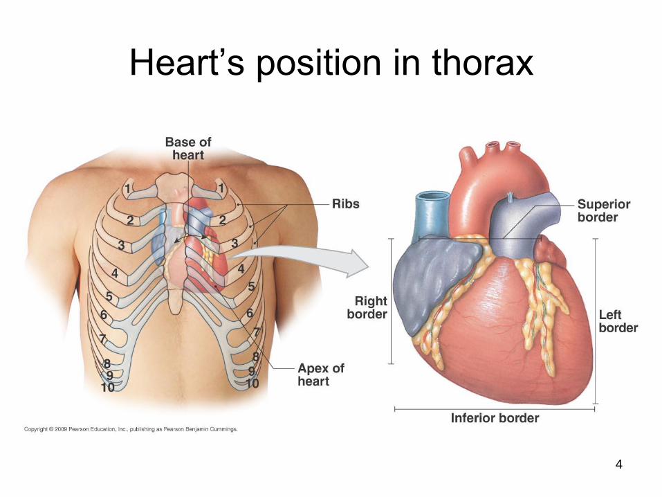
Heart’s position in thorax
5
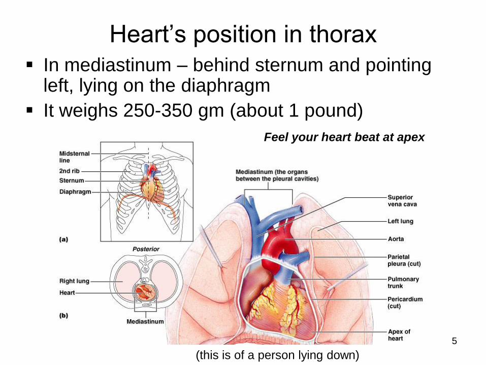
Heart’s position in thorax
In mediastinum – behind sternum and pointing left, lying on the diaphragm
It weighs 250-350 gm (about 1 pound)
Feel your heart beat at apex
(this is of a person lying down)
6
7
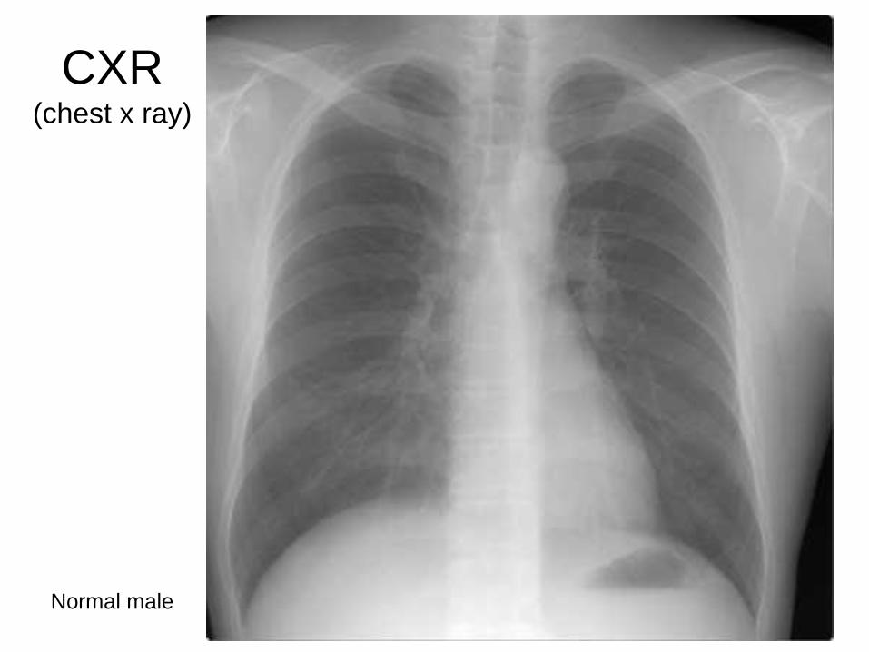
CXR(chest x ray)
Normal male
8
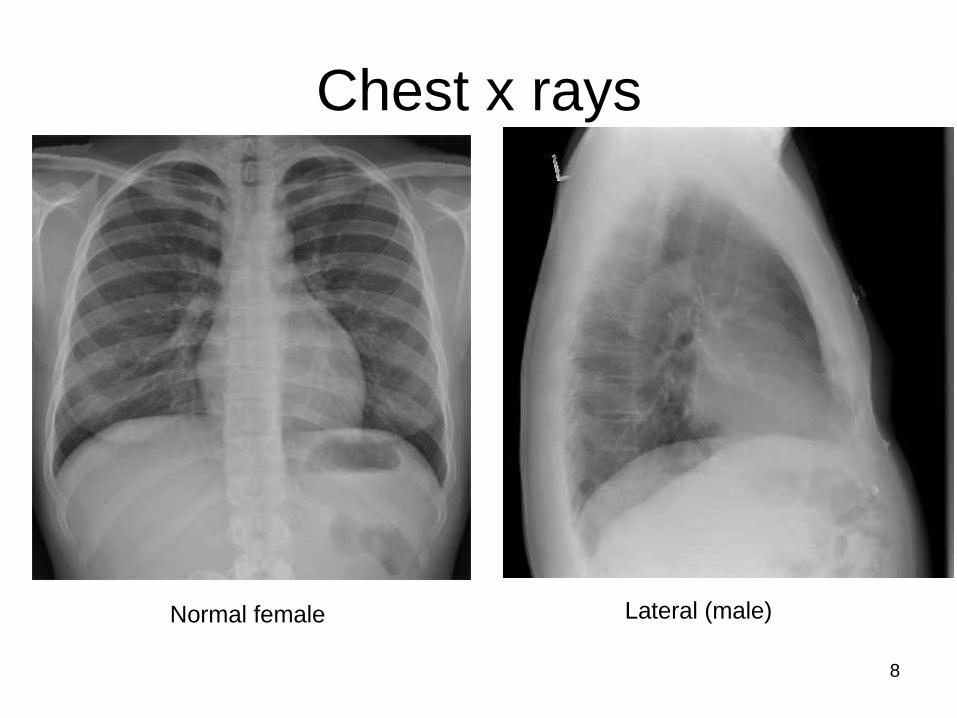
Chest x rays
Normal female Lateral (male)

9
Pericardium(see next slide)
Starting from the outside…
Without most of pericardial layers
10
Coverings of the heart: pericardium
Three layered:
(1) Fibrous pericardium
Serous pericardium of layers (2) & (3)
(2) Parietal layer of serous pericardium
(3) Visceral layer of serous pericardium =
epicardium: on heart and is part of its wall
(Between the layers is pericardial cavity)

11
How pericardium is formed around heart

12
Layers of the heart wall
Muscle of the heart with inner and outer
membrane coverings
Muscle of heart = “myocardium”
The layers from out to in:
Epicardium = visceral layer of serous
pericardium
Myocardium = the muscle
Endocardium lining the chambers

13
Layers of pericardium and heart wall

14
Chambers of the heartsides are labeled in reference to the patient facing you
Two atria
Right atrium
Left atrium
Two ventricles
Right ventricle
Left ventricle
--------------------------------------------------------------------------------
15
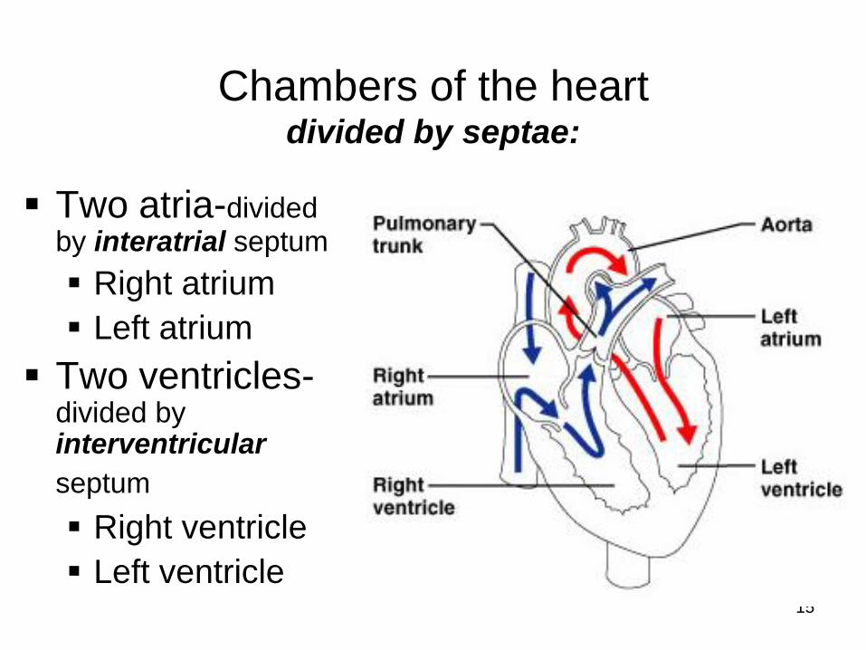
Chambers of the heartdivided by septae:
Two atria-divided by interatrial septum
Right atrium
Left atrium
Two ventricles-divided by interventricular
septum
Right ventricle
Left ventricle
16
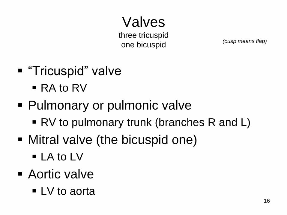
Valvesthree tricuspid
one bicuspid
“Tricuspid” valve
RA to RV
Pulmonary or pulmonic valve
RV to pulmonary trunk (branches R and L)
Mitral valve (the bicuspid one)
LA to LV
Aortic valve
LV to aorta
(cusp means flap)
17
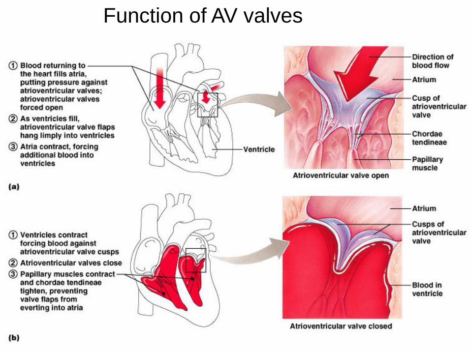
Function of AV valves

18
Function of semilunar valves
(Aortic and pulmonic valves)

19
Pattern of flow(simple to more detailed)
Body
RA
RV
Lungs
LA
LV
Boby
Body to right heart to lungs to
left heart to body
Body, then via vena cavas and
coronary sinus to RA, to RV, then to
lungs via pulmonary arteries, then to
LA via pulmonary veins, to LV, then to
body via aorta
From body via SVC, IVC & coronary
sinus to RA; then to RV through tricuspid
valve; to lungs through pulmonic valve
and via pulmonary arteries; to LA via
pulmonary veins; to LV through mitral
valve; to body via aortic valve then aorta
LEARN THIS
20
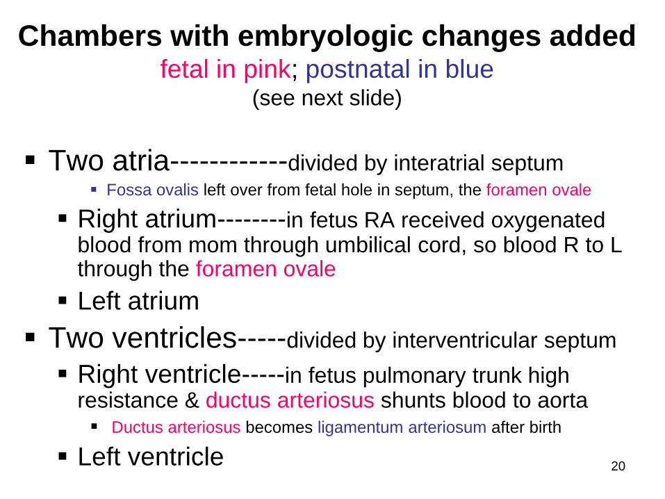
Chambers with embryologic changes addedfetal in pink; postnatal in blue
(see next slide)
Two atria------------divided by interatrial septum Fossa ovalis left over from fetal hole in septum, the foramen ovale
Right atrium--------in fetus RA received oxygenated blood from mom through umbilical cord, so blood R to L through the foramen ovale
Left atrium
Two ventricles-----divided by interventricular septum
Right ventricle-----in fetus pulmonary trunk high resistance & ductus arteriosus shunts blood to aorta Ductus arteriosus becomes ligamentum arteriosum after birth
Left ventricle

21
In the fetus, the RA received oxygenated blood from mom through umbilical cord, so blood R to L through the foramen ovale: fossa ovalis is left after it closes
The pulmonary trunk had high resistance (because lungs not functioning yet) & ductus arteriosusshunted blood to aorta; becomes ligamentum arteriosum after birth
22
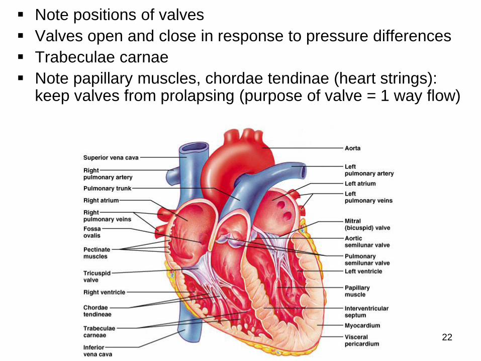
Note positions of valves
Valves open and close in response to pressure differences
Trabeculae carnae
Note papillary muscles, chordae tendinae (heart strings): keep valves from prolapsing (purpose of valve = 1 way flow)
23
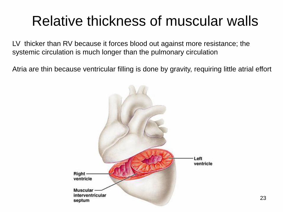
Relative thickness of muscular walls
LV thicker than RV because it forces blood out against more resistance; the
systemic circulation is much longer than the pulmonary circulation
Atria are thin because ventricular filling is done by gravity, requiring little atrial effort

24

25
more on valves

26
Simplified flow: print and fill in details
27
Heartbeat
Systole: contraction
Diastole: filling
Normal rate: 60-100
Slow: bradycardia
Fast: tachycardia
***Note: blood goes to RA, then RV, then lungs, then LA, then LV, then
body; but the fact that a given drop of blood passes through the heart
chambers sequentially does not mean that the four chambers contract in
that order; the 2 atria always contract together, followed by the
simultaneous contraction of the 2 ventricles
Definition: a single sequence of atrial contraction followed by ventricular contraction
See http://www.geocities.com/Athens/Forum/6100/1heart.html
28
Heart sounds
Called S1 and S2
S1 is the closing of AV (Mitral and Tricuspid) valves at the start of ventricular systole
S2 is the closing of the semilunar (Aortic and Pulmonic) valves at the end of ventricular systole Separation easy to hear on inspiration therefore S2
referred to as A2 and P2
Murmurs: the sound of flow Can be normal
Can be abnormal
29
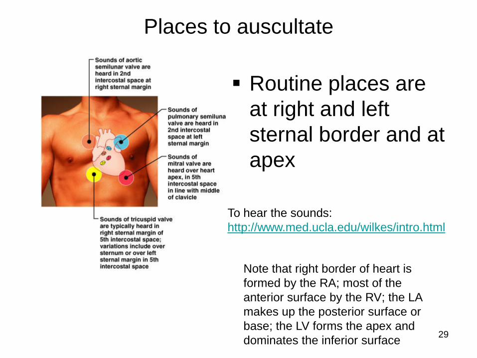
Places to auscultate
Routine places are
at right and left
sternal border and at
apex
To hear the sounds:
http://www.med.ucla.edu/wilkes/intro.html
Note that right border of heart is
formed by the RA; most of the
anterior surface by the RV; the LA
makes up the posterior surface or
base; the LV forms the apex and
dominates the inferior surface
30
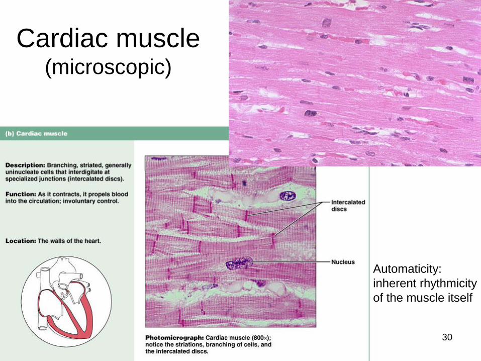
Cardiac muscle(microscopic)
Automaticity:
inherent rhythmicity
of the muscle itself

31
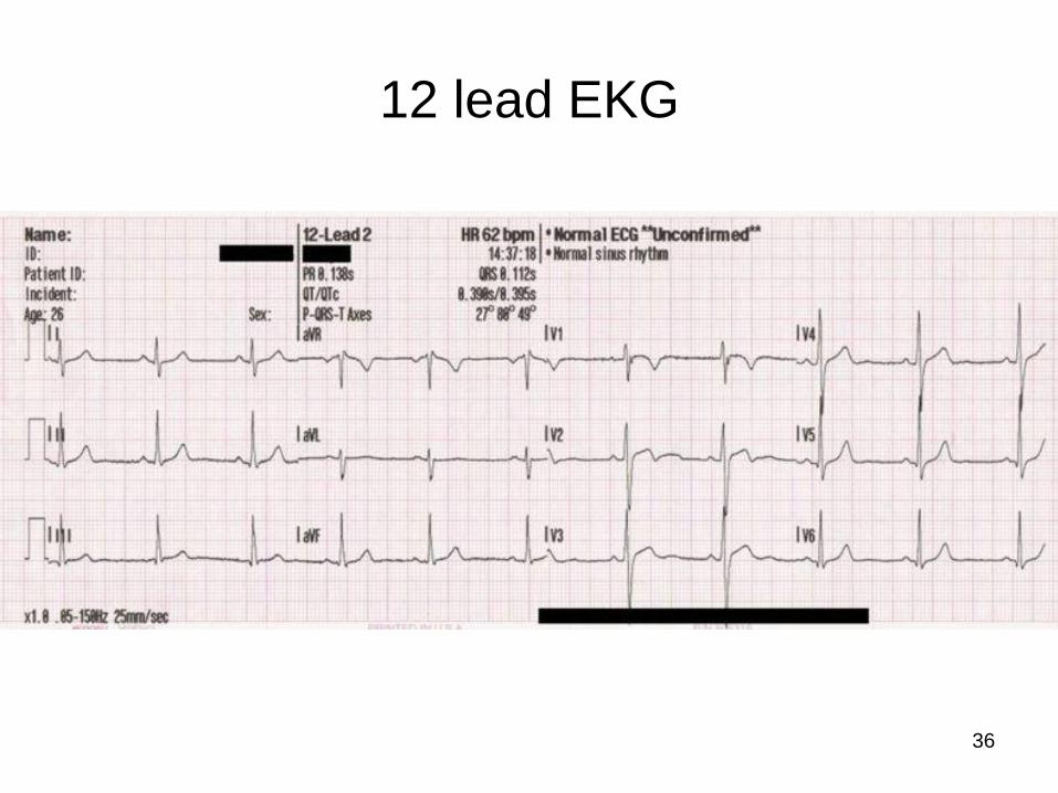
“EKG”
(or ECG, electrocardiogram)
Electrical
depolarization is
recorded on the body
surface by up to 12
leads
Pattern analyzed in
each lead
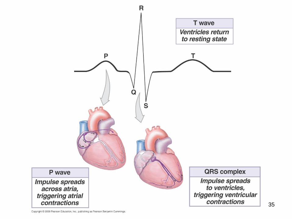
P wave=atrial depolarization
QRS=ventricular depolarization
T wave=ventricular repolarization

32
Electrical conduction system:
(Explanation in next slides)
specialized cardiac muscle cells that carry
impulses throughout the heart
musculature, signaling the chambers to
contract in the proper sequence

33
Conduction system
SA node (sinoatrial)
In wall of RA
Sets basic rate: 70-80
Is the normal pacemaker
Impulse from SA to atria
Impulse also to AV node via internodal pathway
AV node
In interatrial septum

34
Conduction continued
SA node through AV bundle (bundle of His)
Into interventricular septum
DividesR and L bundle branches
become subendocardial
branches (“Purkinje
fibers”)
Contraction begins
at apex
35

37
Artificial
Pacemaker
38
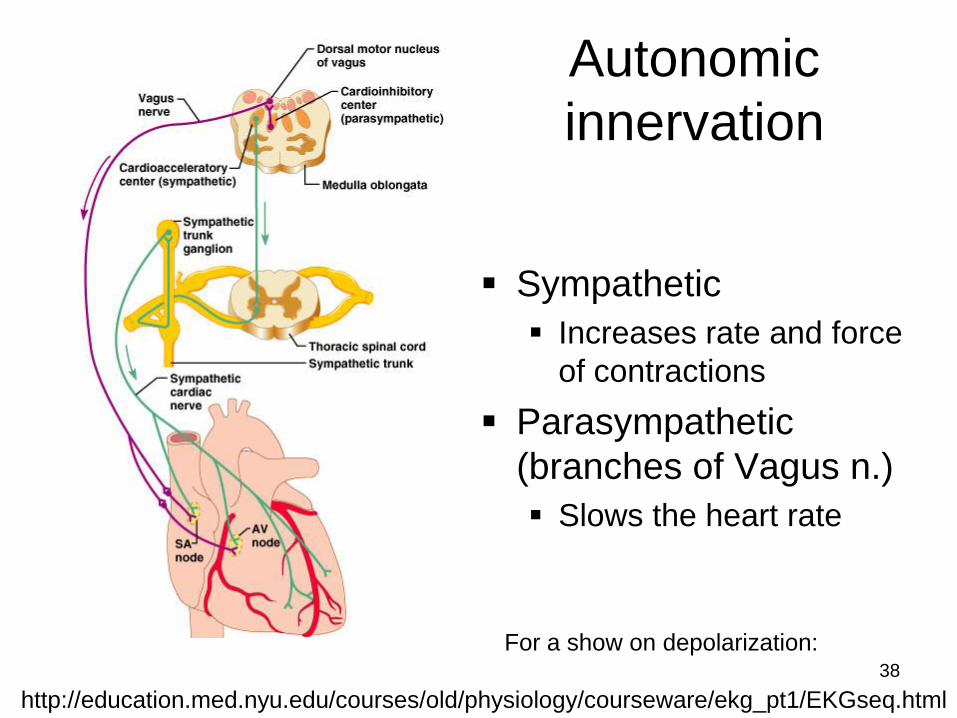
Autonomic
innervation
Sympathetic
Increases rate and force
of contractions
Parasympathetic
(branches of Vagus n.)
Slows the heart rate
http://education.med.nyu.edu/courses/old/physiology/courseware/ekg_pt1/EKGseq.html
For a show on depolarization:

39
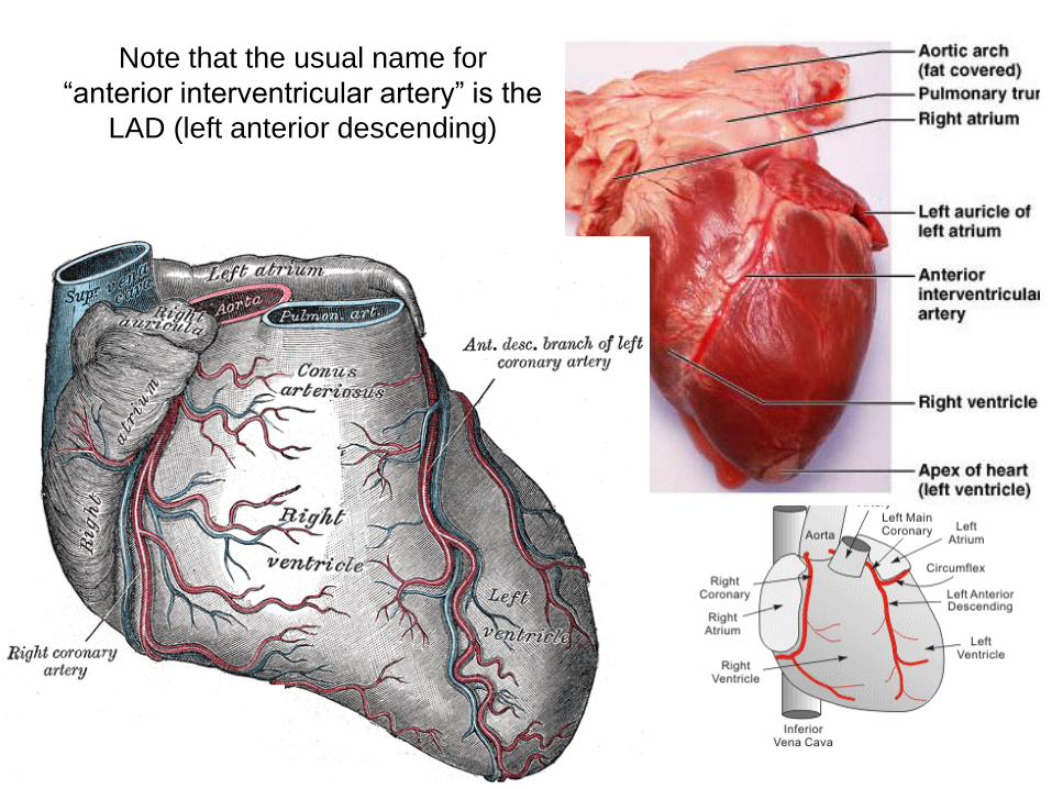
Blood supply to the heart(there’s a lot of variation)
A: Right Coronary Artery; B: Left Main Coronary Artery; C: Left Anterior Descending (LAD, or Left
Anterior Interventricular);
D: Left Circumflex Coronary Artery; G: Marginal Artery; H: Great Cardiac Vein; I: Coronary sinus,
Anterior Cardiac Veins.

40
Anterior viewL main coronary artery arises from the left side of the aorta
and has 2 branches: LAD and circumflex
R coronary artery emerges from right side of aorta
41
Note that the usual name for
“anterior interventricular artery” is the
LAD (left anterior descending)
42
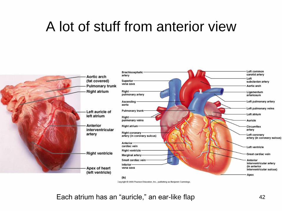
A lot of stuff from anterior view
Each atrium has an “auricle,” an ear-like flap

43
44
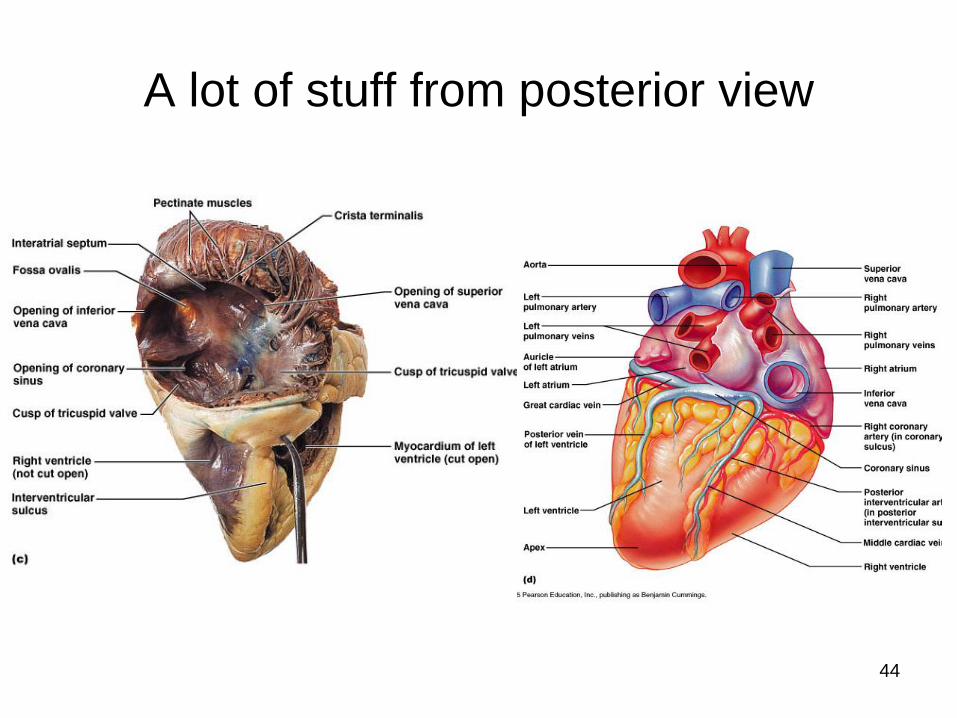
A lot of stuff from posterior view

45
Again posterior view Note: the coronary sinus (largest cardiac vein) –
delivers blood from heart wall to RA, along with SVC & IVC)

46
another flow chart

47
Embryological development during week 4 (helps to understand heart defects)
Day 22, (b) in diagram, heart starts pumping
(day 24)
(day 28)
(day 23)

48
Normal and
abnormal
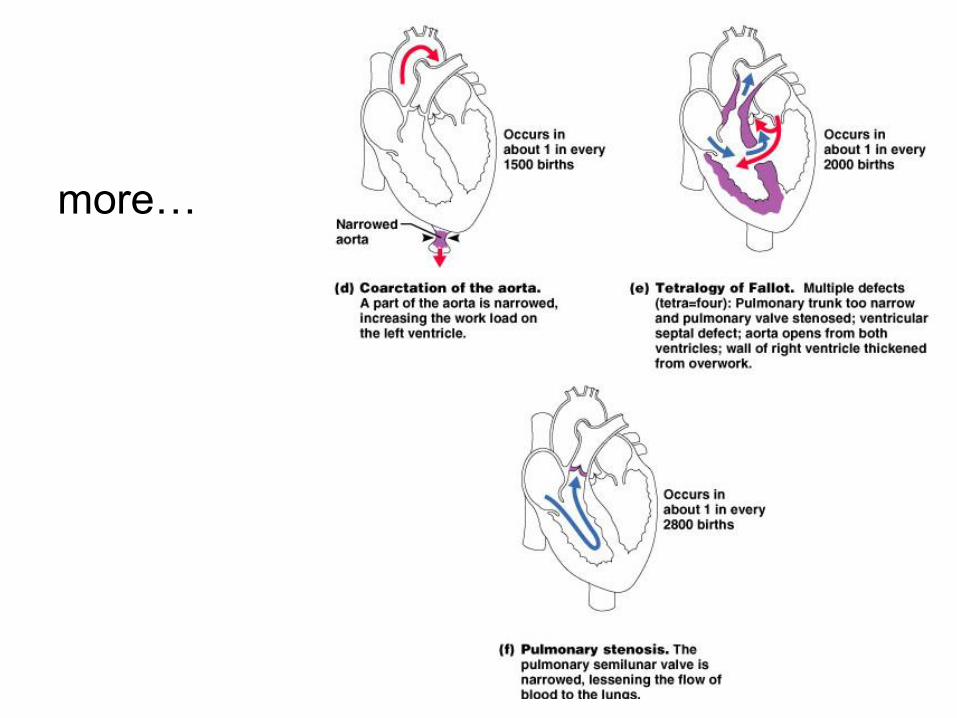
Congenital (means born with)
abnormalities account for nearly half
of all deaths from birth defects
One of every 150 newborns has some
congenital heart defect
49
more…

50
See Paul Wissman’s website: main
link; then Anatomy and Physiology
then Human heart: http://homepage.smc.edu/wissmann_paul/
http://homepage.smc.edu/wissmann_paul/anatomy1/
http://homepage.smc.edu/wissmann_paul/anatomy1/1he
art.html
Then from this site:
click-on from the following list of Human
Heart Anatomy Web Sites:
1) SMC pictures of the Human Heart:
http://homepage.smc.edu/wissmann_paul/heartpics/
3) Human Heart Anatomy
7) NOVA PBS animation of Heart Cycle:
http://www.geocities.com/Athens/Forum/6100/1heart.html
51
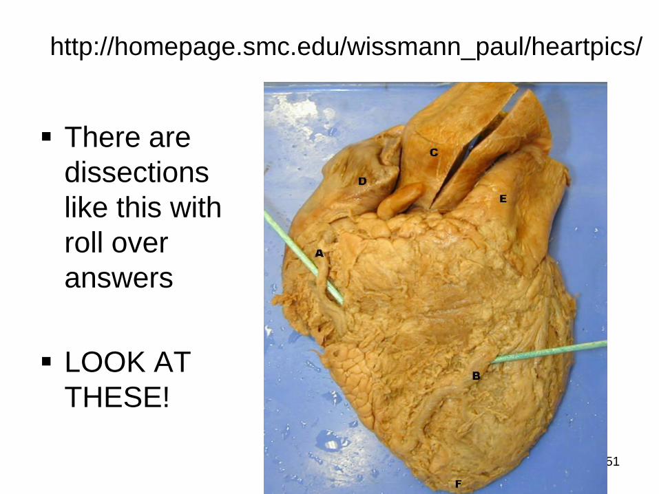
http://homepage.smc.edu/wissmann_paul/heartpics/
There are
dissections
like this with
roll over
answers
LOOK AT
THESE!
52
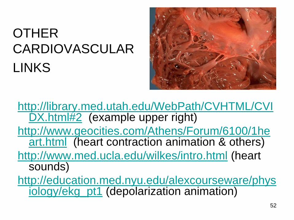
OTHER
CARDIOVASCULAR
LINKS
http://library.med.utah.edu/WebPath/CVHTML/CVIDX.html#2 (example upper right)
http://www.geocities.com/Athens/Forum/6100/1heart.html (heart contraction animation & others)
http://www.med.ucla.edu/wilkes/intro.html (heart sounds)
http://education.med.nyu.edu/alexcourseware/physiology/ekg_pt1 (depolarization animation)
53
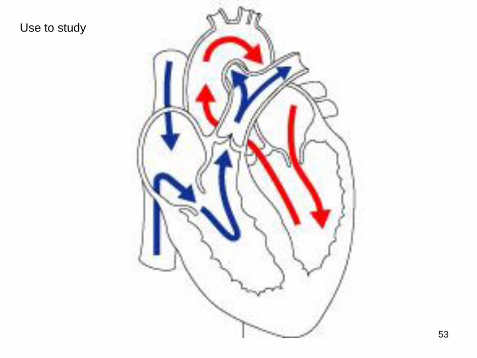
Use to study