embryological development of lachnanthes caroliniana
TRANSCRIPT

Embryological Development of Lachnanthes caroliniana (Haemodoraceae)Author(s): Michael G. SimpsonSource: American Journal of Botany, Vol. 75, No. 9 (Sep., 1988), pp. 1394-1408Published by: Botanical Society of AmericaStable URL: http://www.jstor.org/stable/2444463 .Accessed: 05/10/2011 17:31
Your use of the JSTOR archive indicates your acceptance of the Terms & Conditions of Use, available at .http://www.jstor.org/page/info/about/policies/terms.jsp
JSTOR is a not-for-profit service that helps scholars, researchers, and students discover, use, and build upon a wide range ofcontent in a trusted digital archive. We use information technology and tools to increase productivity and facilitate new formsof scholarship. For more information about JSTOR, please contact [email protected].
Botanical Society of America is collaborating with JSTOR to digitize, preserve and extend access to AmericanJournal of Botany.
http://www.jstor.org

Amer. J. Bot. 75(9): 1394-1408. 1988.
EMBRYOLOGICAL DEVELOPMENT OF LACHNANTHES CAROLINIANA (HAEMODORACEAE)1
MICHAEL G. SIMPSON
Department of Biology, San Diego, State University, San Diego, California 92182
AB3STRACT
Embryological development of Lachnanthes caroliniana was studied utilizing standard an- atomical techniques and SEM. Lachnanthes has a monocotyledonous anther wall development (endothecial cells with spiral secondary wall thickenings), successive microsporogenesis, and amoeboid (periplasmodial) tapetal development. Mature pollen grains are 2-nucleate with a proximal, fusiform generative cell. Ovules are initiated as 5-7 cylindrical primordia from a common placental base. Basal ovular swellings collectively contribute to the enlarged, peltate placenta. Mature ovules are pleurotropous, anatropous, bitegmic, and crassinucellate; the nu- cellus consists of a chalazal hypostase, radially elongate lateral cells, and a prominent micropylar nucellar cap. Megasporogenesis is successive, forming a linear tetrad of megaspores. Mega- gametogenesis is monosporic; the female gametophyte is of the Polygonum-type with relatively large, pyriform antipodals. Endosperm formation is helobial, resulting in the establishment of a ring of four thick-walled basal endosperm cells (the chalazal chamber) and numerous free nuclei (in the micropylar chamber). The mature cellular endosperm is filled with starch grains and has a chalazal cavity and a thick-walled peripheral layer. The discoid, peltately attached seeds have marginal wings derived by anticlinal divisions and buckling of the outer integument alone. Inner and middle cuticular layers are present in the seed coat. Lachnanthes is similar to all other investigated members of the Haemodoraceae in major embryological features. The significance of embryological evidence with regard to interfamilial classification is discussed. Future studies of ovule and seed development may prove valuable in phylogenetic studies in assessing the homology of placental, ovule, and seed morphology and anatomy.
EMBRYOLOGICAL STUDIES have provided char- acters of significant value in angiosperm sys- tematics (see Maheshwari, 1950, 1964; Davis, 1966; Johri, 1984). Yet our knowledge of em- bryological processes in numerous higher plant taxa consists of an extremely small data base. Many plant taxa of doubtful systematic affin- ities remain embryologically unknown, partic- ularly among the monocotyledons (Dahlgren and Clifford, 1982). Embryological studies may aid in our understanding of developmental processes and taxonomic relationships; they may also, in conjunction with phylogenetic analyses, provide a means for hypothesizing specific past evolutionary events and give in- sight into the possible adaptive significance of those events.
The Haemodoraceae (Bloodwort family), as recently circumscribed and described by Simp- son (in press a, b), are a monophyletic monocot family consisting of 14 genera and approxi- mately 70 species with distributions in eastern Australia, New Guinea, southern Africa,
I Received for publication 1 September 1987; revision accepted 6 January 1988.
This study was supported in part by National Science Foundation Grant DEB-8 109909. I thank J. M. Herr, Jr., and an anonymous reviewer for comments made on the manuscript.
northern South America, Central America and Mexico, Cuba, and eastern North America. The family is divided into two purported mono- phyletic tribes (sensu Simpson, in press b): tribe Haemodoreae (8 genera), with 3 (rarely 1) sta- mens per flower and monosulcate, verrucate (foveolate) pollen grains; and tribe Conostyl- ideae (6 genera), with 6 stamens per flower and porate, rugulate pollen grains.
Embryological studies have been conducted previously on only four species (in four of four- teen family genera): Anigozanthos flavidus (Stenar, 1927) of the tribe Conostylideae; and Dilatris pilansii (De Vos, 1956), Wachendorfia paniculata (Dellert, 1933; De Vos, 1956), and Xiphidium coeruleum (= X. album; Stenar, 1938) of the tribe Haemodoreae. As noted by De Vos (1956, 1961), these four genera have several similar embryological features: an amoeboid (plasmodial) tapetum, successive microsporogenesis, two integuments, crassi- nucellate ovules, a Polygonum-type female ga- metophyte, and helobial endosperm devel- opment (observed only in Dilatris and Wachendorfia). These embryological data have not contradicted the monophylesis of the fam- ily, supported more definitively from studies of chemistry (Cooke and Edwards, 1981) and pollen ultrastructure (Simpson, 1983). [Three
1394

September 1988] SIMPSON-EMBRYOLOGY OF LACHNANTHES 1395
TABLE 1. Embryological characters of the Haemodoraceae and relatives
Taxon Tapetal type Microspore division Nucellus type Documentation
Haemodoraceae Lachnanthes Amoeboid Successive Crassinucellate Present study Anigozanthos Amoeboid Successive Crassinucellate Stenar, 1927 Dilatris Amoeboid Successive Crassinucellate De Vos, 1956 Wachendorfia Amoeboid Successive Crassinucellate Dellert, 1933; De Vos, 1956 Xiphidium Amoeboid Successive Crassinucellate Stenar, 1938
Bromeliaceae Glandular Successive Crassinucellate Dahlgren and Clifford, 1982
Cyanastraceae Cyanastrum Glandular Simultaneous Crassinucellate Fries, 1919; Nietsch, 1941
Hypoxidaceae Hypoxis Glandular Successive Tenuinucellate De Vos, 1948 Pauridia Glandular Successive Tenuinucellate De Vos, 1949
Lanariaceae Lanaria Glandular Simultaneous Crassinucellate De Vos, 1961, 1963
Melanthiaceae (in part) Lophiola Glandular Successive Crassinucellate Simpson, 1981
Philydraceae Helmholtzia Glandular Successive Crassinucellate Hamann, 1966 Orthothylax Glandular Successive Crassinucellate Hamann, 1966 Philydrella Glandular Successive Crassinucellate Hamann, 1966 Philydrum Glandular Successive Crassinucellate Hamann, 1966
Pontederiaceae Eichhornia Amoeboid Successive Crassinucellate Banerji and Gangulee, 1937;
Schurhoff, 1922 Monochoria Amoeboid Successive Crassinucellate Banerji and Haldar, 1942
Sparganiaceae Sparganium Amoeboid Successive Crassinucellate Dahlgren and Clifford, 1982
Taccaceae Schizocapsa Glandular Simultaneous Crassinucellate HAkansson, 1921
Tecophilaeaceae Cyanella Glandular Simultaneous Crassinucellate De Vos, 1950 Odontostomum Glandular Simultaneous Crassinucellate Cave, 1952
Typhaceae Typha Amoeboid Successive Crassinucellate Dahlgren and Clifford, 1982
Velloziaceae Vellozia Glandular Successive Tenuinucellate Schnarf, 1931; Stenar, 1925
(Pseudocrassi- nucellate)
genera-Lanaria, Lophiola, and Pauridia- which have been placed in the Haemodoraceae in many recent classifications, differ from all investigated Haemodoraceae (sensu Simpson, in press b) in one or more major embryological features (Table 1). These embryological differ- ences, along with differences in anatomy- Schulze, 1893; Ambrose, 1980,1985; Simpson and Dickison, 1981-and palynology-Erdt- man, 1966; Simpson, 1983; Zavada, Xu, and Edwards, 1983-argue firmly for the place- ment of these three genera in other, more dis- tantly related families (see Simpson, in press b).]
Lachnanthes S. Elliott, the subject of this study, is a monotypic genus in the tribe Hae- modoreae consisting of the species L. carolin- iana (Lam.) Dandy [synonomous with L. car- oliana (Lam.) Dandy and L. tinctoria Elliott; see Robertson, 1976]. The genus is the only North American member of the Haemodor- aceae north of Mexico and occurs in Nova Scotia, the coastal plain of eastern and south- eastern U.S., and Cuba. Lachnanthes was first placed in the Haemodoraceae by Lindley (1830) and has remained in the tribe Haemodoreae (= Euhaemodoreae) since the definitive treat- ment of the family by Bentham and Hooker

1396 AMERICAN JOURNAL OF BOTANY [Vol. 75
pr en ppc spC pint
o ml
1~~ ~ ~ en 2 cp
t~~~P
e n
t~~~~~~~~~~~~~~~~~
8~~ ~ ~~~~~~~~~~~~~~~~~ nc ------
e a
ept 13s Sn
an
8 ~~~1 0
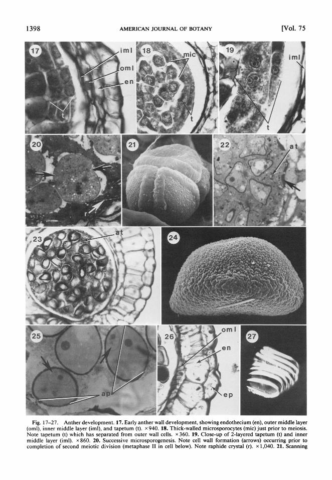
1 2 1 3 4 516 Fig. 1-16. Anther, ovule, and embryo development. 1. Portion of anther cross section, early developmental stage.
Note outer protoderm (pr) and subexterior primary parietal cell (ppc), the latter with some cells having divided periclinally into an endothecial layer (en) and secondary parietal cell layer (spc). x 260. 2. Later developmental stage. Note endothecial layer (en) and recent periclinal divisions of secondary parietal cell (spc) layer, forming the primary middle layer (pml) and primary tapetal layer (pt). x 260. 3. Later stage. Note primary middle layer (pml) and recent divisions of primary

September 1988] SIMPSON-EMBRYOLOGY OF LACHNANTHES 1397
(1883). Although no controversy exits is to the familial or tribal placement of Lachnanthes, its phylogenetic relationship to other members of the tribe Haemodoreae is somewhat ques- tionable; much of the uncertainty of its rela- tionship arises from ambiguity of the homol- ogy of ovule and seed morphology (Simpson, in press b). The purpose of this study is to provide a broader data base for assessing the interfamilial relationships of Lachnanthes and to initiate a comparative study of placental, ovule, and seed development in the family. Toward the latter objective, preliminary ob- servations of the ovule and placental mor- phology of Anigozanthos, Dilatris, and Hae- modorum of the Haemodoraceae are included in the present study.
MATERIALS AND METHODS-Flowering and fruiting material was dissected open, fixed in FAA for at least 48 hours, and stored in 70% ethanol. Whole anthers and placentae bearing ovules or seeds were removed and processed according to standard anatomical techniques (Johansen, 1940; Berlyn and Miksche, 1976). Following infiltration with tertiary butyl al- cohol, the material was paraffin-embedded with the aid of a dissecting microscope to facilitate orientation. Anthers were also dehydrated in an ethanol series and embedded in Spurr's res- in (Spurr, 1969). Anthers were mostly sec- tioned transversely (2-10,um); ovules and seeds were mostly sectioned along the sagittal plane at thicknesses of 7-20 ,um (depending on size and developmental stage). Sectioned material was stained either with iron hematoxylin/saf- ranin/fast green (Johansen, 1940) or with to- luidine blue. In addition, some ovules and an- ther wall components were cleared in Herr's solution (Herr, 1971) and viewed using differ- ential interference contrast (DIC) and phase contrast optics. Line drawings were made using a camera lucida attachment on a Wild bright- field microscope. Photographs were taken with
either a Leitz Wetzlar, Zeiss Photomicroscope, or Nikon Microphot-FX, using Panatomic-X film.
For scanning electron microscope (SEM) ob- servations, placentae containing ovules and seeds at various developmental stages were re- moved, dehydrated in an ethanol series, and infiltrated with Freon 1 13. The material, placed in a metal capsule, was critical-point dried us- ing CO2 as the transition fluid. Placentae were mounted on a stub using double-stick tape, sputter-coated with gold/palladium, and viewed and photographed with a JEOL T20 scanning electron microscope. Documentation for species examined is as follows:
Lachnanthes caroliniana (Lam.) Dandy-M. G. Simpson 14VI80A (DUKE) and M. G. Simpson 7VII84A (SDSU); Anigozanthos flavidus DC.-M. G. Simpson 24IX8 1J (DUKE); Dilatris pilansii Barker-P. V. D. Meriwe 30X8 1-2 (STEU); Haemodorum spicatum R. Br.-M. G. Simpson 16IX81C (DUKE).
RESULTS-Morphology-Lachnanthes car- oliniana has perfect and actinomorphic flow- ers, with a homochlamydeous imbricate peri- anth (of 3 outer and 3 inner tepals), 3 basifixed stamens (opposite the inner tepals), and an in- ferior, globose, slightly 3-lobed ovary. There are 3 carpels and locules. The mature placentae are quite thickened and peltiform, each bearing generally 5-7 ovules (see Fig. 37). Ovules are anatropous and are positioned pleurotropously in a ring along the margin of the peltate pla- centa (Fig. 5, 37, 40). The fruit is a globose, 3- lobed, loculicidal capsule. Seeds are reddish, discoid, convex/concave, minutely scabrate, and peltate in attachment with a central hilum on the concave surface (Fig. 44, 45). (See Simp- son and Dickison, 1981, for a description of vegetative anatomy, floral anatomy, and sto- mate ontogeny.)
tapetal layer, forming two layers of tapetal cells (t). x 260. 4. Later stage, preperiplasmodial. Note slight separation of tapetal layers (t) and recently divided cells of primary middle layer to form inner middle layer (iml) and outer middle layer (oml). x 260. 5. Ovary cross section, showing pleurotropous orientation of ovules (ov) on thickened placenta (p). [Xylem = black; phloem = white (within vascular bundles); transverse vasculature = stippled.] x 18. 6. Metaphase II of megasporocyte meiosis. Note successive wall formation. x 490. 7. Four linear megaspores. Note two degenerating micropylar megaspores (at right) and large chalazal megaspore (at left), indicative of monosporic megasporogenesis. x 490. 8. Two-nucleate stage of megagametogenesis. x 490. 9. Four-nucleate stage of megagametogenesis. x 490. 10. Whole mature ovule, sagittal section, illustrating female gametophyte (fg), hypostase (hy), radial cells (rc), nucellar cap (nc), inner integument (ii), outer integument (oi), and placental epithelium (ept). x 13 5. 1 1. Nucellar cap (nc) and mature female gametophyte, the latter with egg apparatus (ea), secondary nucleus (sn), and antipodal cells (an). x 270. 12. Zygote, showing nucleus in distal (chalazal) region of cell. (Micropyle is below in this and Fig. 13-16.) x 160. 13. Linear, 4-celled stage of embryo development. x 160. 14. Early anticlinal divisions of two terminal cells. Note two undivided micropylar suspensor cells. x 160. 15. Early globuiar stage. x 160. 16. Mature embryo. Note short suspensor (below) and relatively undifferentiated embryo body. x 160.
1398 AMERICAN JOURNAL OF BOTANY [Vol. 75
17 im 1 19
omi~~~~~~~~~~~m
en
t~~~~~~~~~~~
t
123 2
omi 25 27~~~~~~~
ep
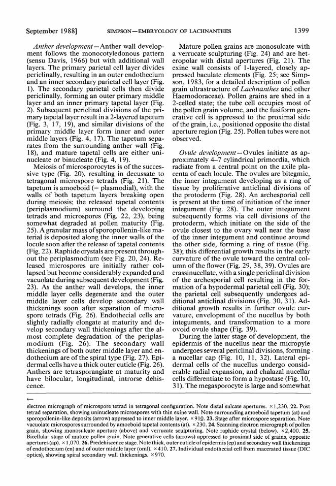
Fig. 17-27. Anther development. 17. Early anther wall development, showing endothecium (en), outer middle layer (oml), inner middle layer (iml), and tapetum (t). x 940. 18. Thick-walled microsporocytes (mic) just prior to meiosis. Note tapetum (t) which has separated from outer wall cells. x 360. 19. Close-up of 2-layered tapetum (t) and inner middle layer (iml). x 860. 20. Successive microsporogenesis. Note cell wall formation (arrows) occurring prior to completion of second meiotic division (metaphase II in cell below). Note raphide crystal (r). x 1,040. 21. Scanning
September 1988] SIMPSON-EMBRYOLOGY OF LACHNANTHES 1399
Anther development -Anther wall develop- ment follows the monocotyledonous pattern (sensu Davis, 1966) but with additional wall layers. The primary parietal cell layer divides periclinally, resulting in an outer endothecium and an inner secondary parietal cell layer (Fig. 1). The secondary parietal cells then divide periclinally, forming an outer primary middle layer and an inner primary tapetal layer (Fig. 2). Subsequent periclinal divisions of the pri- mary tapetal layer result in a 2-layered tapetum (Fig. 3, 17, 19), and similar divisions of the primary middle layer form inner and outer middle layers (Fig. 4, 17). The tapetum sepa- rates from the surrounding anther wall (Fig. 18), and mature tapetal cells are either uni- nucleate or binucleate (Fig. 4, 19).
Meiosis of microsporocytes is of the succes- sive type (Fig. 20), resulting in decussate to tetragonal microspore tetrads (Fig. 21). The tapetum is amoeboid (-plasmodial), with the walls of both tapetum layers breaking open during meiosis; the released tapetal contents (periplasmodium) surround the developing tetrads and microspores (Fig. 22, 23), being somewhat degraded at pollen maturity (Fig. 25). A granular mass of sporopollenin-like ma- terial is deposited along the inner walls of the locule soon after the release of tapetal contents (Fig. 22). Raphide crystals are present through- out the periplasmodium (see Fig. 20, 24). Re- leased microspores are initially rather col- lapsed but become considerably expanded and vacuolate during subsequent development (Fig. 23). As the anther wall develops, the inner middle layer cells degenerate and the outer middle layer cells develop secondary wall thickenings soon after separation of micro- spore tetrads (Fig. 26). Endothecial cells are slightly radially elongate at maturity and de- velop secondary wall thickenings after the al- most complete degradation of the periplas- modium (Fig. 26). The secondary wall thickenings of both outer middle layer and en- dothecium are of the spiral type (Fig. 27). Epi- dermal cells have a thick outer cuticle (Fig. 26). Anthers are tetrasporangiate at maturity and have bilocular, longitudinal, introrse dehis- cence.
Mature pollen grains are monosulcate with a verrucate sculpturing (Fig. 24) and are het- eropolar with distal apertures (Fig. 21). The exine wall consists of 1-layered, closely ap- pressed baculate elements (Fig. 25; see Simp- son, 1983, for a detailed description of pollen grain ultrastructure of Lachnanthes and other Haemodoraceae). Pollen grains are shed in a 2-celled state; the tube cell occupies most of the pollen grain volume, and the fusiform gen- erative cell is appressed to the proximal side of the grain, i.e., positioned opposite the distal aperture region (Fig. 25). Pollen tubes were not observed.
Ovule development-Ovules initiate as ap- proximately 4-7 cylindrical primordia, which radiate from a central point on the axile pla- centa of each locule. The ovules are bitegmic, the inner integument developing as a ring of tissue by proliferative anticlinal divisions of the protoderm (Fig. 28). An archesporial cell is present at the time of initiation of the inner integument (Fig. 28). The outer integument subsequently forms via cell divisions of the protoderm, which initiate on the side of the ovule closest to the ovary wall near the base of the inner integument and continue around the other side, forming a ring of tissue (Fig. 38); this differential growth results in the early curvature of the ovule toward the central col- umn of the flower (Fig. 29, 38, 39). Ovules are crassinucellate, with a single periclinal division of the archesporial cell resulting in the for- mation of a hypodermal parietal cell (Fig. 30); the parietal cell subsequently undergoes ad- ditional anticlinal divisions (Fig. 30, 31). Ad- ditional growth results in further ovule cur- vature, envelopment of the nucellus by both integuments, and transformation to a more ovoid ovule shape (Fig. 39).
During the latter stage of development, the epidermis of the nucellus near the micropyle undergoes several periclinal divisions, forming a nucellar cap (Fig. 10, 11, 32). Lateral epi- dermal cells of the nucellus undergo consid- erable radial expansion, and chalazal nucellar cells differentiate to form a hypostase (Fig. 10, 31). The megasporocyte is large and somewhat
electron micrograph of microspore tetrad in tetragonal configuration. Note distal sulcate apertures. x 1,230. 22. Post tetrad separation, showing uninucleate microspores with thin exine wall. Note surrounding amoeboid tapetum (at) and sporopollenin-like deposits (arrow) appressed to inner middle layer. x 919. 23. Stage after microspore separation. Note vacuolate microspores surrounded by amoeboid tapetal contents (at). x 230. 24. Scanning electron micrograph of pollen grain, showing monosulcate aperture (above) and verrucate sculpturing. Note raphide crystal (below). x 2,400. 25. Bicellular stage of mature pollen grain. Note generative cells (arrows) appressed to proximal side of grains, opposite apertures (ap). x 1,070. 26. Predehiscence stage. Note thick, outer cuticle of epidermis (ep) and secondary wall thickenings of endothecium (en) and of outer middle layer (oml). x 410. 27. Individual endothecial cell from macerated tissue (DIC optics), showing spiral secondary wall thickenings. x 970.

1400 AMERICAN JOURNAL OF BOTANY [Vol. 75
rectangular in shape (Fig. 32). Meiosis is suc- cessive, in which a cell wall develops between the dyad members after meiosis I (Fig. 6, 33). The second meiotic division (Fig. 6) results in a linear tetrad of megaspores (Fig. 34). Female gametophyte formation is monosporic by deg- radation of the three micropylar megaspores, only the chalazal megaspore persisting (Fig 7).
Placental growth initiates via ring-like tissue proliferations at the base of each ovule (Fig. 39). These individual placental swellings ul- timately become contiguous (Fig. 40), forming a large, peltate, hemispheric placental mass (Fig. 37, 40).
The mature ovule is somewhat globose in shape, with a prominent and discrete "neck" formed by the relative narrowing of the integ- uments in the micropylar region (Fig. 10, 41); lateral flanges of tissue are present near the base of this micropylar neck (Fig. 41). A conspic- uous swelling is present in the chalazal region (Fig. 10, 40, 41). Ovules are anatropous (sensu Bocquet, 1959) and bitegmic, with inner and outer integuments proximally composed of two cell layers, becoming 3- to 4-layered in the region of the micropylar neck (Fig. 10). The ovules are pleurotropously oriented (such that the micropyle is directed toward the central ovary column) around the circumference of the
peltate placenta (Fig. 5, 37, 40). The micropyle is amphistomal and "zig-zag," in that the open- ing of the outer integument is displaced nearer to the funiculus than is that of the inner in- tegument (Fig. 10). The surface of the placenta is papillate (Fig. 41); an epithelium occurs at the base of the funiculus near the micropyle (Fig. 10). Early megagametogenesis undergoes a 2-nucleate (Fig. 8) and 4-nucleate (Fig. 9) stage. The mature female gametophyte is ini- tially 7-celled and 8-nucleate (Fig. 35, 36). The two polar nuclei apparently fuse later, forming a secondary nucleus and resulting in a 7-nu- cleate condition (Fig. 1 1). The three antipodals are relatively large and pyriform in shape, with large, densely staining nuclei (Fig. 11, 36).
Embryogeny and seed development - Soon after fertilization the antipodals degenerate (Fig. 46, 47). During endosperm formation, the sin- gle (presumably fertilized) endosperm nucleus divides, initially forming another endosperm nucleus and a single ovoid cell (primary basal endosperm cell), immediately distal to the an- tipodal region (Fig. 47). Further divisions re- sult in the formation of a ring of four, wall- bound endosperm basal cells (as observed in cross section) and numerous free endosperm nuclei (Fig. 48). The four endosperm cells, in-
Fig. 28-37. Ovule development. (Fig. 28-34 are sagittal sections; Fig. 35, 36 are transverse sections.) 28. Immature ovule, showing archesporial cell (a), inner integument (ii), and outer integument (oi). x 510. 29. Later stage. Note inner integument (ii) and outer integument (oi). x 410. 30. Close-up of Fig. 29, showing megasporocyte (mec), parietal cell (pc), and protoderm (pr). x 990. 31. More mature ovule. Note megasporocyte (mec) and differentiation of nucellus into radially elongate cells (rc) and chalazal hypostase (hy). x 400. 32. First meiotic division of megasporocyte (mec). Note nucellar cap (nc). x 510. 33. Post meiosis I stage, showing megaspore dyad. x 1,300. 34. Post meiosis II stage, showing large chalazal megaspore (me) of linear tetrad. x 1,300. 35. Mature ovule, prefertilization, showing female gametophyte (fg). x 230. 36. Close-up of Fig. 35. Note three pyriform antipodal cells (an), two polar nuclei (pn), and egg apparatus (ea). x 380. 37. Mature placenta (p), bearing 5 ovules (locule wall removed). Pedicel below, style above. x 16.
Fig. 38-45. Scanning electron micrographs of ovule and seed development. 38. Immature ovule, showing inner integument (ii) and initiation of outer integument (oi). x 360. 39. Later stage, showing ring of five pleurotropously curved, ovoid ovules. Note ring-like placental thickenings (arrow) at base of each ovule. x 95. 40. Later developmental stage of placenta and ovules, showing coalesced thickenings of placental region (arrow). Note swollen chalazal region (cr) of ovule. x 50. 41. Close-up of ovule in Fig. 40. Note globose shape, peripheral flanges (f), micropylar neck (n), and papillate placental surface. x 96. 42. Early seed development, showing wings (arrows), initiating near micropylar neck. x 97. 43. Close-up of developing seed, showing point of buckling of outer integument (arrow). Note micropylar neck (n). x 50. 44. Detached seed, outer (distal) surface. Note micropylar region (m). x 22. 45. Detached seed, inner (proximal) surface. Note central hilum (hi) and micropylar region (m). x 22.
Fig. 46-53. Endosperm and seed development. 46. Postfertilization. Note egg apparatus (ea), degenerated antipodal cells (an), and hypostase (hy). x 110. 47. Endosperm nucleus (en) and endosperm cell (ec) near degenerated antipodal cells (an). x 390. 48. Later stage, showing two of four endosperm cells (ec) and one of many free endosperm nuclei (en). Note intact hypostase. x 210. 49. Later stage. Note ring of (four total) enlarged, chalazal endosperm cells (ec) and free endosperm nuclei (en) distal to degenerated hypostase (hy). x 520. 50. Early seed development, showing growth and buckling of outer integument (arrow), forming seed wing. Note cellular endosperm, including three (of four) enlarged chalazal endosperm cells. x 74. 51. Close-up of mature embryo, medial section, showing suspensor (s). Note degenerated endosperm cells immediately surrounding embryo. x 90. 52. Seed coat, near wing of seed. Note that wing is formed from buckling (arrow) of 2-layered outer integument. x 98. 53. Close-up of seed coat, showing inner cuticle (ic), 2- layered inner integument (ii), middle cuticle (mc), and 2-layered outer integument (oi). Note thin-walled endosperm cells, devoid of starch grains, immediately adjacent to inner cuticle. x 200.

September 1988] SIMPSON -EMBRYOLOGY OF LACHNANTHES 1401
'28
31 32
mc z
nrc
A 4. /~~~3
V ~~~~~~36 f 37
la ~ea ' 7 > e " e e _

1402 AMERICAN JOURNAL OF BOTANY [Vol. 75
38k
_-~~~~4 _ - _~~~~~~~~
40 41_
fi
iVR-'t''>
42: 43.z" .>_
_lFii~~~~~~~~~~~~~~~~~~~~~~~~~~~~~~~~~~~~~~~~~~~~~~~~~~~~~~~e
44 45E_**

September 1988] SIMPSON -EMBRYOLOGY OF LACHNANTHES 1403
Up -~~~~~~~~~~~~~~~~~

1404 AMERICAN JOURNAL OF BOTANY [Vol. 75
57
Ak
Fig. 54-58. Ovule morphology of other Haemodoraceae genera. 54. Dilatris pilansai, SEM. Note ovule neck (n) and surrounding, ring-like placenta (p). x 53. 55. Haemodorum spicatum, SEM, showing two ovules embedded in thickened placenta (p). Note ovule neck (n) and line of demarcation (arrow) between two placental halves. x 39. Fig. 56-58. Anigozanthos flavidus. 56. Entire placenta and ovules of carpel (pedicel below, style above), SEM. x 24. 57. Close-up of single ovule, SEM. Note swollen tissue of chalazal region (cr) and ovule neck (n). x 104. 58. Sagittal section of ovule, showing chalazal region (cr) proximal to nucellus. x 81.
cluding the nuclei, enlarge considerably, to- gether forming a globose mass of cells at the chalazal end of the ovule, accompanied by de- generation of the hypostase and radial cells of the nucellus and proliferation of the free en- dosperm nuclei in the micropylar chamber (Fig. 49). At about this time the globose, immature seed begins to develop marginal wings, which initiate near the micropylar neck (Fig. 42) but eventually encircle the seed margin (Fig. 43). The marginal wings develop from the lateral growth (presumably by anticlinal divisions) of the outer integument alone, which "buckles" at the point of contact with the inner integu- ment (Fig. 50, 52). At the time of marginal wing development, the endosperm is entirely cellular, the four original endosperm cells being
much larger than the other endosperm cells (Fig. 50). Growth in the ovule is concomitant with massive expansion of the placental tissue. Lateral growth in the body and margins of the ovule eventually obscures the micropylar neck (Fig. 43, 44, 45).
The zygote, just prior to embryogenesis, is medially constricted with the zygote nucleus positioned at the chalazal end of the cell (Fig. 12). Transverse divisions of the zygote result in a linear 4-celled stage (Fig. 13). The two distal cells subsequently divide anticlinally (Fig. 14) to form a terminal globose mass of cells (Fig. 15); the two proximal (micropylar) proembryo cells undergo only few subsequent cell divisions, forming a suspensor. At seed maturity the embryo is relatively undifferen-

September 1988] SIMPSON-EMBRYOLOGY OF LACHNANTHES 1405
tiated, obovoid, and micropylar in position, having a short, ill-defined suspensor (Fig. 16, 51).
The original enlarged ring of four endosperm cells degenerate in the mature seed, leaving an empty chalazal chamber. The nucellus also completely degenerates, and no perisperm is present (Fig. 51). Endosperm cells, which are isodiametric to slightly radially elongate, con- tain numerous starch grains (Fig. 52, 53). En- dosperm cells immediately surrounding the embryo are somewhat degenerated or have large intercellular spaces (Fig. 51); those immedi- ately adjacent to the seed coat have thick, non- lignified outer tangential cell walls and are de- void of starch grains (Fig. 52, 53). The seed coat is derived from both inner and outer in- teguments (Fig. 53). The inner seed coat is composed of two layers of thin-walled, non- lignified, tangentially elongate cells (Fig. 52, 5 3); the outer seed coat consists of thin-walled, tangentially elongate cells in two cell layers (Fig. 52, 53), except in the micropylar region ofthe wings where cells may be in several layers (Fig. 51). A thick (ca. 8 ,um) cuticle occurs on the proximal surface of the inner seed coat and a thin cuticle (ca. 3 Am) is secreted between the inner and outer seed coats (Fig. 52, 53).
The mature seeds are arranged edge to edge, completely covering the outer placental sur- face. Individual seeds are discoid, being convex on the outer surface (Fig. 44) and concave on the inner surface (Fig. 45); the seeds are pel- tately attached by means of a rudimentary fu- niculus (Fig. 45).
Ovule morphology of other family mem- bers-Dilatris pilansii has one globose ovule per carpel (Fig. 54) with a prominent micro- pylar neck and a surrounding ring-shaped pla- centa (Fig. 54). Haemodorum spicatum has two ovoid ovules per carpel (Fig. 55). The ovules have a narrow micropylar neck and are embed- ded in recesses of an enlarged placenta; a line of demarcation is evident dividing the placenta into two halves (Fig. 5 5). Anigozanthosflavidus has numerous ovules per carpel, arising from the surface of a thickened placenta (Fig. 56). Each ovule is ovoid, consisting of a short mi- cropylar neck, a nucellar region, and a swollen chalazal region (Fig. 57, 58).
DISCUSSION-Several major embryological similarities are apparent among Lachnanthes caroliniana and the four previously investi- gated members of the Haemodoraceae. All in- vestigated family members have an amoeboid tapetum and successive microsporogenesis (see Table 1). In Dilatris, Lachnanthes, and Wach-
endorfia the pollen grains are binucleate at the time of release, with the fusiform generative cell appressed to the proximal pollen grain wall (cf. Fig. 25). De Vos (1956), described the gen- erative cells in Dilatris and Wachendorfia as "distal" in position (see fig. 26, 45 of De Vos, 1956). However, based on a comparison of her illustrations with pollen grain shape and wall morphology (as revealed from the electron mi- croscopic studies of Simpson, 1983), it is ev- ident that they are actually proximal, i.e., op- posite the convex, monosulcate aperture. All investigated family members have crassinu- cellate, bitegmic ovules with monosporic (the chalazal megaspore functional) megasporogen- esis and a Polygonum-type (= "normal type") female gametophyte with relatively large, ob- pyriform antipodal cells. Additional features of similarity between Lachnanthes, Dilatris, and Wachendorfia are the development of 1) a nucellar cap, formed by divisions of the pa- rietal cell and micropylar epidermal cells; 2) distinctive lateral, radially elongate nucellar cells; and 3) helobial endosperm formation with a 4-celled whorl of basal endosperm cells (the "chalazal haustorium," sensu De Vos, 1956). Most of these embryological features were not investigated for either Anigozanthos (Stenar, 1927) or Xiphidium (Stenar, 1938); thus, their comparative significance for these genera is unknown.
The monocotyledonous anther wall devel- opmental pattern in Lachnanthes differs from the standard pattern in having a 2-layered ta- petum and both inner and outer middle layers; cells of the inner middle layer develop spiral secondary wall thickenings at maturity, which presumably function similarly to those of the endothecial cells during anther dehiscence. An- ther wall development is unknown in other family members and is, thus, of no compara- tive value. The endothecial thickenings are spi- ral (not girdling), similar to that described for Haemodorum (see Dahlgren and Clifford, 1982). The monocotyledonous anther wall di- vision reported here for Lachnanthes is found in the great majority of investigated mono- cotyledons (Davis, 1966; Dahlgren and Clif- ford, 1982), although the data base for this character is quite small (and reported for no other members of the Haemodoraceae; see Dahlgren and Clifford, 1982).
The ovule type in Lachnanthes is best de- scribed as anatropous (in the strict sense of Bocquet, 1959) in that the ovular vascular sup- ply is curved within a definable funiculus be- fore terminating at the chalazal end of the nu- cellus (see Fig. 10). The ovule type in Wachendorfia appears similarly anatropous,

1406 AMERICAN JOURNAL OF BOTANY [Vol. 75
although it is described as "hemitropous" by De Vos (1956). Ovules of Dilatris, Xiphidium, and Anigozanthos have been described as or- thotropous (Stenar, 1927, 1938; De Vos, 1956) by virtue of the vasculature traversing directly to the chalaza without curvature within a fu- niculus "fused" to the ovule body. However, Stenar (1927, 1938) reported that in Anigo- zanthos and Xiphidium the nucellar axis does not always coincide with the funicular axis. In addition, De Vos (1956) stated that in Dilatris the presence of continuous histological con- nection between the outer integument and the placenta may indicate that the orthotropous condition in this genus is derived from an an- cestral anatropous ovule type. In any case, the different ovule types reported in the family all seem to intergrade, differing only slightly in the degree of curvature of the vascular supply. The discrete micropylar neck of Lachnanthes ovules is closely appressed to the placenta, below which occurs a densely staining (and presumably met- abolically active) epithelium. This "neck" may, thus, function in pollen tube attraction or ori- entation. Most other species of the Haemo- doraceae have such an ovular "neck," although ovule shape is variable within the family, rang- ing from globose to lanceoloid (Simpson, in press b).
Lachnanthes has a helobial endosperm de- velopment, in which wall formation occurs af- ter the first division of the fertilized (presum- ably triploid) endosperm nucleus (see Fig. 47). The smaller wall-bound cell apparently divides in two sequences to form a whorl of four wall- bound cells (basal endosperm cells) adjacent to the now degenerated antipodals. De Vos (1956) described the basal endosperm cells of both Dilatris and Wachendorfia as "haustori- al" because they appear to function in the rapid degeneration of the nucellus in the chalazal region, forming a "chalazal chamber"; a sim- ilar developmental pattern occurs in Lach- nanthes. Free endosperm nuclei, confined to the micropylar chamber, continue to divide unaccompanied by cytokinesis. After mitosis is completed, cell-wall formation and copious starch grain accumulation occur throughout the endosperm.
An ovule of Lachnanthes undergoes a dra- matic change in form during seed develop- ment, transforming from a globose structure to a flattened, convex/concave, winged, pel- tately attached seed. The ovule "neck" dis- appears during early seed development, ap- parently both by degeneration and by overgrowth of the marginal ovule wings; (cf. Fig. 43, 44). The seed coat in Lachnanthes is
identical to that described for both Dilatris and Wachendorfia (De Vos, 1956); each integu- ment is composed oftwo cell layers at maturity, and has a cuticular layer between endosperm and inner integument and between the inner and outer integuments. Of all family members, only Dilatris and Haemodorum have flattened, winged seeds like those of Lachnanthes. The seed wing of both Dilatris and Haemodorum appears (from sections of mature seeds) to de- velop similarly to that of Lachnanthes, i.e., via anticlinal divisions and "buckling" ofthe outer integument (Simpson, personal observation). Almost all other members of the tribe Hae- modoreae have ovoid to globose seeds with a tomentum of trichomes. Seeds of the genus Pyrrorhiza, which are somewhat flattened with numerous marginal trichomes, may possibly represent a condition intermediate between the above two seed types in the tribe (Simpson, in press b). A study of seed development in other members of the family might prove quite in- teresting with regard to refining intrafamilial phylogenetic relationships.
The foregoing embryological studies provide several features of significance with regard to interfamilial systematics. Of the monocot fam- ilies presumed closely related to the Haemo- doraceae in recent systems of classification (e.g., Dahlgren and Clifford, 1982; Dahlgren and Rasmussen, 1983; Walker, 1986), it is inter- esting that members of the Pontederiaceae, Typhaceae, and Sparganiaceae are similar with regard to type of microsporogenesis, tapetal type, and ovule type (Table 1). Of these em- bryological features only an amoeboid tape- tum is likely an apomorphic feature (Dahlgren and Rasmussen, 1983). Thus, the occurrence of an amoeboid tapetum in these four families may argue for their constituting a monophy- letic group. In addition, Hamann (personal communication to Dahlgren and Rasmussen, 1983) reports that a "characteristic type of he- lobial endosperm ... where the chalazal cham- ber is cellular, small in relation to the micro- pylar chamber, and differs from this in contents
." is present in members of the Bromeli- aceae, Haemodoraceae, Philydraceae, Ponte- deriaceae, Sparganiaceae, Typhaceae, and Vel- loziaceae. Dahlgren and Rasmussen (1983) hypothesized that this characteristic helobial endosperm (e.g., as seen in Lachnanthes) is synapomorphic for these seven families, war- ranting their classification together in the su- perorder Bromeliiflorae. More comprehensive evidence will be needed to corroborate the monophyly of the Bromeliiflorae based on this single embryological character.

September 1988] SIMPSON-EMBRYOLOGY OF LACHNANTHES 1407
In conclusion, Lachnanthes is similar to all other investigated taxa of the Haemodoraceae in major embryological features. Although this certainly corroborates the placement of the ge- nus within the family, these studies have pro- vided little data that would elucidate its intra- familial classification. Of all family members, only Dilatris, Haemodorum, and Lachnanthes have similar discoid, winged seeds with ap- parently similar wing development (above). In addition, only Dilatris (see Fig. 54) has a strict- ly globose ovule similar to that of Lach- nanthes, perhaps supportive of the sister group status between the two genera (Simpson, in press b). Despite these evident similarities in form, the intrafamilial phylogeny of the Hae- modoraceae still remains uncertain because of intergrading ovular and seed characters and generally conflicting patterns of character state change (Simpson, in press b). However, two features noticed in the present study may be quite valuable in elucidating the evolutionary history of the family. First, significant placen- tal growth occurs from individual tissue swell- ings at the bases of ovules in Lachnanthes (see Fig. 39). These swellings expand and at ma- turity coalesce into one massive structure, the enlarged, peltate placenta (Fig. 40). Prelimi- nary studies indicate that a similar enlarged placenta occurs in other members of the Hae- modoraceae. For example, Dilatris has a thick- ened ring-like mass of placental tissue sur- rounding the single ovule per carpel (Fig. 54). Species of Haemodorum have a thickened pla- centa bearing two, partially embedded ovules (Fig. 55). Interestingly, the placenta of Hae- modorum appears to be derived from separate basal swellings (similar to that of Lach- nanthes); in fact, a line of demarcation can be seen on the mature placenta. A second feature of interest concerns the swollen chalazal region of ovules in Lachnanthes (see Fig. 40). Several other genera in the family (including those with numerous ovules) have a similar swelling in the chalazal region of the ovule itself, seen, e.g., in Anigozanthos (Fig. 57, 58). This cha- lazal swelling (Fig. 57) is proximal to the nu- cellus and is comprised solely of parenchyma and vascular tissue (see Fig. 58) and appears homologous with that observed in Lach- nanthes (see Fig. 37, 40, 41). A detailed in- vestigation of both placental ontogeny and changes in form during ovule and seed devel- opment may prove quite useful systematically. Morphometric studies are planned by the au- thor to investigate the above-mentioned ho- mologies and to assess the specific evolution- ary changes in both placental and ovule
morphology in order to better elucidate the phylogenetic relationships within the Hae- modoraceae.
LITERATURE CITED
AMBROSE, J. D. 1980. A re-evaluation of the Melan- thioideae (Liliaceae) using numerical analysis. In C. D. Brickell, D. F. Cutler, and M. Gregory [eds.], Pet- aloid monocotyledons, Linn. Soc. Symp. Ser. No. 8: 65-82.
1985. Lophiola, familial affinity with the Lili- aceae. Taxon 34:140-150.
BANERJI, I., AND H. C. GANGULEE. 1937. Spermatogen- esis in Eichhornia crassipes. J. Indian Bot. Soc. 16: 289-295.
, AND S. HALDAR. 1942. A contribution to the morphology and cytology of Monochoria hastaefolia. Proc. Indian Acad. Sci. 16: 91-106.
BENTHAM, G., AND J. D. HOOKER. 1883. Genera Plan- tarum 3: 671-681. L. Reeve, London.
BERLYN, G. P., AND J. P. MIKSCHE. 1976. Botanical mi- crotechnique and cytochemistry. Iowa State Univer- sity Press, Ames.
BOCQUET, G. 1959. The campylotropous ovule. Phyto- morphology 9: 222-227.
CAVE, M. S. 1952. Sporogenesis and gametogenesis in Odontostomum hartwegii. Phytomorphology 2: 210- 214.
CooKE, R. G., AND J. M. EDWARDS. 1981. Naturally occurring phenalenones and related compounds. Fortschr. Chem. Org. Naturst. 40: 158-190.
DAHLGREN, R., AND H. T. CLIFFORD. 1982. The mono- cotyledons, a comparative study. Vol. 2 in Botanical systematics: an occasional series of monographs. Ac- ademic Press, New York.
, AND F. N. RASMUSSEN. 1983. Monocotyledon evolution: characters and phylogenetic estimation. In M. K. Hecht, B. Wallace, and G. T. Prance [eds.], Evolutionarybiology 16: 255-395. Plenum, New York.
DAVIS, G. L. 1966. Systematic embryology of the angio- sperms. John Wiley, New York.
DELLERT, R. 1933. Zur systematischen Stellung von Wachendorfia. Oesterr. Bot. Z. 82: 335-345.
DE VOS, M. P. 1948. The development of the ovule and seed in the Hypoxideae. 1. Ianthe. J. S. African Bot. 14: 159-169.
1949. The development of the ovule and the seed in the Hypoxideae. 2. The genera Pauridia and For- besia. J. S. African Bot. 15: 13-22.
1950. Die Ontwikkeling van die Saadknop en Saad by Cyanella capensis: 'n Gefal von Polyem- bryonie. S. African J. Sci. 46: 220-226.
1956. Studies on the embryology and relation- ships of South African genera of the Haemodoraceae: Dilatris Berg. and Wachendorfia Burm. J. S. African Bot. 22: 41-63.
1961. On the embryology and relationships of the South African genera of the Haemodoraceae. In Recent Advances Bot. 1: 694-698. University of To- ronto Press, Toronto.
1963. Studies on the embryology and relation- ships of South African genera of the Haemodoraceae: Lanaria Ait. J. S. African Bot. 29: 79-90.
ERDTMAN, G. 1966. Pollen morphology and plant tax- onomy. Angiosperms. Corrected reprint and new ad- dendum. Hafner, New York.
1408 AMERICAN JOURNAL OF BOTANY [Vol. 75
FRIES, T. C. E. 1919. Der Samenbau bei Cyanastrum. Svensk Bot. Tidskr. 13: 295-304.
HAKANSSON, A. 1921. Beitrage zur Entwicklungsge- schichte der Taccaceen. Bot. Not. 1921: 189-268.
HAMANN, U. 1966. Embryologische, morphologisch-an- atomische und systematische untersuchungen an Phi- lydraceen. Willdenowia 4: 1-178.
HERR, J. M., JR. 1971. A new clearing-squash technique for the study of ovule development in angiosperms. Amer. J. Bot. 58: 785-790.
JOHANSEN, D. A. 1940. Plant microtechnique. McGraw- Hill, New York.
JOHRI, B. M. (ed.) 1984. Embryology of angiosperms. Springer-Verlag, Berlin.
LINDLEY, J. 1830. An introduction to the natural system of botany. Longman, Rees, Orme, Brown, and Green, London.
MAHESHWARI, P. 1950. An introduction to the embryol- ogy of angiosperms. McGraw-Hill, New York.
1964. Embryology in relation to taxonomy. In W. B. Turrill [ed.], Vistas in botany IV. Macmillan, New York.
NIETSCH, H. 1941. Zur systematischen Stellung von Cy- anastrum. Oesterr. Bot. Z. 90: 31-52.
ROBERTSON, K. R. 1976. The genera of Haemodoraceae in the southeastern United States. J. Arnold Arbor. 57: 205-216.
SCHNARF, K. 1931. Vergleichende Embryologie der An- giospermen. Bebruder Borntraeger, Berlin.
SCHULZE, R. 1893. Beitrage zurvergleichendenAnatomie Liliaceen, Haemodoraceen, Hypoxidaceen, und Vel- loziaceen. Bot. Jahrb. Syst. 17: 295-394.
SCHURHOFF, P. N. 1922. Die Teilung des vegetativen
Pollenkerns bei Eichhornia crassipes. Ber. Deutsch. Bot. Ges. 40: 60-63.
SIMPSON, M. G. 1981. Embryological development of Lachnanthes caroliniana and Lophiola aurea (Hae- modoraceae) and its taxonomic significance. Ab- stracts, Botanical Society of America. Miscellaneous Series. Publication 160: 78.
1983. Pollen ultrastructure of the Haemodora- ceae and its taxonomic significance. Grana 22: 79- 103.
In press a. Haemodoraceae. Tecophilaeaceae. In R. Dahlgren and P. Goldblatt [eds.], Families and genera of flowering plants. Vol. 2. Monocotyledons.
In press b Phylogeny and classification of the Haemodoraceae. Ann. Missouri Bot. Gard.
, AND W. C. DICIUSON. 1981. Comparative anat- omy of Lachnanthes and Lophiola (Haemodoraceae). Flora 171: 95-113.
SpuRR, A. 1969. A low-viscosity epoxy resin embedding medium for electron microscopy. J. Ultrastruct. Res. 26: 31-43.
STENAR, H. 1925. Embryologische Studien. 1. Zur Em- bryologie eniger Columniferen. 2. Die Embryologie der Amaryllidaceen. Ph.D. dissertation, Uppsala.
1927. Zur entwicklungsgeschichte der Gattung Anigozanthos Labill. Bot. Not. 1927: 104-114.
1938. Die systematische Stellung der Gattung Xiphidium. Svensk Bot. Tidskr. 32: 274.
WALKER, J. W. 1986. Classification and evolution of the monocotyledons. Amer. J. Bot. 73: 746.
ZAVADA, M. S., XUE-LIN Xu, AND J. M. EDWARDS. 1983. On the taxonomic status of Lophiola aurea Ker-Gaw- ler. Rhodora 85: 73-8 1.