the health evects of chrysotile: current perspective … & hoskins rtp 2006 the...the external...
TRANSCRIPT
Regulatory Toxicology and Pharmacology xxx (2006) xxx–xxx
www.elsevier.com/locate/yrtph
ARTICLE IN PRESS
The health eVects of chrysotile: Current perspective based upon recent data �
David M. Bernstein a,¤, John A. Hoskins b
a Consultant in Toxicology, Geneva, Switzerlandb Consultant in Toxicology, Haslemere, UK
Received 22 August 2005
Abstract
This review substantiates kinetically and pathologically the diVerences between chrysotile and amphiboles. The serpentine chrysotile isa thin walled sheet silicate while the amphiboles are double-chain silicates. These diVerent chemistries result in chrysotile clearing veryrapidly from the lung (T1/2D 0.3 to 11 days) while amphiboles are among the slowest clearing Wbers known (T1/2 D 500 days to 1). Acrossthe range of mineral Wber solubilities chrysotile lies towards the soluble end of the scale. Chronic inhalation toxicity studies with chryso-tile in animals have unfortunately been performed at very high exposure concentrations resulting in lung overload. Consequently theirrelevance to human exposures is extremely limited. Chrysotile following subchronic inhalation at a mean exposure of 76 Wbers L > 20 �m/cm3 (3413 total Wbers/cm3) resulted in no Wbrosis (Wagner score 1.8–2.6), at any time point and no diVerence with controls in BrdUresponse or biochemical and cellular parameters. The long chrysotile Wbers were observed to break apart into small particles and smallerWbers. Toxicologically, chrysotile which rapidly falls apart in the lung behaves more like non-Wbrous mineral dusts while response toamphibole asbestos reXects its insoluble Wbrous structure. Recent quantitative reviews of epidemiological studies of mineral Wbers havedetermined the potency of chrysotile and amphibole asbestos for causing lung cancer and mesothelioma in relation to Wber type have alsodiVerentiated between these two minerals. The most recent analyses also concluded that it is the longer, thinner Wbers that have the great-est potency as has been reported in animal inhalation toxicology studies. However, one of the major diYculties in interpreting these stud-ies is that the original exposure estimates rarely diVerentiated between chrysotile and amphiboles. Not unlike some other respirableparticulates, to which humans are, or have been heavily occupationally exposed, there is evidence that heavy and prolonged exposure tochrysotile can produce lung cancer. The value of the present and other similar studies is that they show that low exposures to pure chrys-otile do not present a detectable risk to health. Since total dose over time decides the likelihood of disease occurrence and progression,they also suggest that the risk of an adverse outcome may be low if even any high exposures experienced were of short duration.© 2006 Elsevier Inc. All rights reserved.
Keywords: Chrysotile; Serpentine; Amphiboles; Asbestos; Biopersistence; Carcinogenicity; Epidemiology; Fiber
1. Introduction
Chrysotile asbestos is often included with other asbestosminerals in evaluation and classiWcation. ‘Asbestos’ is not amineral in itself. It is a collective term given to a group ofminerals whose crystals occur in Wbrous forms. The term
� This study was supported by grants from the Government of Québecand The Asbestos Institute, Montréal, QC, Canada.
* Corresponding author. Fax: +41227351463.E-mail address: [email protected] (D.M. Bernstein).
0273-2300/$ - see front matter © 2006 Elsevier Inc. All rights reserved.doi:10.1016/j.yrtph.2006.04.008
‘asbestos’ was adopted for the purposes of commercialidentiWcation alone.
The six minerals commonly referred to as asbestos comefrom two groups of minerals known as serpentines (chryso-tile, white asbestos) and amphiboles (amosite, brown asbes-tos; crocidolite, blue asbestos; anthophyllite; tremolite; andactinolite). While they are all silicate minerals, the twogroups are chemically and mineralogically distinct. In par-ticular, their mineralogical structures are remarkably diVer-ent and result in a notable diVerence in the manner they areprocessed by the lung once inhaled. Today, only one

2 D.M. Bernstein, J.A. Hoskins / Regulatory Toxicology and Pharmacology xxx (2006) xxx–xxx
ARTICLE IN PRESS
mineral chrysotile, or white asbestos, is currently mined inany quantity. This serpentine mineral has always been theprincipal asbestos of commerce.
This review provides a systematic analysis and assess-ment of the available mineralogical, toxicological, and epi-demiological data on those studies which diVerentiate theserpentine mineral chrysotile from amphiboles.
2. Mineralogical structure chemistry of chrysotile and amphibole
Chemically all of the asbestos minerals are silicates butmineralogically and crystallographically the serpentine andamphibole groups are quite diVerent (Deer et al., 1966).
2.1. Chrysotile
Chrysotile is a sheet silicate and instead of forming intorods (Wbers) as do the amphiboles, the mismatch in spacingbetween the magnesium ions and the silica ions causes chrys-otile to curl into eVectively a thin rolled sheet (Fig. 1A).
When chrysotile Wbres are disaggregated as happensduring milling, other comminution or just admixture withwater, the chrysotile Wbre structure breaks down to produceseparated unit Wbrils.
The external surface of a chrysotile Wbril is the magne-sium mineral brucite. Hargreaves and Taylor (1946)
reported that if Wbrous chrysotile is treated with dilute acidthe magnesia can be completely removed. The hydrated sil-ica which remains, though Wbrous in form, had completelylost the elasticity characteristic of the original chrysotileand had a structure that was “amorphous” or “glassy” intype. Wypych et al. (2005) recently examined what happensto natural chrysotile Wbers when acid-leached under con-trolled conditions. The authors reported that the leachedproducts consisted of layered hydrated disordered silicawith a “distorted” structure resembling the silicate layerexisting in the original minerals. Extensive characterizationtechniques conWrmed the removal of the brucite-like sheets,leaving silica with an eminently amorphous structure.
Removal of magnesium from the brucite layer by acidweakens the chrysotile Wbrils and eventually destroys theirdimensional stability. The sensitivity of chrysotile to aciddissolution is particularly important in the lung where themacrophages in the lung are capable of generating a milieuat a pH of »4.5 (Fig. 1B). Chrysotile Wbers which arecleared from the lung and swallowed would be readilyattacked by the hydrochloric acid in the stomach whichkeeps the lumen of that organ below pH 2.
2.2. Amphiboles
The chemical composition of the amphiboles Wbers ismore complex and the idealized chemical formulae of the Wve
Fig. 1. A: Chrysotile Fiber Structure: Schematic representation of the structural formation of the sheet silica chrysotile asbestos showing the positioning ofthe Mg molecule on the outside of the curl. (a) The relative spacing of the Mg and Si sheets. (b) Illustrates how the sheets have to curl in order for the Mgand Si atoms to line up. (c) and (d) Show how this results in the chrysotile Wber being formed as a rolled thin sheet (»8 angstroms thick). (adopted withpermission from: http://academic.brooklyn.cuny.edu/geology/powell/core_asbestos/asbestoshome.htm). B: Chrysotile Fiber Disintegration: The magne-sium is dissolved at neutral pH and the silica matrix is broken up at acid pH (Pundsack, 1955; Wypych, 2005).

D.M. Bernstein, J.A. Hoskins / Regulatory Toxicology and Pharmacology xxx (2006) xxx–xxx 3
ARTICLE IN PRESS
amphiboles are shown below. Although their structures arethe same this variability in composition is a direct conse-quence of the fact that the silicate framework can accommo-date a mixture of many diVerent ions (as determined by thehost rock) in the space between the silicate ribbons whichform the Wbers (Speil and Leineweber, 1969).
Crocidoliteƒ(Na2Fe32+Fe2
3+) Si8O22(OH)2Amositeƒ(Fe2+, Mg)7 Si8O22(OH)2TremoliteƒCa2Mg5 Si8O22(OH)2Anthophylliteƒ(Mg, Fe2+)7 Si8O22(OH)2ActinoliteƒCa2(Mg, Fe2+)5 Si8O22(OH)2
The external surface of the crystal structures of the amphi-boles is quartz-like, and has the chemical resistance of quartz.This structure is illustrated for tremolite in Figs. 2(A,B).
Each of the blue Wber in Fig. 2A represents a doublechain of tetrahedral silicate structures. With tremolite, theorange spheres represent the magnesium and calcium cat-ions that eVectively ‘glue’ one Wber chain to its neighbour.The chains bond the Wbers weakly and it is along these sur-faces that the mineral will likely break. This is illustrated inFig. 2B which shows that the double-chain silicates canbreak into a set of fragments with Wbrous shape. In thelung, the weak bonds holding the individual Wbers togetherwould quickly dissolve, however, the amphibole Wbersthemselves would not dissolve either at neutral or acid pH.
2.3. In-vitro toxicology
In-vitro toxicology studies are often very helpful in eluci-dating possible mechanisms involved in pathogenesis.
However, as used in the assessment of Wber toxicology, theyare diYcult to interpret. This stems from several factors.The in-vitro test system is a static system and thus is notsensitive to diVerences in Wber solubility. High doses ofWbers are used to obtain a positive response and it is diY-cult to extrapolate from these large short-term cellularexposures to lower-dose chronic exposures that occurin vivo. In addition, the number of Wbers and size distribu-tion are often not quantiWed. Most important, however, isthat these endpoints have not been validated as screeningassays that are predictive of long-term pathological eVectsin vivo. While in-vitro tests may be useful tools to identifyand evaluate possible mechanisms, with Wbers, these in-vitro test systems are of limited use in diVerentiating Wbertypes (ILSI, 2005).
2.4. Biopersistence
Recent publications have clearly shown for syntheticmineral Wbers the relationship of biopersistence to bothchronic inhalation toxicity and chronic intraperitonealinjection tumour response in the rat (Bernstein et al.,2001a,b). In essence, if the longer Wbers which the macro-phage cannot fully engulf dissolve or break rapidly and dis-appear from the lung, they do not cause a carcinogeniceVect. This concept was incorporated, in 1997, into theEuropean Commissions Directive on man made mineralWbers (European Commission, 1997).
Chrysotile has been shown to be rapidly removed fromthe lung following inhalation in experimental animals(Bernstein et al., 2003a,b, 2004, 2005a,b). In addition, lunganalysis of workers exposed to predominately chrysotile
Fig. 2. A: Amphibole Fiber Structure: The structural formation of the double-chain silica tremolite asbestos is illustrated. The amphibole Wbers are weaklybonded by the magnesium cations shown here as orange spheres between the Wbers which are double chain tetrahedrals (SiO2). B: Amphibole Fiber Disas-sociation: It is along these weakly bonded surfaces that the mineral will most likely break apart. With tremolite, these weak bonds are associated with theMg. At neutral pH the Mg would dissolve releasing individual Wbers. These Wbers are highly resistant to either neutral or acid dissolution.
4 D.M. Bernstein, J.A. Hoskins / Regulatory Toxicology and Pharmacology xxx (2006) xxx–xxx
ARTICLE IN PRESS
show low levels of chrysotile compared to amphiboles(Albin et al., 1994) even when amphibole exposure was onlya trace impurity (Rowlands et al., 1982).
As chrysotile is a naturally occurring mined Wber, it isnot surprising that there are some slight diVerences in biop-ersistence depending on the origin and commercial gradetested. However, across the range of mineral Wber solubili-ties chrysotile lies towards the soluble end of the scale andranges from the least biopersistent Wber to a Wber withbiopersistence in the range of glass and stone wools. It isless biopersistent than the ceramic Wbers tested or the spe-cial-purpose glasses (Hesterberg et al., 1998a) and consider-ably less biopersistent than amphiboles.
2.4.1. Biopersistence of Wber structureA Wber is unique among inhaled particles in that the
Wbers’ aerodynamic diameter is largely related to threetimes the Wber diameter. Because of this, long thin Wbers canpenetrate into the deep lung eVectively bypassing the Wltra-tion which occurs for non-Wbrous particles. Within thelung, Wbers which can be fully engulfed by the macrophagecan be removed as with any other particle. However, thoseWbers which are too long to be fully engulfed by the macro-phage cannot be cleared by this route.
Fibers less than 5�m in length are eVectively not diVer-ent from non-Wbrous particles and are cleared with similarkinetics and mechanism as isomorphous particles. Whilelonger Wbers may also be cleared eVectively if the macro-phage can fully phagocytise them, the 5�m cut-oV was cho-sen to mirror the use by the WHO of a 5 �m cut-oV in theircounting schemes for Wbers. Recent reviews of these sizeWbers have concluded that the shorter Wbers present verylittle or no risk to human health (ATSDR, 2003).
Fibers between 5 and 20 �m in length represent the tran-sition range between those Wbers which are cleared as parti-cles and the longer Wbers that the macrophage cannot fullyphagocytise. The actual limit as to what length Wber can befully phagocytised has been proposed for the rat as rangingfrom 15 �m (Miller, 2000) to 20 �m (Luoto et al., 1995;Morimoto et al., 1994; Zeidler-Erdely et al., 2006). Thevalue of 20 �m has been used in animal studies as an indexfor Wbers that cannot be fully phagocytised and cleared bythe macrophages.
In the lung, modelling the dissolution of synthetic vitre-ous Wbers (SVF) using in-vitro dissolution techniques andinhalation biopersistence has shown that the lung has avery large Xuid buVer capacity (Mattson, 1994). An equiva-lent in-vitro Xow rate of up to 1 ml/min is required to pro-vide the same dissolution rate of SVF as that which occursin the lung. This large Xuid Xow within the lung results inthe dissolution of the more soluble Wbers.
To standardise the evaluation of the biopersistence ofWbers a protocol has been developed by a working groupfor the European Commission which involves a 5 day inha-lation exposure followed by analysis of the lungs at peri-odic intervals up to 1 year post-exposure (Bernstein andRiego-Sintes, 1999). For mineral Wbers, the clearance half-
time of Wbers longer than 20�m ranges from a few days tomore than 100 days (Table 1).
The chrysotile Wber is physically a very thin rolled sheet.This thin sheet is much more fragile than the silicate doublechains of the amphiboles and can break. The brucite (Mg2+)layer can dissolve in water or the lung Xuid, and the remain-ing structure is attacked in an acid environment such as isencountered with the macrophage. Deterioration of thechrysotile surface can result in the loss of the structuralintegrity of the Wber and disintegration.
Earlier studies have shown chrysotile to clear less rapidly(e.g., Coin et al., 1992; KauVer et al., 1987). In the Coin et al.(1992) study, the NIEHS chrysotile used was derived froma chrysotile product called Plastibest-20 which was a chrys-otile used in the plastics industry. The sample was groundthree times using a “hurricane pulveriser” which is a com-mercial device designed to grind material under steel. In theCoin et al studies, an exposure concentration of 10 mg/m3
was used. Although not stated in the publications, due tothe extensive grinding of the Plastibest-20, many moreshort Wbers would be expected. As presented below, inchronic studies at a concentration of 10 mg/m3, this expo-sure meets the criteria presented by Oberdörster (2002) forlung overload. In the KauVer et al. (1987) study, a singlemass concentration of 5 mg/m3 UICC chrysotile wasreported with no indication of the Wber number or distribu-tion in the aerosol. In this study, the aerosol was generatedby a Xuidized bed generator which would preferentially aer-osolise the shorter/lighter Wbers, thus it is possible that asimilarly high number of total particles/Wbers were presentin the aerosol. The authors reported that this was “a levelsuYciently high to produce cell responses in BAL Xuids,and in which lung Wbres were examined for their diameterand length distribution at diVerent post-exposure times,suggests that lung Wbres may be progressively separatedinto individual Wbrils and that their number can increase inthe lung.” The fact that macrophage mediated clearancecould not remove the shorter Wbers suggests as well that“lung overload” could have occurred in this study as well.Besides the biopersistence studies cited above, there havebeen no other studies examining the clearance of commer-cial chrysotile products at exposure concentrations even afew orders of magnitude higher than the concentrationsfound in the workplace.
2.5. Chronic inhalation toxicology studies
While many chronic inhalation toxicology studies ofWbers ranging from amphibole asbestos, to soluble glassWbers and to organic Wbers have been performed, theirdesign and subsequent interpretation are often confoundedby the Wber size distribution and the ratio of longer Wbers toshorter Wbers and non-Wbrous particles present in the expo-sure aerosol. It is now recognised that high concentrationsof insoluble nuisance dusts will compromise the clearancemechanisms of the lung, cause inXammation and a tumori-genic response in the rat, phenomena which taken together
D.M. Bernstein, J.A. Hoskins / Regulatory Toxicology and Pharmacology xxx (2006) xxx–xxx 5
ARTICLE IN PRESS
are referred to as lung overload (Bolton et al., 1983; Muhleet al., 1988; Morrow, 1988, 1992; Oberdörster, 1995).
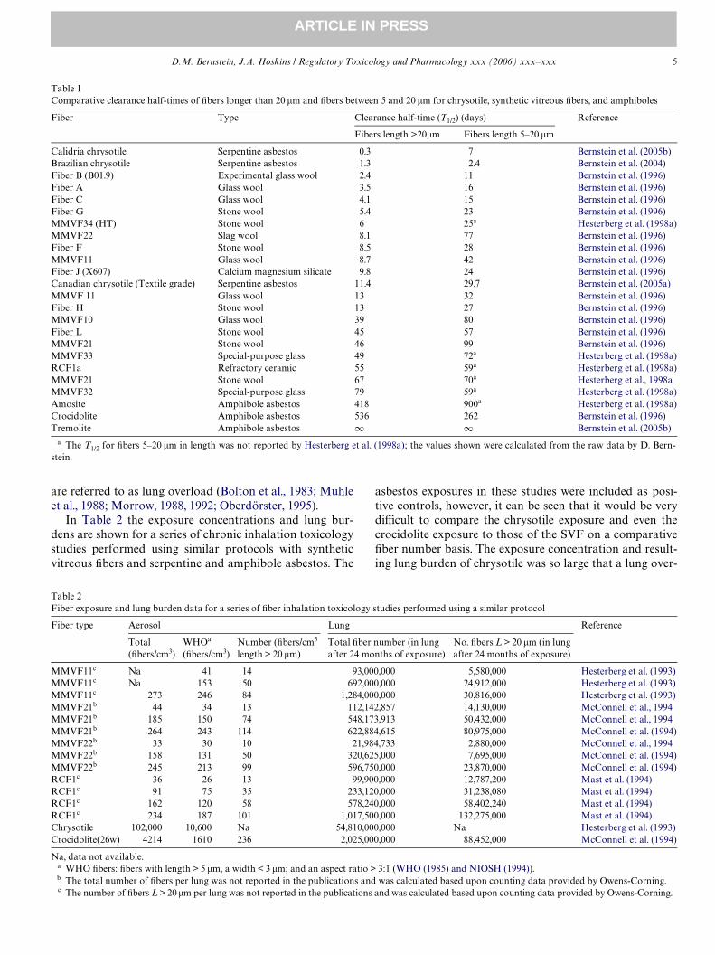
In Table 2 the exposure concentrations and lung bur-dens are shown for a series of chronic inhalation toxicologystudies performed using similar protocols with syntheticvitreous Wbers and serpentine and amphibole asbestos. The
asbestos exposures in these studies were included as posi-tive controls, however, it can be seen that it would be verydiYcult to compare the chrysotile exposure and even thecrocidolite exposure to those of the SVF on a comparativeWber number basis. The exposure concentration and result-ing lung burden of chrysotile was so large that a lung over-
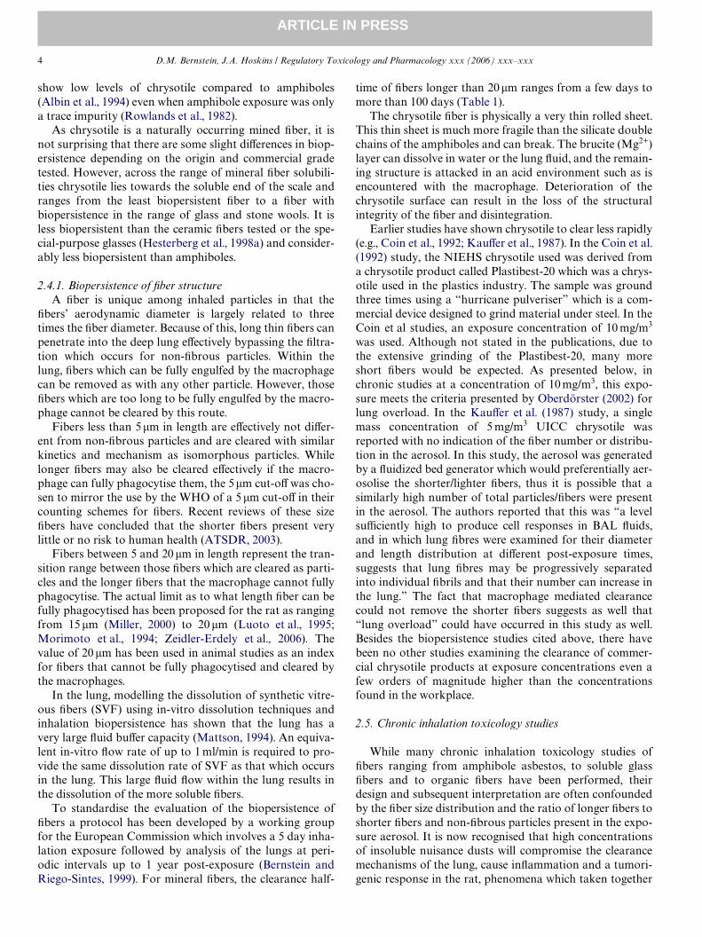
Table 1Comparative clearance half-times of Wbers longer than 20 �m and Wbers between 5 and 20 �m for chrysotile, synthetic vitreous Wbers, and amphiboles
a The T1/2 for Wbers 5–20 �m in length was not reported by Hesterberg et al. (1998a); the values shown were calculated from the raw data by D. Bern-stein.
Fiber Type Clearance half-time (T1/2) (days) Reference
Fibers length >20�m Fibers length 5–20 �m
Calidria chrysotile Serpentine asbestos 0.3 7 Bernstein et al. (2005b)Brazilian chrysotile Serpentine asbestos 1.3 2.4 Bernstein et al. (2004)Fiber B (B01.9) Experimental glass wool 2.4 11 Bernstein et al. (1996)Fiber A Glass wool 3.5 16 Bernstein et al. (1996)Fiber C Glass wool 4.1 15 Bernstein et al. (1996)Fiber G Stone wool 5.4 23 Bernstein et al. (1996)MMVF34 (HT) Stone wool 6 25a Hesterberg et al. (1998a)MMVF22 Slag wool 8.1 77 Bernstein et al. (1996)Fiber F Stone wool 8.5 28 Bernstein et al. (1996)MMVF11 Glass wool 8.7 42 Bernstein et al. (1996)Fiber J (X607) Calcium magnesium silicate 9.8 24 Bernstein et al. (1996)Canadian chrysotile (Textile grade) Serpentine asbestos 11.4 29.7 Bernstein et al. (2005a)MMVF 11 Glass wool 13 32 Bernstein et al. (1996)Fiber H Stone wool 13 27 Bernstein et al. (1996)MMVF10 Glass wool 39 80 Bernstein et al. (1996)Fiber L Stone wool 45 57 Bernstein et al. (1996)MMVF21 Stone wool 46 99 Bernstein et al. (1996)MMVF33 Special-purpose glass 49 72a Hesterberg et al. (1998a)RCF1a Refractory ceramic 55 59a Hesterberg et al. (1998a)MMVF21 Stone wool 67 70a Hesterberg et al., 1998aMMVF32 Special-purpose glass 79 59a Hesterberg et al. (1998a)Amosite Amphibole asbestos 418 900a Hesterberg et al. (1998a)Crocidolite Amphibole asbestos 536 262 Bernstein et al. (1996)Tremolite Amphibole asbestos 1 1 Bernstein et al. (2005b)
Table 2Fiber exposure and lung burden data for a series of Wber inhalation toxicology studies performed using a similar protocol
Na, data not available.a WHO Wbers: Wbers with length > 5 �m, a width < 3 �m; and an aspect ratio > 3:1 (WHO (1985) and NIOSH (1994)).b The total number of Wbers per lung was not reported in the publications and was calculated based upon counting data provided by Owens-Corning.c The number of Wbers L > 20 �m per lung was not reported in the publications and was calculated based upon counting data provided by Owens-Corning.
Fiber type Aerosol Lung Reference
Total (Wbers/cm3)
WHOa (Wbers/cm3)
Number (Wbers/cm3 length > 20 �m)
Total Wber number (in lung after 24 months of exposure)
No. Wbers L > 20 �m (in lung after 24 months of exposure)
MMVF11c Na 41 14 93,000,000 5,580,000 Hesterberg et al. (1993)MMVF11c Na 153 50 692,000,000 24,912,000 Hesterberg et al. (1993)MMVF11c 273 246 84 1,284,000,000 30,816,000 Hesterberg et al. (1993)MMVF21b 44 34 13 112,142,857 14,130,000 McConnell et al., 1994MMVF21b 185 150 74 548,173,913 50,432,000 McConnell et al., 1994MMVF21b 264 243 114 622,884,615 80,975,000 McConnell et al. (1994)MMVF22b 33 30 10 21,984,733 2,880,000 McConnell et al., 1994MMVF22b 158 131 50 320,625,000 7,695,000 McConnell et al. (1994)MMVF22b 245 213 99 596,750,000 23,870,000 McConnell et al. (1994)RCF1c 36 26 13 99,900,000 12,787,200 Mast et al. (1994)RCF1c 91 75 35 233,120,000 31,238,080 Mast et al. (1994)RCF1c 162 120 58 578,240,000 58,402,240 Mast et al. (1994)RCF1c 234 187 101 1,017,500,000 132,275,000 Mast et al. (1994)Chrysotile 102,000 10,600 Na 54,810,000,000 Na Hesterberg et al. (1993)Crocidolite(26w) 4214 1610 236 2,025,000,000 88,452,000 McConnell et al. (1994)

6 D.M. Bernstein, J.A. Hoskins / Regulatory Toxicology and Pharmacology xxx (2006) xxx–xxx
ARTICLE IN PRESS
load eVect likely occurred based upon the number ofshorter Wbers present. It would have been much more usefulin these studies if the exposure concentrations and Wber sizedistributions were comparable between the positive con-trols and the SVFs.
More recently, Oberdörster (2002) reviewed the toxic-okinetics and eVects of Wbers and of non-Wbrous poorly sol-uble particles and related how high exposureconcentrations of poorly soluble particles can induce lungparticle overload in the rat which can result in the induc-tion of lung tumours. He proposed that high-dose eVectsobserved in rats may be associated with two thresholds.
1. The Wrst threshold is the pulmonary dose that results ina reduction in macrophage mediated clearance.
2. The second threshold, occurring at a higher dose thanthe Wrst, is the dose at which antioxidant defences areoverwhelmed and pulmonary tumours develop.
The reduction in macrophage mediated clearance wasplotted against retained dust volume in the lung as shownin Fig. 3 (reproduced from Figure 3, p. 34 of Oberdörster,2002 adding a data point for chrysotile (see below)). Theauthor stated that a threshold for the retained dust bur-den seems to exist above which the clearance rate beginsto decrease.
Neither the retained dust volume nor the clearance ratewere presented by Hesterberg et al. (1993)1 for the chronicinhalation study of chrysotile listed in Table 2. It was,however, possible to estimate this from measurements of
1 The same study reported by Hesterberg et al. (1993) was also reportedin Mast et al. (1994); Hesterberg et al. (1994) and Hesterberg et al. (1998b).
total chrysotile volume retained in the lung as determinedby Bernstein et al. (2006). In this study the total chrysotilelung volume was found to be 28 �l at one third the expo-sure concentration used in the chronic studies. This valuehas been added to Fig. 3 and suggests that at the retaineddust volume used in the chronic inhalation of chrysotilethat the macrophage mediated clearance would bestrongly decreased. If macrophage activity is inhibited,then their interaction in creating an acid environmentwhich would break apart the chrysotile Wbers would alsobe aVected.
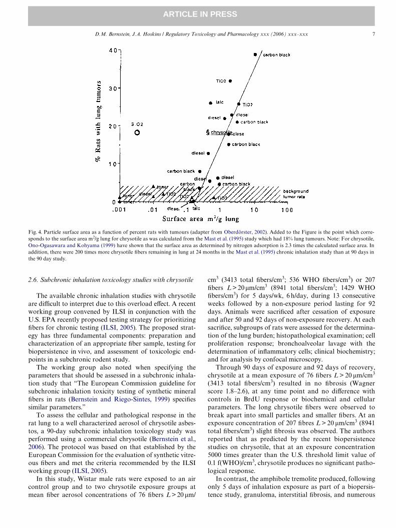
The second threshold postulated by Oberdörster occursat a higher dose than the Wrst, and is the dose at which anti-oxidant defences are overwhelmed and pulmonary tumoursdevelop. This relationship was illustrated by Oberdörster(2002, Figure 6, p. 37) adapted as Fig. 4 (with the additionof a data point for chrysotile) with the surface area ofretained dusts as a function of the percent lung tumours inchronic inhalation studies. The surface area was calculatedusing the same dataset from the 90 day inhalation toxicitystudy extrapolated to the chronic studies of chrysotile. Theparticle and short Wber chrysotile lung dose in the chronicinhalation study falls within the “overload” range shown inFig. 4 for poorly soluble low toxicity particles.
While this comparison shows that the results of thechronic inhalation toxicology studies of chrysotile couldhave occurred as a result of the number of short Wbers andparticles alone, it cannot exclude the possibility that thesmall percentage of longer Wbers present could havecaused the tumorigenic response. However, the biopersis-tence results indicate that fewer if any long Wbers wouldhave been present if overload conditions were not presentin these studies.
Fig. 3. Retained dust volume in lungs and fractional clearance rate (adapted from Oberdörster, 2002, Figure 3). Added to the Figure is the point which cor-responds to the retained lung volume (�l) for chrysotile. This value was calculated from measurements of total chrysotile volume retained in the lung asdetermined in a 90 day inhalation toxicity study of chrysotile (see text).
D.M. Bernstein, J.A. Hoskins / Regulatory Toxicology and Pharmacology xxx (2006) xxx–xxx 7
ARTICLE IN PRESS
2.6. Subchronic inhalation toxicology studies with chrysotile
The available chronic inhalation studies with chrysotileare diYcult to interpret due to this overload eVect. A recentworking group convened by ILSI in conjunction with theU.S. EPA recently proposed testing strategy for prioritizingWbers for chronic testing (ILSI, 2005). The proposed strat-egy has three fundamental components: preparation andcharacterization of an appropriate Wber sample, testing forbiopersistence in vivo, and assessment of toxicologic end-points in a subchronic rodent study.
The working group also noted when specifying theparameters that should be assessed in a subchronic inhala-tion study that “The European Commission guideline forsubchronic inhalation toxicity testing of synthetic mineralWbers in rats (Bernstein and Riego-Sintes, 1999) speciWessimilar parameters.”
To assess the cellular and pathological response in therat lung to a well characterized aerosol of chrysotile asbes-tos, a 90-day subchronic inhalation toxicology study wasperformed using a commercial chrysotile (Bernstein et al.,2006). The protocol was based on that established by theEuropean Commission for the evaluation of synthetic vitre-ous Wbers and met the criteria recommended by the ILSIworking group (ILSI, 2005).
In this study, Wistar male rats were exposed to an aircontrol group and to two chrysotile exposure groups atmean Wber aerosol concentrations of 76 Wbers L > 20 �m/
cm3 (3413 total Wbers/cm3; 536 WHO Wbers/cm3) or 207Wbers L > 20�m/cm3 (8941 total Wbers/cm3; 1429 WHOWbers/cm3) for 5 days/wk, 6 h/day, during 13 consecutiveweeks followed by a non-exposure period lasting for 92days. Animals were sacriWced after cessation of exposureand after 50 and 92 days of non-exposure recovery. At eachsacriWce, subgroups of rats were assessed for the determina-tion of the lung burden; histopathological examination; cellproliferation response; bronchoalveolar lavage with thedetermination of inXammatory cells; clinical biochemistry;and for analysis by confocal microscopy.
Through 90 days of exposure and 92 days of recovery,chrysotile at a mean exposure of 76 Wbers L > 20 �m/cm3
(3413 total Wbers/cm3) resulted in no Wbrosis (Wagnerscore 1.8–2.6), at any time point and no diVerence withcontrols in BrdU response or biochemical and cellularparameters. The long chrysotile Wbers were observed tobreak apart into small particles and smaller Wbers. At anexposure concentration of 207 Wbres L > 20 �m/cm3 (8941total Wbers/cm3) slight Wbrosis was observed. The authorsreported that as predicted by the recent biopersistencestudies on chrysotile, that at an exposure concentration5000 times greater than the U.S. threshold limit value of0.1 f(WHO)/cm3, chrysotile produces no signiWcant patho-logical response.
In contrast, the amphibole tremolite produced, followingonly 5 days of inhalation exposure as part of a biopersis-tence study, granuloma, interstitial Wbrosis, and numerous
Fig. 4. Particle surface area as a function of percent rats with tumours (adapter from Oberdörster, 2002). Added to the Figure is the point which corre-sponds to the surface area m2/g lung for chrysotile as was calculated from the Mast et al. (1995) study which had 18% lung tumours. Note: For chrysotile,Ono-Ogasawara and Kohyama (1999) have shown that the surface area as determined by nitrogen adsorption is 2.3 times the calculated surface area. Inaddition, there were 200 times more chrysotile Wbers remaining in lung at 24 months in the Mast et al. (1995) chronic inhalation study than at 90 days inthe 90 day study.
8 D.M. Bernstein, J.A. Hoskins / Regulatory Toxicology and Pharmacology xxx (2006) xxx–xxx
ARTICLE IN PRESS
macrophage aggregates as well as multinucleated giant cells(Bernstein et al., 2005a).
Bellmann et al. (2003) reported a similar 90-day sub-chronic inhalation toxicity study of SVF with a range ofbiopersistence and amosite. One of the Wbers was a cal-cium–magnesium–silicate (CMS) Wber for which the stock
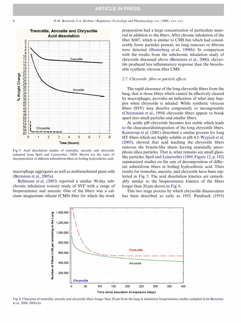
Fig. 5. Acid dissolution studies of tremolite, amosite and chrysotile(adapted from Speil and Leineweber, 1969). Shown are the rates ofdecomposition of diVerent asbestiform Wbers in boiling hydrochloric acid.
preparation had a large concentration of particulate mate-rial in addition to the Wbers. After chronic inhalation of theWber X607, which is similar to CMS but which had consid-erably fewer particles present, no lung tumours or Wbrosiswere detected (Hesterberg et al., 1998b). In comparisonwith the results from the subchronic inhalation study ofchrysotile discussed above (Bernstein et al., 2006), chryso-tile produced less inXammatory response than the biosolu-able synthetic vitreous Wber CMS.
2.7. Chrysotile: Wber or particle eVects
The rapid clearance of the long chrysotile Wbers from thelung, that is those Wbers which cannot be eVectively clearedby macrophages, provides an indication of what may hap-pen when chrysotile is inhaled. While synthetic vitreousWbers (SVF) may dissolve congruently or incongruently(Christensen et al., 1994) chrysotile Wbers appear to breakapart into small particles and smaller Wbers.
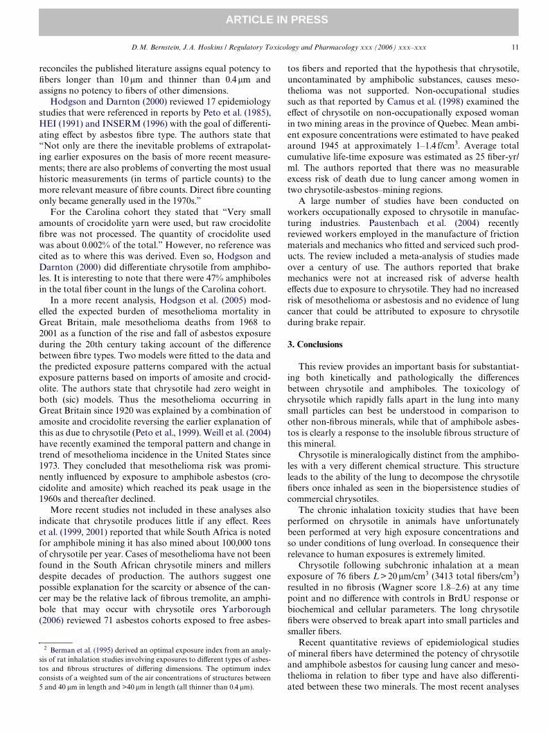
At acidic pH chrysotile becomes less stable which leadsto the clearance/disintegration of the long chrysotile Wbers.Kamstrup et al. (2001) described a similar process for longHT Wbers which are highly soluble at pH 4.5. Wypych et al.(2005), showed that acid leaching the chrysotile Wbersremoves the brucite-like sheets leaving essentially amor-phous silica particles. That is, what remains are small glass-like particles. Speil and Leineweber (1969, Figure 12, p. 182)summarized studies on the rate of decomposition of diVer-ent asbestiform Wbers in boiling hydrochloric acid. Theirresults for tremolite, amosite, and chrysotile have been rep-lotted in Fig. 5. The acid dissolution kinetics are remark-ably similar to the biopersistence kinetics of the Wberslonger than 20 �m shown in Fig. 6.
This two stage process by which chrysotile disassociateshas been described as early as 1955. Pundsack (1955)
Fig. 6. Clearance of tremolite, amosite and chrysotile Wbers longer than 20 �m from the lung in inhalation biopersistence studies (adapted from Bernsteinet al., 2004, 2005a,b).

D.M. Bernstein, J.A. Hoskins / Regulatory Toxicology and Pharmacology xxx (2006) xxx–xxx 9
ARTICLE IN PRESS
explained that: “From a chemical point of view chrysotilebehaves in certain aspects as if it were magnesium hydrox-ide. This is not unexpected when one considers that thestructure generally ascribed to the mineral consists of fun-damental layers made up in terms of a unit cell of O6–Si4–O4(OH)2–Mg6–(OH)6 planes.”
He found that the behavior of chrysotile Wbers can beunderstood as a magnesium hydroxide layer on a silica sub-strate. He explained as well the two step process in whichinitially at neutral pH such as would be found in the lungsurfactant “in contact with relatively pure water the Wbersurface dissociates partially until an equilibrium of theorder of that attained by pure magnesium hydroxide isreached.”
The second step in the lung is associated with the acidenvironment created by the macrophage. In an acid envi-ronment Pundsack (1955) stated that “It is important tonote that chrysotile reacts with strong acids to form eventu-ally a hydrated silica residue. Therefore, the particles sus-pended in initially acid solutions are not chrysotile in thestrict sense, but they represent instead intermediate reactionproducts of the acid and the Wber.” In an acid environmentPundsack found that dissociation of the surface is morepronounced because of the interaction of surface hydroxylgroups with hydrogen ions. Disintegration of the Wbers pro-vides a basis for understanding the potential toxicity ofchrysotile. The rapid disintegration of chrysotile Wbersresults in exposure to a larger number of amorphous silicaparticles and shorter Wbers. This is illustrated in Table 2where the high chrysotile exposure results in a huge numberof particles/Wbers in the lung most of them smaller than5 �m in length. Like any mineral dust at high exposure con-centrations, there is the potential for producing disease andeventually cancer with suYciently high and long exposure.
However, chrysotile at lower exposures levels leads tolevels of exposure to shorter Wbers and particles which thelung can handle. The contrast in the response between theserpentine chrysotile and amphiboles is most clearly illus-trated by the histopathological response in the inhalationstudies. Fig. 7 (reproduced from Bernstein et al., 2006)
shows the histopathological response in the subchronicstudy with chrysotile presented above (90-days of exposureand 92 days recovery) in which no inXammatory responsewas observed in the lung. In contrast, Fig. 8 (reproducedfrom Bernstein et al., 2005a) shows the histopathologicalresponse following a 5-day exposure to tremolite whichproduced marked inXammation, granulomas and even mildinterstitial Wbrosis.
2.8. Epidemiology
While it is clear that exposure to ‘asbestos’ has resulted inlung cancer and mesothelioma the epidemiological studieswhich have quantiWed these relationships have had toattempt to deal with sometimes important limitations in theavailable data. These limitations would be less important ifas considered many years ago all asbestos mineral types wereof equal potency. However, as presented above, the animal
Fig. 8. (Reproduced from Figure 7, Bernstein et al., 2005a) Photomicro-graph of a lung histopathological section from rats exposed 5-days totremolite (at 90 days following cessation of exposure). The severity of theWbrosis in the granulomas was increased from earlier time points, and thegranuloma can be seen interlaced with collagen. By this time the collagenhad progressed into the interstitium and interstitial Wbrosis is seen as well.Numerous macrophage aggregates are also observed, as well as multinu-cleated giant cells.
Fig. 7. (Reproduced from Figure 5, Bernstein et al., 2006) Photomicrographs showing of histopathology of the medium dose lungs after cessation of the 90day exposure. Trichrome stain for collagen speciWc analysis. Frame A is at 63£ and from B at 160£ magniWcation. A few small microgranulomas withslight collagen and a macrophages are seen.

10 D.M. Bernstein, J.A. Hoskins / Regulatory Toxicology and Pharmacology xxx (2006) xxx–xxx
ARTICLE IN PRESS
studies indicate that there is an important diVerence betweenchrysotile, a serpentine, and amphibole asbestos. For chryso-tile, this is often a pivotal issue and the limitations and associ-ated errors merit a more complete understanding.
In a recent analysis of available epidemiological data onthe diVerent asbestos types, Berman and Crump (2003)have summarised the various limitations that could inXu-ence the epidemiological evaluations and that had to beaddressed. These included:
• limitations in air measurements and other data availablefor characterizing historical exposures;
• limitations in the manner that the character of exposure(i.e., the mineralogical types of Wbers and the range anddistribution of Wber dimensions) was delineated;
• limitations in the accuracy of mortality determinationsor incompleteness in the extent of tracing of cohortmembers;
• limitations in the adequacy of the match between cohortsubjects and the selected control population; and
• inadequate characterization of confounding factors,such as smoking histories for individual workers.
In addition, the authors reviewed the capabilities andlimitations of the analytical techniques used for asbestosmeasurements (Table 3). Midget impinger (MI) andphase contrast microscopy (PCM) were the two analyti-cal techniques used to derive exposure estimates in themajority of epidemiology studies from which the existingrisk factors were derived. However, the manner in whichasbestos was quantiWed in the available epidemiologystudies (i.e., MI and PCM) may not have adequatelyreXected the characteristics of the inhaled aerosol thatrelate to biological activity.
With few exceptions little or no sampling was conductedprior to the 1950s when exposure concentrations werethought generally to be higher than those monitored morerecently, due to lack of use of dust control equipment at thetime and procedures to reduce dust levels that were intro-duced only later. For many studies, therefore, early expo-sures had to be estimated by extrapolation from latermeasurements.
In particular, as a result of the measurement techniquesthere was often little quantitative mineralogical exposureinformation on the types of Wbers to which workers wereexposed. The nature of the industrial process dictated inpart what type of Wber was used, however, in the past therewas little attempt to diVerentiate serpentine from amphi-bole asbestos, and as a result amphibole was often substi-tuted or mixed with serpentine without detaileddocumentation. The use of amphibole in place of serpentineresulted from such factors as availability, cost, and eVec-tiveness in the process. In addition, work histories ofemployees were not always as well documented as mightoccur today. While all uncertainty factors are important inassessing the diVerence between chrysotile and amphiboles,the diVerentiation of the Wber type in the exposure atmo-sphere is obviously critical in determining possible eVectsassociated with each type of Wber.
As an example, in Berman and Crump (2003), retroac-tive exposure indices were determined using TEM analy-ses of samples conducted in the same environment inwhich an epidemiological study was conducted or from anenvironment involving a similar operation (e.g., mining,textile manufacture, etc.). However, herein lies a signiW-cant diYculty in interpretation. If, for example, purechrysotile was used or the worker was exposed to amphi-bole elsewhere during the time the TEM samples weretaken and if during the time of the epidemiology studyamphibole was also used, then the TEM exposure indiceswill attribute any eVect to chrysotile when in fact amphi-bole was present.
Berman and Crump summarised that the residual incon-sistency in both the lung cancer and mesothelioma potencyvalues is primarily driven by those calculated from Quebecchrysotile miners and from South Carolina chrysotile tex-tile workers. While the present review will not resolve this,it is still interesting to note that amphiboles have beenfound in the lungs of the few South Carolina chrysotile tex-tile workers that have been examined (Case et al., 2000).Unfortunately, due to the few Wbers analysed per sample,the statistical power of this study is low.
It is interesting to note as well that Berman and Crump(2003) reported that the optimal exposure index2 that best
Table 3Capabilities and limitations of analytical techniques used for asbestos measurementsa (reproduced from Berman and Crump, 2003)
a The capabilities and limitations in this table are based primarily on the physical constraints of the indicated instrumentation. DiVerences attributableto the associated procedures and practices of methods in common use over the last 25 years are highlighted in Table 4–2 in Berman and Crump, 2003.
b Fibrous structures are deWned here as particles exhibiting aspect ratios (the ratio of length to width) greater than 3 (see Walton, 1982).c TEM counts frequently resolve individual Wbrous structures within larger, complex structures. Based on internal discussion of methods presented
below.d Most SEM and TEM instruments are equipped with the capability to record selected area electron diVraction (SAED) spectra and perform energy dis-
persive X-ray analysis (EDXA), which are used to distinguish the mineralogy of structures observed.
Parameter Midget impinger Phase contrast microscopy Scanning electron microscopy Transmission electron microscopy
Range of magniWcation 100 400 2000–10,000 5000–20,000Particles counted All Fibrous structuresb Fibrous structuresb Fibrous structuresb,c
Minimum diameter (size) visible 1 �m 0.3 �m 0.1 �m <0.01 �mResolve internal structure No No Maybe YesDistinguish mineralogyd No No Yes Yes
D.M. Bernstein, J.A. Hoskins / Regulatory Toxicology and Pharmacology xxx (2006) xxx–xxx 11
ARTICLE IN PRESS
reconciles the published literature assigns equal potency toWbers longer than 10 �m and thinner than 0.4�m andassigns no potency to Wbers of other dimensions.
Hodgson and Darnton (2000) reviewed 17 epidemiologystudies that were referenced in reports by Peto et al. (1985),HEI (1991) and INSERM (1996) with the goal of diVerenti-ating eVect by asbestos Wbre type. The authors state that“Not only are there the inevitable problems of extrapolat-ing earlier exposures on the basis of more recent measure-ments; there are also problems of converting the most usualhistoric measurements (in terms of particle counts) to themore relevant measure of Wbre counts. Direct Wbre countingonly became generally used in the 1970s.”
For the Carolina cohort they stated that “Very smallamounts of crocidolite yarn were used, but raw crocidoliteWbre was not processed. The quantity of crocidolite usedwas about 0.002% of the total.” However, no reference wascited as to where this was derived. Even so, Hodgson andDarnton (2000) did diVerentiate chrysotile from amphibo-les. It is interesting to note that there were 47% amphibolesin the total Wber count in the lungs of the Carolina cohort.
In a more recent analysis, Hodgson et al. (2005) mod-elled the expected burden of mesothelioma mortality inGreat Britain, male mesothelioma deaths from 1968 to2001 as a function of the rise and fall of asbestos exposureduring the 20th century taking account of the diVerencebetween Wbre types. Two models were Wtted to the data andthe predicted exposure patterns compared with the actualexposure patterns based on imports of amosite and crocid-olite. The authors state that chrysotile had zero weight inboth (sic) models. Thus the mesothelioma occurring inGreat Britain since 1920 was explained by a combination ofamosite and crocidolite reversing the earlier explanation ofthis as due to chrysotile (Peto et al., 1999). Weill et al. (2004)have recently examined the temporal pattern and change intrend of mesothelioma incidence in the United States since1973. They concluded that mesothelioma risk was promi-nently inXuenced by exposure to amphibole asbestos (cro-cidolite and amosite) which reached its peak usage in the1960s and thereafter declined.
More recent studies not included in these analyses alsoindicate that chrysotile produces little if any eVect. Reeset al. (1999, 2001) reported that while South Africa is notedfor amphibole mining it has also mined about 100,000 tonsof chrysotile per year. Cases of mesothelioma have not beenfound in the South African chrysotile miners and millersdespite decades of production. The authors suggest onepossible explanation for the scarcity or absence of the can-cer may be the relative lack of Wbrous tremolite, an amphi-bole that may occur with chrysotile ores Yarborough(2006) reviewed 71 asbestos cohorts exposed to free asbes-
2 Berman et al. (1995) derived an optimal exposure index from an analy-sis of rat inhalation studies involving exposures to diVerent types of asbes-tos and Wbrous structures of diVering dimensions. The optimum indexconsists of a weighted sum of the air concentrations of structures between5 and 40 �m in length and >40 �m in length (all thinner than 0.4 �m).
tos Wbers and reported that the hypothesis that chrysotile,uncontaminated by amphibolic substances, causes meso-thelioma was not supported. Non-occupational studiessuch as that reported by Camus et al. (1998) examined theeVect of chrysotile on non-occupationally exposed womanin two mining areas in the province of Quebec. Mean ambi-ent exposure concentrations were estimated to have peakedaround 1945 at approximately 1–1.4 f/cm3. Average totalcumulative life-time exposure was estimated as 25 Wber-yr/ml. The authors reported that there was no measurableexcess risk of death due to lung cancer among women intwo chrysotile-asbestos–mining regions.
A large number of studies have been conducted onworkers occupationally exposed to chrysotile in manufac-turing industries. Paustenbach et al. (2004) recentlyreviewed workers employed in the manufacture of frictionmaterials and mechanics who Wtted and serviced such prod-ucts. The review included a meta-analysis of studies madeover a century of use. The authors reported that brakemechanics were not at increased risk of adverse healtheVects due to exposure to chrysotile. They had no increasedrisk of mesothelioma or asbestosis and no evidence of lungcancer that could be attributed to exposure to chrysotileduring brake repair.
3. Conclusions
This review provides an important basis for substantiat-ing both kinetically and pathologically the diVerencesbetween chrysotile and amphiboles. The toxicology ofchrysotile which rapidly falls apart in the lung into manysmall particles can best be understood in comparison toother non-Wbrous minerals, while that of amphibole asbes-tos is clearly a response to the insoluble Wbrous structure ofthis mineral.
Chrysotile is mineralogically distinct from the amphibo-les with a very diVerent chemical structure. This structureleads to the ability of the lung to decompose the chrysotileWbers once inhaled as seen in the biopersistence studies ofcommercial chrysotiles.
The chronic inhalation toxicity studies that have beenperformed on chrysotile in animals have unfortunatelybeen performed at very high exposure concentrations andso under conditions of lung overload. In consequence theirrelevance to human exposures is extremely limited.
Chrysotile following subchronic inhalation at a meanexposure of 76 Wbers L > 20�m/cm3 (3413 total Wbers/cm3)resulted in no Wbrosis (Wagner score 1.8–2.6) at any timepoint and no diVerence with controls in BrdU response orbiochemical and cellular parameters. The long chrysotileWbers were observed to break apart into small particles andsmaller Wbers.
Recent quantitative reviews of epidemiological studiesof mineral Wbers have determined the potency of chrysotileand amphibole asbestos for causing lung cancer and meso-thelioma in relation to Wber type and have also diVerenti-ated between these two minerals. The most recent analyses

12 D.M. Bernstein, J.A. Hoskins / Regulatory Toxicology and Pharmacology xxx (2006) xxx–xxx
ARTICLE IN PRESS
also concluded that it is the longer, thinner Wbers that havethe greatest potency as has been reported in animal inhala-tion toxicology studies. However, one of the major diYcul-ties in interpreting these studies is that the original exposureestimates rarely diVerentiated between chrysotile andamphiboles.
Not unlike some other respirable particulates (e.g., silica,diesel fume particles, etc.), to which humans are, or havebeen heavily occupationally exposed, there is evidence thatheavy and prolonged exposure to chrysotile can producelung cancer.
The value of the present and other similar studies is thatthey show that low exposures to pure chrysotile do notpresent a detectable risk to health. Since total dose overtime decides the likelihood of disease occurrence and pro-gression, they also suggest that the risk of an adverse out-come may be low if even any high exposures experiencedwere of short duration.
References
Albin, M., Pooley, F.D., Stromberg, U., Attewell, R., Mitha, R., Johansson,L., Welinder, H., 1994. Retention patterns of asbestos Wbres in lung tis-sue among asbestos cement workers. Occup. Environ. Med. Mar. 51(3), 205–211.
ATSDR, 2003. Report on the Expert Panel on Health EVects of Asbestosand Synthetic Vitreous Fibers: The InXuence of Fiber Length. Atlanta,GA.: Prepared for: Agency for Toxic Substances and Disease RegistryDivision of Health Assessment and Consultation.
Bellmann, B., Muhle, H., Creutzenberg, O., Ernst, H., Muller, M., Bern-stein, D.M., Riego Sintes, J.M., 2003. Calibration study on subchronicinhalation toxicity of man-made vitreous Wbers in rats. Inhal. Toxicol.15 (12), 1147–1177.
Berman, D.W., Crump, K.S., 2003. Draft technical support document for aprotocol to assess asbestos-related risk. Washington, DC 20460: OYceof Solid Waste and Emergency Response U.S. Environmental Protec-tion Agency.
Berman, D.W., Crump, K.S., ChatWeld, E.J., Davis, J.M., Jones, A.D., 1995.The sizes, shapes, and mineralogy of asbestos structures that inducelung tumors or mesothelioma in AF/HAN rats following inhalation.Risk Anal. 15 (2), 181–195.
Bernstein, D.M., Morscheidt, C., Grirnm, H.-G., Thevenaz, P., Teichert, U.,1996. Evaluation of soluble Wbers using the inhalation Biopersistencemodel, a nine-Wber comparison. Inhal. Toxicol. 8, 345–385.
Bernstein, D.M., Riego-Sintes, J.M.R., 1999. Methods for the determinationof the hazardous properties for human health of man made mineral Wbers(MMMF). Vol. EUR 18748 EN, April. 93, http://ecb.ei.jrc.it/DOCU-MENTS/Testing-Methods/mmmfweb.pdf: European Commission JointResearch Centre, Institute for Health and Consumer Protection, Unit:Toxicology and Chemical Substances, European Chemicals Bureau.
Bernstein, D.M., Riego Sintes, J.M., Ersboell, B.K., Kunert, J., 2001a. Biop-ersistence of synthetic mineral Wbers as a predictor of chronic inhala-tion toxicity in rats. Inhal. Toxicol. 13 (10), 823–849.
Bernstein, D.M., Riego Sintes, J.M., Riego-Sintes, J.M., Ersboell, B.K.,Kunert, J., 2001b. Biopersistence of synthetic mineral Wbers as a predic-tor of chronic intra-peritoneal injection tumor response in rats. Inhal.Toxicol. 13 (10), 851–875.
Bernstein, D.M., Chevalier, J., Smith, P., 2003a. Comparison of Calidriachrysotile asbestos to pure tremolite: inhalation biopersistence and his-topathology following short-term exposure. Inhal. Toxicol. 15 (14),1387–1419.
Bernstein, D.M., Rogers, R., Smith, P., 2003b. The biopersistence of Cana-dian chrysotile asbestos following inhalation. Inhal. Toxicol. 15 (13),1247–1274.
Bernstein, D.M., Rogers, R., Smith, P., 2004. The biopersistence of Brazil-ian chrysotile asbestos following inhalation. Inhal. Toxicol. 16 (9), 745–761.
Bernstein, D.M., Rogers, R., Smith, P., 2005a. The biopersistence of Cana-dian chrysotile asbestos following inhalation: Wnal results through 1year after cessation of exposure. Inhal. Toxicol. 17 (1), 1–14.
Bernstein, D.M., Chevalier, J., Smith, P., 2005b. Comparison of Calidriachrysotile asbestos to pure tremolite: Wnal Results of the inhalationbiopersistence and histopathology following short-term exposure.Inhal. Toxicol. 17 (9), 427–449.
Bernstein, David M., Rogers, Rick, Chevalier, Jörg, Smith, Paul, 2006. Thetoxicological response of Brazilian chrysotile asbestos: A multidosesub-chronic 90-day inhalation toxicology study with 92 day recoveryto assess cellular and pathological response. Inhal. Toxicol. 18 (5), 1–22.
Bolton, R.E., Vincent, J.H., Jones, A.D., Addison, J., Beckett, S.T., 1983. Anoverload hypothesis for pulmonary clearance of UICC amosite Wbresinhaled by rats. Br. J. Ind. Med. 40, 264–272.
Camus, M., Siemiatycki, J., Meek, B., 1998. Non-occupational exposure tochrysotile asbestos and the risk of lung cancer. N. Engl. J. Med. 338(22), 1565–1571.
Case, B.W., Dufresne, A., McDonald, A.D., McDonald, J.C., Sebastien, P.,2000. Asbestos Wber type and length in lungs of chrysotile textile andproduction workers: Wbers longer than 18 �m. Inhal. Toxicol. 12 (S3),411–418.
Christensen, V.R., Lund Jensen, S., Guldberg, M., Kamstrup, O., 1994.EVect of chemical composition of man-made vitreous Wbers on the rateof dissolution in vitro at diVerent pHs. Environ. Health Perspect. 102(Suppl. 5), 83–86.
Coin, P.G., Roggli, V.L., Brody, A.R., 1992. Deposition, clearance andtranslocation of chrysotile asbestos from peripheral and centralregions of the rat lung. Environ. Res. 58, 97–116.
Deer, W.A., Howie, R.A., Zussman, J., 1966. An introduction to the rockforming minerals. Longman Group, Harlow, Essex.
European Commission. 1997. O.J. L 343/19 of 13 December 1997. Com-mission Directive 97/69/EC of 5 December 1997 adapting to technicalprogress for the 23rd time Council Directive 67/ 548/EEC on theapproximation of the laws regulations and administrative provisionsrelating to the classiWcation, packaging and labelling of dangerous sub-stances.
Hargreaves, A., Taylor, W.H., 1946. An X-ray Examination of decomposi-tion products of chrysotile (asbestos) and serpentine. Miner. Mag. 27,204–216.
HEI, 1991. Asbestos in public and commercial buildings. Health EVectsInstitute, Cambridge, MA.
Hesterberg, T.W., Miiller, W.C., McConnell, E.E., Chevalier, J., Hadley,J.G., Bernstein, D.M., Thevenaz, P., Anderson, R., 1993. Chronic inha-lation toxicity of size-separated glass Wbers in Fischer 344 rats. Fun-dam. Appl. Toxicol. 20 (4), 464–476.
Hesterberg, T.W., Miiller, W.C., Mast, R., McConnell, E.E., Bernstein,D.M., Anderson, R., 1994. Relationship between lung biopersistenceand biological eVects of man-made vitreous Wbers after chronic inhala-tion in rodents. Environ. Health Perspect. 102 (Suppl. 5), 133–138.
Hesterberg, T.W., Chase, G., Axten, C., Miiller, W.C., Musselman, R.P.,Kamstrup, O., Hadley, J., Morscheidt, C., Bernstein, D.M., Thevenaz,P., 1998a. Biopersistence of synthetic vitreous Wbers and amosite asbes-tos in the rat lung following inhalation. Toxicol. Appl. Pharmacol. 151(2), 262–275.
Hesterberg, T.W., Hart, G.A., Chevalier, J., Miiller, W.C., Hamilton, R.D.,Bauer, J., Thevenaz, P., 1998b. The importance of Wber biopersistence andlung dose in determining the chronic inhalation eVects of X607, RCF1,and chrysotile asbestos in rats. Toxicol. Appl. Pharmacol. 153 (1), 68–82.
Hodgson, J.T., Darnton, A., 2000. The quantitative risks of mesotheliomaand lung cancer in relation to asbestos exposure. Ann. Occup. Hyg. 44(8), 565–601.
Hodgson, J.T., McElvenny, D.M., Darnton, A.J., Price, M.J., Peto, J., 2005.The expected burden of mesothelioma mortality in Great Britain from2002 to 2050. Br. J. Cancer 92, 587–593.
D.M. Bernstein, J.A. Hoskins / Regulatory Toxicology and Pharmacology xxx (2006) xxx–xxx 13
ARTICLE IN PRESS
ILSI, 2005. Testing of Wbrous particles: short term assays and strategies.Inhal. Toxicol. 17, 497–537.
INSERM, 1996. Institut National de la Santé et de la Recherche Médicale.EVets sur la santé des principaux types d’exposition à l’amiante—rap-port de synthèse. INSERM, Paris.
Kamstrup, O., Ellehauge, A., Chevalier, J., Davis, J.M., McConnell, E.E.,Thevenaz, P., 2001. Chronic inhalation studies of two types of stonewool Wbers in rats. Inhal. Toxicol. 13 (7), 603–621.
KauVer, E., Vigneron, J.C., Hesbert, A., Lemonnier, M., 1987. A study ofthe length and diameter of Wbres, in lung and in broncho-alveolarlavage Xuid, following exposure of rats to chrysotile asbestos. Ann.Occup. Hyg. 31 (2), 233–240.
Luoto, K., Holopainen, M., Kangas, J., Kalliokoski, P., Savolainen, K.,1995. The eVect of Wber length on the dissolution by macrophages ofrockwool and glasswool Wbers. Environ. Res. 70 (1), 51–61.
Mast, R.W., Hesterberg, T.W., Glass, L.R., McConnell, E.E., Anderson, R.,Bernstein, D.M., 1994. Chronic inhalation and biopersistence of refrac-tory ceramic Wber in rats and hamsters. Environ. Health Perspect. 102(Suppl. 5), 207–209.
Mast, R.W., McConnell, E.E., Anderson, R., Chevalier, J., Kotin, P., Bern-stein, D.M., Thevenaz, P., Glass, L.R., Miiller, W.C., Hesterberg, T.W.,1995. Studies on the chronic toxicity (inhalation) of four types of refrac-tory ceramic Wber in male Fischer 344 rats. Inhal. Toxicol. 7 (4), 425–467.
Mattson, S.M., 1994. Glass Wbres in simulated lung Xuid: dissolutionbehavior and analytical requirements. Ann. Occup. Hyg. 38, 857–877.
McConnell, E.E., Kamstrup, O., Musselman, R., Hesterberg, T.W., Cheva-lier, J., Miiller, W.C., Thievenaz, P., 1994. Chronic inhalation study ofsize-separated rock and slag wool insulation Wbers in Fischer 344/Nrats. Inhal. Toxicol. 6, 571–614.
Miller, F.J., 2000. Dosimetry of particles: Critical factors having riskassessment implications. Inhal. Toxicol. 12 (Suppl. 3), 389–395.
Morimoto, Y., Yamato, H., Kido, M., Tanaka, I., Higashi, T., Fujino, A.,Yokosaki, Y., 1994. EVects of inhaled ceramic Wbres on macrophagefunction of rat lungs. Occup. Environ. Med. 51 (1), 62–67.
Morrow, P.E., 1988. Possible mechanisms to explain dust overloading ofthe lung. Fundam. Appl. Toxicol. 10, 369–384.
Morrow, P.E., 1992. Dust overloading of the lungs: update and appraisal.Toxicol. Appl. Pharmacol. 113, 1–12.
Muhle, H., Bellman, B., Heinrich, U., 1988. Overloading of lung clearanceduring chronic exposure of experimental animals to particles. Ann.Occup. Hyg. 32 (Suppl. 1), 141–147.
NIOSH, 1994. Manual of Analytical Methods (NMAM®), fourth ed. Gov-ernment Printing OYce, Washington, DC.
Oberdörster, G., 1995. Lung particle overload: implications for occupationalexposures to particles. Regul. Toxicol. Pharmacol. 21 (1), 123–135.
Oberdörster, G., 2002. Toxicokinetics and eVects of Wbrous and nonWbrousparticles. Inhal. Toxicol. 14 (1), 29–56.
Ono-Ogasawara, M., Kohyama, N., 1999. Evaluation of surface roughnessof Wbrous minerals by comparison of BET surface area and calculatedone. Ann. Occup. Hyg. 43, 505–511.
Paustenbach, D.J., Finley, B.L., Lu, E.T., Brorby, G.P., Sheehan, P.J., 2004.Environmental and occupational health hazards associated with thepresence of asbestos in brake linings and pads (1900 to present): A“state-of-the-art” review. J. Toxicol. Environ. Health B Crit. Rev. 7 (1),33–110.
Peto, J., Doll, R., Hermon, C., Binns, W., Clayton, R., GoVe, T., 1985. Rela-tionship of mortality to measures of environmental asbestos pollutionin an asbestos textile factory. Ann occup. Hyg. 29, 305–355.
Peto, J., Decarli, A., La Vecchia, C., Levi, F., Negri, E., 1999. The Europeanmesothelioma epidemic. Br. J. Cancer 79, 666–672.
Pundsack, F.L., 1955. The properties of asbestos. I. The colloidal and sur-face chemistry of chrysotile. J. Phys. Chem. 59 (9), 892–895.
Rees, D., Goodman, K., Fourie, F., Chapman, R., Blignaut, C., Bachman,O., Myer, M.J., 1999. Asbestos exposure and mesothelioma in SouthAfrica. S. Afr. Med. J. 89, 627–634.
Rees, D., Phillips, J.I., Garton, E., Pooley, F.D., 2001. Asbestos lung Wbreconcentration in South African chrysotile mine workers. Ann. Occup.Hyg. 45 (6), 473–477.
Rowlands, N., Gibbs, G.W., McDonald, A.D., 1982. Asbestos Wbres in thelungs of chrysotile miners and millers—a preliminary report. Ann.Occup. Hyg. 26, 411–415.
Speil, S., Leineweber, J.P., 1969. Asbestos minerals in modern technology.Environ. Res. 2, 166–208.
Walton, W.H., 1982. The nature, hazards, and assessment of occupationalexposure to airborne asbestos dust: a review. Ann. Occup. Hyg. 25,117–247.
Weill, H., Hughes, J.M., Churg, A.M., 2004. Changing trends in US meso-thelioma incidence. Occup. Environ. Med. 61, 438–441.
WHO, 1985. Reference methods for measuring airborne man-made min-eral Wber (MMMF), Copenhagen: World Health Organization.
Wypych, F., Adad, L.B., Mattoso, N., Marangon, A.A., Schreiner, W.H.,2005. Synthesis and characterization of disordered layered silicaobtained by selective leaching of octahedral sheets from chrysotile andphlogopite structures. J. Colloid Interface Sci. 283 (1), 107–112.
Yarborough, C.M., 2006. Chrysotile asbestos and mesothelioma. Crit.Toxicol. Rev. 36 (2), 165–187.
Zeidler-Erdely, P.C., Calhoun, W.J., Ameredes, B.T., Clark, M.P., Deye,G.J., Baron, P., Jones, W., Blake, T., Castranova, V., 2006. In vitro cyto-toxicity of Manville Code 100 glass Wbers: eVect of Wber length onhuman alveolar macrophages. Part Fibre Toxicol. 3, 5.