nucleation and growth of chrysotile nanotubes in … · doi: 10.1002/chem.201204105 nucleation and...
TRANSCRIPT
DOI: 10.1002/chem.201204105
Nucleation and Growth of Chrysotile Nanotubes in H2SiO3/MgCl2/NaOHMedium at 90 to 300 8C
Romain Lafay,*[a] German Montes-Hernandez,*[a] Emilie Janots,[a] Rodica Chiriac,[b]
Nathaniel Findling,[a] and FranÅois Toche[b]
Introduction
Serpentine is a widespread mineral that results from hydro-thermal alteration of the oceanic lithosphere.[1] Various ser-pentine polymorphs, such as antigorite, lizardite, chrysotile,and polygonal serpentine, have been observed in naturalsystems.[2] Chrysotile is the most common fibrous serpentine(Mg3Si2O5(OH)4). Preliminary studies on chrysotile haveshown a significant curvature of the unit cell compared toconventional crystal structures.[3] Chrysotile fibrils are com-posed of layers that are either curved concentrically or spi-rally with fivefold symmetry[4] into a nanometric tubularstructure (22–27 nm)[5] around either the x axis (clinochryso-tile[6] and orthochrysotile[7]) or around the y axis (parachry-sotile[8]). The hollow cores of the nanotubes have a diameterof about 5–8 nm.[9] The association between chrysotile and
polygonal serpentine has also been addressed[10] and, in nat-ural systems, proto-serpentine has been recognized as a po-tential chrysotile precursor[11,12] during the early dissolution/precipitation reactions in the alteration of the oceanic litho-sphere. However, this so-called proto-serpentine precursorhas rarely been observed in experimental systems.[12] Onerecent study reported that glass alteration under hydrother-mal conditions was dominated by the formation of proto-chrysotile, which does not have the fully well-defined crys-tallinity and cylindrical shape like chrysotile.[13] Naturalchrysotile minerals are highly heterogeneous and typicallycorrespond to an assembly of different chrysotile polytypesand/or serpentine polymorphs than can contain abundanttrace elements or mineral inclusions.[14–16] These heterogenei-ties account for the variety of chrysotile morphologies thatare observed in nature (e.g., cylindrical, tube-in-tube, orconical)[17] and the multitude of serpentine assemblies.
Chrysotile syntheses have been investigated for severaldecade.[17,18] The health hazards of this asbestos, especiallythe carcinogenicity, is known.[19] Recently, scientists/engi-neers have developed innovative routes to obtain nanosized-to-submicrometric chrysotile particles of various shapes andsizes.[20–23] For this purpose, the most popular reported reac-tants are MCM41 (specific surface area: about 900 m2 g�1) orenstatite (MgSiO3) as a Si source[24–26] and synthetic brucite,MgO periclase, or MgCl2 soluble salt as a Mg source.[27]
Most of the previous studies were performed in a moveablevessel with a volume of around 500 cm3 under stirring, but
Abstract: Herein, we report new in-sights into the nucleation and growthprocesses of chrysotile nanotubes byusing batch and semi-continuous ex-periments. For the synthesis of thishighly carcinogenic material, the influ-ences of temperature (90, 200, and300 8C), Si/Mg molar ratio, and reac-tion time were investigated. From thesemi-continuous experiments (i.e. , sam-pling of the reacting suspension overtime) and solid-state characterizationof the collected samples by XRPD,TGA, FTIR spectroscopy, andFESEM, three main reaction stepswere identified for chrysotile nuclea-
tion and growth at 300 8C: 1) formationof the proto-serpentine precursorwithin the first 2 h of the reaction, ac-companied by the formation of bruciteand residual silica gel; 2) spontaneousnucleation and growth of chrysotile be-tween about 3 and 8 h reaction time,through a progressive dissolution of theproto-serpentine, brucite, and residualsilica gel; and 3) Ostwald ripeninggrowth of chrysotile from 8 to 30 h re-
action time, as attested to by BET andFESEM measurements. Complementa-ry results from batch experiments con-firmed a significant influence of the re-action temperature on the kinetics ofchrysotile formation. However,FESEM observations revealed someformation of chrysotile nanotubes atlow temperatures (90 8C) after 14 daysof reaction. Finally, doubling the Si/Mgmolar ratio promoted the precipitationof pure smectite (stevensite-type)under the same P (8.2 MPa)/T(300 8C)/pH (13.5) conditions.
Keywords: chrysotile · growth fac-tors · nanotubes · solvent effects ·thermogravimetric analysis
[a] R. Lafay, Dr. G. Montes-Hernandez, Dr. E. Janots, N. FindlingInstitut des Sciences de la Terre (ISTerre)UJF-CNRS, 38041, Grenoble, Cedex 9 (France)Fax: (+33) 476-635-252E-mail : [email protected]
[b] Dr. R. Chiriac, F. TocheLaboratoire des Multimat�riaux et InterfacesUMR CNRS 5615, 43 bd du 11 novembre 191869622 Villeurbanne Cedex (France)
Chem. Eur. J. 2013, 19, 5417 – 5424 � 2013 Wiley-VCH Verlag GmbH & Co. KGaA, Weinheim 5417
FULL PAPER

they generally used pre-treated reactants. Typically, all ofthese reported chrysotile syntheses were performed in alka-line medium (NaOH) to promote the incorporation of �OHions. The influence of several parameters, such as tempera-ture,[20, 28] pH value,[28] and Mg substitution (e.g., Ni andFe),[29–36] and the role of trace elements (e.g., Li)[37] have alsobeen investigated. Most of the experimental studies synthe-sized chrysotile at a temperature of 300 8C and some ofthese studies revealed an optimal temperature of between300 and 400 8C.[21–23,31, 35] The longest chrysotile nanotubes re-ported so far were obtained at 400 8C after a reaction timeof 168 h. Conversely, the reaction kinetic and crystallinity ofchrysotile decrease at temperatures <300 8C.[28] However,very few studies have focused on the nucleation and growthprocesses of chrysotile because batch (or discontinuous) re-actors are generally used.[22,23, 29]
In some studies, the transformation from proto-serpentineinto chrysotile has been interpreted as the curvature ofnanoflakes beyond a certain threshold, thus resulting in theformation of nanotubes through a solid-state-transition proc-ess.[28] In 2012, Bloise et al. suggested that the experimentalconditions played a major role in determining the stabilityof the proto-chrysotile.[13] Conversely, our semi-continuousexperiments (i.e., sampling of the reacting suspension overtime) suggested the dissolution of the proto-serpentine pre-cursor, followed by the precipitation of chrysotile (thisstudy). This controversial interpretation has direct implica-tions on the presence/absence/role of a pre-nucleation proc-ess during the hydrothermal or solvothermal formation of agiven mineral or bio-mineral, which is a current “hot topic”in science.[38]
This study has a twofold objective: Firstly, to determinethe reaction steps and kinetics of chrysotile formation froma H2SiO3/MgCl2/NaOH slurry by using batch and semi-con-tinuous experiments and, secondly, to determine the influ-ence of reaction temperature and Si/Mg molar ratio onchrysotile formation. We have developed an innovativeroute for chrysotile formation by directly using commercialsilica gel (H2SiO3; i.e. , without pre-treatment) instead ofMCM41 as a Si source. Solid-state experimental productswere characterized by using X-ray powder diffraction(XRPD), Fourier-transform IR spectroscopy (FTIR), field-emission gun scanning electron microscopy (FESEM), N2-adsorption isotherms, and thermogravimetric analysis(TGA/SDTA). This latter analytical tool was particularly ef-ficient to accurately determine the abundance of chrysotileand brucite as a function of time.
Experimental Section
Reactants : Silica gel material H2SiO3 was purchased from Strem Chemi-cals in 99% chemical purity and high specific surface area (830 m2 g�1);however, additional N2-adsorption isotherm measurements revealed alower specific surface area (600 m2 g�1) and XRPD diffraction confirmedthe dominant amorphous material. Magnesium chloride hexahydrate(MgCl2·6H2O) and sodium hydroxide (NaOH) were purchased from
ROTH in �99 % chemical purity. Reactants were used with no prelimi-nary treatment.
Batch experiments : A 1m solution of NaOH (250 mL), silica gel (H2SiO3,1.302 g), and magnesium chloride hexahydrate (MgCl2·6H2O, 5.082 g)were placed in a Parr copper-alloy reactor (autoclave with an internalvolume of 0.5 L). This aqueous reaction system was immediately stirredunder constant mechanical agitation (300 rpm) during the reaction. Then,the aqueous system was heated at 300 8C for 30 h by using a heatingjacket that was fitted to the reactor, followed by preliminary experimentson chrysotile syntheses. To evaluate the influence of temperature on thesynthesis, three supplementary experiments were performed, at 90 8C for14 and 30 days and at 200 8C for 2 days. Finally, the influence of the Si/Mg molar ratio (from 1 to 1.33) was evaluated by modifying only the ini-tial amount of silica gel in the system. These latter experiments were per-formed at 300 8C for 30 h.
At the end of the experiment, the autoclave was removed from the heat-ing system and immersed in cold water. After cooling in water at 30 8C(for about 15 min), the autoclave was disassembled and the solid productwas carefully recovered and separated by centrifugation (20 min at11500 rpm), with decantation of the supernatant solutions. The solidproduct was washed twice through re-dispersion/centrifugation processesto remove any soluble compounds (e.g., NaCl and Na2CO3) that were co-formed during the synthesis and subsequent quenching. Finally, the solidproduct was dried directly in the centrifugation flasks at 90 8C for 48 h.The dry solid product was manually recovered, weighed, and stored inplastic flasks for further characterization (FESEM, XRPD, TGA, N2-ad-sorption isotherms, and FTIR spectroscopy).
Semi-continuous experiments : The semi-continuous system (samplingwith time) was operated under optimized conditions (300 8C, Si/Mg=
0.67) to monitor the composition of the solid product in the samplesex situ. About 10 mL of the dispersion was sampled in the reactor as afunction of time during the nucleation and growth of chrysotile. Two ex-periments were performed: The dispersion was collected after 1, 2, 3, 4,6, and 8 h and after 12, 16, 20, and 30 h in the two semi-continuous ex-periments, respectively. For the two experiments, the collected dispersionwas cooled by water circulation in the sampling system. The solid-prod-uct-recovery/drying procedures were similar to those described above forthe batch experiments.
The experimental conditions and mineral composition of the solid prod-ucts from all of the syntheses are summarized in Table 1.
Solid-state characterization
X-ray powder diffraction (XRPD): All samples were manually crushedbefore analysis. Powders were carefully placed and manually compactedin borosilicate capillaries (diameter: 500 mm), which corresponded toabout 5 mg of sample. XRPD patterns were recorded on a Bruker D8powder diffractometer that was equipped with a SolX Si (Li) solid-statedetector from Baltic Scientific Instruments by using CuKa1-Ka2 radiationand a Gçbel mirror. Intensities were recorded from 5 to 808 with an 8 scounting time per 0.0248 2q step for the of determination bulk mineralo-gy. For experiments that contained clays, complementary oriented thinsections were prepared and treated in an atmosphere of ethylene glycolto determine the change in interlayer space by XRPD measurements.
Field-emission gun scanning electron microscopy (FESEM): The reactionproducts were characterized by using secondary or backscattered elec-trons. Microimaging was performed on a Zeiss Ultra 55 FESEM with aspatial resolution of approximately 1 nm at 15 kV. The samples were dis-persed in absolute EtOH by ultrasonic treatment for at least 5 min to de-aggregate the particles. One or two drops of the dispersion were placedonto an aluminum support and coated with a thin film of Pt for the SEMobservations.
Differential thermal analysis and thermogravimetric analysis (DTA/TGA):Experimental solid products were characterized on a TGA/SDTA 851e
www.chemeurj.org � 2013 Wiley-VCH Verlag GmbH & Co. KGaA, Weinheim Chem. Eur. J. 2013, 19, 5417 – 54245418

Mettler Toledo instrument under thefollowing conditions: Sample mass:about 10 mg, platinum crucible(150 mL) with a pinhole, heating rate:10 8C min�1, inert N2 atmosphere:50 mL min�1. The mass loss in thesample and the associated thermal ef-fects were determined by TGA/DTAin the temperature range 30–1200 8C.To identify the different mass-losssteps, the first derivatives of theTGA data (rate of mass loss) wereused. The TGA apparatus was cali-brated in terms of mass and tempera-ture; calcium oxalate was used to cal-ibrate the sample mass and the melt-ing points of three compounds(indium, aluminum, and copper) thatwere obtained from the DTA signalswere used to calibrate the sampletemperature. The temperature accu-racy of the TGA/SDTA system wasabout �0.25 8C. The weighting accu-racy was about 0.1 mg, which corre-sponded to 0.01 % for a 10 mgsample. The losses of mass that wereassociated to the brucite and serpen-
tine phases, as deduced by TGA, are summarized in Table 1.
Fourier-transform IR spectroscopy (FTIR): Measurements (in transmis-sion mode) were performed on a Bruker Hyperion 3000 IR microscope.The IR beam was focused through a � 15 lens and the typical size of theIR aperture was 50� 50 mm2. The light source was a Globar(TM) and thebeam splitter was in KBr. The spectra were measured from 700 to4000 cm�1 (resolution: 4 cm�1) with a MCT monodetector that wascooled by liquid nitrogen.
Samples were prepared with respect to a flat thickness of <100 mm.Sample preparation involved careful sample crushing in a mortar andmanual compaction of the finely crushed particles between two KBr win-dows. Five spectra per sample were recorded and OPUS software wasused to fit the results and to compare the spectra from the experimentalproducts.
N2-sorption isotherms : N2-sorption isotherms for Runs 6, 10, 12, 14, and15 were performed on a Micrometrics ASAP 2010 system. The specificsurface areas of the powdered samples were estimated by applying theBrunauer–Emmett–Teller (BET) equation in the relative-pressure range0.05<P/P0<0.35 and by using the cross-sectional area of molecular N2
(16.2 �2). In addition, the Barrett, Joyner, and Halenda (BJH) method,which take into account capillary condensation by using the Kelvin equa-tion, was used for the determination of the pore-size distribution.
Results and Discussion
A detailed characterization of the textural properties (size,morphology, specific surface area) of chrysotile that was syn-thesized under the optimized conditions (T=300 8C, Si/Mg=0.67), the reaction steps during the crystal-growthprocess, and the influence of temperature and Si/Mg molarratio on the synthesis are given below. Herein, we reportnew insights into the nucleation and growth processes ofchrysotile nanotubes by using semi-continuous experiments.
Textural properties and purity of chrysotile : Figure 1 showsthe solid-state characterization (XRPD, TGA, FTIR spec-
Figure 1. Characterization of synthetized chrysotile after 30 h reactiontime (Run 10): a) Experimental XRPD pattern, Ctl=chrysotile; b) TGfirst derivative (DTG, B), and the corresponding DTA spectra (C);c) FTIR spectrum..
Table 1. Summary of the experimental conditions and mineral composition of the products.[a]
Run t[h]
T[8C]
SiACHTUNGTRENNUNG[mol L�1]MgACHTUNGTRENNUNG[mol L�1]
Si/Mgmolar ratio
Products pHvalue
TGA mass loss [%]
Serpentine Brucite
1 1 300 0.067 0.1 0.67 P-S @ B 13.3 5.90 9.542 2 300 0.067 0.1 0.67 P-S @ B 13.38 5.45 8.643 3 300 0.067 0.1 0.67 Ctl+P-S @ B 13.38 11.84 6.014 4 300 0.067 0.1 0.67 Ctl @ B 13.46 8.05 3.435 6 300 0.067 0.1 0.67 Ctl @ B 13.39 10.57 2.526 8 300 0.067 0.1 0.67 Ctl 13.4 10.90 –7 12 300 0.067 0.1 0.67 Ctl 13.39 10.03 –8 16 300 0.067 0.1 0.67 Ctl 13.38 10.53 –9 20 300 0.067 0.1 0.67 Ctl 13.39 10.91 –10 30 300 0.067 0.1 0.67 Ctl 13.39 12.02 –12 48 200 0.067 0.1 0.67 P-S @Ctl @B 13.4 6.90 7.9413 336 90 0.067 0.1 0.67 B @P-S @Ctl 13.35 n.d. 15.1414 720 90 0.067 0.1 0.67 B @P-S @Ctl 13.38 n.d. 13.6215 30 300 0.133 0.1 1.33 Sm 13.45 n.d. n.d.16 30 300 0.1 0.1 1 Ctl @Sm @ B 13.38 n.d. n.d.
[a] All of the experiments were performed on unreacted starting material at pH�13.5 (measured at 25 8C atthe beginning and end of the experiments). The saturated pressures were 8.2, 1.6, and 0.1 MPa at temperaturesof 300, 200, and 90 8C, respectively. B=brucite, Ctl=chrysotile, P-S =proto-serpentine, Sm = smectite, n.d.=not determined.
Chem. Eur. J. 2013, 19, 5417 – 5424 � 2013 Wiley-VCH Verlag GmbH & Co. KGaA, Weinheim www.chemeurj.org 5419
FULL PAPERNucleation and Growth of Chrysotile Nanotubes

troscopy) of chrysotile that was precipitated under opti-mized conditions, that is, 300 8C (water-saturation pressure:about 80 bar) for 30 h with a stoichiometric molar ratio of Sito Mg with respect to the structural formula of serpentine(Table 1, Run 10).
The experimental XRPD pattern is characterized bybroad peaks and it successfully matches with InternationalCentre for Diffraction Data (ICDD) card #27-1275, whichcorresponds to the chrysotile mineral (Figure 1 a). Thebroad peaks indirectly indicate the presence of very smallparticles/crystallites, as confirmed by FESEM observations(Figure 2).
Herein, the typical length of the chrysotile nanotubes isabout 450 nm, for a width of about 16 nm, based on an aver-age analysis of 200 particles. Moreover, TGA indicates onesingle dehydroxylation event, which corresponds to a globalloss of mass that is close to 11 %, peaking at 596 8C (Fig-ure 1 b and Table 1).
This loss of mass involves a broad endothermic peak atabout 600 8C and a sharp exothermic peak at 820 8C in theDTA pattern (Figure 1 b), thus indicating the crystallizationof the remaining amorphous anhydrous material (the so-called meta-chrysotile Mg3Si2O7) into forsterite (Mg2SiO4).
This result is in agreement with literature reports.[39] Final-ly, the FTIR spectrum (Figure 1 c) shows stretching bandsfor the hydroxy group at 3692 cm�1. The band for the Si�Ogroup in chrysotile is characterized by three specific peaks(976, 1020, and 1088 cm�1).[40] The two less-intense peaks, at1450 and 1640 cm�1, indicate the presence of a carbonatestretching band and a bending vibration of molecular water,respectively.
Complementary N2-sorption isotherms revealed a highspecific surface area (SBET = 185 m2 g�1, Figure 3). The twobranches of the hysteresis loop converge at P/P0 =0.6. Thisresult indicates a wide distribution of pore sizes, as alreadyseen in synthetic chrysotile.[41] The BJH pore-size distribu-tion was in the range 2–70 nm, with a median that wasequivalent to 21.5 nm, which was indicative of a mesoporousmaterial. The corresponding cumulative pore volume(0.64 cm3 g�1) is higher than the values reported in the litera-ture for synthetic chrysotile.[38,39]
Reaction steps and kinetics of chrysotile precipitation : At350 8C under water-saturation pressure, Korytkova et al.[29]
determined by using XRPD that chrysotile spontaneouslyco-precipitated with brucite between 2 and 3 h reaction timeand that, after about 6 h reaction time, only chrysotile wasobserved. Jancar and Suvorov28] proposed that the formationof chrysotile nanotubes resulted from the curvature of thenanoflakes serpentine precursor beyond a certain thresholdsize, that is, through a solid-state transition. In 2012, Bloiseet al. suggested that proto-chrysotile and chrysotile couldco-exist in experimental systems.[13] Conversely, our semi-continuous experiments suggest dissolution of the proto-ser-pentine precursor, followed by the precipitation of chryso-tile, as explained below.
The mechanism for the precipitation of chrysotile from aH2SiO3/MgCl2/NaOH slurry at 300 8C can be summarized inthree main sequential reaction steps: 1) formation of theproto-serpentine precursor and brucite within the first 2 hreaction time; 2) spontaneous nucleation and growth ofchrysotile between about 3 and 8 h reaction time, throughthe progressive dissolution of proto-serpentine, brucite, andresidual silica gel; and 3) Ostwald ripening growth of chrys-otile from 8 to 30 h reaction time.
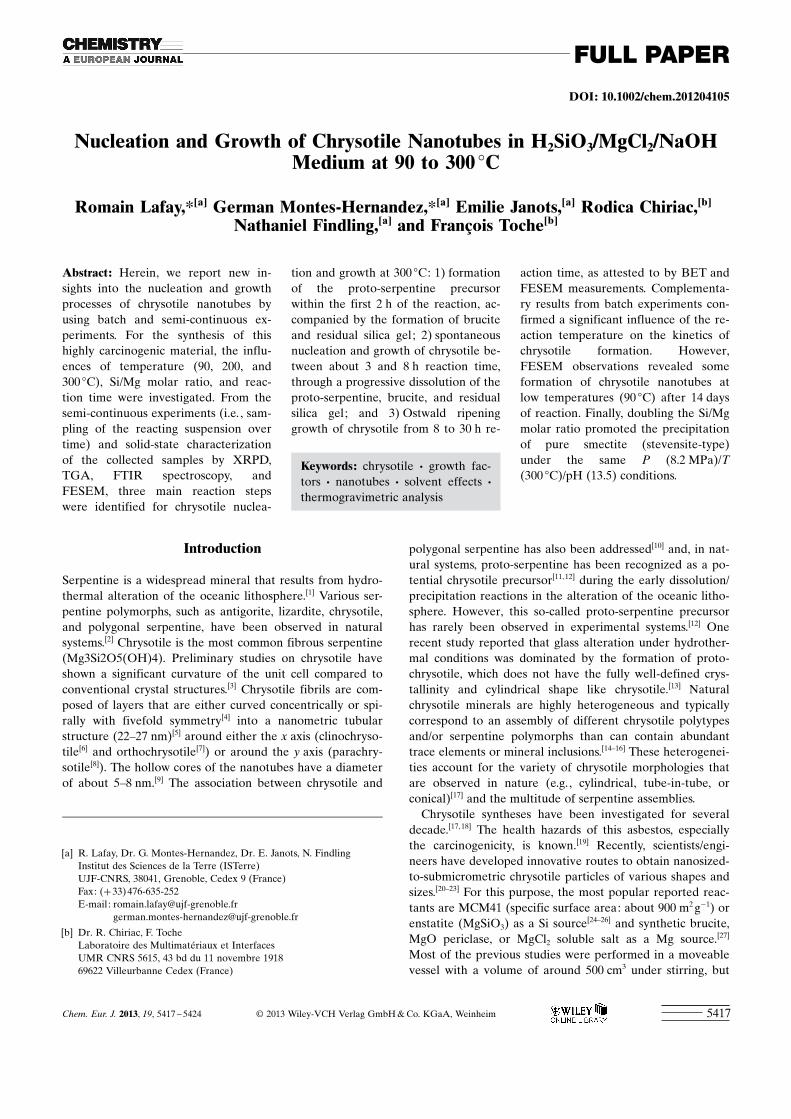
Formation of proto-serpentine precursor : The broad featuresin the XRPD pattern after 2 h reaction time indicate the for-mation of a poorly crystallized material (Figure 4 a). Thisresult is attributed to the formation of proto-serpentine, assupported by the flake-like morphology observed byFESEM (Figure 5 a) and TGA (Figure 6). DTG curves after1 and 2 h reaction time show a broad feature between 450and 650 8C, which can be interpreted as the dehydroxylationof proto-serpentine and corresponds to a loss of mass ofabout 5.5–5.9 %. Well-crystallized brucite was also formedwithin this time (Figure 5 a) and the DTG curve showed asingle peak close to 400 8C. In agreement with the XRPD
Figure 2. FESEM image showing the synthesis of chrysotile after 30 h at300 8C (Run 10).
Figure 3. N2-adsorption/desorption isotherms of synthetic chrysotile after8h (Run 6) and 30 h reaction time (Run 10). Inset: the range 0<P/P0<
0.2.
www.chemeurj.org � 2013 Wiley-VCH Verlag GmbH & Co. KGaA, Weinheim Chem. Eur. J. 2013, 19, 5417 – 54245420
R. Lafay, G. Montes-Hernandez et al.
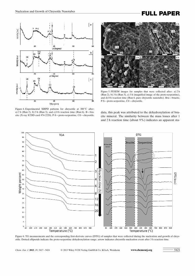
data, this peak was attributed to the dehydroxylation of bru-cite mineral. The similarity between the mass losses after 1and 2 h reaction time (about 9 %) indicates an apparent sta-
Figure 4. Experimental XRPD patterns for chrysotile at 300 8C after:a) 2 h (Run 2), b) 3 h (Run 3), and c) 8 h reaction time (Run 6). B=bru-cite (X-ray ICDD card #74-2220), P-S= proto-serpentine, Ctl=chrysotile.
Figure 5. FESEM images for samples that were collected after: a) 2 h(Run 2), b) 3 h (Run 3), c) 3 h (magnified image of the proto-serpentine),and d) 8 h reaction time (Run 6, pure chrysotile nanotube). Bru =brucite,P-S=proto-serpentine, Ctl =chrysotile.
Figure 6. TG measurements and the corresponding first-derivate curves (DTG) of samples that were collected during the nucleation and growth of chrys-otile. Dotted ellipsoids indicate the proto-serpentine dehydroxylation range; arrow indicates chrysotile-nucleation event after 3 h reaction time.
Chem. Eur. J. 2013, 19, 5417 – 5424 � 2013 Wiley-VCH Verlag GmbH & Co. KGaA, Weinheim www.chemeurj.org 5421
FULL PAPERNucleation and Growth of Chrysotile Nanotubes

bilization during this period. In summary, we have providedclear evidence for the formation of proto-serpentine with ananometric flake-like morphology within the first 2 h of thereaction, accompanied by fast precipitation of brucite.
Nucleation and growth of chrysotile : The experimentalXRPD pattern of the solid product after 3 h reaction timereveals the formation of chrysotile (ICDD card #27-1275),which co-exists with the so-called proto-serpentine and bru-cite (Figure 4 b). The presence of proto-serpentine induces
persistent broad features thatoverlap the chrysotile peaks.FESEM observations confirmthe presence of chrysotile nano-tubes (length: 260 nm, width:17 nm, average of 50 particles)that are well-dispersed in proto-serpentine and micrometricbrucite assemblies (Fig-ure 5 b, c). TG measurementsshow the typical dehydroxyla-tion peak for serpentine, butshifted to lower temperaturescompared to the batch experi-ments (shift from 600 to555 8C). No exothermic peakwas identified at 820 8C (Fig-ure 1 b), probably owing tosmall particle size and/or lowamount of chrysotile mixedwith residual proto-serpentine(Figure 6). Conversely, a typicaldehydroxylation-temperaturepeak was observed close to600 8C, with its associated exo-thermic peak after 4 h reactiontime.
The X-ray diffraction peaksthat correspond to brucite de-crease significantly in intensitybetween 3 and 4 h reactiontime. This result is in agreementwith the TG measurements, inwhich the brucite content de-creases progressively and nobrucite is detected after 8 h(Figure 4 c and Figure 6). Proto-serpentine is still observed after3 h reaction time, but FESEMobservations indicate that itcompletely disappears within4 h. Single chrysotile was de-tected/observed by XRPD,TGA, and FESEM after 8 h re-action time; the TGA measure-ments indicate strong variationsin the content of the serpentine
phase between 3 and 8 h reaction time (Table 1).Based on these results, we assume that the crystal growth
process of chrysotile proceeds through the simultaneous dis-solution of proto-serpentine and brucite. In our system, theidentified proto-serpentine could play two important roles:firstly, it could act as a nucleation agent for chrysotile and,secondly, it could provide the required chemical elementsfor chrysotile growth. As mentioned above, this process is indisagreement with a solid-state-transition process previouslysuggested by Jancar and Suvorov.28]
Figure 7. Influence of reaction temperature on chrysotile formation at 200 8C (a–d, Run 12) and 90 8C (e–h,Run 14). a,e) Experimental XRPD patterns, b, f) TG curves, c–h) FESEM images. B=brucite, Ctl= chrysotile.The positions of the DTG peaks for reference chrysotile and brucite that were synthetized at 300 8C (Run 10)are reported for comparison.
www.chemeurj.org � 2013 Wiley-VCH Verlag GmbH & Co. KGaA, Weinheim Chem. Eur. J. 2013, 19, 5417 – 54245422
R. Lafay, G. Montes-Hernandez et al.

Ostwald ripening growth of chrysotile : As discussed above,only the chrysotile mineral was detected and/or observedafter 8 h reaction time (Figure 4 c, Figure 5 d, and Figure 6),that is, an apparent equilibrium was reached between chrys-otile and the interacting solution after this reaction time inthe system. However, when different particle-size popula-tions co-exist in a given interacting fluid, Ostwald ripeninggrowth can be active and it is often promoted at high tem-peratures. Thus, conventional BET measurements revealedthat the specific surface area decreased from 206 m2 g�1 (8 hreaction time) to 185 m2 g�1 (30 h, Figure 3). Based on thisresult and some FESEM observations, we concluded thatthe Ostwald ripening growth particularly took place be-tween 8 and 30 h at 300 8C, that is, the smaller chrysotilecrystals dissolved and the dissolved species were re-deposit-ed on the surfaces of larger chrysotile crystals.
Influence of the temperature : In agreement with literaturereports,[20,28] the reaction temperature plays a crucial role indetermining the formation kinetics of chrysotile. For exam-ple, Figure 7 summarizes the XRPD, TGA, and FESEM re-sults at two different reaction temperatures (90 and 200 8C).Herein, chrysotile particles with tubular morphology wereobserved by FESEM at both temperatures. However, noclear evidence for the formation of chrysotile was obtainedin the XRPD and TGA measurements on the solid-stateproducts, possibly owing to its low proportion after the in-vestigated reaction time of 2 days at 200 8C or after 14 and30 days at 90 8C. Only brucite was clearly identified as thecrystalline phase in both cases. In analogy with the reactionsteps that were deduced from semi-continuous experiments(see above), we suspect that we reach the chrysotile nuclea-tion event at 200 8C after 2 days reaction time. The evidencefor chrysotile-nucleation events at lower temperatures(90 8C) after 14 and 30 days reaction time is less convincing.In summary, the reaction temperature has a strong influenceon the formation kinetics of chrysotile, but it could alsohave a significant effect on the reaction pathway (e.g., theinhibition of chrysotile formation and the stabilization ofother silicate phases).
On the other hand, N2-sorption isotherms revealed a highspecific surface area, that is, SBET =133.5 m2 g�1 after 2 daysat 200 8C and SBET = 233 and 125.62 m2 g�1 after 14 and30 days at 90 8C, respectively. The adsorption and desorptionbranches present a large hysteresis (Figure 8) and do notcompletely close until the relative pressure in the desorptionbranch returns to P/P0 =0.4, thus indicating a microporousmaterial. This result was confirmed by applying the BJHmethod on the sorption isotherms. Herein, the pore size islower than 8 nm, with a median at around 3.8 nm, for bothsyntheses (Figure 8, insets). The cumulative pore volumewas 0.32 cm3 g�1 after 30 days at 90 8C and 0.20 cm3 g�1 after2 days at 200 8C.
Influence of the Si/Mg ratio : As described above, purechrysotile was obtained at 300 8C after 30 h reaction time by
using a Si/Mg molar ratio of about 0.67, which correspondedto the Si/Mg ratio in the serpentine mineral(Mg3Si2O5(OH)4). The initial amount of the Si and Mg sour-ces and the Si/Mg molar ratio significantly influenced thepurity of chrysotile. Higher Si concentrations (Si/Mg>0.70)systematically induced the precipitation of a swelling claywith a stevensite (Mg smectite) structural formula(Na)x(Mg)3�xACHTUNGTRENNUNG(Si4O10)(OH)2·n H2O. This smectite co-precipi-tated with chrysotile and brucite. Mg smectite was found tobe the sole mineral phase when the Si/Mg ratio was con-strained to 1.33. The N2-sorption isotherm revealed a moder-ate specific surface area, SBET =83.75 m2 g�1, and the conven-tional “ethylene glycol test” confirmed the swelling proper-ties of this synthesized material (Figure 9). FESEM analysis(Figure 9, inset) shows a cornflake-like morphology, which istypical of smectite clay.
Conclusion
We showed that chrysotile synthesis at 300 8C is the result ofa complex reaction pathway. Proto-serpentine and brucite
Figure 8. N2-adsorption/desorption isotherms of the synthesized productsafter 2 days at 200 8C (Run 12) and 14 days at 90 8C (Run 13).
Figure 9. Experimental XRPD patterns for an experiment at 300 8C withSi/Mg =1.33 (Run 14) before and after treatment with ethylene glycol(E.G.) and the corresponding FESEM image.
Chem. Eur. J. 2013, 19, 5417 – 5424 � 2013 Wiley-VCH Verlag GmbH & Co. KGaA, Weinheim www.chemeurj.org 5423
FULL PAPERNucleation and Growth of Chrysotile Nanotubes

appear as transient phases during the first step of the reac-tion (first 2 h). Then, chrysotile is spontaneously nucleatedfrom suspension and growth through the simultaneous disso-lution of proto-serpentine and brucite from 3 to 8 h reactiontime. The last step of the reaction is characterized by Ost-wald ripening growth of chrysotile from 8 to 30 h reactiontime. Complementary experiments confirmed a strong influ-ence of the reaction temperature on the kinetic formation ofchrysotile. For example, chrysotile can be formed at 90 8C,but only some particles of chrysotile were observed after14 days reaction time. Finally, variation of the Si/Mg molarratio constrained the mineral composition in the final prod-uct under same P/T/pH conditions.
Acknowledgements
The authors are grateful to the French National Center for Scientific Re-search (CNRS) and the University Joseph Fourier (UJF) in Grenoble forfinancial support. R.L. was supported by a PhD grant from the FrenchEducation Ministry.The authors are grateful to O. Vidal, who allowed theuse of a sophisticated autoclave to perform various experiments.
[1] J. B. Moody, Lithos 1976, 9, 125 –138.[2] C. M�vel, Comptes Rendus Geosciences 2003, 335, 825 – 852.[3] E. J. W. Whittaker, Acta Crystallogr. 1955, 8, 571 –574.[4] G. Cressey, B. A. Cressey, F. J. Wicks, K. Yada, Mineralogical Maga-
zine 2010, 74, 29– 37.[5] K. Yada, Acta Crystallogr. Sect. A 1971, 27, 659 –664.[6] E. Whittaker, Acta Crystallogr. 1956, 9, 855 – 862.[7] E. Whittaker, Acta Crystallogr. 1956, 9, 862 – 864.[8] E. Whittaker, Acta Crystallogr. 1956, 9, 865 – 867.[9] B. A. Cressey, E. J. W. Whittaker, Mineralogical Magazine 1993, 57,
729 – 732.[10] A. Baronnet, B. Devouard, Can. Mineral. 2005, 43, 513 –542.[11] M. Andreani, A. Baronnet, A.-M. Boullier, J.-P. Gratier, Eur. J.
Mineral. 2004, 16, 585 – 595.[12] M. Andreani, O. Grauby, A. Baronnet, M. MuÇoz, Eur. J. Mineral.
2008, 20, 159 –171.[13] A. Bloise, E. Belluso, M. catalano, E. Barrese, D. Miriello, C. Apol-
laro, J. Am. Ceram. Soc. 2012, 95, 3050 – 3055.[14] D. R. Veblen, P. R. Buseck, Science 1979, 206, 1398 –1400.[15] D. S. O�Hanley, J. V. Chernosky, F. J. Wicks, Can. Mineral. 1989, 27,
483 – 493.[16] F. J. Wicks, D. S. O�Hanley, Rev. Mineral. Geochem. 1988, 19, 91 –
167.
[17] K. Yada, K. Iishi, Am. Miner. 1977, 62, 958 –965.[18] K. Yada, K. Iishi, J. Cryst. Growth 1974, 24– 25, 627 –630.[19] E. Foresti, E. Fornero, I. G. Lesci, C. Rinaudo, T. Zuccheri, N.
Roveri, J. Hazard. Mater. 2009, 167, 1070 – 1079.[20] E. Korytkova, A. Brovkin, T. Maslennikova, L. Pivovarova, I. Droz-
dova, Glass Phys. Chem. 2011, 37, 161 –171.[21] E. N. Korytkova, A. V. Maslov, L. N. Pivovarova, Y. V. Polegotchen-
kova, V. F. Povinich, V. V. Gusarov, Inorg. Mater. 2005, 41, 743 –749.[22] G. Falini, E. Foresti, M. Gazzano, A. F. Gualtieri, M. Leoni, I. G.
Lesci, N. Roveri, Chem. Eur. J. 2004, 10, 3043 –3049.[23] G. Falini, E. Foresti, G. Lesci, N. Roveri, Chem. Commun. 2002,
1512 – 1513.[24] C.-F. Cheng, W. Zhou, D. Ho Park, J. Klinowski, M. Hargreaves,
L. F. Gladden, J. Chem. Soc. Faraday Trans. 1997, 93, 359 –363.[25] Q. Cai, W.-Y. Lin, F.-S. Xiao, W.-Q. Pang, X.-H. Chen, B.-S. Zou,
Microporous Mesoporous Mater. 1999, 32, 1 –15.[26] M. Gr�n, I. Lauer, K. K. Unger, Adv. Mater. 1997, 9, 254 – 257.[27] E. N. Korytkova, A. V. Maslov, L. N. Pivovarova, I. A. Drozdova,
V. V. Gusarov, Glass Phys. Chem. 2004, 30, 51 –55.[28] B. Jancar, D. Suvorov, “The Influence of Hydrothermal-Reaction Pa-
rameters on the Formation of Chrysotile Nanotubes,” can be foundunder http://iopscience.iop.org/0957-4484/17/1/005, 2006.
[29] E. Korytkova, L. Pivovarova, V. Gusarov, Geochem. Int. 2007, 45,825 – 831.
[30] E. Korytkova, L. Pivovarova, I. Drosdova, V. Gusarov, Russian Jour-nal of General Chemistry 2007, 77, 1669 –1676.
[31] E. Korytkova, L. Pivovarova, Glass Phys. Chem. 2010, 36, 53 –60.[32] A. Bloise, E. Belluso, E. Barrese, D. Miriello, C. Apollaro, Cryst.
Res. Technol. 2009, 44, 590 – 596.[33] A. Bloise, E. Belluso, E. Fornero, C. Rinaudo, E. Barrese, S. Capel-
la, Microporous Mesoporous Mater. 2010, 132, 239 – 245.[34] A. Bloise, E. Barresse, C. Apollaro, Neues Jahrb. Mineral. Abh.
2009, 185, 297 –304.[35] E. Foresti, M. F. Hochella, H. Kornishi, I. G. Lesci, A. S. Madden, N.
Roveri, H. Xu, Adv. Funct. Mater. 2005, 15, 1009 –1016.[36] F. Turci, M. Tomatis, I. G. Lesci, N. Roveri, B. Fubini, Chem. Eur. J.
2011, 17, 350 –358.[37] B. Wunder, F. Deschamps, A. Watenphul, S. Guillot, A. Meixner, R.
Romer, R. Wirth, Contrib. Mineral. Petrol. 2010, 159, 781 – 790.[38] D. Gebauer, A. Vçlkel, H. Cçlfen, Science 2008, 322, 1819 –1822.[39] A. Cattaneo, F. Gualtieri, G. Artioli, Phys. Chem. Miner. 2003, 30,
177 – 183.[40] G. Anbalagan, G. Sivakumar, A. R. Prabakaran, S. Gunasekaran,
Vib. Spectrosc. 2010, 52, 122 – 127.[41] A. McDonald, B. Scott, G. Villemure, Microporous Mesoporous
Mater. 2009, 120, 263 –266.
Received: November 16, 2012Published online: February 27, 2013
www.chemeurj.org � 2013 Wiley-VCH Verlag GmbH & Co. KGaA, Weinheim Chem. Eur. J. 2013, 19, 5417 – 54245424
R. Lafay, G. Montes-Hernandez et al.