the epidemiology of contact lens related...
TRANSCRIPT
REVIEW
The Epidemiology of Contact Lens Related InfiltratesFIONA STAPLETON, MCOptom, PhD, FAAO, LISA KEAY, BOptom, PhD, FAAO,
ISABELLE JALBERT, OD, PhD, FAAO, and NERIDA COLE, PhD
Vision Cooperative Research Centre, University of New South Wales, Sydney, Australia (FS, LK), Institute for Eye Research, University of New South Wales,Sydney, Australia (FS, LK, IJ, NC), and School of Optometry and Vision Science, University of New South Wales, Sydney, Australia (FS, LK)
ABSTRACTWith estimated numbers of contact lens wearers worldwide exceeding 140 million, even complications with a lowincidence will affect a significant number of individuals. Although contact lenses clearly have many advantages forwearers, certain risks have been associated with their use. Differences in risk for different types of contact lenses andwearing patterns have been demonstrated for both rare and common lens related complications. This review particularlyfocuses on the incidence and etiology of contact lens related corneal infection and inflammation. An understanding ofthe risks and contributory factors to these conditions is important for practitioners and will enable an informed choice ofsafer lens wear modalities, wear schedules, and hygiene regimes to be made.(Optom Vis Sci 2007;84:257–272)
Key Words: contact lenses, microbial keratitis, corneal infiltrates, epidemiology
Epidemiological studies of contact lens related complicationsprovide information on their frequency and distribution andon their associated risk factors. Estimates of the total number
of contact lens wearers worldwide in 2005 were as high as 140million, such that even complications with a low incidence mayaffect a large number of individuals. Knowledge of the incidence andrisk factors of individual contact lens complications enables practitio-ners to accurately inform their patients on the risks of developing theseconditions. This information may also assist in the management andin understanding the pathogenesis of contact lens related disease.
Contact lens related complications occur because of a widerange of causes, and clearly the epidemiology of complications withdifferent pathogenesis will be different. Attempts at classifyingcontact lens related complications have previously been made onthe basis of the underlying etiology,1–3 the primary location of thecondition,4 or the clinical subtype.5 Because of the diversity ofclassifications used by different authors, it is difficult to give anexact estimate of the overall complication rate associated withcontact lens wear, although one study has estimated that 6% ofcontact lens wearers develop a complication each year.4 Thisreview will focus on the epidemiology of inflammatory/infec-tious complications of contact lens wear, including (1) micro-bial keratitis and (2) sterile/aseptic keratitis.
Microbial Keratitis
Corneal infection is a rare but severe complication of contact lenswear. In severe cases, it is associated with visual loss because of scarring
and perforation. Less severe cases may also be associated with signifi-cant morbidity, for example, in hospital admission, the cost of treat-ment, outpatient visits, time off needed from work, inability to wearcontact lenses, severe pain, and temporary visual loss experienced.6
Microbial keratitis in contact lens wearers predominantly ap-pears to be a bacterial process,7 although amoebae, particularlyAcanthamoeba have been associated with contact lens related infec-tions. Historically, fungal infections in contact lens wearers havebeen infrequently reported, although a recent series of contact lensrelated cases in Singapore8 and across multiple states in the UnitedStates9 have been reported in association with use of a particularmultipurpose solution. The association between viruses and con-tact lens related keratitis is poorly understood.
The Epidemiology of Contact Lens Related MicrobialKeratitis. Before the widespread use of contact lenses, microbialkeratitis was predominantly associated with trauma, ocular surfacedisease, ocular surgery, or with contact lens wear for aphakic or ther-apeutic indications. During the 1970s and 1980s, there was increasedanecdotal reporting of cases of lens related infections.10–15 In studies ofhospital cases, the proportion of cases of microbial keratitis because ofcontact lens wear varies with the severity of disease. In severe diseaserequiring hospital admission, 20 to 44% of cases were caused by con-tact lens wear.16–18 In studies which have examined all cases of micro-bial keratitis, 34 to 65% of cases could be attributed to contact lenswear for the correction of low refractive errors.19–22
In determining the incidence of contact lens related microbialkeratitis, several study designs have been proposed.23 Randomized
1040-5488/07/8404-0257/0 VOL. 84, NO. 4, PP. 257–272OPTOMETRY AND VISION SCIENCECopyright © 2007 American Academy of Optometry
Optometry and Vision Science, Vol. 84, No. 4, April 2007
clinical trials provide the gold standard in level of research evidenceand these designs reduce the effects of confounding factors byrandomly allocating a treatment or exposure. Randomized trials areonly feasible when the complication of interest is not rare and there areno randomized trials of contact lens related microbial keratitis as thesewould require an unfeasibly large sample size. For example, given arate of microbial keratitis of 0.2% per year in EW lenses, to measure areduction of 0.1% with a certain exposure, with a power of 80%, therequired sample size would be in excess of 24,000.
As an alternative to randomized trials, observational studies al-low estimation of the incidence of disease, where the investigatorobserves the outcome of contact lens wear on a suitably large num-ber of individuals without assigning contact lens type and mode ofwear.24 The numerator (incident cases of disease) and denomina-tor (number of wearers in the cohort) by the duration of wearexperience are used to establish the incidence of disease (new casesper 10,000 wearers per unit time). This approach requires a largecohort of individuals wearing the lens type or types of interest.23
A farther approach involves surveying all practitioners or pri-mary eye care centers involved in the management of disease, in aselected area to determine the number of new cases of microbialkeratitis over a period of time. An estimate of the total contact lenswearing population in that area is used as the denominator. Thedenominator can be derived from surveying the population in thearea, where a representative sample may be derived, for example,from the relevant postcode regions, general practitioner patientlists, or electoral registers. Other strategies for deriving the denom-inator might include manufacturers’ contact lens sales data or fromdata from local contact lens practitioners, which may be applied toestimates of the total population in the region of interest. Differentmethods are associated with different sources of bias. For example,surveys of contact lens prescribing reflect entry of lens modalitiesinto the community and such estimates show trends in advance ofcommunity surveys. This has been illustrated in the United King-dom where community surveys have shown the penetrance ofsilicone hydrogel lenses to be 7% of wearers and contact lens pre-scribing surveys have shown silicone hydrogel lenses are prescribedfor 13% of refits.25 Conversely, lens types or modalities which areinfrequently prescribed (such as hydrogel EW) would show a lowpenetrance in fitting surveys, but a higher penetrance in the lenswearing community. Surveys of lens wearers in the community arepreferred because of greater accuracy for modalities with low pen-etration rates and reflection of actual wear practice.26
An additional consideration in all study designs is the diagnosticcriteria used. Inclusion criteria are usually based on a diagnosis ofpresumed microbial keratitis, rather than a positive corneal cul-ture, because of the low sensitivity of microbial investigations,27
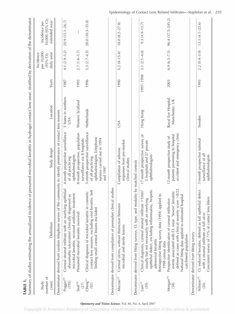
and more recently, the diminishing reliance on culture in the man-agement of mild and moderate disease.28 Table 1 describes thediagnostic criteria and derivation of the denominator in studies ofthe incidence of presumed microbial keratitis.29–35 Given the mor-bidity associated with microbial keratitis, it would seem reasonableto include equivocal diagnoses as presumed microbial keratitis.The impact of diagnostic criteria on calculated incidence rates hasbeen illustrated by applying diagnostic criteria retrospectively to anexisting data set36 and this clearly supports the need for rigor in meth-odological considerations and for the use of criteria which are in placeprospectively and for which specific variables have been collected.
In addition to diagnostic difficulties, there has been confusion interminology between studies.37 The term “ulcerative keratitis”may include both presumed infected and presumed sterile lesionsand “suppurative keratitis” describes a spectrum of corneal infiltra-tive lesions.27,34,38 Morgan et al., in 200534 used a scoring system,modified from that proposed by Aasuri et al., 2003,38 to stratifycorneal infiltrates into “nonsevere” and “severe” keratitis, where“severe” keratitis is likely to be analogous to the historical defini-tions of presumed microbial keratitis. Schein et al., 200539 recentlyused an endpoint adjudication committee to classify infiltrativeevents by severity. Outcome measures (vision loss, disease dura-tion, and direct and indirect cost of disease) have also provided ameans to validate grading of disease severity.40
Studies from the United States,29 Sweden,35 the Netherlands,32
and Hong Kong33 estimated incidences of ulcerative keratitis indaily wear (DW) soft contact lens users and EW hydrogel contactlens users based on identifying new cases within a defined area andusing population based studies to estimate numbers of contact lenswearers in the region to establish the denominator. These studies allshowed incidences that were broadly similar (Table 1). Minor differ-ences between estimates across studies may be based on the studymethodology, selection of cases and controls, and diagnostic criteria.On the basis of the results from such studies, approximately 1 in every2500 daily wear soft lens users and 1 in every 500 EW soft lens userswill develop presumed microbial keratitis every year.
Since these early studies, high oxygen transmissibility silicone hy-drogel and daily disposable contact lenses have been released in manymarkets. Although a cause relation effect has not been convincinglydemonstrated between hypoxia and corneal infection,41 the higherrisk of disease in overnight lens wear has led to speculation that contactlens induced corneal hypoxia predisposes contact lens wearers to agreater rate of corneal infection because of compromised cornealepithelial integrity,42 impaired wound healing,43 and an increasedsusceptibility of corneal epithelial cells to bacterial binding.44–46 Allcontact lens wear slows normal corneal epithelial homeostasis by sup-pressing cell proliferation,47 impairing cell migration,48 and by reduc-ing the rate of cell exfoliation.49–51 These effects are reduced but noteliminated with highly oxygen permeable contact lenses made fromsilicone hydrogel materials.47,52 Compared with other soft contactlenses, silicone hydrogel contact lenses do provide considerably im-proved corneal oxygen permeability and significantly reduce the overtclinical manifestations of corneal hypoxia.53 However, the impact ofthis reduced hypoxia on either the absolute risk or severity of microbialkeratitis with silicone hydrogel lens wear could only be investigated inlarge scale epidemiological studies.
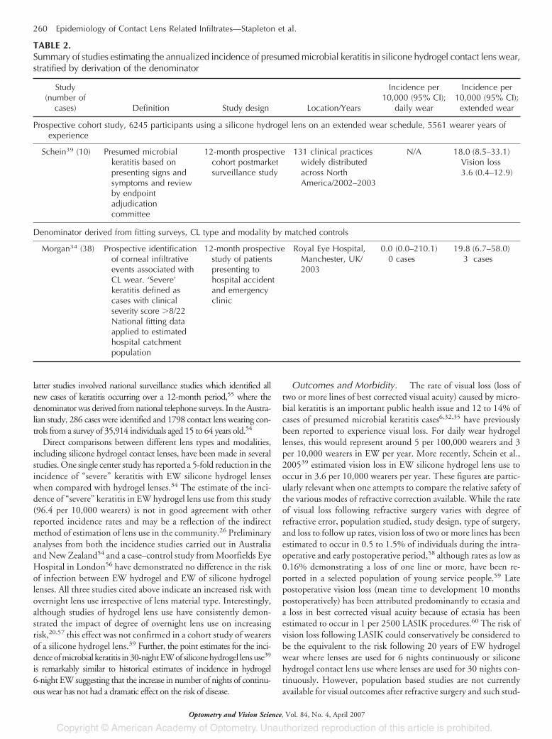
Recent epidemiological studies evaluating contact lens related pre-sumed microbial keratitis have included newly introduced lens types(Table 2). A 12-month prospective cohort study involving 5561 pa-tient years of wear of a silicone hydrogel lens on a 30-night EW basis,has reported an overall risk of 18.0 per 10,000 wearers per year.39
Morgan et al., in 200534 reported similar absolute risk data of 19.8 per10,000 wearers per year developing “severe” keratitis, which is likely tobe analogous to presumed microbial keratitis. These data were basedon a 12-month prospective study of patients presenting to a hospitalaccident and emergency clinic, with controls derived from fittingstudy estimates extrapolated to an estimate of the hospital catchmentpopulation. Preliminary analysis from the Australian and New Zea-land surveillance studies complement these early estimates.54 These
258 Epidemiology of Contact Lens Related Infiltrates—Stapleton et al.
Optometry and Vision Science, Vol. 84, No. 4, April 2007
TAB
LE1.
Sum
mar
yof
stud
ies
estim
atin
gth
ean
nual
ized
inci
denc
eof
pres
umed
mic
robi
alke
ratit
isin
hydr
ogel
cont
actl
ens
wea
r,st
ratif
ied
byde
riva
tion
ofth
ede
nom
inat
or
Stud
y(n
umbe
rof
case
s)D
efin
ition
Stud
yde
sign
Loca
tion
Yea
rs
Inci
denc
epe
r10
,000
(95%
CI);
daily
wea
r
Inci
denc
epe
r10
,000
(95%
CI);
exte
nded
wea
r
Den
omin
ator
deri
ved
from
rand
omte
leph
one
surv
eyof
the
com
mun
ityto
iden
tify
pene
tran
ceof
cont
act
lens
wea
rers
Pogg
io29
(195
)C
orne
alst
rom
alin
filtr
ate
with
anov
erly
ing
epith
elia
lab
norm
ality
(ulc
erat
ion)
clin
ical
lydi
agno
sed
asm
icro
bial
kera
titis
,re
ceiv
edan
tibio
tictr
eatm
ent
4-m
onth
pros
pect
ive
surv
eilla
nce
ofal
lpr
actic
ing
opht
halm
olog
ists
5St
ates
inno
rthe
rnU
SA19
874.
1(2
.9–5
.2)
20.9
(15.
1–26
.7)
Seal
30
(27)
Pres
umed
mic
robi
alke
ratit
is(n
onvi
ral)
8-m
onth
pros
pect
ive,
popu
latio
nsu
rvei
llanc
evi
a8
hosp
itals
Wes
tern
Scot
land
1995
2.7
(1.6
–3.7
)—
Che
ng31
(92)
Clin
ical
diag
nosi
sof
mic
robi
alke
ratit
isin
cosm
etic
cont
act
lens
wea
rers
,ex
clud
ing
vira
lke
ratit
is.
Self
limiti
ngsm
all
corn
eal
lesi
ons
excl
uded
3-m
onth
pros
pect
ive
surv
eilla
nce
ofal
lpr
actic
ing
opht
halm
olog
ists
Tele
phon
esu
rvey
sca
rrie
dou
tin
1994
and
1997
Net
herl
ands
1996
3.5
(2.7
–4.5
)20
.0(1
0.3–
35.0
)
Den
omin
ator
deri
ved
from
com
pila
tion
ofpr
emar
ket
clin
ical
stud
ies
Mac
rae3
2C
orne
alul
cers
,w
ithou
tdi
stin
ctio
nbe
twee
nm
icro
bial
and
ster
ilele
sion
sC
ompi
latio
nof
adve
rse
resp
onse
sfr
ompr
e-m
arke
tcl
inic
alst
udie
s
USA
1990
5.2
(0–1
5.4)
18.0
(8.2
–27.
8)
Den
omin
ator
deri
ved
from
fittin
gsu
rvey
s,C
Lty
pe,
and
mod
ality
bym
atch
edco
ntro
ls
Lam
33
(59)
Clin
ical
diag
nosi
s,co
rnea
lst
rom
alin
filtr
ate
�1m
m2
usua
llybu
tno
tne
cess
arily
with
anov
erly
ing
epith
elia
lde
fect
,ex
clud
ing
infla
mm
ator
y,he
rpet
icad
enov
iral
kera
titis
Ret
rosp
ectiv
efit
ting
surv
eyda
ta(1
994)
appl
ied
to19
98ce
nsus
data
17-m
onth
pros
pect
ive
surv
eyof
2ho
spita
lsan
d27
priv
ate
opht
halm
olog
ists
Hon
gK
ong
1997
–199
83.
1(2
.1–4
.0)
9.3
(4.9
–13.
7)
Mor
gan3
4
(38)
Pros
pect
ive
iden
tific
atio
nof
corn
eal
infil
trat
ive
even
tsas
soci
ated
with
CL
wea
r.‘S
ever
e’ke
ratit
isde
fined
asca
ses
with
clin
ical
seve
rity
scor
e�
8/22
Nat
iona
lfit
ting
data
appl
ied
toes
timat
edho
spita
lca
tchm
ent
popu
latio
n
12-m
onth
pros
pect
ive
stud
yof
patie
nts
pres
entin
gto
hosp
ital
acci
dent
and
emer
genc
ycl
inic
Roy
alEy
eH
ospi
tal,
Man
ches
ter,
UK
2003
6.9
(6.3
–7.5
)96
.4(3
7.5–
245.
2)
Den
omin
ator
deri
ved
from
fittin
gsu
rvey
Nils
son3
5
(26)
CL
indu
ced
kera
titis
,de
fined
asfu
llep
ithel
ial
defe
ctw
itha
stro
mal
infil
trat
eor
full
ulce
rC
oncu
rren
tsu
rvey
of71
%of
cont
act
lens
fitte
rs
3-m
onth
pros
pect
ive
natio
nal
surv
eilla
nce
ofal
lop
htha
lmol
ogis
ts
Swed
en19
932.
2(0
.4–3
.9)
13.3
(4.1
–22.
6)
Epidemiology of Contact Lens Related Infiltrates—Stapleton et al. 259
Optometry and Vision Science, Vol. 84, No. 4, April 2007
latter studies involved national surveillance studies which identified allnew cases of keratitis occurring over a 12-month period,55 where thedenominator was derived from national telephone surveys. In the Austra-lian study, 286 cases were identified and 1798 contact lens wearing con-trols from a survey of 35,914 individuals aged 15 to 64 years old.54
Direct comparisons between different lens types and modalities,including silicone hydrogel contact lenses, have been made in severalstudies. One single center study has reported a 5-fold reduction in theincidence of “severe” keratitis with EW silicone hydrogel lenseswhen compared with hydrogel lenses.34 The estimate of the inci-dence of “severe” keratitis in EW hydrogel lens use from this study(96.4 per 10,000 wearers) is not in good agreement with otherreported incidence rates and may be a reflection of the indirectmethod of estimation of lens use in the community.26 Preliminaryanalyses from both the incidence studies carried out in Australiaand New Zealand54 and a case–control study from Moorfields EyeHospital in London56 have demonstrated no difference in the riskof infection between EW hydrogel and EW of silicone hydrogellenses. All three studies cited above indicate an increased risk withovernight lens use irrespective of lens material type. Interestingly,although studies of hydrogel lens use have consistently demon-strated the impact of degree of overnight lens use on increasingrisk,20,57 this effect was not confirmed in a cohort study of wearersof a silicone hydrogel lens.39 Further, the point estimates for the inci-denceofmicrobial keratitis in30-nightEWof siliconehydrogel lensuse39
is remarkably similar to historical estimates of incidence in hydrogel6-night EW suggesting that the increase in number of nights of continu-ous wear has not had a dramatic effect on the risk of disease.
Outcomes and Morbidity. The rate of visual loss (loss oftwo or more lines of best corrected visual acuity) caused by micro-bial keratitis is an important public health issue and 12 to 14% ofcases of presumed microbial keratitis cases6,32,35 have previouslybeen reported to experience visual loss. For daily wear hydrogellenses, this would represent around 5 per 100,000 wearers and 3per 10,000 wearers in EW per year. More recently, Schein et al.,200539 estimated vision loss in EW silicone hydrogel lens use tooccur in 3.6 per 10,000 wearers per year. These figures are partic-ularly relevant when one attempts to compare the relative safety ofthe various modes of refractive correction available. While the rateof visual loss following refractive surgery varies with degree ofrefractive error, population studied, study design, type of surgery,and loss to follow up rates, vision loss of two or more lines has beenestimated to occur in 0.5 to 1.5% of individuals during the intra-operative and early postoperative period,58 although rates as low as0.16% demonstrating a loss of one line or more, have been re-ported in a selected population of young service people.59 Latepostoperative vision loss (mean time to development 10 monthspostoperatively) has been attributed predominantly to ectasia anda loss in best corrected visual acuity because of ectasia has beenestimated to occur in 1 per 2500 LASIK procedures.60 The risk ofvision loss following LASIK could conservatively be considered tobe the equivalent to the risk following 20 years of EW hydrogelwear where lenses are used for 6 nights continuously or siliconehydrogel contact lens use where lenses are used for 30 nights con-tinuously. However, population based studies are not currentlyavailable for visual outcomes after refractive surgery and such stud-
TABLE 2.Summary of studies estimating the annualized incidence of presumed microbial keratitis in silicone hydrogel contact lens wear,stratified by derivation of the denominator
Study(number of
cases) Definition Study design Location/Years
Incidence per10,000 (95% CI);
daily wear
Incidence per10,000 (95% CI);
extended wear
Prospective cohort study, 6245 participants using a silicone hydrogel lens on an extended wear schedule, 5561 wearer years ofexperience
Schein39 (10) Presumed microbialkeratitis based onpresenting signs andsymptoms and reviewby endpointadjudicationcommittee
12-month prospectivecohort postmarketsurveillance study
131 clinical practiceswidely distributedacross NorthAmerica/2002–2003
N/A 18.0 (8.5–33.1)Vision loss3.6 (0.4–12.9)
Denominator derived from fitting surveys, CL type and modality by matched controls
Morgan34 (38) Prospective identificationof corneal infiltrativeevents associated withCL wear. ‘Severe’keratitis defined ascases with clinicalseverity score �8/22National fitting dataapplied to estimatedhospital catchmentpopulation
12-month prospectivestudy of patientspresenting tohospital accidentand emergencyclinic
Royal Eye Hospital,Manchester, UK/2003
0.0 (0.0–210.1)0 cases
19.8 (6.7–58.0)3 cases
260 Epidemiology of Contact Lens Related Infiltrates—Stapleton et al.
Optometry and Vision Science, Vol. 84, No. 4, April 2007
ies would be required for meaningful comparison of the risks asso-ciated with different correction modalities.
Other than the incidence of the disease and associated visualloss, other outcome parameters related to disease severity are ofimportance. Microbial keratitis may be associated with hospitaladmission, time off needed from work, and the cost of medicationsand back up spectacles. A population study has examined factorsaffecting the morbidity of contact lens related microbial keratitis.6
Disease severity was strongly influenced by culture result and by adelay in receiving appropriate treatment. After adjustment forthese factors in a paired analysis, wearers of silicone hydrogel lenseshad a shorter disease duration (median 4, interquartile range 4days) than those of hydrogel lens wearers (median 7, interquartilerange 10 days), although the rate of vision loss and disease cost weresimilar. The distribution of disease severity in a study of symptom-atic corneal infiltrates, including lesions presumed to be microbial,has also suggested that disease severity, based on a clinical scoringscheme may also be reduced in EW of silicone hydrogel lenseswhen compared with hydrogel lenses.61
Risk Factors for Disease. From these incidence data, it isclear that the risk of presumed microbial keratitis differs for differ-ent lens types and wear schedules and these relationships wereinvestigated in the late 1980s to 1990s and recently in a series ofstudies in 2003 to 2005. Case–control studies have also been usedto establish relative risk of microbial keratitis for different lensmodalities and to estimate the impact of potential risk factors suchas lens wear practice, patient demographics, and lens wear history.
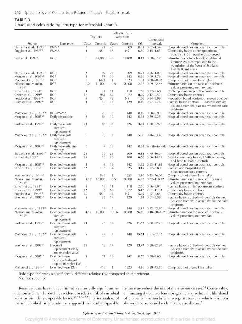
Table 3 summarizes the crude relative risks for microbial kera-titis for different lens types and modes of wear. Reliable differencesin risk have not been reported between daily use of rigid gas perme-able, PMMA, and daily wear soft contact lens use. In hydrogel contactlens use, a progressive increase in risk from daily wear, to occasionalovernight to EW has been consistently reported.20,54,56,57,66 Recentstudies including silicone hydrogel contact lenses have confirmedthe excess risk associated with overnight contact lens use whencompared with daily use,34,54,56,62 however, debate persists regard-ing differences between EW hydrogel and EW silicone hydrogelcontact lenses.
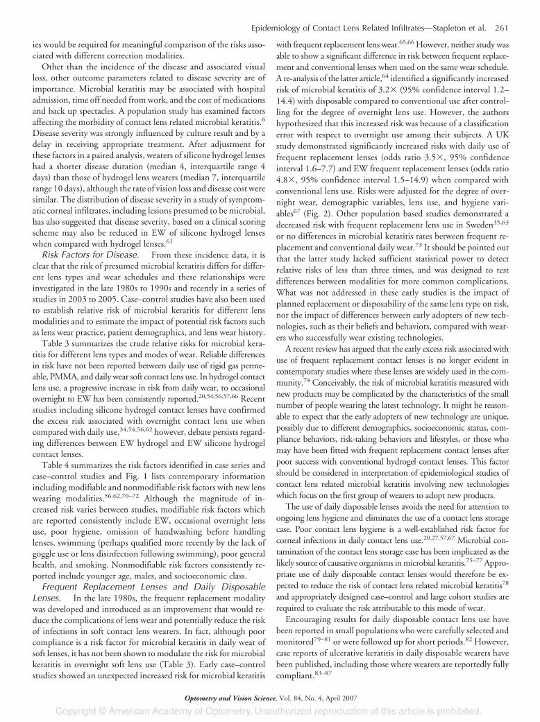
Table 4 summarizes the risk factors identified in case series andcase–control studies and Fig. 1 lists contemporary informationincluding modifiable and nonmodifiable risk factors with new lenswearing modalities.56,62,70–72 Although the magnitude of in-creased risk varies between studies, modifiable risk factors whichare reported consistently include EW, occasional overnight lensuse, poor hygiene, omission of handwashing before handlinglenses, swimming (perhaps qualified more recently by the lack ofgoggle use or lens disinfection following swimming), poor generalhealth, and smoking. Nonmodifiable risk factors consistently re-ported include younger age, males, and socioeconomic class.
Frequent Replacement Lenses and Daily DisposableLenses. In the late 1980s, the frequent replacement modalitywas developed and introduced as an improvement that would re-duce the complications of lens wear and potentially reduce the riskof infections in soft contact lens wearers. In fact, although poorcompliance is a risk factor for microbial keratitis in daily wear ofsoft lenses, it has not been shown to modulate the risk for microbialkeratitis in overnight soft lens use (Table 3). Early case–controlstudies showed an unexpected increased risk for microbial keratitis
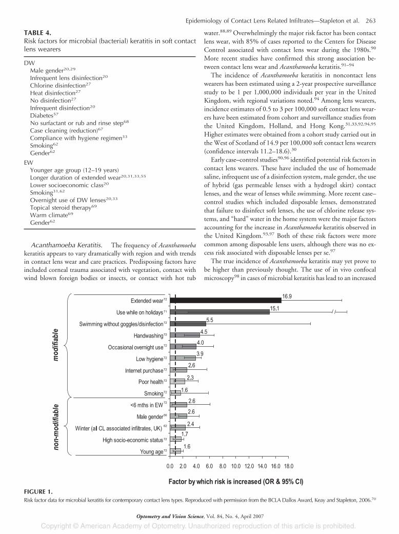
with frequent replacement lens wear.65,66 However, neither study wasable to show a significant difference in risk between frequent replace-ment and conventional lenses when used on the same wear schedule.A re-analysis of the latter article,64 identified a significantly increasedrisk of microbial keratitis of 3.2� (95% confidence interval 1.2–14.4) with disposable compared to conventional use after control-ling for the degree of overnight lens use. However, the authorshypothesized that this increased risk was because of a classificationerror with respect to overnight use among their subjects. A UKstudy demonstrated significantly increased risks with daily use offrequent replacement lenses (odds ratio 3.5�, 95% confidenceinterval 1.6–7.7) and EW frequent replacement lenses (odds ratio4.8�, 95% confidence interval 1.5–14.9) when compared withconventional lens use. Risks were adjusted for the degree of over-night wear, demographic variables, lens use, and hygiene vari-ables67 (Fig. 2). Other population based studies demonstrated adecreased risk with frequent replacement lens use in Sweden35,63
or no differences in microbial keratitis rates between frequent re-placement and conventional daily wear.73 It should be pointed outthat the latter study lacked sufficient statistical power to detectrelative risks of less than three times, and was designed to testdifferences between modalities for more common complications.What was not addressed in these early studies is the impact ofplanned replacement or disposability of the same lens type on risk,nor the impact of differences between early adopters of new tech-nologies, such as their beliefs and behaviors, compared with wear-ers who successfully wear existing technologies.
A recent review has argued that the early excess risk associated withuse of frequent replacement contact lenses is no longer evident incontemporary studies where these lenses are widely used in the com-munity.74 Conceivably, the risk of microbial keratitis measured withnew products may be complicated by the characteristics of the smallnumber of people wearing the latest technology. It might be reason-able to expect that the early adopters of new technology are unique,possibly due to different demographics, socioeconomic status, com-pliance behaviors, risk-taking behaviors and lifestyles, or those whomay have been fitted with frequent replacement contact lenses afterpoor success with conventional hydrogel contact lenses. This factorshould be considered in interpretation of epidemiological studies ofcontact lens related microbial keratitis involving new technologieswhich focus on the first group of wearers to adopt new products.
The use of daily disposable lenses avoids the need for attention toongoing lens hygiene and eliminates the use of a contact lens storagecase. Poor contact lens hygiene is a well-established risk factor forcorneal infections in daily contact lens use.20,27,57,67 Microbial con-tamination of the contact lens storage case has been implicated as thelikely source of causative organisms in microbial keratitis.75–77 Appro-priate use of daily disposable contact lenses would therefore be ex-pected to reduce the risk of contact lens related microbial keratitis78
and appropriately designed case–control and large cohort studies arerequired to evaluate the risk attributable to this mode of wear.
Encouraging results for daily disposable contact lens use havebeen reported in small populations who were carefully selected andmonitored79–81 or were followed up for short periods.82 However,case reports of ulcerative keratitis in daily disposable wearers havebeen published, including those where wearers are reportedly fullycompliant.83–87
Epidemiology of Contact Lens Related Infiltrates—Stapleton et al. 261
Optometry and Vision Science, Vol. 84, No. 4, April 2007
Recent studies have not confirmed a statistically significant re-duction in either the absolute incidence or relative risk of microbialkeratitis with daily disposable lenses.34,54,56,62 Interim analysis ofthe unpublished latter study has suggested that daily disposable
lenses may reduce the risk of more severe disease.54 Conceivably,eliminating the contact lens storage case may reduce the likelihoodof lens contamination by Gram-negative bacteria, which have beenshown to be associated with more severe disease.6
TABLE 3.Unadjusted odds ratio by lens type for microbial keratitis
Source Lens type
Test lensReferent (daily
wear soft)
ORConfidence
intervals CommentCases Controls Cases ControlsStapleton et al., 199327 PMMA 2 71 28 309 0.31 0.07–1.34 Hospital-based contemporaneous controlsPoggio et al., 198929 PMMA 4 NS 48 NS 0.50 0.15–1.65 Community-based contemporaneous
controls. 4178 households surveyedSeal et al., 199930 RGP 1 24,980 25 14100 0.02 0.00–0.17 Estimate for controls based on National
Opinion Polls extrapolated to thepopulation of the West of ScotlandHealth Board areas
Stapleton et al., 199327 RGP 2 92 28 309 0.24 0.06–1.03 Hospital-based contemporaneous controlsMorgan et al., 200562 RGP 2 38 19 142 0.39 0.09–1.76 Hospital-based contemporaneous controlsMacrae et al., 199132 RGP 1 1471 1 1923 1.31 0.08–20.92 Compilation of premarket studiesNilsson and Montan,
199463RGP 1.21 10,000 0.51 10,000 2.37 0.09–62.57 Estimate based on the ratio of incidence
values presented, not raw dataSchein et al., 199464 RGP 4 37 11 110 1.08 0.32–3.60 Contemporaneous practice based controlsCheng et al., 199931 RGP 17 961 63 1072 0.30 0.17–0.52 Community based controlsPoggio et al., 198929 RGP 5 NS 48 NS 1.00 0.34–2.89 Population based contemporaneous controlsBuehler et al., 199265 RGP 4 43 14 129 0.86 0.27–2.74 Practice-based controls—5 controls derived
per case from the practice where the caseoriginated
Matthews et al., 199266 RGP/PMMA 1 79 2 140 0.89 0.08–9.93 Hospital-based contemporaneous controlsMorgan et al., 200562 Daily disposable
soft8 64 19 142 0.93 0.39–2.25 Hospital-based contemporaneous controls
Radford et al., 199867 Daily wear soft(frequentreplacement)
23 86 34 426 3.35 1.88–5.97 Hospital-based contemporaneous controls
Matthews et al., 199266 Daily wear soft(frequentreplacement)
1 13 2 140 5.38 0.46–63.46 Hospital-based contemporaneous controls
Morgan et al., 200562 Daily wear siliconehydrogel
0 4 19 142 0.03 Infinite–infinite Hospital-based contemporaneous controls
Stapleton et al., 199327 Extended wear soft 28 35 28 309 8.83 4.70–16.57 Hospital-based contemporaneous controlsLam et al., 200233 Extended wear soft 25 19 20 100 6.58 3.06–14.15 Mixed community based, LASIK screening
and hospital based controlsMorgan et al., 200562 Extended wear soft 4 9 19 142 3.32 0.93–11.84 Hospital-based contemporaneous controlsSchein et al., 198957 Extended wear soft 52 136 34 325 3.65 2.27–5.89 Practice and hospital-based
contemporaneous controlsMacrae et al., 199132 Extended wear soft 1 549 1 1923 3.50 0.22–56.09 Compliation of premarket studiesNilsson and Montan,
199463Extended wear soft 3.12 10,000 0.51 10,000 6.12 0.32–118.12 Estimate based on the ratio of incidence
values presented, not raw dataSchein et al., 199464 Extended wear soft 5 18 11 110 2.78 0.86–8.94 Practice based contemporaneous controlsCheng et al., 199931 Extended wear soft 12 36 63 1072 5.67 2.81–11.43 Community based controlsPoggio et al., 198929 Extended wear soft 80 NS 48 NS 5.15 3.47–7.65 Community based controlsBuehler et al., 199265 Extended wear soft 5 25 14 129 1.84 0.61–5.58 Practice based controls—5 controls derived
per case from the practice where the caseoriginated
Matthews et al., 199266 Extended wear soft 1 19 2 140 3.68 0.32–42.60 Hospital-based contemporaneous controlsNilsson and Montan,
199463Extended wear soft
(frequentreplacement)
4.17 10,000 0.16 10,000 26.06 0.18–3841.79 Estimate based on the ratio of incidencevalues presented, not raw data
Radford et al., 199867 Extended wear soft(frequentreplacement)
24 26 34 426 11.57 6.00–22.28 Hospital-based contemporaneous controls
Matthews et al., 199266 Extended wear soft(frequentreplacement)
5 22 2 140 15.91 2.91–87.12 Hospital-based contemporaneous controls
Buehler et al., 199265 Frequentreplacement (dailyand extended wear)
19 13 14 129 13.47 5.50–32.97 Practice based controls—5 controls derivedper case from the practice where the caseoriginated
Morgan et al., 200562 Extended wearsilicone hydrogel(up to 30-nights EW)
3 31 19 142 0.72 0.20–2.60 Hospital-based contemporaneous controls
Macrae et al., 199132 Extended wear RGP 1 418 1 1923 4.60 0.29–73.70 Compliation of premarket studies
Bold type indicates a significantly different relative risk compared to the referent.NS, not specified.
262 Epidemiology of Contact Lens Related Infiltrates—Stapleton et al.
Optometry and Vision Science, Vol. 84, No. 4, April 2007
Acanthamoeba Keratitis. The frequency of Acanthamoebakeratitis appears to vary dramatically with region and with trendsin contact lens wear and care practices. Predisposing factors haveincluded corneal trauma associated with vegetation, contact withwind blown foreign bodies or insects, or contact with hot tub
water.88,89 Overwhelmingly the major risk factor has been contactlens wear, with 85% of cases reported to the Centers for DiseaseControl associated with contact lens wear during the 1980s.90
More recent studies have confirmed this strong association be-tween contact lens wear and Acanthamoeba keratitis.91–94
The incidence of Acanthamoeba keratitis in noncontact lenswearers has been estimated using a 2-year prospective surveillancestudy to be 1 per 1,000,000 individuals per year in the UnitedKingdom, with regional variations noted.94 Among lens wearers,incidence estimates of 0.5 to 3 per 100,000 soft contact lens wear-ers have been estimated from cohort and surveillance studies fromthe United Kingdom, Holland, and Hong Kong.31,33,92,94,95
Higher estimates were obtained from a cohort study carried out inthe West of Scotland of 14.9 per 100,000 soft contact lens wearers(confidence intervals 11.2–18.6).30
Early case–control studies90,96 identified potential risk factors incontact lens wearers. These have included the use of homemadesaline, infrequent use of a disinfection system, male gender, the useof hybrid (gas permeable lenses with a hydrogel skirt) contactlenses, and the wear of lenses while swimming. More recent case–control studies which included disposable lenses, demonstratedthat failure to disinfect soft lenses, the use of chlorine release sys-tems, and “hard” water in the home system were the major factorsaccounting for the increase in Acanthamoeba keratitis observed inthe United Kingdom.93,97 Both of these risk factors were morecommon among disposable lens users, although there was no ex-cess risk associated with disposable lenses per se.97
The true incidence of Acanthamoeba keratitis may yet prove tobe higher than previously thought. The use of in vivo confocalmicroscopy98 in cases of microbial keratitis has lead to an increased
FIGURE 1.Risk factor data for microbial keratitis for contemporary contact lens types. Reproduced with permission from the BCLA Dallos Award, Keay and Stapleton, 2006.70
TABLE 4.Risk factors for microbial (bacterial) keratitis in soft contactlens wearers
DWMale gender20,29
Infrequent lens disinfection20
Chlorine disinfection27
Heat disinfection27
No disinfection27
Infrequent disinfection20
Diabetes57
No surfactant or rub and rinse step68
Case cleaning (reduction)67
Compliance with hygiene regimen33
Smoking62
Gender62
EWYounger age group (12–19 years)Longer duration of extended wear20,31,33,55
Lower socioeconomic class20
Smoking31,62
Overnight use of DW lenses20,33
Topical steroid therapy69
Warm climate69
Gender62
Epidemiology of Contact Lens Related Infiltrates—Stapleton et al. 263
Optometry and Vision Science, Vol. 84, No. 4, April 2007
detection rate of Acanthamoeba keratitis, particularly in mild cul-ture negative cases.99 The use of confocal microscopy in futureepidemiological studies may result in a revised estimate.
New Issues in Contact Lens Related Microbial Keratitis.Recently there have been a number of reports describing fungalkeratitis associated with soft contact lens wear, particularly inFusarium species. Alfonso et al. (2006) reported a doubling inincidence from 2004 to 2005 at the Bascom Palmer Eye Institute(FL)100 following closely on a report of an outbreak of Fusariumkeratitis in 66 contact lens wearers in Singapore.8 The Singaporeanalysis comprised a national case series, and the numbers of wear-ers in the community was estimated from a 1998 wearing surveywith the numbers extrapolated to recent census data. The nationalannual incidence was estimated to be 2.35 cases per 10,000 contactlens wearers (95% confidence interval, 0.62–7.22) per year. Anepidemiological study identified 164 confirmed cases in theUnited States between June 2005 and June 20069 and a case–control study design was used. Forty-five cases identified before thewidespread publicity about the disease in April 2006 were com-pared with 78 neighborhood-matched contemporaneous contactlens wearing controls. Univariate analysis established a higher riskassociated with the use of ReNu with MoistureLoc only (OR13.3�, 95% CI 3.1–119.5) and a higher risk associated with reuseof solution in the storage case (OR 3.2�, 95%CI 1.2–9.4). Mul-tivariable analysis identified the use of ReNu with MoistureLoconly. Species of causative organisms were consistent with localenvironmental sources. Although poor hygiene showed an associ-
ation with disease in univariate analysis, multiple other factorsincluding possibly the effects of a novel disinfectant (Alexidine)and surfactants (Poloxomer 407) in this particular solution onenvironmental isolates of Fusarium may be relevant.
Resurgence in the popularity of orthokeratology (OK) contactlens fitting, particularly in countries where myopia is reachingepidemic proportions has been noted recently.101 Concerns havebeen raised about the risk of microbial keratitis and vision lossassociated with overnight OK wear, particularly given the targetdemographic of children and adolescents.102 Watt and Swarbrick(2005)103 have provided an analysis of the first 50 cases of micro-bial keratitis, although others have subsequently been reported.Their findings showed that 60% of the affected OK patients were15 years of age or younger. Of interest is that 30% of these casesduring overnight OK were caused by Acanthamoeba when com-pared with 5% of infections reported in regular contact lens wear-ers. The incidence or relative risk of OK when compared withother lens wear modalities has not yet been determined becausereliable estimates of patients fitted with OK lenses are not easilyobtained. The relatively severe cases reported in the literature likelyrepresent an underestimation of the true number of cases of mi-crobial keratitis associated with OK. It has been suggested that thefitting relationship of OK lenses is more likely to compromise thecorneal epithelium.104 The refractive change in OK appears to bebecause of the central corneal epithelial thinning (Swarbrick, 2006for review),105 which may compromise the epithelial barrier. Ad-
FIGURE 2.Odds ratios and 95% confidence intervals for the risk of frequent replacement contact lenses worn on a daily wear schedule. All odds ratios arecalculated in comparison to conventional soft contact lenses also worn on a daily wear schedule. The studies are listed on the x axis with year of datacollection and the % use of frequent replacement contact lenses in the population at the time of the study. Figure reproduced with permission from EyeContact Lens, 33, 2007; in press.74 * � univariate analysis.
264 Epidemiology of Contact Lens Related Infiltrates—Stapleton et al.
Optometry and Vision Science, Vol. 84, No. 4, April 2007
herence of P. aeruginosa to the corneal epithelium is increased after24 h of closed eye wear of reverse-geometry lenses in an animalmodel.106 These findings suggest that OK wear may alter the sus-ceptibility of the cornea to infection, however there are no datacurrently available on the risk of microbial keratitis in OK contactlens wear. Clearly, appropriately designed prospective populationstudies are necessary to provide robust estimates of the incidence ofand risk factors for microbial keratitis during OK.
Sterile/“Aseptic” Corneal Infiltrates
From a clinical decision-making perspective, it is important todifferentiate between corneal infiltrates that result from a diseasebecause of replicating microorganisms when compared with con-ditions resulting from noninfectious inflammation from a range ofcauses. The cornea has a limited range of responses to insult andcorneal infiltrates can range from mild, asymptomatic, self limitingdisease to frank microbial keratitis with the potential for visual lossor significant morbidity, which requires prompt and appropriatetreatment. Debate in the literature has focused on whether symp-tomatic infiltrates are best considered as a continuum of suppura-tive keratitis,27,37–39 which may be graded for severity according topreestablished clinical guidelines or scoring system or which maygrouped according to possible etiology5 to facilitate management.A classification scheme should be valid if it is based on basic scien-tific and clinical research and if it is applied prospectively by clini-cians familiar with the system. Retrospective application of ascheme is problematic as complete data are not always available.Notwithstanding these discussions, as a minimum, a distinctionbetween infective keratitis and sterile keratitis must be made toensure treatment is received to manage a corneal infection.
Epidemiology of Sterile Infiltrates in Contact Lens Wear.Clinical criteria have been used to distinguish presumed microbialand sterile infiltrates,107 and this is supported by epidemiologicaldata.27 As previously discussed, however, the disease definition andstudy design have major impact on reported disease frequency.
More frequent observation of inflammatory infiltrates in con-junction with hydrogel contact lens wear was first reported byJosephson in 1979.1 As soft contact lenses became more popular,infiltrates were observed more frequently and interest in their in-cidence, risk factors, and pathogenesis grew (Robboy et al., 2003for review).108 Because sterile infiltrates may be asymptomatic,1
they may not lead to patients consulting their practitioner. In aCasualty setting, where only acute symptomatic episodes wouldpresent, sterile infiltrates accounted for only 8.4% of contact lenswearers presenting to the Emergency Department.109 Josephsonreported that 4% of their soft contact lens wearers presented withsterile infiltrates over a 2-year period to their practice.1
The clinical picture of sterile infiltrates can vary tremendouslyfor a small single peripheral asymptomatic focal infiltrate to amuch more severe symptomatic inflammatory reaction, involvingwidespread focal and diffuse infiltrates. Depending on whethersymptomatic or asymptomatic infiltrates are included, estimates ofthe frequency of infiltrates will vary. The incidence of symptomaticsterile infiltrates has ranged from 0.5 to 3.3% per year in hydrogel lensuse, with higher rates associated with overnight lens use.73,110–113
Clinical trials have quoted an incidence figure of sterile (symptom-atic and asymptomatic) infiltrates in EW disposable hydrogel
wearers of 10%114 in Australia per year and as high as 44% inIndia.115 In a series of hospital presenting acute corneal infiltrates,estimates of incidence was derived for nonsevere keratitis fromcontact lens fitting survey data extrapolated to the calculated hos-pital catchment population.34 For daily and EW hydrogel use,estimates were 0.14 and 0.48 per 100 wearers per year, respective-ly.34 It is likely however that this approach underestimates the totalincidence because a proportion of such self-limiting conditionswould be expected to be managed through eyecare practitioners,pharmacies, or general medical practitioners rather than a localcasualty department. Clearly incidence rates are affected by diseasedefinition, population under review, and environmental factors.Some studies are contralateral producing rates by “eye years,”whereas in others, lenses are worn in both eyes. The schedule forevaluation of subjects in a study will also influence the rate ofdetection of asymptomatic events. The differences in study designand the infiltrate rates are summarized in Table 5.
Effect of Lens Type and Modality. Among contemporarylens types, in a prospective clinical trial of daily disposable hydrogelwearers carried out in India, symptomatic infiltrates were reported in4 per 100 eyes per year and asymptomatic infiltrates in 20.5 per 100eyes per year,116 compared with symptomatic infiltrates in a UK hos-pital casualty population with an estimated incidence of 9.1 (95%confidence interval 5.5–15.1) per 10,000 wearers per year. High in-filtrate rates in India may be associated with environmental condi-tions, also higher habitual levels of bacterial colonization of contactlenses have been reported in India compared with Australia.119
Two studies report 12 month-randomized clinical trials of a singlesilicone hydrogel lens worn on an EW basis. The rate of sterile infil-trates in silicone hydrogel wearers is 4.7 per 100 eyes in a study carriedout in Sweden117 and 5 per 100 wearers in a US based study.120 In anonrandomized open label observational study of 317 wearers, thecumulative incidence of corneal infiltrates in silicone hydrogel EW ina nonrandomized observational study was 5.7 per 100 in year 1 andrising to 10.3 per 100 at the end of the third year of wear.121 Theannualized rates of infiltrates (criteria not defined) in a 212-patientstudy involving the wear of a silicone hydrogel lens in one eye and anhydrogel lens in the contralateral eye were slightly lower at 1.1 per 100and 0.5 per 100, respectively and differences between the two modal-ities were not significant.118 A similar rate of nonsevere symptomaticinfiltrates in EW silicone hydrogel use was reported in a hospital ca-sualty population, although this study would have been unlikely tohave captured mild cases of disease.34 A recent large scale postmarketsurveillance study involving continuous wear of a single silicone hy-drogel lens type reported symptomatic infiltrates in 2.6 per 100 peryear in 6245 participants.39
Cases involving unusually severe presentations of sterile infil-trates have been described,122 although other investigators havesuggested that inflammatory conditions associated with siliconehydrogel lens wear are typically less severe than previously encoun-tered with hydrogel lens wear.123
Estimates of relative risks for the different lens types and modal-ity of wear have been evaluated for sterile peripheral infiltrates inhospital studies.27, 62, 66, 109, 127 Although an increased risk for thedevelopment of sterile infiltrates in daily and EW soft lens use hasbeen demonstrated when compared with gas permeable lenses, themagnitude of increased risk and associated risk factors differ fromthose associated with microbial keratitis. Compared with hard gas
Epidemiology of Contact Lens Related Infiltrates—Stapleton et al. 265
Optometry and Vision Science, Vol. 84, No. 4, April 2007
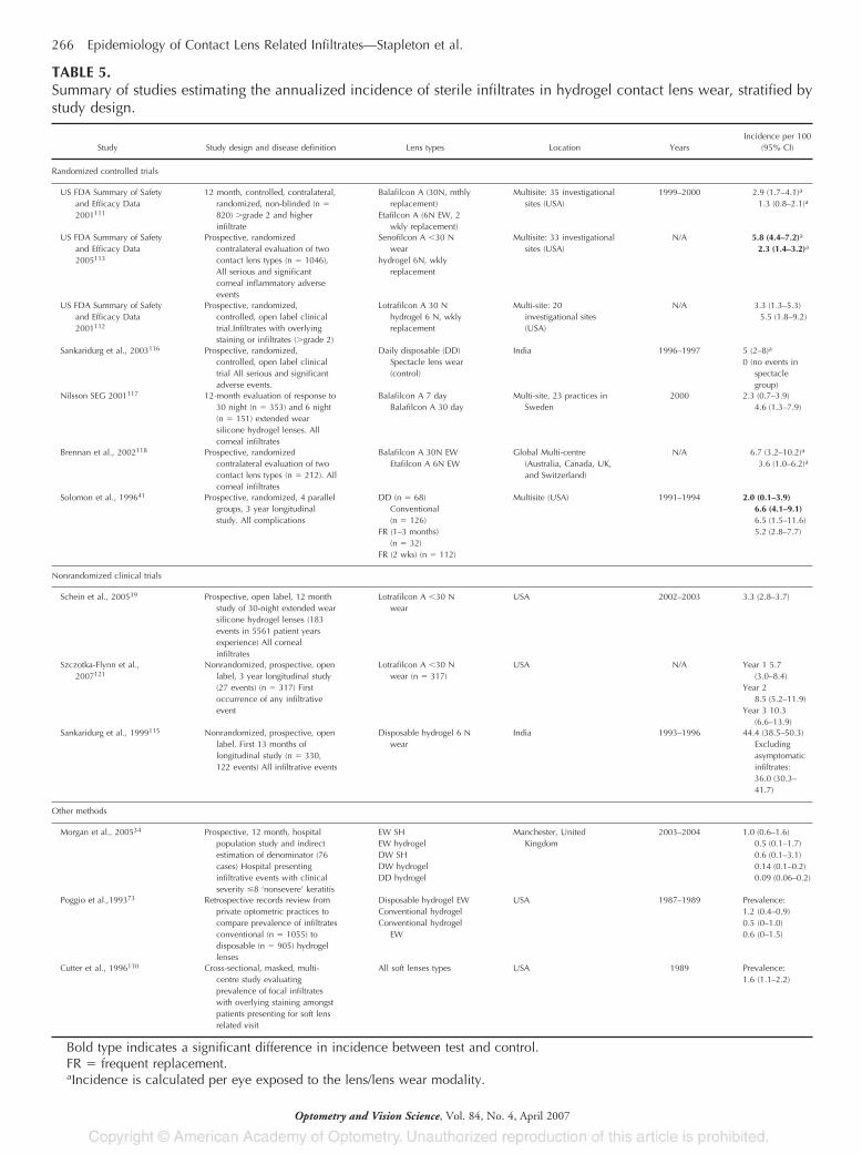
TABLE 5.Summary of studies estimating the annualized incidence of sterile infiltrates in hydrogel contact lens wear, stratified bystudy design.
Study Study design and disease definition Lens types Location YearsIncidence per 100
(95% CI)
Randomized controlled trials
US FDA Summary of Safetyand Efficacy Data2001111
12 month, controlled, contralateral,randomized, non-blinded (n �
820) �grade 2 and higherinfiltrate
Balafilcon A (30N, mthlyreplacement)
Etafilcon A (6N EW, 2wkly replacement)
Multisite: 35 investigationalsites (USA)
1999–2000 2.9 (1.7–4.1)a
1.3 (0.8–2.1)a
US FDA Summary of Safetyand Efficacy Data2005113
Prospective, randomizedcontralateral evaluation of twocontact lens types (n � 1046),All serious and significantcorneal inflammatory adverseevents
Senofilcon A �30 Nwear
hydrogel 6N, wklyreplacement
Multisite: 33 investigationalsites (USA)
N/A 5.8 (4.4–7.2)a
2.3 (1.4–3.2)a
US FDA Summary of Safetyand Efficacy Data2001112
Prospective, randomized,controlled, open label clinicaltrial.Infiltrates with overlyingstaining or infiltrates (�grade 2)
Lotrafilcon A 30 Nhydrogel 6 N, wklyreplacement
Multi-site: 20investigational sites(USA)
N/A 3.3 (1.3–5.3)5.5 (1.8–9.2)
Sankaridurg et al., 2003116 Prospective, randomized,controlled, open label clinicaltrial All serious and significantadverse events.
Daily disposable (DD)Spectacle lens wear(control)
India 1996–1997 5 (2–8)a
0 (no events inspectaclegroup)
Nilsson SEG 2001117 12-month evaluation of response to30 night (n � 353) and 6 night(n � 151) extended wearsilicone hydrogel lenses. Allcorneal infiltrates
Balafilcon A 7 dayBalafilcon A 30 day
Multi-site, 23 practices inSweden
2000 2.3 (0.7–3.9)4.6 (1.3–7.9)
Brennan et al., 2002118 Prospective, randomizedcontralateral evaluation of twocontact lens types (n � 212). Allcorneal infiltrates
Balafilcon A 30N EWEtafilcon A 6N EW
Global Multi-centre(Australia, Canada, UK,and Switzerland)
N/A 6.7 (3.2–10.2)a
3.6 (1.0–6.2)a
Solomon et al., 199641 Prospective, randomized, 4 parallelgroups, 3 year longitudinalstudy. All complications
DD (n � 68)Conventional(n � 126)
FR (1–3 months)(n � 32)
FR (2 wks) (n � 112)
Multisite (USA) 1991–1994 2.0 (0.1–3.9)6.6 (4.1–9.1)6.5 (1.5–11.6)5.2 (2.8–7.7)
Nonrandomized clinical trials
Schein et al., 200539 Prospective, open label, 12 monthstudy of 30-night extended wearsilicone hydrogel lenses (183events in 5561 patient yearsexperience) All cornealinfiltrates
Lotrafilcon A �30 Nwear
USA 2002–2003 3.3 (2.8–3.7)
Szczotka-Flynn et al.,2007121
Nonrandomized, prospective, openlabel, 3 year longitudinal study(27 events) (n � 317) Firstoccurrence of any infiltrativeevent
Lotrafilcon A �30 Nwear (n � 317)
USA N/A Year 1 5.7(3.0–8.4)
Year 28.5 (5.2–11.9)
Year 3 10.3(6.6–13.9)
Sankaridurg et al., 1999115 Nonrandomized, prospective, openlabel. First 13 months oflongitudinal study (n � 330,122 events) All infiltrative events
Disposable hydrogel 6 Nwear
India 1993–1996 44.4 (38.5–50.3)Excludingasymptomaticinfiltrates:36.0 (30.3–41.7)
Other methods
Morgan et al., 200534 Prospective, 12 month, hospitalpopulation study and indirectestimation of denominator (76cases) Hospital presentinginfiltrative events with clinicalseverity �8 ‘nonsevere’ keratitis
EW SHEW hydrogelDW SHDW hydrogelDD hydrogel
Manchester, UnitedKingdom
2003–2004 1.0 (0.6–1.6)0.5 (0.1–1.7)0.6 (0.1–3.1)0.14 (0.1–0.2)0.09 (0.06–0.2)
Poggio et al.,199373 Retrospective records review fromprivate optometric practices tocompare prevalence of infiltratesconventional (n � 1055) todisposable (n � 905) hydrogellenses
Disposable hydrogel EWConventional hydrogelConventional hydrogel
EW
USA 1987–1989 Prevalence:1.2 (0.4–0.9)0.5 (0–1.0)0.6 (0–1.5)
Cutter et al., 1996110 Cross-sectional, masked, multi-centre study evaluatingprevalence of focal infiltrateswith overlying staining amongstpatients presenting for soft lensrelated visit
All soft lenses types USA 1989 Prevalence:1.6 (1.1–2.2)
Bold type indicates a significant difference in incidence between test and control.FR � frequent replacement.aIncidence is calculated per eye exposed to the lens/lens wear modality.
266 Epidemiology of Contact Lens Related Infiltrates—Stapleton et al.
Optometry and Vision Science, Vol. 84, No. 4, April 2007
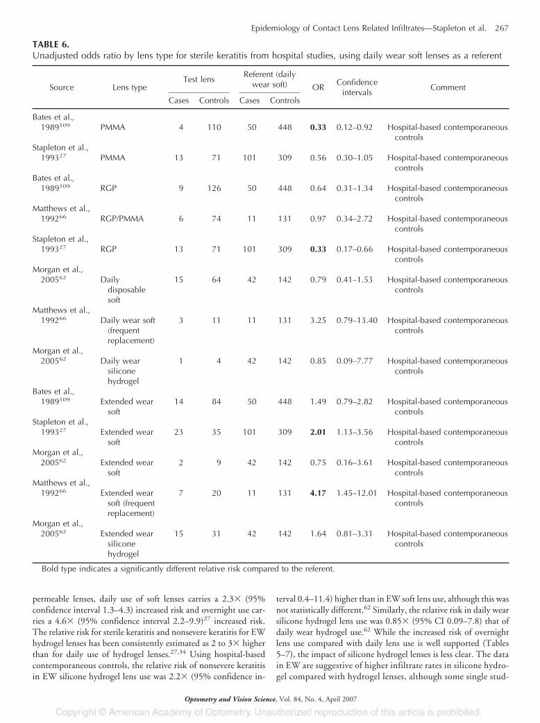
permeable lenses, daily use of soft lenses carries a 2.3� (95%confidence interval 1.3–4.3) increased risk and overnight use car-ries a 4.6� (95% confidence interval 2.2–9.9)27 increased risk.The relative risk for sterile keratitis and nonsevere keratitis for EWhydrogel lenses has been consistently estimated as 2 to 3� higherthan for daily use of hydrogel lenses.27,34 Using hospital-basedcontemporaneous controls, the relative risk of nonsevere keratitisin EW silicone hydrogel lens use was 2.2� (95% confidence in-
terval 0.4–11.4) higher than in EW soft lens use, although this wasnot statistically different.62 Similarly, the relative risk in daily wearsilicone hydrogel lens use was 0.85� (95% CI 0.09–7.8) that ofdaily wear hydrogel use.62 While the increased risk of overnightlens use compared with daily lens use is well supported (Tables5–7), the impact of silicone hydrogel lenses is less clear. The datain EW are suggestive of higher infiltrate rates in silicone hydro-gel compared with hydrogel lenses, although some single stud-
TABLE 6.Unadjusted odds ratio by lens type for sterile keratitis from hospital studies, using daily wear soft lenses as a referent
Source Lens typeTest lens
Referent (dailywear soft) OR
Confidenceintervals
Comment
Cases Controls Cases Controls
Bates et al.,1989109 PMMA 4 110 50 448 0.33 0.12–0.92 Hospital-based contemporaneous
controlsStapleton et al.,
199327 PMMA 13 71 101 309 0.56 0.30–1.05 Hospital-based contemporaneouscontrols
Bates et al.,1989109 RGP 9 126 50 448 0.64 0.31–1.34 Hospital-based contemporaneous
controlsMatthews et al.,
199266 RGP/PMMA 6 74 11 131 0.97 0.34–2.72 Hospital-based contemporaneouscontrols
Stapleton et al.,199327 RGP 13 71 101 309 0.33 0.17–0.66 Hospital-based contemporaneous
controlsMorgan et al.,
200562 Dailydisposablesoft
15 64 42 142 0.79 0.41–1.53 Hospital-based contemporaneouscontrols
Matthews et al.,199266 Daily wear soft
(frequentreplacement)
3 11 11 131 3.25 0.79–13.40 Hospital-based contemporaneouscontrols
Morgan et al.,200562 Daily wear
siliconehydrogel
1 4 42 142 0.85 0.09–7.77 Hospital-based contemporaneouscontrols
Bates et al.,1989109 Extended wear
soft14 84 50 448 1.49 0.79–2.82 Hospital-based contemporaneous
controlsStapleton et al.,
199327 Extended wearsoft
23 35 101 309 2.01 1.13–3.56 Hospital-based contemporaneouscontrols
Morgan et al.,200562 Extended wear
soft2 9 42 142 0.75 0.16–3.61 Hospital-based contemporaneous
controlsMatthews et al.,
199266 Extended wearsoft (frequentreplacement)
7 20 11 131 4.17 1.45–12.01 Hospital-based contemporaneouscontrols
Morgan et al.,200562 Extended wear
siliconehydrogel
15 31 42 142 1.64 0.81–3.31 Hospital-based contemporaneouscontrols
Bold type indicates a significantly different relative risk compared to the referent.
Epidemiology of Contact Lens Related Infiltrates—Stapleton et al. 267
Optometry and Vision Science, Vol. 84, No. 4, April 2007
ies do not reach significance in these estimates and there ispotential for confounding because of the duration of continu-ous wear in silicone hydrogel lens use. A summary of relativerisk by lens type using daily wear soft lens use as the referent isshown in Table 6.
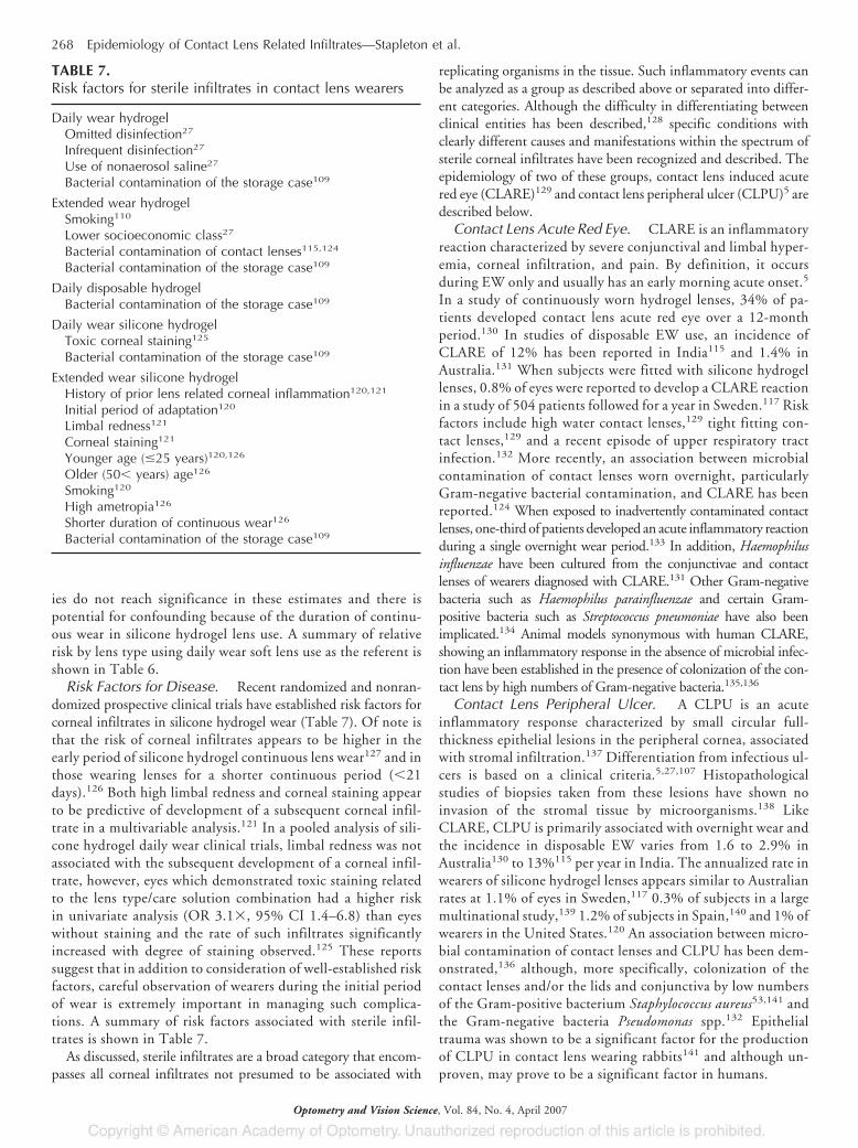
Risk Factors for Disease. Recent randomized and nonran-domized prospective clinical trials have established risk factors forcorneal infiltrates in silicone hydrogel wear (Table 7). Of note isthat the risk of corneal infiltrates appears to be higher in theearly period of silicone hydrogel continuous lens wear127 and inthose wearing lenses for a shorter continuous period (�21days).126 Both high limbal redness and corneal staining appearto be predictive of development of a subsequent corneal infil-trate in a multivariable analysis.121 In a pooled analysis of sili-cone hydrogel daily wear clinical trials, limbal redness was notassociated with the subsequent development of a corneal infil-trate, however, eyes which demonstrated toxic staining relatedto the lens type/care solution combination had a higher riskin univariate analysis (OR 3.1�, 95% CI 1.4–6.8) than eyeswithout staining and the rate of such infiltrates significantlyincreased with degree of staining observed.125 These reportssuggest that in addition to consideration of well-established riskfactors, careful observation of wearers during the initial periodof wear is extremely important in managing such complica-tions. A summary of risk factors associated with sterile infil-trates is shown in Table 7.
As discussed, sterile infiltrates are a broad category that encom-passes all corneal infiltrates not presumed to be associated with
replicating organisms in the tissue. Such inflammatory events canbe analyzed as a group as described above or separated into differ-ent categories. Although the difficulty in differentiating betweenclinical entities has been described,128 specific conditions withclearly different causes and manifestations within the spectrum ofsterile corneal infiltrates have been recognized and described. Theepidemiology of two of these groups, contact lens induced acutered eye (CLARE)129 and contact lens peripheral ulcer (CLPU)5 aredescribed below.
Contact Lens Acute Red Eye. CLARE is an inflammatoryreaction characterized by severe conjunctival and limbal hyper-emia, corneal infiltration, and pain. By definition, it occursduring EW only and usually has an early morning acute onset.5
In a study of continuously worn hydrogel lenses, 34% of pa-tients developed contact lens acute red eye over a 12-monthperiod.130 In studies of disposable EW use, an incidence ofCLARE of 12% has been reported in India115 and 1.4% inAustralia.131 When subjects were fitted with silicone hydrogellenses, 0.8% of eyes were reported to develop a CLARE reactionin a study of 504 patients followed for a year in Sweden.117 Riskfactors include high water contact lenses,129 tight fitting con-tact lenses,129 and a recent episode of upper respiratory tractinfection.132 More recently, an association between microbialcontamination of contact lenses worn overnight, particularlyGram-negative bacterial contamination, and CLARE has beenreported.124 When exposed to inadvertently contaminated contactlenses, one-third of patients developed an acute inflammatory reactionduring a single overnight wear period.133 In addition, Haemophilusinfluenzae have been cultured from the conjunctivae and contactlenses of wearers diagnosed with CLARE.131 Other Gram-negativebacteria such as Haemophilus parainfluenzae and certain Gram-positive bacteria such as Streptococcus pneumoniae have also beenimplicated.134 Animal models synonymous with human CLARE,showing an inflammatory response in the absence of microbial infec-tion have been established in the presence of colonization of the con-tact lens by high numbers of Gram-negative bacteria.135,136
Contact Lens Peripheral Ulcer. A CLPU is an acuteinflammatory response characterized by small circular full-thickness epithelial lesions in the peripheral cornea, associatedwith stromal infiltration.137 Differentiation from infectious ul-cers is based on a clinical criteria.5,27,107 Histopathologicalstudies of biopsies taken from these lesions have shown noinvasion of the stromal tissue by microorganisms.138 LikeCLARE, CLPU is primarily associated with overnight wear andthe incidence in disposable EW varies from 1.6 to 2.9% inAustralia130 to 13%115 per year in India. The annualized rate inwearers of silicone hydrogel lenses appears similar to Australianrates at 1.1% of eyes in Sweden,117 0.3% of subjects in a largemultinational study,139 1.2% of subjects in Spain,140 and 1% ofwearers in the United States.120 An association between micro-bial contamination of contact lenses and CLPU has been dem-onstrated,136 although, more specifically, colonization of thecontact lenses and/or the lids and conjunctiva by low numbersof the Gram-positive bacterium Staphylococcus aureus53,141 andthe Gram-negative bacteria Pseudomonas spp.132 Epithelialtrauma was shown to be a significant factor for the productionof CLPU in contact lens wearing rabbits141 and although un-proven, may prove to be a significant factor in humans.
TABLE 7.Risk factors for sterile infiltrates in contact lens wearers
Daily wear hydrogelOmitted disinfection27
Infrequent disinfection27
Use of nonaerosol saline27
Bacterial contamination of the storage case109
Extended wear hydrogelSmoking110
Lower socioeconomic class27
Bacterial contamination of contact lenses115,124
Bacterial contamination of the storage case109
Daily disposable hydrogelBacterial contamination of the storage case109
Daily wear silicone hydrogelToxic corneal staining125
Bacterial contamination of the storage case109
Extended wear silicone hydrogelHistory of prior lens related corneal inflammation120,121
Initial period of adaptation120
Limbal redness121
Corneal staining121
Younger age (�25 years)120,126
Older (50� years) age126
Smoking120
High ametropia126
Shorter duration of continuous wear126
Bacterial contamination of the storage case109
268 Epidemiology of Contact Lens Related Infiltrates—Stapleton et al.
Optometry and Vision Science, Vol. 84, No. 4, April 2007
CONCLUSIONS
Contact lenses clearly have optical, occupational, sporting, andcosmetic advantages for millions of wearers; however, certain riskshave been associated with their use. Given the large populationcurrently wearing contact lenses worldwide, even rare reactions canaffect large numbers of wearers. This becomes an issue for thedelivery of primary eye care and for practitioners involved in thefitting of lenses and in the management of lens related disease.Differences in risk for different types of contact lenses and wearingpatterns have been demonstrated for both rare and common lensrelated complications. This article has reviewed the epidemiologyof both microbial keratitis and sterile keratitis for contemporarycontact lens types.
Understanding the epidemiology of lens related disease, partic-ularly with the introduction of new lens types and modalities, iscrucial for practitioners to enable an informed choice of lens mo-dality, wear schedule and hygiene regimes to be made. Emergingrisk data have indicated that careful observation is important dur-ing the early period of lens wear and that early adopters of newtechnologies may show different patterns of risk. Epidemiologicaldata also provides information on the etiology of lens related com-plications, which is required to enable safer lens wear modalities tobe developed.
ACKNOWLEDGEMENTS
The authors thank Arthur Ho for graphical and technical assistance.The authors are supported by the Institute for Eye Research (FS, LK, IJ,
NC), the University of New South Wales (FS, LK), the CommonwealthGovernment through the Cooperative Research Centres Programme (VisionCooperative Research Centre; FS, LK), and the National Health and MedicalResearch Council (LK).
Received January 4, 2007; accepted February 5, 2007.
REFERENCES
1. Josephson JE, Caffery BE. Infiltrative keratitis in hydrogel lenswearers. Int Contact Lens Clin 1979;6:223–41.
2. Franks WA, Adams GG, Dart JK, Minassian D. Relative risks ofdifferent types of contact lenses. BMJ 1988;297:524–5.
3. Stapleton F, Dart J, Minassian D. Nonulcerative complications ofcontact lens wear. Relative risks for different lens types. Arch Oph-thalmol 1992;110:1601–6.
4. Stamler JF. The complications of contact lens wear. Curr OpinOphthalmol 1998;9:66–71.
5. Sweeney DF, Jalbert I, Covey M, Sankaridurg PR, Vajdic C,Holden BA, Sharma S, Ramachandran L, Willcox MD, Rao GN.Clinical characterization of corneal infiltrative events observed withsoft contact lens wear. Cornea 2003;22:435–42.
6. Keay L, Edwards K, Naduvilath T, Forde K, Stapleton F. Factorsaffecting the morbidity of contact lens-related microbial keratitis: apopulation study. Invest Ophthalmol Vis Sci 2006;47:4302–8.
7. Schein OD, Ormerod LD, Barraquer E, Alfonso E, Egan KM, PatonBG, Kenyon KR. Microbiology of contact lens-related keratitis. Cornea1989;8:281–5.
8. Khor WB, Aung T, Saw SM, Wong TY, Tambyah PA, Tan AL,Beuerman R, Lim L, Chan WK, Heng WJ, Lim J, Loh RS, Lee SB,Tan DT. An outbreak of Fusarium keratitis associated with contactlens wear in Singapore. JAMA 2006;295:2867–73.
9. Chang DC, Grant GB, O’Donnell K, Wannemuehler KA, Noble-
Wang J, Rao CY, Jacobson LM, Crowell CS, Sneed RS, Lewis FM,Schaffzin JK, Kainer MA, Genese CA, Alfonso EC, Jones DB,Srinivasan A, Fridkin SK, Park BJ. Multistate outbreak of Fusariumkeratitis associated with use of a contact lens solution. JAMA 2006;296:953–63.
10. Golden B, Fingerman LH, Allen HF. Pseudomonas corneal ulcersin contact lens wearers. Epidemiology and treatment. Arch Oph-thalmol 1971;85:543–7.
11. Cooper RL, Constable IJ. Infective keratitis in soft contact lenswearers. Br J Ophthalmol 1977;61:250–4.
12. Galentine PG, Cohen EJ, Laibson PR, Adams CP, Michaud R,Arentsen JJ. Corneal ulcers associated with contact lens wear. ArchOphthalmol 1984;102:891–4.
13. Alfonso E, Mandelbaum S, Fox MJ, Forster RK. Ulcerative keratitisassociated with contact lens wear. Am J Ophthalmol 1986;101:429–33.
14. Dart JK. Predisposing factors in microbial keratitis: the significanceof contact lens wear. Br J Ophthalmol 1988;72:926–30.
15. Koidou-Tsiligianni A, Alfonso E, Forster RK. Ulcerative keratitisassociated with contact lens wear. Am J Ophthalmol 1989;108:64–7.
16. Gebauer A, McGhee CN, Crawford GJ. Severe microbial keratitis intemperate and tropical Western Australia. Eye 1996;10 (Part 5):575–80.
17. Wong T, Ormonde S, Gamble G, McGhee CN. Severe infectivekeratitis leading to hospital admission in New Zealand. Br J Oph-thalmol 2003;87:1103–8.
18. Fong CF, Tseng CH, Hu FR, Wang IJ, Chen WL, Hou YC. Clin-ical characteristics of microbial keratitis in a university hospital inTaiwan. Am J Ophthalmol 2004;137:329–36.
19. Erie JC, Nevitt MP, Hodge DO, Ballard DJ. Incidence of ulcerativekeratitis in a defined population from 1950 through 1988. ArchOphthalmol 1993;111:1665–71.
20. Dart JK, Stapleton F, Minassian D. Contact lenses and other riskfactors in microbial keratitis. Lancet 1991;338:650–3.
21. Bourcier T, Thomas F, Borderie V, Chaumeil C, Laroche L. Bacte-rial keratitis: predisposing factors, clinical and microbiological re-view of 300 cases. Br J Ophthalmol 2003;87:834–8.
22. Keay L, Edwards K, Naduvilath T, Taylor HR, Snibson GR, FordeK, Stapleton F. Microbial keratitis predisposing factors and morbid-ity. Ophthalmology 2006;113:109–16.
23. Stapleton F. Contact lens-related microbial keratitis: what can epi-demiologic studies tell us? Eye Contact Lens 2003;29:S85–S89.
24. Lilienfield DE, Stolley PD. Foundations of Epidemiology, 3rd ed.New York: Oxford University Press; 1994.
25. Bowden T, Harknett T. Contact lens wearer profile 2004. ContLens Anterior Eye 2005;28:37–45.
26. Stapleton F, Keay L, Edwards K, Naduvilath T, Radford C, HoldenB, Dart J. Incidence of keratitis of varying severity amongst contactlens wearers [letter]. Br J Ophthalmol April 19, 2005. Available at:www.bjo.bmjjournals.com/cgi/eletters/89/4/430. Accessed Febru-ary 5, 2007.
27. Stapleton F, Dart JK, Minassian D. Risk factors with contact lensrelated suppurative keratitis. CLAO J 1993;19:204–10.
28. Daniell M. Overview: initial antimicrobial therapy for microbialkeratitis. Br J Ophthalmol 2003;87:1172–4.
29. Poggio EC, Glynn RJ, Schein OD, Seddon JM, Shannon MJ,Scardino VA, Kenyon KR. The incidence of ulcerative keratitisamong users of daily-wear and extended-wear soft contact lenses.N Engl J Med 1989;321:779–83.
30. Seal DV, Kirkness CM, Bennett HG, Peterson M. Population-based cohort study of microbial keratitis in Scotland: incidence andfeatures. Cont Lens Anterior Eye 1999;22:49–57.
Epidemiology of Contact Lens Related Infiltrates—Stapleton et al. 269
Optometry and Vision Science, Vol. 84, No. 4, April 2007
31. Cheng KH, Leung SL, Hoekman HW, Beekhuis WH, Mulder PG,Geerards AJ, Kijlstra A. Incidence of contact-lens-associated micro-bial keratitis and its related morbidity. Lancet 1999;354:181–5.
32. MacRae S, Herman C, Stulting RD, Lippman R, Whipple D,Cohen E, Egan D, Wilkinson CP, Scott C, Smith R, et al. Cornealulcer and adverse reaction rates in premarket contact lens studies.Am J Ophthalmol 1991;111:457–65.
33. Lam DS, Houang E, Fan DS, Lyon D, Seal D, Wong E. Incidenceand risk factors for microbial keratitis in Hong Kong: comparisonwith Europe and North America. Eye 2002;16:608–18.
34. Morgan PB, Efron N, Hill EA, Raynor MK, Whiting MA, TulloAB. Incidence of keratitis of varying severity among contact lenswearers. Br J Ophthalmol 2005;89:430–6.
35. Nilsson SE, Montan PG. The annualized incidence of contact lensinduced keratitis in Sweden and its relation to lens type and wearschedule: results of a 3-month prospective study. CLAO J 1994;20:225–30.
36. Efron N, Morgan PB. Impact of differences in diagnostic criteriawhen determining the incidence of contact lens-associated keratitis.Optom Vis Sci 2006;83:152–9.
37. Efron N, Morgan PB. Rethinking contact lens associated keratitis.Clin Exp Optom 2006;89:280–98.
38. Aasuri MK, Venkata N, Kumar VM. Clinical (differential) diagno-sis of microbial keratitis (MK) and contact lens induced peripheralulceration. Eye Contact Lens 2003;29:S60–S62.
39. Schein OD, McNally JJ, Katz J, Chalmers RL, Tielsch JM, AlfonsoE, Bullimore M, O’Day D, Shovlin J. The incidence of microbialkeratitis among wearers of a 30-day silicone hydrogel extended-wearcontact lens. Ophthalmology 2005;112:2172–9.
40. Keay L, Edwards K, Dart JK, Stapleton F. Validation of a clinicalgrading system for presumed contact lens related microbial keratitis(Abstract). Optom Vis Sci 2006;83:E-abstract 060003.
41. Solomon OD, Loff H, Perla B, Kellis A, Belkin J, Roth AS, ZuckerJ. Testing hypotheses for risk factors for contact lens-associated in-fectious keratitis in an animal model. CLAO J 1994;20:109–13.
42. Madigan MC, Holden BA. Reduced epithelial adhesion after ex-tended contact lens wear correlates with reduced hemidesmosomedensity in cat cornea. Invest Ophthalmol Vis Sci 1992;33:314–23.
43. Mauger TF, Hill RM. Corneal epithelial healing under contactlenses. Quantitative analysis in the rabbit. Acta Ophthalmol(Copenh) 1992;70:361–5.
44. Imayasu M, Petroll WM, Jester JV, Patel SK, Ohashi J, CavanaghHD. The relation between contact lens oxygen transmissibility andbinding of Pseudomonas aeruginosa to the cornea after overnightwear. Ophthalmology 1994;101:371–88.
45. Cavanagh HD, Ladage PM, Li SL, Yamamoto K, Molai M, RenDH, Petroll WM, Jester JV. Effects of daily and overnight wear of anovel hyper oxygen-transmissible soft contact lens on bacterial bind-ing and corneal epithelium: a 13-month clinical trial. Ophthalmol-ogy 2002;109:1957–69.
46. Latkovic S, Nilsson SE. The effect of high and low Dk/L soft contactlenses on the glycocalyx layer of the corneal epithelium and on themembrane associated receptors for lectins. CLAO J 1997;23:185–91.
47. Ladage PM, Ren DH, Petroll WM, Jester JV, Bergmanson JP,Cavanagh HD. Effects of eyelid closure and disposable and siliconehydrogel extended contact lens wear on rabbit corneal epithelialproliferation. Invest Ophthalmol Vis Sci 2003;44:1843–9.
48. Ladage PM, Jester JV, Petroll WM, Bergmanson JP, Cavanagh HD.Vertical movement of epithelial basal cells toward the corneal sur-face during use of extended-wear contact lenses. Invest OphthalmolVis Sci 2003;44:1056–63.
49. Ladage PM, Yamamoto K, Ren DH, Li L, Jester JV, Petroll WM,
Cavanagh HD. Effects of rigid and soft contact lens daily wear oncorneal epithelium, tear lactate dehydrogenase, and bacterial bind-ing to exfoliated epithelial cells. Ophthalmology 2001;108:1279–88.
50. Ren DH, Petroll WM, Jester JV, Ho-Fan J, Cavanagh HD. Therelationship between contact lens oxygen permeability and bindingof Pseudomonas aeruginosa to human corneal epithelial cells afterovernight and extended wear. CLAO J 1999;25:80–100.
51. Stapleton F, Kasses S, Bolis S, Keay L. Short term wear of high Dksoft contact lenses does not alter corneal epithelial cell size or viabil-ity. Br J Ophthalmol 2001;85:143–6.
52. Ren DH, Yamamoto K, Ladage PM, Molai M, Li L, Petroll WM,Jester JV, Cavanagh HD. Adaptive effects of 30-night wear of hyper-O(2) transmissible contact lenses on bacterial binding and cornealepithelium: a 1-year clinical trial. Ophthalmology 2002;109:27–39.
53. Jalbert I, Willcox MD, Sweeney DF. Isolation of Staphylococcusaureus from a contact lens at the time of a contact lens-inducedperipheral ulcer: case report. Cornea 2000;19:116–20.
54. Stapleton F, Edwards K, Keay L, Naviduluth T, Dart JKG, Brian G,Sweeney D, Holden BA. The incidence of contact lens related mi-crobial keratitis (Abstract). Invest Ophthalmol Vis Sci 2005;46:E-abstract 5025.
55. Keay L, Edwards K, Brian G, Stapleton F. Surveillance of contactlens related microbial keratitis in Australia and New Zealand: multi-source case capture and cost-effectiveness. Ophthalmic Epidemiol,in press.
56. Radford CF, Stapleton F, Minassian DC, Dart JKG. Risk factors forcontact lens related microbial keratitis: interim analysis ofcase–control study (Abstract). Invest Ophthalmol Vis Sci 2005;46:E-abstract 5026.
57. Schein OD, Glynn RJ, Poggio EC, Seddon JM, Kenyon KR, theMicrobial Keratitis Study Group. The relative risk of ulcerative ker-atitis among users of daily-wear and extended-wear soft contactlenses. A case-control study. N Engl J Med 1989;321:773–8.
58. Watson SL, Bunce C, Allan BD. Improved safety in contemporaryLASIK. Ophthalmology 2005;112:1375–80.
59. Hammond MD, Madigan WP Jr., Bower KS. Refractive surgery inthe United States Army, 2000–2003. Ophthalmology 2005;112:184–90.
60. Randleman JB, Russell B, Ward MA, Thompson KP, Stulting RD.Risk factors and prognosis for corneal ectasia after LASIK. Ophthal-mology 2003;110:267–75.
61. Efron N, Morgan PB, Hill EA, Raynor MK, Tullo AB. Incidenceand morbidity of hospital-presenting corneal infiltrative events as-sociated with contact lens wear. Clin Exp Optom 2005;88:232–9.
62. Morgan PB, Efron N, Brennan NA, Hill EA, Raynor MK, TulloAB. Risk factors for the development of corneal infiltrative eventsassociated with contact lens wear. Invest Ophthalmol Vis Sci 2005;46:3136–43.
63. Nilsson SE, Montan PG. The hospitalized cases of contact lensinduced keratitis in Sweden and their relation to lens type and wearschedule: results of a three-year retrospective study. CLAO J 1994;20:97–101.
64. Schein OD, Buehler PO, Stamler JF, Verdier DD, Katz J. Theimpact of overnight wear on the risk of contact lens-associated ul-cerative keratitis. Arch Ophthalmol 1994;112:186–90.
65. Buehler PO, Schein OD, Stamler JF, Verdier DD, Katz J. Theincreased risk of ulcerative keratitis among disposable soft contactlens users. Arch Ophthalmol 1992;110:1555–8.
66. Matthews TD, Frazer DG, Minassian DC, Radford CF, Dart JK.Risks of keratitis and patterns of use with disposable contact lenses.Arch Ophthalmol 1992;110:1559–62.
67. Radford CF, Minassian DC, Dart JK. Disposable contact lens use as
270 Epidemiology of Contact Lens Related Infiltrates—Stapleton et al.
Optometry and Vision Science, Vol. 84, No. 4, April 2007
a risk factor for microbial keratitis. Br J Ophthalmol 1998;82:1272–5.
68. Efron N, Wohl A, Jones L, Toma NG, Lowe R. Pseudomonascorneal ulcers associated with daily wear of disposable hydrogel con-tact lenses. Int Contact Lens Clin 1991;18:46–51.
69. Chalupa E, Swarbrick HA, Holden BA, Sjostrand J. Severe cornealinfections associated with contact lens wear. Ophthalmology 1987;94:17–22.
70. Keay L, Stapleton F. Clinical guidelines. Contact lens related micro-bial keratitis: Dallos Award Lecture, 2006. British Contact LensAssociation 2006 Clinical Conference, Birmingham, England, May19–21, 2006.
71. Edwards K, Keay L, Naduvilath T, Taylor HR, Snibson G, Staple-ton F. The relative risk of ulcerative keratitis with different lenswearing modalities at an Australian tertiary referral hospital. Pre-sented at the 22nd Cornea and Eyebank Meeting, Sydney Eye Hos-pital, Sydney, Australia, February 24–25, 2005.
72. Stapleton F, Keay L, Edwards K, Naduvilath T, Dart J, Holden BA.Risks of contact lens related microbial keratitis. Optom Vis Sci2005;82:E-Abstract 050068.
73. Poggio EC, Abelson M. Complications and symptoms in disposableextended wear lenses compared with conventional soft daily wearand soft extended wear lenses. CLAO J 1993;19:31–9.
74. Keay L, Radford CF, Dart JK, Stapleton F. Perspective on 15 yearsof research: reduced risk of microbial keratitis in frequent replace-ment contact lenses with wider use. Eye Contact Lens, in press.
75. Mayo MS, Schlitzer RL, Ward MA, Wilson LA, Ahearn DG. Asso-ciation of Pseudomonas and Serratia corneal ulcers with use of con-taminated solutions. J Clin Microbiol 1987;25:1398–400.
76. Wilson LA, Sawant AD, Simmons RB, Ahearn DG. Microbial con-tamination of contact lens storage cases and solutions. Am J Oph-thalmol 1990;110:193–8.
77. McLaughlin-Borlace L, Stapleton F, Matheson M, Dart JK. Bacte-rial biofilm on contact lenses and lens storage cases in wearers withmicrobial keratitis. J Appl Microbiol 1998;84:827–38.
78. Guillon M, Benjamin WJ. Contact lenses and keratitis. Lancet1991;338:1146–7.
79. Nason RJ, Boshnick EL, Cannon WM, Dubow BW, Freeman MI,Kame RT, Lanier JC, Lopanik RW, Quinn TG, Rigel LE, SherrillDD, Solomon OD, Stiefemeier MJ, Teiche R, Zigler LG, Yi FP.Multisite comparison of contact lens modalities. Daily disposablewear vs conventional daily wear in successful contact lens wearers.J Am Optom Assoc 1994;65:774–80.
80. Solomon OD, Freeman MI, Boshnick EL, Cannon WM, DubowBW, Kame RT, Lanier JC Jr, Lopanik RW, Quinn TG, Rigel LE,Sherrill DD, Stiegmeier MJ, Teiche RS, Zigler LG, Mertz GW,Nason RJ. A 3-year prospective study of the clinical performance ofdaily disposable contact lenses compared with frequent replacementand conventional daily wear contact lenses. CLAO J 1996;22:250–7.
81. Suchecki JK, Ehlers WH, Donshik PC. A comparison of contactlens-related complications in various daily wear modalities. CLAO J2000;26:204–13.
82. Nilsson S, Soderqvist M. Clinical performance of a daily disposablecontact lens: a 3-month prospective study. J Br Contact Lens Assoc1995;18:81–6.
83. Hingorani M, Christie C, Buckley RJ. Ulcerative keratitis in a per-son wearing daily disposable contact lenses. Br J Ophthalmol 1995;79:1138.
84. Woodruff SA, Dart JK. Acanthamoeba keratitis occurring with dailydisposable contact lens wear. Br J Ophthalmol 1999;83:1088–9.
85. Choi DM, Goldstein MH, Salierno A, Driebe WT. Fungal keratitis
in a daily disposable soft contact lens wearer. CLAO J 2001;27:111–12.
86. Su DH, Chan TK, Lim L. Infectious keratitis associated with dailydisposable contact lenses. Eye Contact Lens 2003;29:185–6.
87. Munneke R, Lash SC, Prendiville C. A case of a pseudomonascorneal ulcer in an occasional use daily disposable contact lenswearer. Eye Contact Lens 2006;32:94–5.
88. Auran JD, Starr MB, Jakobiec FA. Acanthamoeba keratitis. A reviewof the literature. Cornea 1987;6:2–26.
89. Sharma S, Srinivasan M, George C. Acanthamoeba keratitis in non-contact lens wearers. Arch Ophthalmol 1990;108:676–8.
90. Stehr-Green JK, Bailey TM, Visvesvara GS. The epidemiology ofAcanthamoeba keratitis in the United States. Am J Ophthalmol1989;107:331–6.
91. Schaumberg DA, Snow KK, Dana MR. The epidemic of Acan-thamoeba keratitis: where do we stand? Cornea 1998;17:3–10.
92. Radford CF, Lehmann OJ, Dart JK, National Acanthamoeba Ker-atitis Study Group. Acanthamoeba keratitis: multicentre survey inEngland 1992–6. Br J Ophthalmol 1998;82:1387–92.
93. Seal DV, Kirkness CM, Bennett HG, Peterson M. Acanthamoebakeratitis in Scotland: risk factors for contact lens wearers. Cont LensAnterior Eye 1999;22:58–68.
94. Radford CF, Minassian DC, Dart JK. Acanthamoeba keratitis inEngland and Wales: incidence, outcome, and risk factors. Br J Oph-thalmol 2002;86:536–42.
95. Seal DV, Beattie TK, Tomlinson A, Fan D, Wong E. Acan-thamoeba keratitis. Br J Ophthalmol 2003;87:516–17.
96. Stehr-Green JK, Bailey TM, Brandt FH, Carr JH, Bond WW,Visvesvara GS. Acanthamoeba keratitis in soft contact lens wearers.A case-control study. JAMA 1987;258:57–60.
97. Radford CF, Bacon AS, Dart JK, Minassian DC. Risk factors forAcanthamoeba keratitis in contact lens users: a case-control study.BMJ 1995;310:1567–70.
98. Jalbert I, Stapleton F, Papas E, Sweeney DF, Coroneo M. In vivoconfocal microscopy of the human cornea. Br J Ophthalmol 2003;87:225–36.
99. Mathers WD, Sutphin JE, Folberg R, Meier PA, Wenzel RP, ElginRG. Outbreak of keratitis presumed to be caused by Acanthamoeba.Am J Ophthalmol 1996;121:129–42.
100. Alfonso EC, Miller D, Cantu-Dibildox J, O’Brien T P, Schein OD.Fungal keratitis associated with non-therapeutic soft contact lenses.Am J Ophthalmol 2006;142:154–5.
101. Cho P, Cheung SW, Edwards MH. Practice of orthokeratology by agroup of contact lens practitioners in Hong Kong, Part 1: Generaloverview. Clin Exp Optom 2002;85:365–71.
102. Kwok LS, Pierscionek BK, Bullimore M, Swarbrick HA, Mountford J,Sutton G. Orthokeratology for myopic children: wolf in sheep’s cloth-ing? Clin Exp Ophthalmol 2005;33:343–7.
103. Watt K, Swarbrick HA. Microbial keratitis in overnightorthokeratology: review of the first 50 cases. Eye Contact Lens 2005;31:201–8.
104. Swarbrick HA, Wong G, O’Leary DJ. Corneal response to orthok-eratology. Optom Vis Sci 1998;75:791–9.
105. Swarbrick HA. Orthokeratology review and update. Clin Exp Op-tom 2006;89:124–43.
106. Ladage PM, Yamamoto N, Robertson DM, Jester JV, Petroll WM,Cavanagh HD. Pseudomonas aeruginosa corneal binding after 24-hour orthokeratology lens wear. Eye Contact Lens 2004;30:173–8.
107. Stein RM, Clinch TE, Cohen EJ, Genvert GI, Arentsen JJ, LaibsonPR. Infected vs sterile corneal infiltrates in contact lens wearers.Am J Ophthalmol 1988;105:632–6.
108. Robboy MW, Comstock TL, Kalsow CM. Contact lens-associatedcorneal infiltrates. Eye Contact Lens 2003;29:146–54.
Epidemiology of Contact Lens Related Infiltrates—Stapleton et al. 271
Optometry and Vision Science, Vol. 84, No. 4, April 2007
109. Bates AK, Morris RJ, Stapleton F, Minassian DC, Dart JK. ‘Sterile’corneal infiltrates in contact lens wearers. Eye 1989;3 (Part 6):803–10.
110. Cutter GR, Chalmers RL, Roseman M. The clinical presentation,prevalence, and risk factors of focal corneal infiltrates in soft contactlens wearers. CLAO J 1996;22:30–7.
111. US Food and Drug Administration. Premarket Application Sum-mary of Safety and Effectiveness Data. Pure Vision Visibility tintedcontact lens. November 20, 2001. Available at: http://www.fda.gov/cdrh/pdf/P980006S004b.doc. Accessed Janaury 19, 2007.
112. US Food and Drug Administration. Summary of Safety and Effec-tiveness. CIBAVision Focus Night and Day (lotrafilcon A) soft con-tact lens. October 12, 2001. Available at: http://www.fda.gov/cdrh/pdf/p000030b.pdf. Accessed January 19, 2007.
113. US Food and Drug Administration. Summary of Safety and EfficacyData. Vistakon (senofilcon A) Contact Lens, Clear and VisabilityTinted with UV Blocker. Available at: http://www.fda.gov/cdrh/pdf4/p040045a.pdf. Accessed February 5, 2007.
114. Vajdic CM, Sweeney DF, Cornish R, Terry RL, Hickson SB,Sulaiman S, Chong MS, Grant T, Holden BA. The incidence ofidiopathic corneal infiltrates with disposable and rigid gas permeabledaily and extended wear. Invest Ophthalmol Vis Sci 1995;36:S151.
115. Sankaridurg PR, Sweeney DF, Sharma S, Gora R, Naduvilath T,Ramachandran L, Holden BA, Rao GN. Adverse events with ex-tended wear of disposable hydrogels: results for the first 13 monthsof lens wear. Ophthalmology 1999;106:1671–80.
116. Sankaridurg PR, Sweeney DF, Holden BA, Naduvilath T, Velala I,Gora R, Krishnamachary M, Rao GN. Comparison of adverseevents with daily disposable hydrogels and spectacle wear: resultsfrom a 12-month prospective clinical trial. Ophthalmology 2003;110:2327–34.
117. Nilsson SE. Seven-day extended wear and 30-day continuous wearof high oxygen transmissibility soft silicone hydrogel contact lenses:a randomized 1-year study of 504 patients. CLAO J 2001;27:125–36.
118. Brennan NA, Coles ML, Comstock TL, Levy B. A 1-year prospectiveclinical trial of balafilcon a (Pure Vision) silicone-hydrogel contactlenses used on a 30-day continuous wear schedule. Ophthalmology2002;109:1172–7.
119. Gopinathan U, Stapleton F, Sharma S, Willcox MD, Sweeney DF,Rao GN, Holden BA. Microbial contamination of hydrogel contactlenses. J Appl Microbiol 1997;82:653–8.
120. McNally JJ, McKenney C. A clinical look at a silicone hydrogelextended wear lens. Contact Lens Spectrum 2002;17:38–41.
121. Szczotka-Flynn L, Debanne SM, Cheruvu V, Long B, Dillehay S,Barr J, Bergenske P, Donshik PC, Secor G, Yoakum J. Predictivefactors for corneal infiltrates with continuous wear of silicone hy-drogel lenses. Arch Ophthalmol, in press.
122. Skotnitsky C, Jalbert I, O’Hare N, Sweeney DF, Holden BA. Casereports of three atypical infiltrative keratitis events with high DKsoft contact lens wear. Cornea 2002;21:318–24.
123. Dumbleton K. Adverse events with silicone hydrogel continuouswear. Cont Lens Anterior Eye 2002;25:137–46.
124. Sankaridurg PR, Sharma S, Willcox M, Naduvilath TJ, SweeneyDF, Holden BA, Rao GN. Bacterial colonization of disposable softcontact lenses is greater during corneal infiltrative events than dur-ing asymptomatic extended lens wear. J Clin Microbiol 2000;38:4420–4.
125. Carnt N, Jalbert I, Stretton S, Naduvilath T, Papas EB. Solutiontoxicity in soft contact lens daily wear is associated with cornealinflammation. Optom Vis Sci, in press.
126. Chalmers RL, McNally J, Schein OD, Katz J, Tielsch JM, AlfonsoE, Bullimore M, O’Day D, Shovlin J. Risk factors for corneal infil-
trates with continuous wear of contact lenses. Ophthalmology, inpress.
127. McNally JJ, Chalmers RL, McKenney CD, Robirds S. Risk factorsfor corneal infiltrative events with 30-night continuous wear of sil-icone hydrogel lenses. Eye Contact Lens 2003;29:S153–S156.
128. Efron N, Morgan PB. Can subtypes of contact lens-associated cor-neal infiltrative events be clinically differentiated? Cornea 2006;25:540–4.
129. Zantos SG. Management of corneal infiltrates in extended-wearcontact lens patients. Int Contact Lens Clin 1984;11:604–12.
130. Zantos SG, Holden BA. Ocular changes associated with continuouswear of contact lenses. Aust J Optom 1978;61:418–26.
131. Sankaridurg PR, Holden BA, Jalbert I. Adverse effects andinfections: which ones and how many? In: Sweeney DF, ed. SiliconeHydrogels: Continuous Wear Contact Lenses, 2nd ed. Oxford:Butterworth-Heinemann; 2004:217–74.
132. Sankaridurg PR, Willcox MD, Sharma S, Gopinathan U, Janakira-man D, Hickson S, Vuppala N, Sweeney DF, Rao GN, Holden BA.Haemophilus influenzae adherent to contact lenses associated withproduction of acute ocular inflammation. J Clin Microbiol 1996;34:2426–31.
133. Holden BA, La Hood D, Grant T, Newton-Howes J, Baleriola-Lucas C, Willcox MD, Sweeney DF. Gram-negative bacteria caninduce contact lens related acute red eye (CLARE) responses. CLAOJ 1996;22:47–52.
134. Sankaridurg PR, Sharma S, Willcox MD, Sweeney DF, NaduvilathTJ, Holden BA, Rao GN. Colonization of hydrogel lenses withStreptococcus pneumoniae: risk of development of corneal infiltrates.Cornea 1999;18:289–95.
135. Harmis NY, Sankaridurg PR, Willcox MD. The development of ananimal model for CLARE. Microbiol Aust 2000;21:A152.
136. Willcox MD, Sankaridurg PR, Zhu H, Hume EB, Cole N,Conibear T, Glasson M, Harmis N, Stapleton F. Inflammation andinfection and the effects of the closed eye. In: Sweeney DF, ed.Silicone Hydrogels: Continuous Wear Contact Lenses, 2nd ed.Oxford: Butterworth Heinemann; 2004:90–125.
137. Grant T, Chong MS, Vajdic C, Swarbrick HA, Gauthier C,Sweeney DF, Holden BA. Contact lens induced peripheral ulcersduring hydrogel contact lens wear. CLAO J 1998;24:145–51.
138. Holden BA, Reddy MK, Sankaridurg PR, Buddi R, Sharma S, WillcoxMD, Sweeney DF, Rao GN. Contact lens-induced peripheral ulcerswith extended wear of disposable hydrogel lenses: histopathologic ob-servations on the nature and type of corneal infiltrate. Cornea 1999;18:538–43.
139. Long B, Robirds S, Grant T. Six months of in-practice experiencewith a high Dk lotrafilcon a soft contact lens. Cont Lens AnteriorEye 2000;23:112–18.
140. Montero Iruzubieta J, Nebot Ripoll JR, Chiva J, Fernandez OE,Rubio Alvarez JJ, Delgado F, Villa C, Traverso LM. Practical expe-rience with a high Dk lotrafilcon A fluorosilicone hydrogel extendedwear contact lens in Spain. CLAO J 2001;27:41–6.
141. Wu P, Stapleton F, Willcox MD. The causes of and cures for contactlens-induced peripheral ulcer. Eye Contact Lens 2003;29:S63–S66.
Fiona StapletonSchool of Optometry and Vision Science
University of New South WalesLevel 3, Rupert Myers Building
North Wing, Gate 14, Barker St.Sydney, NSW 2052, Australia
e-mail: [email protected]
272 Epidemiology of Contact Lens Related Infiltrates—Stapleton et al.
Optometry and Vision Science, Vol. 84, No. 4, April 2007