the effect osf sodium-free solutions on the …jeb.biologists.org/content/jexbio/58/1/1.full.pdf ·...
TRANSCRIPT
J. Exp. Biol. (1973), 58, 1-14 IWith 10 text-figuresPrinted in Great Britain
THE EFFECTS OF SODIUM-FREESOLUTIONS ON THE FAST ACTION POTENTIALS OF
VIVIPARUS CONTECTUS (MILLET)(GASTROPODA: PROSOBRANCHIA)
BY D. B. SATTELLE
A.R.C. Unit of Invertebrate Chemistry and Physiology,Department of Zoology, Cambridge
{Received 9 May 1972)
INTRODUCTION
In conventional excitable cells, such as the squid axon, the inward current of theaction potential is attributable to a rapid influx of sodium ions (cf. Hodgkin, 1951,1964). The replacement of sodium in the saline bathing this preparation by electrolyteor non-electrolyte substitutes results predictably in a rapid decline of the actionpotential (Hodgkin & Katz, 1949). Effects of sodium-free solutions on conductionprocesses in a number of other molluscan nerve preparations have also been explored(cf. Kostyuk, 1968; Treherne & Moreton, 1970). The results obtained are compli-cated, however, by the discovery in the nerve cell bodies of several pulmonate andopisthobranch gastropod molluscs of action potentials which depend on a transiententry of one or more species of cation other than sodium. In the cell bodies ofOnchidium verruculatum (Oomura, Ozaki & Maeno, 1961) and Helix pomatia (Gera-simov, Kostyuk & Maisky, 1964, 1965 a, b; Meves, 1966, 1968; Krishtal & Magura,1970), for example, calcium ions appear to account for the inward component of theaction current. Action potentials dependent upon an influx of both sodium andcalcium have been demonstrated in Aplysia californica (Junge, 1967; Geduldig &Junge, i968)and certain neurones of Helix aspersa (Chamberlain & Kerkut, 1967,1969;Moreton, 1972). Sodium, calcium and magnesium ions have been implicated in theactive membrane response recorded from nerve cell bodies of Limnaea stagnalis(Jevelova, Krasts & Veprintsev, 1971; Sattelle, 1973 a). Conventional sodium-dependent action potentials have been reported for certain neurones of Helix aspersa(Moreton, 1968,1972) and Tritonia diomedia (Magura & Gerasimov, 1966; Veprintsevet al. 1966; Krasts & Veprintsev, 1972).
Further problems of interpreting the effects of sodium-free solutions on conductionprocesses in molluscan neurones arise with the demonstration of a functional sodiumreservoir, available to certain axons within the central nervous system of the fresh-water lamellibranch Anodonta cygnea (Treherne, Carlson & Gupta, 1969). Thissodium store has been identified using electronmicroscopical, radioisotopic andspectrophotometric techniques in addition to electrophysiological investigations whichhave explored the different effects of electrolyte and non-electrolyte sodium substi-tutes on the conducted action potentials (Carlson & Treherne, 1969; Mellon &Treherne, 1969; Treherne, Mellon & Carlson, 1969). Sodium from this source is
I EXB J 8
2 D. B. SATTELLE
available only to the larger (2-4 /an diameter) axons of the cerebro-visceral connectives and has tentatively been located as follows: a readily accessible componentassociated with non-diffusible anions in the immediate vicinity of the axon surfaces;also a component not readily accessible, requiring mobilization, and sequestered inthe glial elements which exhibit much stronger association with the relatively fewlarger diameter axons than with the bulk of the smaller axons of the connectives(Gupta, Mellon & Treherne, 1969; Treherne, Carlson & Gupta, 1969).
An investigation into the ionic basis of axonal function in the central nervous systemof the prosobranch gastropod Viviparus contectus has established a strong dependenceof the conducted spike on the presence of sodium in the bathing medium (Sattelle,1972 a). In spite of this, dextran Ringer or isotonic dextran solutions are able to sustaincompound action potentials for extended periods (Sattelle, 1972). Differences haveemerged between the findings for Anodonta connectives and Viviparus connectives.Whilst in the latter preparation both fast and slow fibres can maintain conduction indextran solutions by a sodium-dependent process (Sattelle, 1972), this ability isconfined to the fast fibres of Anodonta (Treherne et al. 1969). Any such reservoir ofsodium in the central nervous system of Viviparus would therefore need to be moreextensive than that postulated to exist in Anodonta connectives. Also, the tentativelocation of the less accessible component of the sodium store in the glial elementsassociated with the larger axons of Anodonta could not apply to Viviparus, where asparse distribution of glial elements has been observed throughout the connective(Sattelle & Lane, 1972). An intimate glial investment of all axons would be expectedif a glial-mediated mechanism controlled the ionic composition of the extracellularfluid in the connective of this prosobranch mollusc. This paper describes an investiga-tion into the effects of electrolyte and non-electrolyte sodium substitutes on the fastaction potentials of the pleural-supraintestinal connective of Viviparus contectus.Experiments have been confined to the fast component of the compound actionpotential because it is much less variable than the slow component (Sattelle, 1972).The purpose of this study is to determine whether or not the maintenance of conductionprocesses under non-electrolyte, sodium-free conditions constitutes a physiologicalmechanism for regulation of the ionic micro-environment of the nervous elements.
MATERIAL AND METHODS
Records of fast action potentials were obtained from isolated pleural-supraintestinalconnectives of Viviparus contectus. Techniques employed for dissection and isolationof the connectives and extracellular recording and display of the fast component ofthe compound action potential were as described in a previous report (Sattelle, 1972).That account included a description of the perspex preparation chamber used inthe present study and details of its perfusion. Changes in weight of the preparationin different bathing media were investigated using dissected lengths (8-10 mm) ofpleural-supraintestinal connective. These were carefully blotted and rapidly weighedon an electrobalance (range 0-2-5 mE)- Each weighing recorded was the average ofthree successive readings. The normal Ringer solution used was based on a salinedevised for Viviparus viviparus (Little, 1965) with the following composition: 24-0 IHM/1NaCl; 9-0 mM/1 NaHCO3; 1-2 min/1 KC1; 5-5 nm/l CaCl2; pH 7-4. This saline main-

Fast action potentials in Viviparus
SodiumRinger Dextran Ringer
Time (min)
Fig. i. Effects on the fast action potentials of prolonged exposure to dextran Ringer. A plot ofrelative conduction velocity (0u»t/0nonn«j) against time following replacement of the sodiumsalts of normal Ringer by dextran. The vertical lines represent twice the standard error. Insetshows records of fast action potentials; scale bars represent ioomsec (horizontally) andioo fiV (vertically).
tained conduction in the fast fibres of Viviparus contectus for extended periods withlittle change in conduction velocity (Sattelle, 1972).
RESULTSEffects of dextran and tris
When the sodium salts of normal Ringer were replaced by dextran (to maintainisosmotic conditions*) fast action potentials were maintained for long periods. Theconduction velocity (8) fell to about 50 % of its value in normal Ringer during a 2 hexposure to dextran Ringer. The decline in 6 was greatest over the first 30 min of theexperiment and thereafter a much steadier decline was observed (Fig. 1).
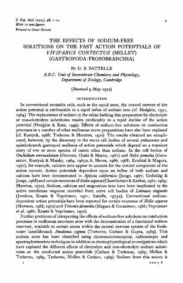
Experiments were conducted in which the sodium salts in normal Ringer werereplaced in stages by increasing concentrations of either tris chloride or dextran, thusmaintaining isosmotic conditions, and the effects on 6 were observed. The resultswere plotted as relative conduction velocity (#test/̂ normai) against the square root ofthe relative external sodium concentration (Na+
test/Na+nonnal)i following the methodused for Anodonta connectives (Carlson & Treherne, 1969; Treherne et al. 1969) andenabling a comparison with the data obtained for squid (Loligo) axons (Hodgkin &Katz, 1949) (Fig. 2). When tris replaced sodium the fast fibres of Viviparus exhibited
• The terms isosmotic and isotonic as used in this paper follow the definitions given by Potts & Parry(1964). ' Isosmotic conditions' describe, for example, the replacement of sodium salts in normal Ringerby a concentration of electrolyte or non-electrolyte substitute calculated to maintain normalosmolarity. 'Isotonic conditions' refer to the use of an experimentally determined concentration ofsubstitute which produces neither swelling nor shrinkage of the connective.
D. B. SATTELLE
1-2 -,
1 0 -
2 0 - 8 -
£
4 . 0 -6-
*" 0 - 4 -
0 - 2 -
0
.•••' Data for squid axon(Hodgkin & Katz, 1949)
l0-2 0-4 0-6
I
0-81
10i
1-2
(Na+ al )
Fig. a. Effects on axonal conduction of varying the external sodium concentration. Trischloride (closed circles) and dextran (open circles) replace the sodium salts of normal Ringer.The curve for the squid (Loligo) axon is derived from measurements of the maTi'mnm rate ofrise of the action potentials based on the assumption that, in a simplified theoretical system,the conduction velocity (ff) is related to the square-root of the rate of rise of the action potential(Hodgkin & Katz, 1949). The vertical lines represent twice the standard error.
a similar response to the squid axon. In fact, the curve obtained was somewhat dis-placed and approximated more closely to the data obtained for Carcinus axons (Katz,1947). With dextran as a substitute for sodium salts, however, a straight-line relation-ship was obtained and when all the sodium had been replaced action potentials werestill conducted (Fig. 2). These effects of tris and dextran on the fast action potentials ofViviparus closely correspond to those observed for the fast action potentials of thecerebro-visceral connective of Anodonta under similar conditions (Carlson & Treherne,1969).
Effects of a range of non-electrolyte sodium substitutes
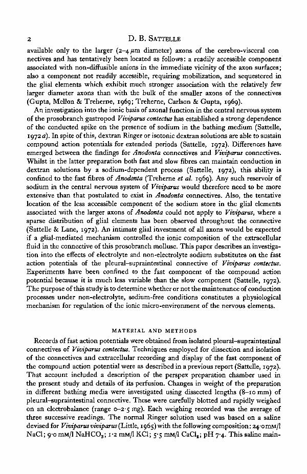
To investigate further the discrepancy between the effects on conduction velocityof tris and dextran, a range of non-electrolyte sodium substitutes was employed. Theresults have been summarized in Fig. 3 as plots of (̂ test/̂ normai) against (Na+^gJNa+normal)i. Replacing the sodium salts in normal Ringer by mannitol, sucrose andraffinose produced much the same effect. In the presence of these non-electrolytes thefast action potentials were maintained at lower concentrations of external sodium ionsthan when tris was used to replace sodium. With inulin as the substitute, this trendcontinued and conduction block was only achieved in the complete absence of sodiumin the bathing medium. Only dextran of the range of non-electrolyte substances in-vestigated supported fast action potentials in the complete absence of sodium in thebathing medium.
Changes in weight of connectives in sodium-free Ringer
To test whether or not truly isotonic* conditions prevailed during exposure tosodium-free solutions, weight changes of connectives were followed. Plots of
\ Test weight {Wt) is the weight of the connective (average of three successive readings on theelectrobalance) in the test (sodium-free) solution and initial weight (Wt) is the weight of the connectivein normal Ringer at the start of the experiment.

Fast action potentials in Viviparus
1 0 -
0 - 8 -
1 0 - 6 -
J 0-4-
0-2 -
Dextran _ _ — •
• / / •
/" /
Tris chlorideMannitolSucroseRaffinoseInulin
0-21
0-4
(Na+tn, /Na
i0-6
normal)
10 8 10
Radius ofequivalent
sphere(A)1-4*4"4*5-7*
i5-3#
22-S+
DTIDfor 2OO Achannel
0 9 5o-860 8 30 6 20-50
Dr/Dfor 100 Achannel
0-91o-7s0-700-400-26
Fig. 3. Effects on axonal conduction of a range of sodium substitutes, each of which replaces instages the sodium salts of normal Ringer. Results are summarized as plots of the relativeconduction velocity (̂ t«t/̂ nonnmi) against the square root of the relative sodium concentrationof the Ringer (Na+taa/Na"1",,,,,,^)'. To facilitate a comparison of the effects of the differentnon-electrolytes, individual points (standard errors less than o-i units on vertical axis) areomitted and the final curves only are represented. The graphs summarize experiments on33 isolated, pleural-supraintestinal connectives.
Table 1. Molecular dimensions and ratios of restricted (Dr) to free (D) diffusioncoefficients for various sodium substitutes
Molecularspecies
NaClSucroseRaffinoseInulinDextran
• Data from Pappenheimer (1953).•J" Data from Ingleman & Hailing (1949).
against time were obtained (Fig. 4). During 30 min exposures to bathing media inwhich sodium salts were replaced by calculated isosmotic concentrations of trischloride, mannitol, sucrose, raffinose, inulin and dextran, a loss in weight of the con-nectives occurred which could be abolished by re-exposure to normal Ringer. Themost pronounced and rapid changes in weight of the connectives were observed whendextran replaced the sodium salts. This result is clearly of interest in the light of theelectrophysiological findings for Viviparus axons in dextran Ringer.
Changes in weight of connectives in mannitol and dextran
To determine the truly isotonic concentrations of two of the non-electrolyte sodiumsubstitutes, changes in weight of Viviparus connectives were studied during exposureto a range of concentrations of mannitol and dextran in distilled water (Figs. 5, 6).Connectives bathed in concentrations of mannitol in the region of 30 mM/l exhibited

D. B. SATTELLE
Ringer , Sodium-free Ringer Ringer
10 20 30Time fmin)
40 50 60
Fig. 4. Changes in weight of connectives during 30 min exposures to sodium-free Ringer. Thevarious sodium substitutes are indicated on the graphs. Results have been expressed as plotsof WtIWt (test weight/initial weight).
200 r
150
- 100to
50
00 20 40 60 80
mM/1 mannitol
Fig. 5. Changes in weight of connectives in mannitol solutions. Weights are recorded as apercentage of the initial weight of the connective in normal Ringer. An equilibration time of15 min i» allowed before weighing.

wei
ght
niti
al
200
150
100
50
0
Fast
- i
K
action potentials
i i
m Viviparus
i
—o
j20 40
rnM,'l dextran60 80
Fig. 6. Changes in weight of connectives in dextran solutions. Weights are recorded as a per-centage of the initial weight of the connective in normal Ringer. An equilibration time ofi s min is allowed before weighing.
Ringer Sodium-free Ringer Ringer
Mannitol30 mil
66 mM
i •—i r
10 20 30
Time (min)
40I
50 60
Fig. 7. Changes in weight of connectives during exposure to Ringer solutions in whichisotonic (open circles) and isosmotic (closed circles) concentrations of mannitol and dextranreplace the sodium salts.
minimal weight changes. When exposed to a range of dextran solutions, connectivesmaintained constant weight at concentrations of about 8 mM/1. Ringer solutions inwhich the truly isotonic concentrations of mannitol and dextran have been used toreplace sodium salts induced, over a period of 30 min, very small changes in weightof the connectives (Fig. 7).
D. B. SATTELLE
1 0 -
0 - 8 -
0 - 6 -
C 0-4 -
0-2 -
8 mM/1 \ _ .i ; D e x t r a n66mM/i
30 mM/1Mannitol
0-2 0-4 0-6 0-8 10
Fig. 8. Effects on axonal conduction of using isosmotic and isotonic concentrations of mannitoland dextran to replace in stages the sodium salts of normal Ringer. Individual points (standarderrors less than o-i units on vertical axis) are omitted and final curves only are represented.
10
0-8
! 0 6
0-2
— • Intermittent stimulation
° Intermittent stimulation withbursts of prolonged stimulation
lsec
1 sec1 sec-
30 60 90Time (min) in dextran Ringer
120
Fig. g. Effects of stimulation on axonal conduction in dextran Ringer. Bursts of stimulation ati sec"1 and of 5 min and 7-5 min duration are applied to a connective exposed to dextranRinger. The resultant changes in relative conduction velocity are recorded (open circles).For comparison, the changes in relative conduction velocity incurred during exposure todextran Ringer with only intermiucr.t stimulation are illustrated (closed circles).
Effects of isotonic (experimentally determined) concentrations of mannitol and dextran onfast action potentials
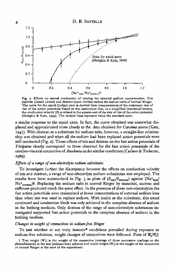
When 30 mM/1 concentrations of mannitol were employed to replace the sodiumsalts in normal Viviparus Ringer, the relationship between (#test/̂ normai) a ndNa+normal)i shown in Fig. 8 was obtained. With this concentration of mannitol as asubstitute, fast action potentials were maintained at somewhat lower sodiumconcentrations than was the case when 66 mM/1 mannitol was used. There was verylittle difference between the effects of 8 and 66 mM/1 concentrations of dextran whenthese replaced the sodium salts of normal Ringer. The findings using 30 mM/1concentrations of mannitol emphasized the difference, partly obscured in earnerwork by the use of higher concentrations, between the effects of mannitol and of trischloride (cf. Fig. 3) on the fast action potentials.

Fast action potentials in Viviparus
1 0 -
0-8-
0-6-
0-4-
0-2-
0 -
~\ I-'-t ( 1 t, , 1 J
-\ i vri
301
60
Time (min)
— I "90
" I120
Fig. io. Effects on axonal conduction of intermittent exposure to dextran Ringer. A connectiveis exposed to tris Ringer for z h apart from 5 min periods (at 15 min intervals) when it issubjected to dextran Ringer. Changes in relative conduction velocity are followed throughoutthe experiment (solid line) and the results have been compared to those obtained when con-nectives are exposed continuously to dextran Ringer (open circles, dotted line). Arrowsindicate the beginning ( J ) and the end (f ) of a period of substitution of tris Ringer bydextran Ringer. Vertical lines represent twice the standard error.
Effects of stimulation on the fast action potentials
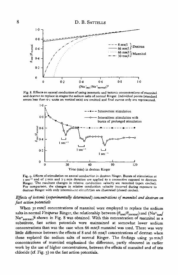
The effect of stimulation on the ability of the fast fibres to maintain their functionin dextran Ringer was investigated (Fig. 9). A 5 min burst of stimulation at 1 sec"1
following a 20 min exposure of connectives to dextran Ringer produced some reduc-tion in the conduction velocity of the fast axons which recovered when stimulationceased. After 45 min, stimulation at the same rate for i\ min produced an irreversibledecline in conduction velocity and a similar reduction was induced by a 5 min burstof stimulation following 75 min of exposure to dextran Ringer. Whatever the sourceof sodium maintaining fast action potentials in dextran Ringer, this cation is clearlyabundant in the extra-neuronal fluid, although stimulation may lead to some depletionof the available sodium.
Effects on the fast action potentials of intermittent exposure to dextran Ringer
Connectives were exposed to sodium-free (tris) Ringer for a 2 h period. Conductionblock in the fast fibres rapidly ensued, but exposure to dextran Ringer for briefperiods at 15 min intervals restored activity (Fig. 10). The decline of conductionvelocity in these restored fast fibres closely followed the normal pattern of declinewhen fibres were maintained in dextran Ringer throughout the same period.
DISCUSSION
The effects of a range of sodium substitutes on the fast action potentials of thepleural-supraintestinal connective of Viviparus contectus have been investigated. Thebehaviour of these action potentials on exposure to a range of sodium-free (tris)solutions in which the proportion of tris is steadily increased up to the point ofcomplete replacement of sodium resembles that of the active membrane responsesrecorded under comparable conditions from conventional excitable cells such as thesquid (Loligo) axon (Hodgkin & Katz, 1949) and the crab {Carcinus) axon (Katz,

io D. B. SATTELLE
1947). With dextran as the sodium substitute, however, the behaviour of the fastfibres departs considerably from the conventional situation and complete replacementof the sodium salts of normal Ringer by this non-electrolyte does not produceconduction block. Fast action potentials are maintained by a sodium-dependentprocess for extended periods under these conditions (cf. Sattelle, 1972); a 2hexposure to dextran Ringer producing only a 50 % decline in the conduction velocity(6) of the fast fibres. It has been observed that the effects of dextran on the fast axonsof Viviparus are atypical for the range of non-electrolyte sodium substitutes employed.When mannitol, sucrose, rafnnose and inulin replace sodium, fast action potentialsare rapidly abolished, in contrast to the findings for connectives of Anodonta cygneawhere both sucrose and dextran are able to maintain the conduction of fast fibres(Treherne et al. 1969; Carlson & Treherne, 1969). The different effects of non-electrolyte sodium substitutes on action potentials of Viviparus contectus are not,therefore, readily explained in terms of the functional sodium store proposed toaccount for the results obtained in the fast axons of Anodonta cygnea (Carlson &Treherne, 1969; Treherne, Carlson & Gupta, 1969).
The anomalous behaviour of Viviparus axons when exposed to various non-electro-lytes may result from shrinkage of the nervous tissue. This possibility is raised withthe demonstration that weight losses of connectives occur when isosmotic concentra-tions of mannitol, sucrose, raffinose, inulin and dextran replace the sodium salts ofnormal Ringer and the largest and most rapid changes accompany substitution bydextran. Adjusting to isotonic concentrations of mannitol and dextran does not, how-ever, greatly affect the response of the fast fibres to these non-electrolytes. For example,axonal function is still maintained when isotonic dextran completely replaces sodium,suggesting that the changes in weight of the connectives associated with the use ofisosmotic concentrations of dextran are not the primary cause of this phenomenon.
Clearly a major problem in interpreting the effects of dextran is that none of theother non-electrolyte sodium substitutes employed produces the same response in thelarge axons of Viviparus. In a first attempt to account for this it is assumed thatdextran (M.W. IOOOO) alone of these non-electrolytes fails to penetrate the extracellularsystem. An explanation of the results may then be sought in terms of the withdrawalof ions and water from the nervous elements in response to concentration and osmoticgradients arising from the exclusion of dextran. The Stokes radius of this dextran,which refers to the hydrated form, has been estimated at 22-5 A (Ingleman & Hailing,1949). The molecular size (45 A in diameter) of this non-electrolyte is not, therefore,insignificant in comparison with the dimensions (100-200 A in width) of the out-wardly facing clefts of the extracellular system (Sattelle & Lane, 1972). Its completeexclusion from the extracellular system does nevertheless seem a remote possibility inthe apparent absence of any ultrastructural evidence for restriction of the inter-cellular clefts (cf. Sattelle & Lane, 1972). Such a view is given support by the demon-stration that the exogenous tracer molecule macroperoxidase (M.W. 40000) penetratesthe extracellular system of the cerebro-visceral connective of Anodonta (Lane &Treherne, 1972) in which the dimensions of the extracellular channels (150 A inwidth) (Gupta et al. 1969) closely resemble those referred to above for Viviparus.
If, as seems likely, dextran penetrates the extracellular system, the sodium involvedin action potential production in dextran Ringer may be derived from sodium bound

Fast action potentials in Viviparus 1i
to fixed negative charges located either on the axonal membrane or in close proximityto its surface. It is well known, for example, that membrane-bound fragments fromvertebrate central nervous tissues contain tightly bound cations, and that boundsodium and potassium are available to the Na+, Reactivated ATPase (Rodnight,Carrera & Goldfarb, 1969). More recently a cell-surface coating of an ionic naturehas been detected as a ' surface potential' of lymphocytes and trophoblastic cells usingconventional microelectrode recording techniques (Hause et al. 1970). Also the poten-tial role of indiffusible anions as cation reservoirs in the central nervous system ofmolluscs has been suggested for Helix aspersa (Chamberlain & Kerkut, 1967, 1969)and Anodonta cygnea (Carlson & Treherne, 1969; Treherne et al. 1969). Sodiumattached to such anion groups would be expected to exchange with tris ions but notwith dextran, which might account for the observed eflFects of these sodium substituteson the fast action potentials of Viviparus. Conduction failure in all the other non-electrolyte sodium substitutes nevertheless remains difficult to reconcile with thisexplanation.
Retaining the assumption that dextran penetrates the extracellular system, it isnecessary to consider whether or not its rate of entry into the connective is retardedeither by its molecular dimensions in relation to the size of the outwardly facingintercellular clefts or by its diffusivity. A simplified analysis of the eflFects of sterichindrance and viscous drag for the case of spherical particles entering circular openingsis discussed by Pappenheimer (1953), who accounts for these eflFects in terms of arestriction to free diffusion given by the equation
DT (i-a/xfD 1 + 2-40/*'
where Dr/D is the ratio of diffusion coefficients for restricted (Dr) and free (D)diffusion, a is the radius of the particle and x is the radius of the channel. The ratiosDJD have been calculated for a number of the non-electrolyte sodium substitutes(Table 1) and allowance is made for the maximum and minimum observed widths ofthe intercellular clefts of Viviparus contectus (cf. Sattelle & Lane, 1972). Restriction tofree diffusion appears to be considerably greater for dextran than for sodium chloridealthough it should be pointed out that the above analysis refers to circular openingsrather than elongate clefts.
A value of 1*483 x io~° cm2/sec* has been obtained for the diffusion coefficient ofsodium chloride at a concentration of o-i M in aqueous solution at 25 °C. To a reason-able approximation, the diffusion coefficient (D) varies inversely as the cube root ofthe molecular weight (M) (cf. Clark, 1952). From a plot of D against M for varioussugars and proteins, a diffusion coefficient of approximately 0-135 x Io~* cm2/sec isobtained for dextran (M.W. 10 000). Thus a combination of steric hindrance, viscousdrag and low diffusivity could markedly slow down the entry of dextran into thecentral nervous tissues of Viviparus, which may contribute to its observed effects onaxonal conduction. A direct effect of dextran in, for example, inducing a leakage ofions into the extracellular spaces even under isotonic conditions should not be dis-counted. It has been shown that dextran Ringer rapidly restores axonal function whenapplied in short bursts at fifteen minute intervals during a 2 h exposure to tris Ringer.
• From Handbook of Chemistry and Physics (44th ed.)( the Chemical Rubber Publishing Co. (196a).
12 D. B. SATTELLE
The relative conduction velocity (̂ test/̂ normai) quickly attains a level very similar tothat produced by continuous perfusion of Viviparus connectives with dextran Ringer.Also, in this context it may be relevant to note the recent observations of Pooler &Oxford (1972) on the lobster axon under sucrose-gap conditions. Careful measure-ments of membrane current indicated the presence of a parallel leakage current attri-butable to a leaching of ions from the axon into the bathing sucrose solution. If asimilar withdrawal of intracellular ions takes place in the connective of Viviparus itmust be much greater in the presence of dextran than in the presence of the lowermolecular weight non-electrolytes.
It nevertheless appears that under normal physiological conditions all the axons ofthe pleural-supraintestinal connective of Viviparus contectus exist in a fluid compart-ment that is readily accessible to ions and small molecules in the bathing medium(cf. also Sattelle, 1972). The maintenance of axonal conduction in sodium-free(dextran) Ringer is not therefore considered to be an example of a physiologicalmechanism of control of the ionic environment of the nervous elements of thisprosobranch mollusc.
SUMMARY
1. A 2 h exposure of connectives of Viviparus contectus to sodium-free Ringer, inwhich dextran maintains osmolarity, results in a decline in the conduction velocityof the fast axons to 50% of the value obtained in normal Ringer.
2. The changes in conduction velocity of the fast axons in response to the replace-ment in stages of the sodium salts of normal Ringer correspond to those exhibitedby conventional excitable cells when tris is the substitute, but depart considerablyfrom this behaviour when dextran is the substitute.
3. Experiments employing mannitol, sucrose, raffinose, inulin and dextran toreplace the sodium salts of normal Ringer reveal that dextran alone of these non-electrolytes maintains fast action potentials in the absence of sodium.
4. Weight losses of connectives under sodium-free (isosmotic) conditions aregreatest and are most rapidly achieved when dextran (of the range of non-electrolytesstudied) replaces the sodium salts of normal Ringer. Adjusting to isotonic conditions(8 mM dextran) does not diminish the ability of this non-electrolyte to maintain fastaction potentials.
5. During prolonged perfusion of connectives by tris Ringer brief (5 min) exposuresto dextran Ringer at 15 min intervals rapidly restore fast action potentials. Thedecline of conduction velocity in these restored fibres closely follows the normalpattern of decline when function is maintained continuously in dextran Ringer.
I am indebted to Dr J. E. Treherne for his advice and constructive criticism through-out the course of this work and I thank Dr R. W. Meech and Dr R. B. Moreton forhelpful discussions. The financial support of the Science Research Council is grate-fully acknowledged.
Fast action potentials in Viviparus 13
REFERENCES
CARLSON, A. D. & TREHERNE, J. E. (1969). The ionic basis of the fast action potentials in the isolatedcerebro-visceral connective of Anodonta cygnea. J. exp. Biol. 51, 297-318.
CHAMBERLAIN, S. G. & KERKUT, G. A. (1967). Voltage clamp studies on snail (Helix aspersa) neurones.Nature, Lond. 316, 89.
CHAMBERLAIN, S. G. & KERKUT, G. A. (1969). Voltage clamp analysis of the sodium and calciuminward currents in snail neurones. Comp. Biochem. Phytiol. 38, 787—801.
CLARK, W. M. (1952). Topics in Physical Chemistry. Baltimore: Williams and Wilkins.GEDULDIG, D. & JUNGE, D. (1968). Sodium and calcium components of action potentials in the Aplysia
giant neurone. J. Physiol., Lond. 199, 347-365.GERASIMOV, V. D., KOSTYUK, P. G. & MAISKY, V. A. (1964). Excitability of giant nerve cells of various
pulmonate mollusks in sodium-free solutions. Byuall. eksp. Biol. Med. 58, 3-7. (Translated in FednProc. M, T676 (1965).)
GERASIMOV, V. D., KOSTYUK, P. G. & MAISKY, V. A. (1965a) Ionic conductance of the membrane ofa giant nerve cell of a snail. Biofhsika 10, 82-9.
GERASIMOV, V. D., KOSTYUK, P. G. & MAISKY, V. A. (1965 A). The influence of divalent cations on theelectric characteristics of the membrane of giant neurons. Biofizika 10, 447-53.
GUPTA, B. L., MELLON, D., Jr., & TREHERNE, J. E. The organization of the central nervousconnectives in Anodonta cygnea (Linnaeus) (Molhisca: Eulamellibranchia).
HAUSE, L. L., PATTILLO, R. A., SANCES, A. & MATTTNGLEY, R. F. (1970). Cell surface coatings andmembrane potentials of malignant and non-malignant cells. Science, N. Y. 169, 601-3.
HODGKTN, A. L. (1951). The ionic basis of electrical activity in nerve and muscle. Biol. Rev. 36, 339—409.HODGKTN, A. L. (1964). The Conduction of the Nervous Impulse. Liverpool University Press.HODGKIN, A. L. & KATZ, B. (1949). The effect of sodium ions on the electrical activity of the giant axon
of the squid. J. Physiol., Lond. 108, 37-77.INGLBMAN, B. & HALLING, M. S. (1949). Some physico-chemical experiments on fractions of dextran.
Ark. Kern 1, 61-80.JERELOVA, O. M., KRASTS, I. V. & VEPRTNTSEV, B. N. (1971). The effect of sodium, calcium and mag-
nesium on the amplitude of the action potential from giant neurones of Lnrmaea stagnalis. Comp.Biochem. Physiol. 40A, 281-93.
JUNGE, D. (1967). Multi-ionic action potentials in molluscan giant neurones. Nature, Lond. 315,546-48.
OOMURA, Y., OZAKI, S. & MAENO, T. (1961). Electrical activity of a giant nerve cell under abnormalconditions. Nature, Lond. 191, 1265-7.
KATZ, B. (1947). The effect of electrolyte deficiency on the rate of conduction in a single nerve fibre.J. Physiol., Lond. 106, 411-17.
KOSTYUK, P. G. (1968). Ionic background of activity in giant neurons of molluscs. In Symposium onNeurobiology of Invertebrates (ed. J. Salanki), pp. 145—67. New York: Plenum.
KRASTS, I. V. & VEPRINTSEV, B. N. (1972). The giant neurones of Tritonia: its electric properties andionic dependence of the action potential. Comp. Biochem. Physiol. 41 A, 280-96.
KRISHTAL, O. A. & MAGURA, I. S. (1970). Calcium ions as inward current carriers in mollusc neurones.Comp. Biochem. Physiol. 35, 857-66.
LITTLE, C. (1965). Osmotic and ionic regulation in the prosobranch gastropod mullusc, Viviparusviviparus Linn. J. exp. Biol. 43, 23-37.
LANE, N. J. & TREHERNE, J. E. (1972). Accessibility of the central nervous connectives of Anodontacygnea to a large molecular weight compound. J. exp. Biol. 56, 493—9.
MAGURA, I. S. & GERASIMOV, V. D. (1966). The action of calcium on the electrical activity of giantneurons of the mollusc Tritonia diomedia. J. evol. Biochem. physiol. 3, 347—52.
MEVES, H. (1966). Das Aktionspotential der Riesennervenzellen der Weinbergschneck Helix pomatia.Pftugers Arch. ga. Physiol. 389, Rio.
MEVES, H. (1968). Elektrophysiologie Untersuchungen an der Riesennervenzellen der Weinberg-schnecke Helix pomatia. Bet. Bunsen ges. Physik. Chem. 71, 831-4.
MORETON, R. B. (1068). Ionic mechanism of the action potentials of giant neurones of Helix aspersa.Nature, Lond. 319, 70-1.
MORETON, R. B. (1972). Electrophysiology and ionic movements in the central nervous system of thesnail Helix aspersa. J. exp. Biol. 57, 513-41.
MELLON, D. & TREHERNE, J. E. (1069). Exchanges of sodium ions in the central nervous system ofAnodonta cygnea. J. exp. Biol. 51, 287-96.
PAPPKNHEIMER, J. R. (1953). Passage of molecules through capillary walls. Physiol. Rev. 33,387-423.
POOLER, J. P. & OXFORD, G. S. (1972). Low membrane resistance in sucrose gap - a parallel leakagepath. Biochim. Biophys. Acta 355, 691-695.
POTTS, W. T. W. & PARRY, G. (1964). Osmotic and Ionic Regulation in Animals. Pergamon: London.
14 D. B. SATTELLE
RODNIOHT, R., CARRERA, D. & GOLDFAHB, P. S. G. (1969). Bound cations and the Na, K-activatedATPase of cerebral membrane fragments. 2nd Meeting Int. Soc. Neurochetmstry (ed. R. Paoletti,R. Fumagalli and C. Galli), P- 345- Milan: Tamburini Editore.
SATTELLE, D . B. (1972). The ionic basis of axonal conduction in the central nervous system of Vivi-parus (Millet) (Gastropoda: Prosobranchia). J. exp. Biol. 57, 41-53.
SATTELLE, D . B. (1973). The ionic basis of resting and action potentials in giant neurones of Limnatastagnalis (L.) (Gastropoda :Pulmonata). (In preparation.)
SATTELLE, D . B. & LANE, N. J. (1972). Architecture of gastropod central nervous tissues in relation toionic movements. Tissue and Cell 4, 253-270.
TREHHRNE, J. E., CARLSON, A. D. & GUPTA, B. L. (1969). Extra-neuronal sodium store in centralnervous system of Anodonta cygnea. Nature, Land. 223, 377-80.
TREHERNE, J. E., MELLON, D. & CARLSON, A. D. (1969). The ionic basis of axonal conduction in thecentral nervous system of Anodonta cygnea (MolluscarEulamellibranchia). J. exp. Biol. 50, 711-22.
TREHERNE, J. E. & MORETON, R. B. (1970). The environment and function of invertebrate nerve cells.Int. Rev. Cytol. a8, 45-88.
VEPRINTBEV, B. N., GERASIMOV, V. D., KRASTS, I. V. & MAGURA, I. S. (1966). Influence of the ioniccomposition of a medium on the action potential of giant neurones of the nudibranchiate molluscTritonia. Biofixika 11, 1000-7.