responsiveness of the parotid salivary gland of...
TRANSCRIPT

J. exp. Biol. 120,309-324 (1986) 3 0 9Printed in Great Britain © The Company ofBiobgists Limited 1986
RESPONSIVENESS OF THE PAROTID SALIVARY GLAND OFRED KANGAROOS (MACROPUS RUFUS) TO
MINERALOCORTICOIDS
BY A. M. BEAL
School of Zoology, University ofN.S.W., P.O. Box 1, Kensington, N.S.W.,Australia 2033
Accepted 11 June 1985
SUMMARY
During both acute and chronic mineralocorticoid administration, parotid salivawas obtained by acetylcholine stimulation at rates of 1-0—1 -5 mlmin"1 from anaes-thetized red kangaroos. The Na/K ratio of saliva from chronically Na-repletekangaroos was virtually unaltered by ipsilateral intracarotid infusion of aldosteroneat rates of 8, 40 or 80/igh"1 for 4h, the ratio falling from 20-1 ± 1-09 to 17-5 ± 0-53(t6 = 2-07; NS) at 80/Jgh~'. Kangaroos given intramuscular injection of themineralocorticoid, deoxycorticosterone (DOCA), at rates of 0-25 or O-SOmgkg"1
12 h"1 showed a progressive fall in salivary Na/K ratio from 19-1 ±0-47 tol-76± 0-41 (t5 = 27-4; P<0-001) over the 21-day period of injection. The DOCAtreatment caused hypertrophy of the ducts, particularly the intralobular ducts of theparotid gland. Aldosterone acetate given intramuscularly at 0-03 mgkg"1 12b."1 for10 days also reduced the Na/K ratio of the saliva. As soon as the salivary Na/K ratiohad returned to replete values, some 3-4 days after cessation of the DOCA injec-tions, the kangaroos were given a 5-h infusion of aldosterone. Intracarotid infusion ofaldosterone at 8 /igh"1 produced a near maximal fall in salivary Na/K ratio after 3 hof infusion, and increasing the infusion to 80/igh"1 had little additional effect. Theminimum Na/K ratio obtained at this time was 5-7 ± 1-04 (t5 = 14-21; P< 0-001),which was equivalent to the ratio obtained at 3-6 days of DOCA injection.Significant regression of the intralobular ducts occurred during the 3 days followingcessation of DOCA administration; 24 days after the end of DOCA treatment ductdevelopment was approaching that of Na-replete, untreated kangaroos.
The results demonstrate that the parotid glands of kangaroos from a sodium-richenvironment are almost unable to respond to acute fluctuations in endogenousaldosterone levels; that chronically high levels of mineralocorticoids cause hyper-trophy of the sodium-transporting ducts of the parotid gland, which results in anincreasing ability to reduce the Na/K ratio of the saliva; and that responsiveness tomineralocorticoids declines rapidly in the absence of high mineralocorticoid levelsdue to regression of the ducts.
INTRODUCTION
In Na-replete red kangaroos, the parotid saliva has been found to have a highconcentration of sodium and a low concentration of potassium (Beal, 1984). The
Key words: parotid saliva, mineralocorticoids, red kangaroo.
310 A. M. BEAL
sodium concentration exceeds the plasma sodium concentration and the potassiumconcentration is less than lOmmolP1 over most of the flow range (i.e. 1 to >4mlmin"1). The tendency of the concentration/flow curves for these ions to plateau atlow flow rates indicates that the rate of ductal transport of these ions is very low.
The parotid gland of Na-replete ruminants also produces a saliva in which thesodium concentration exceeds the plasma concentration and the potassium concen-tration is very low (Coats & Wright, 1957; Bailey & Balch, 1961; Komi & Snyder,1963; Beal, 1979) and, as a result, kangaroo and ruminant parotid salivas are quitesimilar with respect to the concentrations of the two ions. Sheep, both Na-repleteand Na-depleted, respond to the infusion of the mineralocorticoid, aldosterone, byreducing the sodium concentration and increasing the potassium concentration oftheir parotid saliva, the delay in onset of this response varying between 70 and130 min depending on the rate of steroid infusion (Blair-West et al. 1963). Whetherelevated mineralocorticoid levels will alter the Na/K ratio of the parotid saliva ofkangaroos is unknown. Grey kangaroos (Macrvpus giganteus) from mountain areas,a sodium deficient environment, have greater development of the striated or intra-lobular ducts in their salivary glands and higher peripheral-blood aldosterone levelsthan grey kangaroos from sodium-rich coastal areas (Blair-West et al. 1968); thismay indicate that kangaroo parotid glands can respond to elevated mineralocorticoidconcentrations in the blood.
This paper reports an investigation of the effect of mineralocorticoids on salivarysodium and potassium concentrations in the parotid saliva of Na-replete redkangaroos.
METHODS
Experimental procedures
Nine adult red kangaroos were used, four males weighing 37-5—46-0 kg and fivenon-lactating females weighing 23-5-30-0kg. Each animal had one common carotidartery exteriorized in a skin loop, the loops having been prepared months to yearspreviously. The kangaroos were maintained on ad lib. lucerne chaff, supplementcubes and dilute saline solution (25 mmolF1 NaCl+25 mmolP1 NaHCC>3).
Aldosterone infusion experiment (four males and three females)
Two days before the aldosterone infusions, the kangaroos were lightly anaes-thetized with ketamine hydrochloride (Ketalar; Park Davis, Australia) given at ratesof 8—18 mgkg~'. This level was sufficient to tranquillize each animal so that it wouldlie unrestrained for about 30 min in a normal resting position on its side with its headraised and its swallowing reflex unaffected. The skin overlying one superficial lateraltail vein was infiltrated with 1 % Lignocaine hydrochloride in saline (David BullLaboratories, Victoria) to produce local anaesthesia and the vein was then cannulatedwith a vinyl cannula (086 mm i.d., l-27mmo.d.; Dural Plastics, N.S.W.) using the
Kangaroo saliva 311
technique of Seldinger (1953). The cannula was filled with heparinized saline(1000 i.u. ml"1) and covered with a bandage. Food was removed 15—16 h before thecommencement of each experiment, but the saline drinking solution was availableuntil the experiment began.
At the beginning of each experiment the kangaroos were anaesthetized with 5 %sodium pentobarbitone in saline given at rates of 25—36 mg kg"1 by intravenousinjection through the tail vein cannula. Anaesthesia was maintained with sodiumpentobarbitone throughout the experiment using the corneal reflex as a guide to thelevel of anaesthesia. The animals were positioned on one side (carotid loop side up)with an electrically-heated pad under the thorax to maintain normal body tem-perature and with an air cushion under the hind quarters to prevent pressure damageto the hip and thigh region. The trachea was intubated with a cuffed endotrachealtube which was shortened so that the dead space of the respiratory tract was notincreased. A solution of NaCl:KCl (lSO^mmoll"1) was infused intravenously at1-2-2-Omlmin"1 for the duration of each experiment to minimize changes in bodyfluid composition resulting from transpiration and salivary loss. The carotid arteryloop was cannulated with a polyethylene cannula (0-58 mm i.d., 0-96 mm o.d.; DuralPlastics, N.S.W.) which was inserted 10cm in the direction of the heart using thetechnique of Seldinger (1953). The duct of the parotid gland ipsilateral to the carotidartery loop was catheterized with a vinyl tube (1-57 mm i.d., 208 mm o.d.; DuralPlastics, N.S.W.). This catheter was inserted 3 cm into the duct through its orifice inthe mouth. Saliva was collected into polypropylene sample tubes which were closedexcept for a 20 wire gauge air-bleed. The distal end of the salivary catheter waspositioned about 10 cm below the duct orifice and the dead space in the catheter was0-4-0-5 ml.
Salivary secretion was stimulated by ipsilateral, intracarotid infusion of acetyl-choline chloride (Sigma Chemical Co., U.S.A.) at rates sufficient to maintainparotid salivary flow at l-O-l-Smlmin"1 depending on the size of the animal (i.e.approx. 25% of maximum sustainable flow under anaesthesia). Once the desiredflow rate had been established, three or four 15-min serial samples of saliva werecollected. Aldosterone (Aldocorten; Ciba Pharmaceuticals, N.S.W.; or d-aldos-terone; Sigma Chemical Co., U.S.A.) was then infused in saline (01 mlmin"1) atrates of 8, 40 or 80 fig h~' (3, 2 and 7 experiments respectively) for 4 h. Serial 15 -minsamples of saliva were collected throughout each experiment except during the firsthour of the aldosterone infusion. Blood samples (5 ml) were taken beforecommencement of sampling and, thereafter, at hourly intervals throughout theinfusion.
The presence of mineralocorticoid activity in the aldosterone infusates used in fiveof the 80 ^gh"1 experiments was confirmed by measuring the urinary ratio of inertNa/K before and after injection of a volume containing the nominal equivalent of0-1 fig aldosterone into adrenalectomized rats. The protocol used was based on thatof Simpson & Tait (1952). The concentration of aldosterone in the vials of Aldo-corten and <f-aldosterone used in the same five experiments was estimated byradioimmunoassay.

312 A. M. BEAL
Chronic deoxycorticosterone administration (four males and tzvo females)
Each kangaroo was given deoxycorticosterone acetate (DOCA) dissolved in ethyloleate (5 or Gmgml"1) intramuscularly for 21 days. Husbandry of the kangaroosbefore this treatment, cannulation of the tail vein and carotid artery and stimulationof salivation were as described above. The cannulae were left in the artery and veinfor the 21 days of this treatment and were kept patent with heparin (5000 i.u. ml"1)between samplings. On the first day of the treatment (day 0) the kangaroos wereanaesthetized with sodium pentobarbitone, salivation was stimulated by intracarotidacetylcholine infusion to approximately 25 % of maximum sustainable flow and fourserial 10- to 15-min samples of parotid saliva were collected. Immediately followingthis collection, three animals received a 4-h infusion of aldosterone at 80/igh"1
(as above) and the other three were given DOCA at the rate of 25mgkg~'.Thereafter, the kangaroos were injected with DOCA at the rate of 0-25 mg kg"1
12b"1 (3 animals) or O-Smgkg"1 12h-1 (3 animals) at approximately 08.00 and20.00 h each day for 21 days (the rate was increased from 0-3 to 0-5 mgkg"1 12 h"1
from days 15-21 in one relatively unresponsive animal). Under the conditionsoutlined for day 0, four serial 10- to 15-min samples of saliva were collected atthe day 0 flow rate for each animal between 09.30 and 11.00 h on days 1, 3, 6, 10, 14and 21 of DOCA administration. Blood samples (5 ml) were taken before and aftersalivary collection. Several procedures were used to offset the sodium-gaining andpotassium-losing actions of the DOCA injections. Throughout the period of DOCAadministration the kangaroos were fed lucerne chaff only (the supplement cubeshad added salt), were given drinking water containing 50-100 mmolP1 potassiumderived equally from KC1 and K2CO3 and, during each saliva collection, they weregiven an infusion containing potassium at concentrations varying between 5 and125mmoll"1. The concentration of potassium in the drinking solution and in theinfusate depended on the elapsed time through the DOCA treatment and on thechanges in plasma sodium and potassium observed in each animal. The kangarooshad access to food and drinking solution except during saliva collection.
Samples of tissue were taken from the contralateral parotid glands of two of theanimals before, and at 21 days of, DOCA administration. Subsequently, a secondpair of kangaroos was given DOCA for 21 days at 0-25mgkg"1 12h- 1 with glandtissue being biopsied at 21 days DOCA, 3 days post-DOCA and 24 days post-DOCA.
Chronic aldosterone administration (one female)
Husbandry and methods for this experiment were similar to the chronic DOCAexperiment except that the animal was given <f-aldosterone acetate (Sigma ChemicalCo., U.S.A.), dissolved in butyl alcohol and ethyl oleate, intramuscularly at the rateof 0-03 mgkg"1 12h-1 for 10 days.
Post-DOCA infusion of aldosterone (four males and two females)After cessation of DOCA administration, the salivary Na/K ratio returned to
sodium replete values within 3 days (4 days in kangaroo, LB). Over this period the

Kangaroo saliva 313
potassium was progressively reduced in the kangaroos' drinking water and, over thelast 15 h (40h for kangaroo, LB), saline and supplement cubes were made availableto the animals. Once the salivary potassium concentration was below lOmmoll"1 at25 % of the maximal flow rate, the kangaroos were given an aldosterone infusion. Themethods used for this experiment were as described for the initial aldosteroneinfusion except that the aldosterone was infused at 8 pg h"1 for 3 h and then increasedto 80/igh"1 for a further 2h.
At the end of each period of anaesthesia, the kangaroos were kept on the heatedpad and air cushion until 15—30min after the endotracheal tube had been removed.The endotracheal tube was removed when the swallowing reflex had returned andthe animals were monitored over the following 15—30 min to ensure that respirationwas unimpaired. Removal of cannulae and catheters was done while the kangarooswere anaesthetized. At the end of each experiment, the anaesthetized kangaroos weregiven a single intramuscular injection of procaine penicillin and dihydrostreptomycin(Streptopen injection; Glaxo Australia Pty Ltd; Victoria) at a rate of 1 ml 12kg"1 asa safeguard against infection. Since kangaroos are essentially night-feeding animals,experiments during the daylight hours do not interfere with their food intake andthus the experimental animals gained weight during the period of these experiments.
Analytical procedures
Blood samples were taken into plastic syringes heparinized with one drop ofheparin (5000 i.u. ml"1) and centrifuged at 2200^ for 10 min to obtain plasma foranalysis. Microhaematocrit determinations were made in triplicate on blood spun at12 000 £ for 10 min in a microhaematocrit centrifuge (Hawksley). Saliva and plasmawere analysed in duplicate for sodium and potassium by atomic absorption spectro-scopy using appropriate ionization suppressants.
Histological procedures
Changes in the histology of the parotid gland which occurred during and aftercessation of DOCA administration were examined in four animals. Under generalanaesthesia, samples of tissue were taken by excision biopsy from the dorsal cervicalregion of the contralateral gland before, and at 21 days of, DOCA administrationfrom two animals (one male and one female), and at 21 days DOCA, at 3 days post-DOCA and at 24 days post-DOCA from the other two animals (one male and onefemale). The tissue was fixed in formol saline or buffered formalin solution,embedded in paraffin wax or methacrylate to provide two or more blocks per biopsyand, after sectioning, stained by both haematoxylin/eosin and periodic acid Schifftechniques. The mean height of the cells lining the intralobular ducts in each sectionwas obtained by measuring 100 cells from at least 50 duct cross-sections using aneyepiece micrometer. Similarly, the mean diameter of the duct cell nuclei in eachsection was obtained by making two measurements of nuclear diameter at rightangles to one another on the nuclei of 100 cells from at least 50 duct cross-sections.The nuclei to be measured were selected at random, with those having sharp nuclearmargins being measured. Using the mean diameter, the mean nuclear volume was
314 A. M. BEAL
estimated assuming the nuclei were spherical. The area of the sections occupied byserous cells, striated/intralobular ducts and excretory ducts was estimated by tracingthe magnified image of the section projected onto a graphics tablet. The volumeoccupied by the intralobular ducts was expressed as a percentage of lobular volumerather than gland volume since any shrinkage of the tissue during preparationenlarges the interlobular spaces.
Statistical procedures
Responses to the various treatments were compared by <-test or paired f-test asappropriate.
RESULTS
Mdosterone infusion experiment
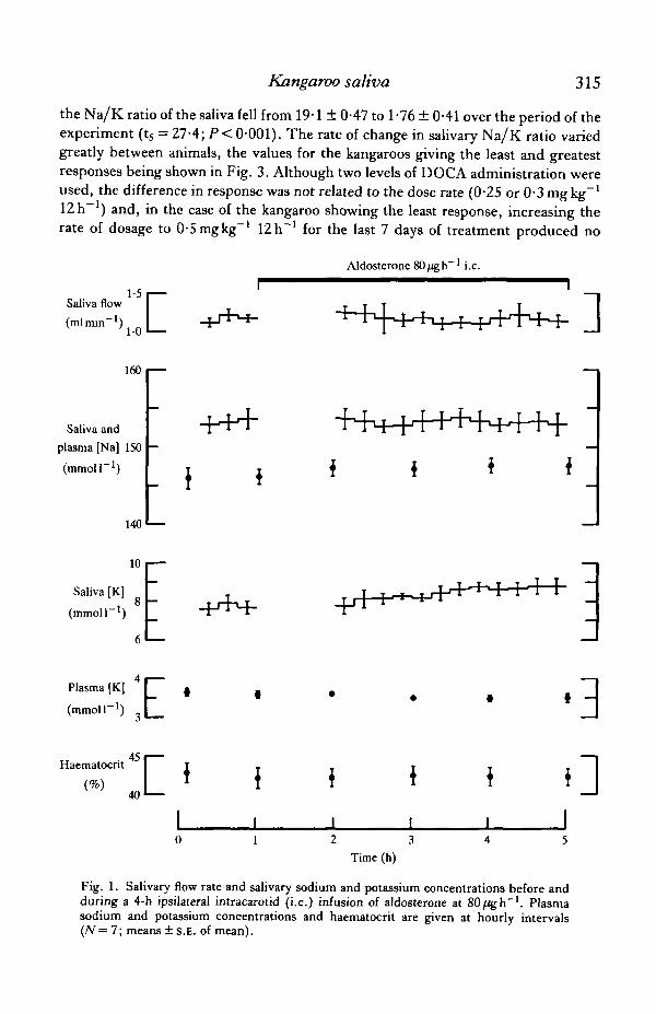
Aldosterone was infused at 8, 40 and 80/Ugh"1. Since the changes in salivarysodium and potassium concentrations associated with the highest rate were minimaland the effects of the lower dosages were less obvious, the data for the highestinfusion rate only are presented in this paper (Fig. 1). Plasma sodium concentrationrose during aldosterone infusion in all seven experiments (P<0-02), the meanincrease in plasma sodium being l^mmoll"1. The potassium concentration in theplasma showed a variable response, rising in some experiments and falling in othersso that the mean potassium concentration fell by (XWmmolF1. No change insalivary sodium concentration was associated with the aldosterone infusion, althoughsome tendency for salivary potassium concentration to rise was observed. In three ofthe seven experiments, salivary potassium was unaltered or fell during the aldos-terone infusion whereas, in the remainder, potassium rose by 1—Zmmoll"1. As aconsequence the mean sodium/potassium ratio of the saliva fell from 20*1 ± 1*09 to17-5 ±0-53 (ts = 2-07; NS). When administered to adrenalectomized rats, thealdosterone infusates from five of the above experiments caused a fall in urinarysodium/potassium ratio from 1-12 ±0-336 to 0-206 ± 0-076 (t̂ = 3-504; P< 0-05).Radioimmunoassay of the five stock solutions of aldosterone used in the bioassaygave an estimate of 0-48 ± 0-062 mgml"1, which was not significantly different fromthe nominal concentration of 0-5 mgrnl"1.
Chronic deoxycorticosterone administration
Changes in plasma sodium and potassium concentrations (Fig. 2) throughout the21 days of DOCA administration were not statistically significant, with the exceptionof the elevated plasma potassium on day 1 of the treatment (t5 = 2-581; P < 0 0 5 ) .The fall in haematocrit during the first 6-10 days of this treatment was significant(t5 = 8-288; P<0-001). Throughout the period of DOCA injection, there was aprogressive fall in salivary sodium concentration and an increase in salivarypotassium concentration (Fig. 2), the concentrations of these ions being significantlydifferent from the non-treated state by day 1 of the treatment (t5 = 5-004 and 6-272respectively; / ) < 0 0 1 ) . A s a consequence of these changes in salivary composition,
Kangaroo saliva 315
the Na/K ratio of the saliva fell from 19-1 ± 047 to 1 -76 ± 0-41 over the period of theexperiment (t5 = 27-4; P< 0-001). The rate of change in salivary Na/K ratio variedgreatly between animals, the values for the kangaroos giving the least and greatestresponses being shown in Fig. 3. Although two levels of DOCA administration wereused, the difference in response was not related to the dose rate (0-25 or 0-3 mgkg"1
12 h ) and, in the case of the kangaroo showing the least response, increasing therate of dosage to 0-5mgkg"1 12h~' for the last 7 days of treatment produced no
Saliva
(ml
1-5 •iva flow Imin"1)
1-0 •
Aldosterone 80/igh 1 i.e.
]160 i
Saliva and
plasma [Na] 150
(mmoir1)
140
10
Saliva [K]
(mmoir1)8
-H+
- \
Plasma [K]
(mmoir1) E- 3• 45Haematocnt
40 0I I
0 1 2 3 4 5Time (h)
Fig. 1. Salivary flow rate and salivary sodium and potassium concentrations before andduring a 4-h ipsilateral intracarotid (i.e.) infusion of aldosterone at 80/igh"1. Plasmasodium and potassium concentrations and haematocrit are given at hourly intervals(N= 7; means ± s.E. of mean).

316 A.M. BEAL
obvious increase in effect. As a subjective observation, it was noted that theresponsiveness to DOCA appeared to be related to whether the animal readilyindulged in thermoregulatory and stress-induced body licking or not.
Saliva flow *'•'
(mlmin ) i n
160 i—
140
Saliva and
plasma [Na]
(mmoir1)110
90
70 i—
50
Saliva [K]
(mmoir1)
20
Plasma [K]
(mmoir1)
Haematocrit
• t
['I
0 1 3
DOCA 0-25 or O-Smgkg-112b"1
i
10 14Time (days)
21
Fig. 2. Salivary flow rate, sodium concentration and potasium concentration (squares)and plasma sodium and potassium concentrations and haematocrit (circles) on days 0, 1,3, 6, 10, 14 and 21 of intramuscular deoxycorticosterone acetate injection at 0-25 or0-3 mgkg"1 12h~' (N = 6; means ± S.E. of mean).

Kangaroo saliva 317
l h - 1 0-3 mg kg"1 ^ h " 1 O-Smgkg"1 U h " 1
20 i
3 6 10 14 21 0 3 6 10 14 21Time (days)
Fig. 3. Salivary Na/K ratio during 21 days of intramuscular DOCA injection into thekangaroos having the most and least responsive parotid glands. Animal LB is a malewhich readily indulged in stress-induced and thermoregulatory licking whereas animalNF is a female which rarely indulged in either stress-induced or thermoregulatory bodylicking.
Chronic aldosterone administration
As with the DOCA treatment, intramuscular injection of aldosterone acetatecaused a progressive fall in salivary Na/K ratio from 19-1 to 6-69 over the 10 days ofthis experiment. This fall in Na/K ratio was less than that of the least responsive ofthe DOCA-treated kangaroos at the equivalent time in that experiment (18-6 to5-49).
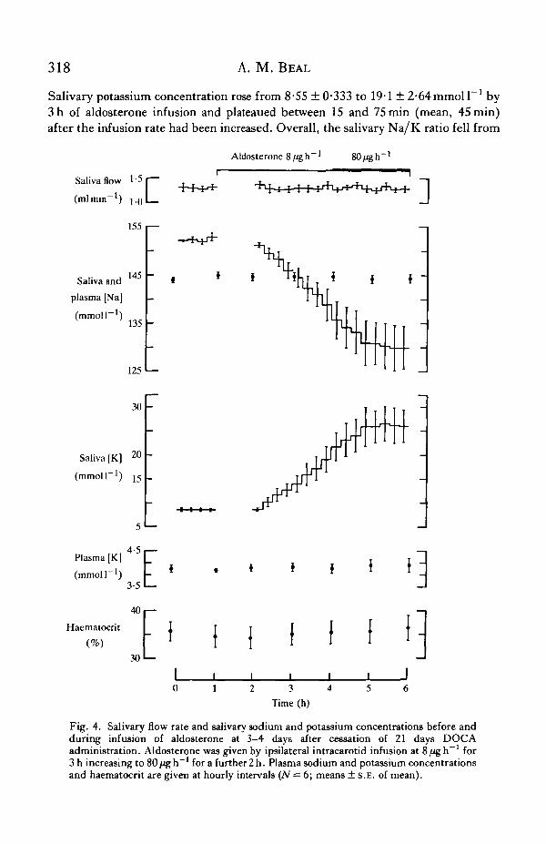
Post-DOCA infusion of aldosterone
No significant changes in haematocrit, or in plasma sodium and potassium con-centrations were associated with this aldosterone infusion (Fig. 4). Salivary sodiumconcentration fell from a mean value of 152-4 ± 046 to 138-9 ± S-iymmoll"1 at 3 hof aldosterone infusion (time of change from 8 to 80/igh"1) and tended to plateaubetween 30 and 75min (mean, 45min) after the change to infusion at 80/igh"1.
318 A. M. BEAL
Salivary potassium concentration rose from 8-55 ± 0-333 to 19-1 ± 2"64mmoll 1 by3 h of aldosterone infusion and plateaued between 15 and 75 min (mean, 45 min)after the infusion rate had been increased. Overall, the salivary Na/K ratio fell from
Saliva and
plasma [Na]
(mmoir1)
Aldosterone -1 -1
Saliva flow
(ml min"1) r155
145
135
125 L -
30 -
Saliva [K] 2°
(mmoll"1) 15
Plasma [K]
(mmoll"1
Haematocrit
30'—
0 1 2 3 4 5 6Time (h)
Fig. 4. Salivary flow rate and salivary sodium and potassium concentrations before andduring infusion of aldosterone at 3-4 days after cessation of 21 days DOCAadministration. Aldosterone was given by ipsilateral intracarotid infusion at 8 ^ g h - 1 for3 h increasing to 80 fig h~' for a further 2 h. Plasma sodium and potassium concentrationsand haematocrit are given at hourly intervals (iV = 6; means ± S.E. of mean).

Kangaroo saliva 319
180 ± 0-72 to 5-7 ± 104 (ts = 14-21; P< 0-001). The magnitude of the response byindividual kangaroos to aldosterone infusion was correlated with the size of theirresponse to chronic DOCA administration (r4 = 0-909; P < 0 0 5 ) . The minimumNa/K ratio achieved as a result of the aldosterone infusion was similar to the ratiosobtained on days 3 and 6 of the DOCA experiment and significantly different fromthe ratios observed on days 1, 10 14 and 21 of DOCA administration.
Gland histology
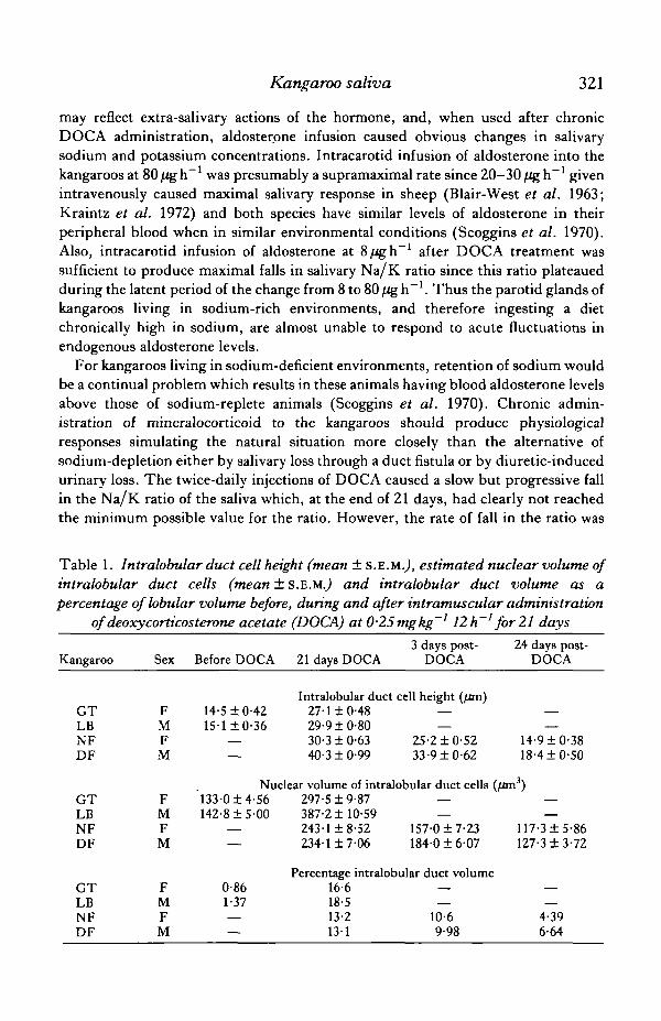
DOCA administration for 21 days was associated with an increase in height of theintralobular duct cells (P< 0-001), in volume of the nuclei in the duct cells(P< 0-001) and in the lobular volume occupied by the ducts (Fig. 5; Table 1). AfterDOCA treatment the duct cell nuclei had a very granular appearance and prominentacidophilic nucleoli. Three days after cessation of DOCA injection, measurableregression of the duct system had occurred, the height of the duct cells having fallenby 16-17% (P<0-001), the volume of their nuclei by 21-35% (P<0-001) and thevolume of ducts by 20—24%. By 24 days post-DOCA, the cell height and nuclearvolume had returned to or were approaching the values found in Na-repletekangaroos prior to DOCA treatment. At this time, the duct volume was on average 5times the duct volume found prior to DOCA administration (Table 1). The nucleoliof the intralobular duct cells remained identifiable but were now no more obviousthan those of the adjacent serous cells. Periodic acid Schiff staining showed thatpolysaccharides were not being accumulated by the duct cells either before or duringthe DOCA treatment.
DISCUSSION
Throughout these experiments, the salivary flow rate was maintained atapproximately 25% (i.e. 1-0—1-5 mlmin"1) of the maximum flow under anaesthesia.This rate was used because the kangaroo parotid gland can achieve and maintain thisrate easily for long periods of time and, as a consequence of the shape of the salivarysodium and potassium concentration/flow curves (Beal, 1984), the sodium andpotassium concentrations in the saliva will vary little with moderate fluctuations inflow but should show obvious changes when ductal transport of these ions isincreased.
In Na-replete sheep, intravenous infusion of aldosterone at 20—30/igh"1 causesmaximal changes in parotid Na/K ratio, the salivary potassium rising to14—45mmoll~1 and the sodium concentration falling by a similar amount (Blair-West et al. 1963; Kraintz et al. 1972). By comparison, the response of thechronically Na-replete kangaroos to acute aldosterone administration was virtuallynegligible. There can be little doubt that the aldosterone infusates were biologicallyactive, for the following reasons: rat renal bioassay demonstrated the presence ofmineralocorticoid activity, radioimmunoassay of the stock aldosterone solutions gaveestimates approximating to their nominal concentrations, aldosterone infusion intothe kangaroos was associated with increased plasma sodium concentrations, which

320 A. M. BEAL
Fig. 5. Parotid gland from the kangaroo which gave the greatest physiological response toDOCA administration (animal LB). Fixed in formol saline; methacrylate sections stainedwith haematoxylin and eosin (X150). (A) represents the Na-replete state and (B) showsthe effect of 21 days injection of DOCA at 3 mgkg"1 IZh"1.
Kangaroo saliva 321
may reflect extra-salivary actions of the hormone, and, when used after chronicDOCA administration, aldosterone infusion caused obvious changes in salivarysodium and potassium concentrations. Intracarotid infusion of aldosterone into thekangaroos at 80/igh"1 was presumably a supramaximal rate since 20-30/igh"1 givenintravenously caused maximal salivary response in sheep (Blair-West et al. 1963;Kraintz et al. 1972) and both species have similar levels of aldosterone in theirperipheral blood when in similar environmental conditions (Scoggins et al. 1970).Also, intracarotid infusion of aldosterone at 8/igh"1 after DOCA treatment wassufficient to produce maximal falls in salivary Na/K ratio since this ratio plateauedduring the latent period of the change from 8 to 80 /ig h~l. Thus the parotid glands ofkangaroos living in sodium-rich environments, and therefore ingesting a dietchronically high in sodium, are almost unable to respond to acute fluctuations inendogenous aldosterone levels.
For kangaroos living in sodium-deficient environments, retention of sodium wouldbe a continual problem which results in these animals having blood aldosterone levelsabove those of sodium-replete animals (Scoggins et al. 1970). Chronic admin-istration of mineralocorticoid to the kangaroos should produce physiologicalresponses simulating the natural situation more closely than the alternative ofsodium-depletion either by salivary loss through a duct fistula or by diuretic-inducedurinary loss. The twice-daily injections of DOCA caused a slow but progressive fallin the Na/K ratio of the saliva which, at the end of 21 days, had clearly not reachedthe minimum possible value for the ratio. However, the rate of fall in the ratio was
Table 1. Intralobular duct cell height (mean ± S.E.M.J, estimated nuclear volume ofintralobular duct cells (mean ± S.E.M.,) and intralobular duct volume as apercentage oflobular volume before, during and after intramuscular administration
of deoxycorticosterone acetate (DOCA) at 025mgkg~' 12h~' for 21 days
Kangaroo
GTLBNFDF
GTLBNFDF
GTLBNFDF
Sex
FMFM
FMFM
FMFM
Before DOCA
14-5 ±0-4215-1 ±0-36
—
21 days DOCA
Intralobular duct27-1 ±0-4829-9 ±0-8030-310-6340-310-99
3 days post-DOCA
cell height (/im)—
25-210-5233-910-62
24 days post-DOCA
—
14-910-3818-410-50
Nuclear volume of intralobular duct cells (fim3)133-0 ±4-56142-8 ± 5 0 0
——
0-861-37
—
297-519-87387-2110-59243-118-52234-117-06
——
157-017-23184-016-07
Percentage intralobular duct volume16-618-513-2131
—
10-69-98
——
117-315-86127-313-72
—
4-396-64

322 A. M. BEAL
probably similar to the maximum rate for each animal under these conditions sincethere was no correlation between the dose rates of 0-25 and 0-3 mgkg"1 12 h"1 andthe rate of fall in the ratio, and increasing the dosage in one animal from 0-3 to0-5 mgkg"1 12 h"1 did not increase the rate of fall in the ratio. The lowest dose ratewas chosen on the basis that it was 10 times the dosage necessary to maintain a 40-kgadrenalectomized sheep (i.e. 2mgday~1) and therefore might be expected toproduce a near-maximal rate of fall in the salivary Na/K ratio.
Sodium deficiency in sheep, grey kangaroos and rabbits is associated with hyper-plasia and hypertrophy of the striated and excretory ducts of the major salivaryglands (Blair-West et al. 1968, 1969; Comptoneia/. 1975). The histological changesobserved in the intralobular ducts of the red kangaroo parotid gland as a consequenceof the DOCA injection were similar to those of the grey kangaroo althoughconsiderably more extreme. Hypertrophy of the duct cells, with histological evidenceof increased nuclear activity, was clearly demonstrated, whereas duct hyperplasiacould not be demonstrated beyond question. In the earlier studies it was uncertainwhether these histological changes were due to the sodium deficiency itself or toconcomitant changes in mineralocorticoid activity. The red kangaroos were Na-replete at the commencement of the DOCA injection and there is no reason to believethat they became Na-deficient during the course of mineralocorticoid injections.Indeed, the tendency for the plasma sodium concentration to rise during the periodof injection would indicate that they remained Na-replete. Hence the duct hyper-trophy seen during Na deficiency must be due to the action of endogenousmineralocorticoids on the duct cells. As a consequence of low mineralocorticoidlevels, parotid glands of chronically Na-replete red kangaroos have short, relativelyinactive ducts, which explains the lack of response to acute aldosterone infusion.However, with the establishment of high mineralocorticoid levels, the duct systemenlarges and the duct cells become more active, which explains the slowly increasingability of the gland to reduce the salivary Na/K ratio. The observation that animalswhich indulge in frequent skin licking appear to be more responsive to DOCAadministration than non-licking animals may reflect a slight priming of this mech-anism due to loss of sodium during this activity. During sodium deficiency in rabbits,glycogen accumulates in the duct cells of the mandibular gland (Compton et al.1975; Young & van Lennep, 1978). This did not occur in the kangaroo parotidgland.
The 3-4 days required for the salivary Na/K ratio to return to Na-replete levelsafter cessation of DOCA administration corresponded to the time necessary for themetabolism of the residual steroid in the muscles since, in terms of sodium balance,the animal should have remained Na-replete throughout the DOCA treatment.Under these conditions, intracarotid infusion of aldosterone reduced the salivaryNa/K ratio to a minimum after 3-4 h which coincided with the latent periodfollowing the increase in infusion rate from 8 to 80 ̂ gh"1. At the higher infusionrate, aldosterone caused no further fall in the Na/K ratio, a further indication thatthis infusion rate was supramaximal. The increased ability to transport Na and K wasapparently very dependent on high-level mineralocorticoid support since, during the

Kangaroo saliva 323
3-4 days between the end of DOCA administration and the post-DOCA aldosteroneinfusion, the minimum Na/K ratios were only equivalent to those observed at 3-6days of DOCA administration. This loss of sodium transporting ability wasassociated with substantial regression of the duct system, indicating that theincreased activity and development of the ducts are rapidly lost when mineralo-corticoid support is withdrawn. Indeed, by 24 days post-DOCA, which is equivalentto 21 days of low mineralocorticoid levels, the regression of the ducts wasapproaching completion. Based on these observations, one would predict that theparotid glands of kangaroos from sodium-deficient environments would lose theirability to respond to acute fluctuations in blood mineralocorticoid levels within amonth of arriving in a sodium-rich environment.
I am indebted to Mr David Hair for skilful technical assistance, to Mrs GlynysForsyth for diligent husbandry of the kangaroos, to Dr Geoffrey G. Duggin and MsLinda Critchley for the radioimmunoassay of the aldosterone solutions and to MrStan Watkins for assistance with histology.
I would like to acknowledge the gift of sodium pentobarbitone by Abbott AustraliaPty Ltd.
REFERENCES
BAILEY, C. B. & BALCH, C. C. (1961). Saliva secretion and its relation to feeding in cattle. I. Thecomposition and rate of secretion of parotid saliva in a small steer. Brit.jf. Nutr. 15, 371—382.
BEAL, A. M. (1979). Parotid salivary flow and composition during infusion of acetylcholine and atro-pine into the carotid artery of conscious sodium-replete sheep. Q.Jl exp. Physiol. 54, 89—107.
BEAL, A. M. (1984). Electrolyte composition of parotid saliva from sodium-replete red kangaroos(Macropus rufus).J. exp. Biol. I l l , 225-237.
BLAIR-WEST, J. R., COGHLAN, J. P., DENTON, D. A., GODING, J. R. & WRIGHT, R. D. (1963).
The effect of aldosterone, cortisol and corticosterone upon the sodium and potassium content ofsheep's parotid saliva. J. din. Invest. 42, 484-496.
BLAIR-WEST, J. R., COGHLAN, J. P., DENTON, D. A., NELSON, J. F., ORCHARD, E., SCOGGINS,
B. A., WRIGHT, R. D., MYERS, K. & JUNQUEIRA, C. L. (1968). Physiological, morphologicaland behavioural adaptation to a sodium deficient environment by wild native Australian andintroduced species of animals. Nature, Land. 217, 922-928.
BLAIR-WEST, J. R., COGHLAN, J. P., DENTON, D. A., NELSON, J., WRIGHT, R. D. & YAMAUCHI,
A. (1969). Ionic, histological and vascular factors in the reaction of the sheep's parotid to highand low mineralocorticoid status. J. Physiol., Land. 205, 563—579.
COATS, D. A. & WRIGHT, R. D. (1957). Secretion by the parotid gland of the sheep: therelationship between salivary flow and composition. J . Physiol., Land. 135, 611-622.
COMPTON, J. S., DENNISS, A. R., HUNG, P., KENNERSON, A. R., VAN LENNEP, E. W. & YOUNG,
J. A. (1975). The effect of a sodium deficient diet on the striated ducts of the rabbit mandibulargland. Proc. Aust. physiol. pharmacol. Soc. 6, 132-133.
KOMI, N. & SNYDER, W. H. (1963). Electrolyte concentrations in saliva of the goat under variousconditions. Am. J. Physiol. 204, 1055-1058.
KRATNTZ, L., BLAIR-WEST, J. R., COGHLAN, J. P., DENTON, D. A. & WRIGHT, R. D. (1972).
Aldosterone action on the parotid gland: sodium replete sheep. Acta endocr., Copnh. 70,510-514.
SCOGGINS, B. A., BLAIR-WEST, J. R., COGHLAN, J. P., DENTON, D. A., MYERS, K., NELSON,
J. F., ORCHARD, ELSPETH & WRIGHT, R. D. (1970). The physiological and morphologicalresponse of mammals to changes in their sodium status. In Hormones and the Environment,(eds G. K. Benson & J. G. Phillips), pp. 577-602. London: Cambridge University Press.

324 A. M. BEAL
SELDLNGER, S. I. (1953). Catheter replacement of the needle in percutaneous anteriography. Actaradiol 39, 368-376.
SIMPSON, S. A. & TAIT, J. F. (1952). A quantitative method for the bioassay of the effect of adrenalcortical steroids on mineral metabolism. Endocrinology 50, 150—161.
YOUNG, J. A. & VAN LENNEP, E. W. (1978). The Morphology of Salivary Glands, p. 39, Fig. 21.London, New York, San Francisco: Academic Press.