the effect of antigen polymorphisms on serological
TRANSCRIPT
University of South FloridaScholar Commons
Graduate Theses and Dissertations Graduate School
June 2017
The Effect of Antigen Polymorphisms onSerological Antibody Detection Assays BasedUpon theKristi M. MileyUniversity of South Florida, [email protected]
Follow this and additional works at: http://scholarcommons.usf.edu/etd
Part of the Biomedical Engineering and Bioengineering Commons, Parasitology Commons, andthe Public Health Commons
This Thesis is brought to you for free and open access by the Graduate School at Scholar Commons. It has been accepted for inclusion in GraduateTheses and Dissertations by an authorized administrator of Scholar Commons. For more information, please contact [email protected].
Scholar Commons CitationMiley, Kristi M., "The Effect of Antigen Polymorphisms on Serological Antibody Detection Assays Based Upon the" (2017). GraduateTheses and Dissertations.http://scholarcommons.usf.edu/etd/6904
1
The Effect of Antigen Polymorphisms on Serological Antibody Detection Assays Based
Upon the Onchocerca volvulus 16kDa Diagnostic Antigen (Ov16)
by
Kristi M. Miley
A thesis submitted in partial fulfillment of the requirements for the degree of
Master of Science Department of Global Health University of South Florida
Major Professor: Thomas R. Unnasch, Ph.D. Ricardo Izurieta, M.D., Dr. PH, M.P.H.
Francis Ntumngia, Ph.D.
Date of Approval June 21, 2017
Keywords: Onchocerciasis, Ov-16 ELISA, Recombinant IgG4, Diagnostics
Copyright © 2017, Kristi M. Miley

2
DEDICATION
I would like to give special thanks to my husband, James Miley, my mother and
stepfather, Rosemary and Brian Lee, and my children. I will be forever grateful for the
numerous sacrifices and countless words of encouragement that made this work
possible. I would also like to dedicate this thesis in honor of my late father, V. Lee
Watson, whose love and compassion lives on in me, forever giving me strength,
courage, and passion to succeed wherever life’s journey may take me.

3
ACKNOWLEDGMENTS
My deepest thanks and fondest regards to Thomas R. Unnasch, Ph.D., for the
vast knowledge and foresight that made this project a reality. It has been an honor to
work under Dr. Unnasch’s tutelage on this project and very rewarding having the
opportunity to work on this research as an investigator. Special thanks to Hassan K.
Hassan for assisting me in navigating the laboratory and reinforcing the skills that were
necessary to aid in this research. I would also like to thank Dr. Canhui Liu for his
assistance, as well as my fellow lab members Johan, and Kati for their support and
comradery. Furthermore, I would like to acknowledge Ricardo Izurieta, M.D., Dr. PH,
M.P.H., and Francis Ntumngia, Ph.D., for their support on my committee during this
project.

i
TABLE OF CONTENTS List of Tables .................................................................................................................... ii List of Figures .................................................................................................................. iii Abstract ........................................................................................................................... iv Chapter One: Introduction ............................................................................................... 1 Chapter Two: Materials and Methods.............................................................................. 9 Isolation and Purification of Mutations for Use in OV-16 ELISA ........................... 9 Monoclonal Standards via OV-16 ELISA Against 4 Experimental Antigens ....... 11 Testing Ov-16 Polymorphisms in Sera via ELISA ............................................... 12 Testing GST Cross-Reactivity in Sera via Ov-16 ELISA ..................................... 15 Chapter Three: Results ................................................................................................. 16 Analysis of Mutations Against the Monoclonal Antibody ..................................... 16 Isolated Mutations for Use in OV-16 ELISA ........................................................ 17 Monoclonal Reactivity to Four Recombinant Antigens ....................................... 18 Serum Reactivity to Four Recombinant Antigens via Ov-16 ELISA .................... 19 OV-16 ELISA Sensitivity of Polymorphisms ....................................................... 21 OV-16 ELISA GST Cross-Reactivity ................................................................... 23 Chapter Four: Discussion .............................................................................................. 24 References .................................................................................................................... 30

ii
LIST OF TABLES
Table 1: List of purified OV-16 proteins with yield concentrations in mg/ml ........................ 11
iii
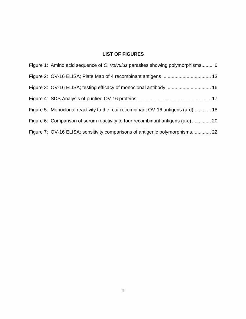
LIST OF FIGURES Figure 1: Amino acid sequence of O. volvulus parasites showing polymorphisms ......... 6 Figure 2: OV-16 ELISA; Plate Map of 4 recombinant antigens ................................... 13
Figure 3: OV-16 ELISA; testing efficacy of monoclonal antibody ................................. 16
Figure 4: SDS Analysis of purified OV-16 proteins ....................................................... 17
Figure 5: Monoclonal reactivity to the four recombinant OV-16 antigens (a-d) ............. 18 Figure 6: Comparison of serum reactivity to four recombinant antigens (a-c) .............. 20
Figure 7: OV-16 ELISA; sensitivity comparisons of antigenic polymorphisms .............. 22
iv
ABSTRACT
Onchocerca volvulus is a filarial parasite transmitted to humans by female
Simulium spp. black flies. Infection with this parasite can cause blindness and severe
skin disease among humans in Africa and the Americas. Enzyme-linked Immunosorbent
Assay serological testing of OV-16 antigen is a diagnostic tool for determining effective
elimination of the parasite. Programs typically rely on OV–16 ELISA to evaluate the
progress towards interruption and/or elimination of disease by mass drug distribution of
ivermectin and vector larvicidal control efforts. As elimination grows closer, monoclonal
antibody positive controls for OV-16 ELISA become important to develop for
Onchocerca testing due to the limited availability of pooled sera positive controls.
Recent evaluation of laboratory designed OV-16 ELISA coating antigen by the Unnasch
Lab (University of South Florida) showed that polymorphisms occurred which may alter
the ability of the humanized monoclonal antibody to recognize the cognate antigen. With
this development, it was important to evaluate these polymorphisms and isolate them
for further testing against the standardized monoclonal antibody and positive sera to
determine the effects antigenic polymorphisms could have on diagnostic testing. Upon
evaluation, the polymorphisms did influence signaling when testing the monoclonal
antibody. However, little effect on the recognition of the antigen was seen when different
isoforms were evaluated against sera from O. volvulus infected individuals. Data
suggest that the epitope recognized by the synthetically produced monoclonal antibody
is not immuno-dominant in infected individuals.
1
CHAPTER ONE:
INTRODUCTION
Onchocerciasis, sometimes referred to as “River Blindness”, is one of the
neglected tropical diseases (NTDs) affecting humankind and has been documented as
one of the most common causes of preventable infectious blindness (WHO, 2016).
However, Onchocerciasis is also one of the NTDs which has the potential for elimination
through vector control measures and the efforts of mass distribution of microfilaricidal
treatments by multiple health response alliances with the support of the World Health
Organization (WHO) (WHO, 2016).
This debilitating disease is caused by a parasitic filarial nematode known as
Onchocerca volvulus (Coffeng, et al., 2013). Although there are other Onchocerca spp.
which have been identified to infect mammals, such as; O. ochengi in cattle, O. lupi in
canines, and O. cervipedis in deer, O. volvulus appears to selectively target humans as
its only host (Boatin & Amazigo, 2016). Onchocerciasis has the potential to manifest in
either an ocular form of illness, lymphatic involvement, or display in the form of filarial
dermatitis, and the severity of disease appears to be linked to repeated exposure to
infective bites (Dobson, 2008).
Human infection of O. volvulus occurs from the bite of a previously infected
blackfly, which upon inoculation the L3 larvae will complete their cycle to reproductive

2
adulthood in approximately 24 months within the human host (Boatin & Amazigo, 2016;
WHO, 2016). Adult female O. volvulus form nodules within the body, usually near bony
prominences, where they have the ability to reside for upwards of 14 years and produce
first stage larvae (microfilariae) at a reproductive potential of “700-1500 per day” (Liu,
2013). The microfilariae that are produced leave these nodules and take up residence in
the skin, and in some cases the eye, where they can survive for up to two years (Liu,
2013). In this human infective stage, the microfilariae are taken up by the black fly
vector during blood-feeding and perpetuate the cycle upon molting to L3 larvae within
the black fly’s thoracic flight muscles and eventually exit the labium during subsequent
feeding to further transmission (McClelland, 1992).
It is estimated that 37 million individuals are infected with O. volvulus (Heymann,
2008). The disease predominantly affects individuals in sub-Saharan Africa wherein
approximately 99% of onchocerciasis cases in the world occur, with a few foci also
noted in the Americas and Eastern Mediterranean (Noma et al., 2014; WHO, 2016).
There does appear to be a geographical component to the distribution and
symptomology of the disease. There are noted divisions in distribution of disease where
“blinding” illnesses appear to be more prevalent in the savannah foci, “non-blinding”
illnesses tend to occur in forest foci and this might be explained by varying vector-
parasite complexes with the different strains of O. volvulus (Boatin & Amazigo, 2016).
The black fly (Simulium spp.) vector for onchocerciasis prefers a habitat of fast
flowing water found in rivers and streams. Unfortunately for developing countries, this
has created a cyclical paradigm between onchocerciasis and malnutrition, as
communities attempt to avoid the risk of disease they also regress to inferior farmland

3
(Dobson, 2008). Unfortunately, the plight of this disease does not stop with the illnesses
that it evokes, it also causes economic strain on the affected communities. However,
great efforts have been made in targeting these black fly environments with control
measures as noted in early achievements of the Onchocerciasis Control Programme in
West Africa (OCP), which initially started targeting habitat control with larvicides in the
1970’s and later added human microfilaricidal treatments to their arsenal using
ivermectin in the late 1980’s (Dobson, 2008; Unnasch, 2004). Other programs such as;
the African Program for Onchocerciasis (APOC) and Onchocerciasis Elimination
Program in the Americas (OEPA) have mass treatment strategies in pursuit of
interruption and eradication of O. volvulus (Eisenbarth et al., 2016).
With control program measures in place and ongoing human treatment with
mass drug administration (MDA) of “Mectizan ® from Merck & Co.”, onchocerciasis is
making its way towards elimination (Schwab, 2007). In fact, recent research indicates
that Colombia, Ecuador, Mexico, and Guatemala have reached elimination status, and
several foci have thought to have interrupted the disease, including foci in Uganda and
the Sudan (Boatin & Amazigo, 2016; Higazi et al., 2013; WHO, 2016). As research
continues to evaluate the progress of the MDA program and vector control measures it
appears that onchocerciasis elimination is possible within endemic regions still affected
by disease. That being said, as interruption and elimination approaches, challenges will
inevitably continue to arise in determining the future risk of reemergence in previously
endemic regions.
The gold standard of diagnosis for O. volvulus has historically been through the
microscopic evaluation of skin biopsies (skin snips) for the appearance of microfilariae
4
after clinical pathology of disease has been determined via ocular damage, palpable
nodules, or perhaps by dermatological symptoms (Liu, 2013). Generally, these skin
snips are obtained using a sclerocorneal biopsy punch that excises approximately
2.5mm of tissue, that is then incubated in culture media or saline wherein the
microfilariae exit the tissue and the fluid can be examined for their presence (Boatin &
Amazigo, 2016; Liu, 2013). Unfortunately, this diagnostic method can be painful and
could potentially result in underestimation of disease burden due to limitations in the
number of accommodating participants.
The paradox of MDA treatment programs is that it may lead to fewer detectable
microfilariae in each patient and could allow for underestimation of disease in foci that
are near or at elimination status. Assays like the Ov-16 enzyme-linked immunosorbent
assay (ELISA) are used to geographically map the distribution of onchocerciasis and
monitor control program progress, but many such filariasis assay applications have
been noted to have issues regarding cross reactivity and deficiencies with
standardization (Weil, et al., 2011). Currently, the Ov-16 ELISA is used to evaluate
successful suppression of the disease by investigating potential exposure to the
parasite in children less than 10 years old (Cupp, et al., 2012). Exposure is determined
by the ELISA detecting IgG4 antibodies against Ov16, a 16kDa immunodominant
antigen. Children are tested as sentinels for continued or emerging exposure, as Ov-16
ELISA is limited to evaluating antibodies that are exhibited post exposure which may
also be present in individuals who were previously infected and treated.
As elimination approaches in a given region, the limited availability of positive
sera for use as controls in diagnostic tests like Ov-16 ELISA adds to the complex

5
challenges of having sufficient diagnostic tools available for monitoring the progress
towards onchocerciasis elimination (Golden et al., 2016). Recently, a humanized
monoclonal antibody was produced as a positive control for use in Ov-16 ELISA to help
solve this dilemma (Golden et al., 2016). As low levels of parasitemia become more
common due to the current MDA programs, the monoclonal antibody design seems an
advantageous alternative to the previous techniques of pooled sera. Designing
standardized positive controls also reflects the need to have universal applications and
reproducibility of these required conjugates in the current onchocerciasis surveillance
programs in order to evaluate various foci and potential elimination and/or transmission
status properly.
Upon evaluating the efficacy of the humanized monoclonal antibody against Ov-
16 ELISA within the Unnasch laboratory (University of South Florida), difficulties arose
in the ability of the Ov-16 antigen to appropriately detect the humanized monoclonal
antibody. It was determined that this monoclonal antibody, produced as a positive
control for the Ov-16 ELISA, reacted to a recombinant version of the Ov-16 antigen
derived from parasites from Guatemala and was unable to bind to a homologue derived
from a sequence obtained from parasites from Cameroon. It is not uncommon to exploit
the antigen antibody interactions in designing surveillance test formats when diagnosing
filarial infections, but there are known drawbacks including cross reactivity with
nematode infections other than the target, in this case O. volvulus (Lammie, 2004).
Therefore, designing a humanized monoclonal antibody that recognizes the
immunodominant antigen appropriately with specificity for an Onchocerca testing
platform is important.
6
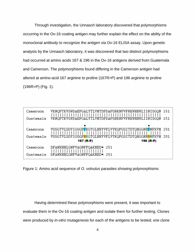
Through investigation, the Unnasch laboratory discovered that polymorphisms
occurring in the Ov-16 coating antigen may further explain the effect on the ability of the
monoclonal antibody to recognize the antigen via Ov-16 ELISA assay. Upon genetic
analysis by the Unnasch laboratory, it was discovered that two distinct polymorphisms
had occurred at amino acids 167 & 196 in the Ov-16 antigens derived from Guatemala
and Cameroon. The polymorphisms found differing in the Cameroon antigen had
altered at amino-acid 167 arginine to proline (167R>P) and 196 arginine to proline
(196R>P) (Fig. 1).
Figure 1: Amino acid sequence of O. volvulus parasites showing polymorphisms
Having determined these polymorphisms were present, it was important to
evaluate them in the Ov-16 coating antigen and isolate them for further testing. Clones
were produced by in-vitro mutagenesis for each of the antigens to be tested; one clone

7
containing no mutations reflecting the Guatemalan strain (parental construct), one clone
containing a single mutation at 167R-P (arginine to proline), one clone containing a
single mutation at 196R-P (arginine to proline), and one clone containing both
mutations. Each of the mutated Ov-16 antigens was expressed with a GST tag and
purified using affinity chromatography. Once purified, these four laboratory designed
Ov-16 antigens (OV-16:167R196R, OV-16:167P196P, OV-16:167P196R, & OV-
16:167R196P) could then be tested against the monoclonal antibody, as well as a set of
known positive sera via Ov-16 ELISA in order to identify the polymorphism responsible
for the loss in monoclonal activity and to determine if the polymorphisms affected the
sensitivity of the Ov-16 ELISA.
The aim of this research was designed to investigate the performance of Ov-16
ELISA with respect to four variant recombinant Ov-16 antigens to evaluate the noted
polymorphisms. Objective comparisons of these Ov-16 antigens containing isolated
polymorphisms to an Ov-16 antigen that contained no polymorphisms were performed
to determine whether or not these isoforms infer functional similarity on serological
testing within the Ov-16 ELISA platform. Investigation regarding reactivity to the
humanized monoclonal antibody was also performed to determine the effects antigenic
polymorphisms may have on such synthetic positive controls. With onchocerciasis
interruption and elimination in sight and fewer infections occurring, future test
applications may soon rely on the development of adequate humanized monoclonal
antibody standardization techniques. However, these synthetic positive controls should
be designed to react appropriately with an immunodominant antigen that is specific to
O. volvulus and sensitive to detection when present in sera if it is to replace the need for

8
pooled sera positive controls in current onchocerciasis surveillance programs (Lucius,
1988). Furthermore, the World Health Organization’s certification of elimination protocol
relies heavily on the Ov-16 ELISA testing platform in their Onchocerciasis surveillance
programs which warrants quality control efforts to ensure that these serological assays
perform to the highest standards possible, as the results of such testing will ultimately
dictate how screening children in each focus will proceed (Cupp, et al., 2012).

9
CHAPTER TWO:
MATERIALS AND METHODS
Isolation and Purification of Mutations for Use in OV-16 ELISA
Four clones were produced, one for each of the laboratory Ov-16 antigens, by in-
vitro mutagenesis and plasmids (pGEX, a commonly used expression vector) were
maintained at -80o C, by Dr. Canhui Liu in the Unnasch laboratory. 1ul of each of the
plasmid was inoculated into 25ul BL-21 competent E-coli cells and incubated on ice for
5 minutes then heat shocked at 42o C 45 seconds. They were immediately placed on ice
for two minutes after which time 900ul of SOC buffer was added and tubes were
incubated in platform shaker at 37o C for one hour. After incubation, a 1:10 dilution of
positive cells was created and 125ul of cells were plated on LB Agar plates containing
Ampicillin at a concentration of 100ug/ml, along with negative control plates, and
incubated upside down at 37o C overnight. Positive plates were examined the next day
for presence of colonies, as well as no colonies confirmed in negative control plates.
Individual E. coli colonies containing the transformed plasmids were chosen from each
of the Ov-16 plasmid clones and grown using LB media containing Ampicillin100ug/ml
in step-wise proportions starting with 5ml of media incubated overnight at 37o C and
shaking at 300 RPM. The 5ml overnight culture was then diluted to 100ml with LB
media containing Ampicillin 100ug/ml and incubated overnight under the same

10
conditions. 50ml of this overnight culture was then added to 1 liter of LB media
containing Ampicillin 100ug/ml, incubated at 37o C and 300 RPM to mid-log phase by
monitoring optical densities at A600 to a value between 0.6 - 1.0. Once these values
were reached, protein expression was induced by adding isopropyl-β D-thiogalactoside
(IPTG) to a final concentration of 0.1mM. An additional OD reading was taken 1 hour
following the addition of IPTG to confirm growth was continuing and the cells were
allowed to grow overnight at 37o C and 300 RPM. After growth was complete, the media
containing each protein/Ov-16 antigen was then divided into 250ml cell centrifuge tubes
and the cell pellet concentrated via centrifugation at 13,000 RPM for 10 minutes at 4o C.
Cells are resuspended in cold GST binding buffer (150mM NaCl, 25mMTris, 1mM
EDTA) containing protease inhibitors (Thermo ® Protease Inhibitor tablets Cat.#78430)
and 100ug/ml lysozyme. This was then followed by the addition of Triton x100 detergent
to a final concentration of 0.5% to assist in lysing cells after which the cell pellets were
placed at -80o C overnight.
Each of the isolated Ov-16 antigens was individually purified by affinity
chromatography using GSTrap HP® columns (GE Healthcare Life Sciences). This
process began by thawing the cell pellets and sonicating them at 15% for 10 seconds
with a one minute pause on ice, over three intervals to lower the viscosity. The cell
debris was then centrifuged at 18,000rpm for 30 minutes at 4o C. and the supernatant
was retained and filtered through a 0.45um filter prior to its application to the affinity
column. Using a separate 1.0ml GSTrap® column for each of the four Ov-16 antigens,
all were purified under the same conditions using an infusion pump at a wash flow-rate
of 1.0ml/min., a sample loading flow-rate of 0.2ml/min., and an elution flow-rate of

11
1.0ml/min. wherein all samples and buffers were maintained on ice. Important to note
that the slower flow-rate for sample loading is recommended for increased binding of
desired protein. The following buffers were used in all four purifications; binding buffer –
10mM sodium phosphate,140mM NaCl, 2.7mM KCl at pH of 7.4 and elution buffer –
50mM Tris/HCl, 10mM reduced glutathione at a pH of 8.0. The elutions retrieved from
the four purifications were evaluated via the Nano-drop® and SDS gel electrophoresis
to confirm presence of desired Ov-16 protein prior to following up with dialysis on each
of them. Dialysis was performed on each protein using 3.0ml Thermo Scientific Slide-A-
Lyzer ® dialysis cassettes immersed in 1X PBS. SDS gel electrophoresis was then
repeated on the four Ov-16 antigens post dialysis. Each of the Ov-16 antigens was
given a label designation based on their mutation characteristics and yield
concentrations were calculated via Bradford ® Protein assay (Table 1).
Table 1: List of purified OV-16 proteins with yield concentrations in mg/ml
Monoclonal Standards via OV-16 ELISA Against 4 Experimental Antigens
In previous research performed by Golden et al. (2016), a humanized
monoclonal antibody was designed for positive controls in the OV-16 ELISA. Using a
Purified OV-16 protein ID Yield Concentration in mg/ml
Polymorphism Characteristics
OV-16:167R196R 2.0 mg/ml Parental construct
OV-16:167P196P 2.0 mg/ml Double mutation; 167R>P & 196R>P
OV-16:167P196R 0.5 mg/ml Isolated single mutation; 167R>P
OV-16:167P196R 1.0 mg/ml Isolated single mutation; 196R>P

12
human combinatorial antibody library (HuCAL & HuCAL Platinum), they were able to
identify 15 unique antibody clones with specificity for Ov-16, which were then narrowed
down to two based on binding affinity in ELISA and nitrocellulose platforms (Golden et
al.,2016). Ultimately one recombinant antibody clone was chosen for development
(AbD19432_hIgG4) based on absorbance range and signal strength (Golden et
al.,2016). As previously noted upon testing in the Unnasch Laboratory, it was found that
this humanized monoclonal antibody produced as a positive control for the Ov16 ELISA
reacted to a recombinant version of the Ov16 antigen derived from parasites from
Guatemala but did not react to a homologue derived from a sequence obtained from
parasites from Cameroon. After careful isolation of the polymorphisms, the experimental
Ov-16 mutations were tested individually against the humanized monoclonal antibody
(AbD19432_hIgG4). The monoclonal antibody was serially diluted and tested via OV-16
ELISA techniques using the four experimental OV-16 antigens to evaluate detection.
Optical densities were evaluated via spectroscopy at 405nm as per OV-16 ELISA
protocols and results of each of the four antigens were compared.
Testing Ov-16 Polymorphisms in Sera via ELISA
To determine the effects that Ov-16 antigenic polymorphisms may have on
serum reactivity regarding naturally acquired human antibodies in endemic regions, the
four recombinant Ov-16 antigens were evaluated via OV-16 ELISA against 704
specimens that were collected from Liberia and Ghana. These serum samples
contained no personal identifiers and the University of South Florida’s IRB ruled on

13
9/16/16 that this work did not meet the definition of human subjects research. Standard
Ov-16 ELISA protocols were used to evaluate the sensitivity of the four experimental
Ov-16 antigens against the 704 serum samples. All 704 samples were previously
determined positives for O. volvulus via skin snip results. For effective comparison of
serum reactivity, each of the ELISA 96-well microtiter plates were divided such to be
coated with all four experimental Ov-16 antigens (100ul per well) at 2.0ug/ml in
carbonate buffer (NaHCO3) (Fig. 2).
Figure 2: OV-16 ELISA; Plate Map of 4 recombinant antigens
Coating Antigens
OV-16:167R196R (NO MUTATIONS)
OV-16:167P196P (BOTH MUTATIONS 167R>P,196R>P)
OV-16:167P196R (SINGLE MUTATION 167R>P)
OV-16:167R196P (SINGLE MUTATION 196R>P)

14
Samples were then tested at 1:80 dilution in 1X PBST/5% BSA (Phosphate
buffered saline, 0.05% Tween20/ Bovine Serum Albumin) and 1ul of the humanized
monoclonal antibody was diluted in 499ul of 1X PBST/5% FBS (Phosphate buffered
saline, 0.05% Tween20/ Fetal Bovine Serum) which was then serially diluted using 1X
PBST/5% FBS. All samples were tested in duplicate with each of the experimental Ov-
16 antigens under the following conditions; plates were coated with 100ul at a
concentration of 2ug/ml of each experimental antigen; Ov-16:167R196R, Ov-
16:167P196P, Ov-16:167P196R, and Ov-16:167R196P as shown in figure 2 and
incubated at 4o C overnight. After incubation, the plates were washed four times with 1X
PBST and dried post fourth wash. All plates were then blocked with 1X PBST/5%BSA
and incubated for one hour at 4o C. During this incubation step samples were diluted
1:80 and monoclonal antibody standards were diluted. After blocking step, the plates
were emptied and dried without washing and sample standards and controls were
added to wells and incubated at room temperature for two hours. After incubation, the
plates were washed four times with 1X PBST, but dried after both the first and last
washes. An anti-human (Mouse) IgG4 antibody conjugated to biotin was then added to
all plates at a dilution of 1:1000 in 1X PBST and incubated at room temperature for one
hour. 1XPBST washes were repeated four times and plates were dried. Streptavidin-AP
(Streptavidin, Alkaline Phosphatase), a conjugate used to detect biotin in signal
amplification in combination with chromogenic or fluorogenic substrates, is added to the
plates in a 1:2000 dilution in 1X PBST and incubated at room temperature for one hour.
Washes were repeated four times, plates were dried, and PNPP (p-Nitrophenyl
phosphate) 1mg/ml solution was added to the plate wells. Plate optical densities were

15
evaluated at 405nm using a BioTek® microplate reader to optimum signal output upon
which the plates exposure was stopped using 3M NaOH. Results were analyzed for
sensitivity and the test efficacy of the non-mutated Ov-16 antigen was compared
between each of the mutated Ov-16 antigens.
Testing GST Cross-Reactivity in Sera via Ov-16 ELISA
Plates were coated with GST at 2.0ug/ml in carbonate buffer, with the exception
of four wells that were coated with Ov-16:167R196R (parental construct) at 2.0ug/ml in
carbonate buffer for use with positive and negative controls. The plates were incubated
overnight at 4o C and the protocols were followed the same as during testing Ov-16
polymorphisms in sera via ELISA. The serum samples were tested in duplicate, with two
negative controls and two positive controls per plate. Results were evaluated for signal
at 405nm and any presence of cross reactivity.

16
CHAPTER THREE:
RESULTS
Analysis of Mutations Against the Monoclonal Antibody
Upon evaluation of the Ov-16 ELISA that was performed using a set of pooled
sera and the monoclonal antibody standards, it confirmed that the antigen containing
the polymorphisms at 167 (R-P) & 196 (R-P) was unable to detect the monoclonal
antibody appropriately (Fig. 3), while the parental antigen detected the monoclonal
Figure 3: OV-16 ELISA; testing efficacy of monoclonal antibody
17
antibody signal as expected (Fig.3). Taking this information into account led to isolating
the polymorphisms so that they could be evaluated further against the monoclonal
antibody.
Isolated Mutations for Use in OV-16 ELISA
After purification of the four experimental Ov-16 antigens was completed via
GSTrap HP® and Dialysis, an SDS gel electrophoresis was performed to identify the
presence of the Ov-16 protein. Ov-16, a 16 kDa protein, which appeared at
approximately 42 kDa when tagged to GST which has a value of 26 kDa (Fig. 4).
Figure 4: SDS Analysis of purified OV-16 proteins
18
The SDS gel confirmed the presence of the desired Ov-16 protein in each of the four
experimental antigens that were required to move forward with testing each of them
against the monoclonal antibody and the 704 positive sera.
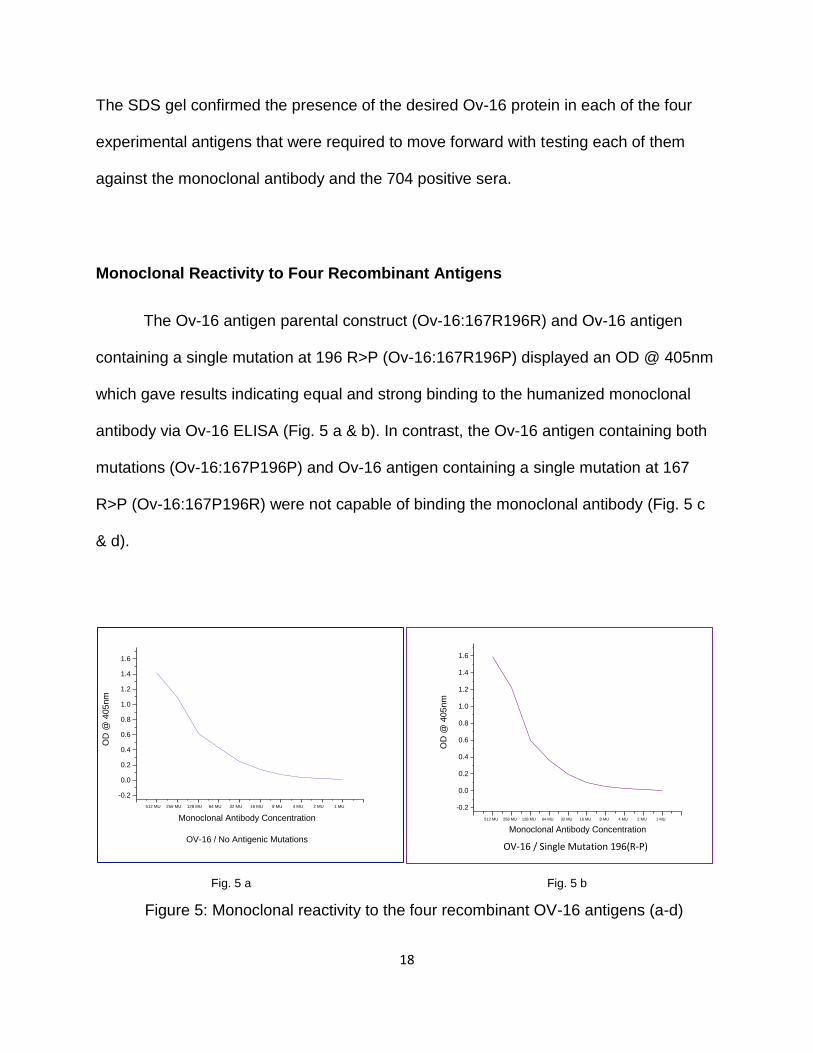
Monoclonal Reactivity to Four Recombinant Antigens
The Ov-16 antigen parental construct (Ov-16:167R196R) and Ov-16 antigen
containing a single mutation at 196 R>P (Ov-16:167R196P) displayed an OD @ 405nm
which gave results indicating equal and strong binding to the humanized monoclonal
antibody via Ov-16 ELISA (Fig. 5 a & b). In contrast, the Ov-16 antigen containing both
mutations (Ov-16:167P196P) and Ov-16 antigen containing a single mutation at 167
R>P (Ov-16:167P196R) were not capable of binding the monoclonal antibody (Fig. 5 c
& d).
Fig. 5 a Fig. 5 b
Figure 5: Monoclonal reactivity to the four recombinant OV-16 antigens (a-d)
512 MU 256 MU 128 MU 64 MU 32 MU 16 MU 8 MU 4 MU 2 MU 1 MU
-0.2
0.0
0.2
0.4
0.6
0.8
1.0
1.2
1.4
1.6
OD
@ 4
05
nm
Monoclonal Antibody Concentration
OV-16 / No Antigenic Mutations
512 MU 256 MU 128 MU 64 MU 32 MU 16 MU 8 MU 4 MU 2 MU 1 MU
-0.2
0.0
0.2
0.4
0.6
0.8
1.0
1.2
1.4
1.6
OD
@ 4
05
nm
Monoclonal Antibody Concentration
OV-16 / Single Mutation #420OV-16 / Single Mutation 196(R-P)
19
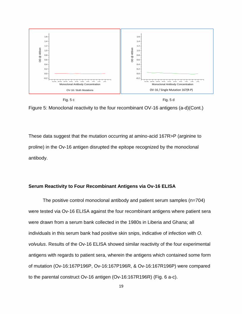
Fig. 5 c Fig. 5 d
Figure 5: Monoclonal reactivity to the four recombinant OV-16 antigens (a-d)(Cont.)
These data suggest that the mutation occurring at amino-acid 167R>P (arginine to
proline) in the Ov-16 antigen disrupted the epitope recognized by the monoclonal
antibody.
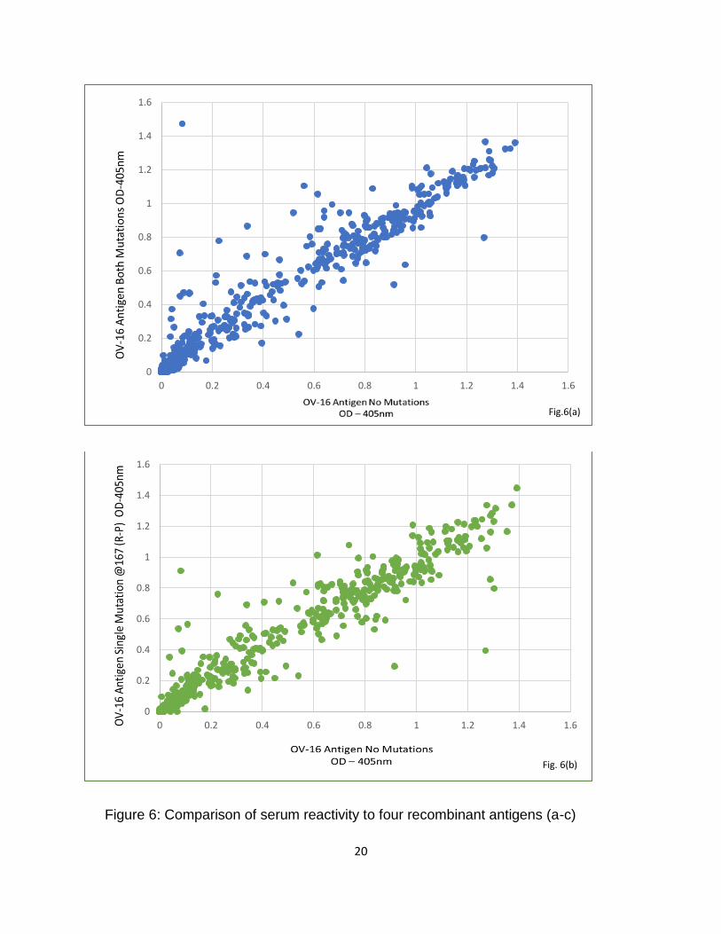
Serum Reactivity to Four Recombinant Antigens via Ov-16 ELISA
The positive control monoclonal antibody and patient serum samples (n=704)
were tested via Ov-16 ELISA against the four recombinant antigens where patient sera
were drawn from a serum bank collected in the 1980s in Liberia and Ghana; all
individuals in this serum bank had positive skin snips, indicative of infection with O.
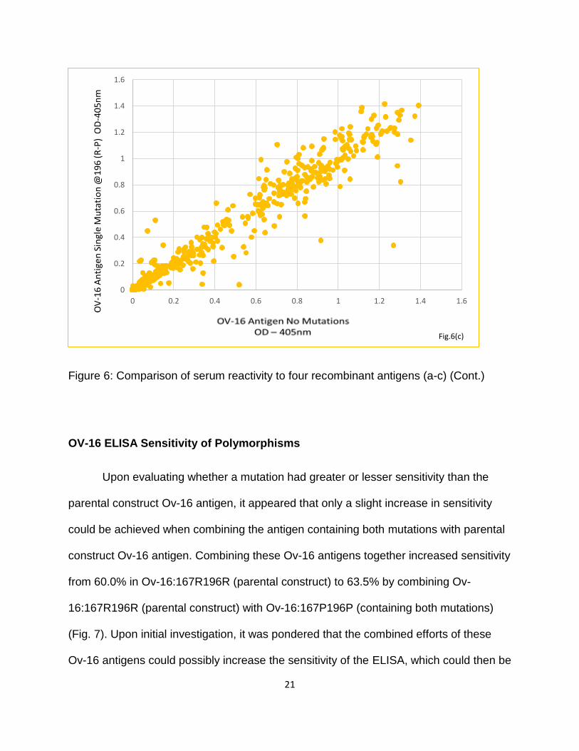
volvulus. Results of the Ov-16 ELISA showed similar reactivity of the four experimental
antigens with regards to patient sera, wherein the antigens which contained some form
of mutation (Ov-16:167P196P, Ov-16:167P196R, & Ov-16:167R196P) were compared
to the parental construct Ov-16 antigen (Ov-16:167R196R) (Fig. 6 a-c).
512 MU 256 MU 128 MU 64 MU 32 MU 16 MU 8 MU 4 MU 2 MU 1 MU
-0.2
0.0
0.2
0.4
0.6
0.8
1.0
1.2
1.4
1.6
OD
@ 4
05nm
Monoclonal Antibody Concentration
OV-16 / Both Mutations
512 MU 256 MU 128 MU 64 MU 32 MU 16 MU 8 MU 4 MU 2 MU 1 MU
-0.2
0.0
0.2
0.4
0.6
0.8
1.0
1.2
1.4
1.6
OD
@ 4
05nm
Monoclonal Antibody Concentration
OV-16 / Single Mutation #333OV-16 / Single Mutation 167(R-P)
20
Figure 6: Comparison of serum reactivity to four recombinant antigens (a-c)
0
0.2
0.4
0.6
0.8
1
1.2
1.4
1.6
0 0.2 0.4 0.6 0.8 1 1.2 1.4 1.6
Fig.6(a)
0
0.2
0.4
0.6
0.8
1
1.2
1.4
1.6
0 0.2 0.4 0.6 0.8 1 1.2 1.4 1.6
Fig. 6(b)
21
Figure 6: Comparison of serum reactivity to four recombinant antigens (a-c) (Cont.)
OV-16 ELISA Sensitivity of Polymorphisms
Upon evaluating whether a mutation had greater or lesser sensitivity than the
parental construct Ov-16 antigen, it appeared that only a slight increase in sensitivity
could be achieved when combining the antigen containing both mutations with parental
construct Ov-16 antigen. Combining these Ov-16 antigens together increased sensitivity
from 60.0% in Ov-16:167R196R (parental construct) to 63.5% by combining Ov-
16:167R196R (parental construct) with Ov-16:167P196P (containing both mutations)
(Fig. 7). Upon initial investigation, it was pondered that the combined efforts of these
Ov-16 antigens could possibly increase the sensitivity of the ELISA, which could then be
0
0.2
0.4
0.6
0.8
1
1.2
1.4
1.6
0 0.2 0.4 0.6 0.8 1 1.2 1.4 1.6
Fig.6(c)

22
initiated into current protocols to optimize testing in the field. After careful evaluation
however, it appears that the combined antigen sensitivity does not imply statistical
significance in this case. Statistical significance was evaluated using Chi-squared test.
In this case, Ov-16:167R196R (parental construct) and the combined Ov-16:167R196R
(parental construct) with Ov-16:167P196P (containing both mutations), both had a
sample size n= 704 with percentages at 60.0% and 63.5% respectively. Based on Chi-
squared test the P value was 0.18 indicating that no statistical significance exists for
increased sensitivity when the Ov-16 antigens are combined.
Figure 7: OV-16 ELISA; sensitivity comparisons of antigenic polymorphisms
60.00%62.60% 63.50%
0.00%
20.00%
40.00%
60.00%
80.00%
100.00%
120.00%
OV-16 Sensitivity
Combined antigensOV16:167R196R
+OV16:167P196P
OV16:167R196R OV16:167P196P
23
Specificity was not established during this research as all 704 serum samples
that were tested via Ov-16 ELISA were from individuals with known positive skin snips
indicative of infection with O. volvulus.
OV-16 ELISA GST Cross-Reactivity
All the serum samples tested negative for the presence of GST via Ov-16 ELISA
upon evaluation of signal at 405nm. It was therefore determined that no cross reactivity
with GST exists in any of the 704 serum samples that were tested indicating that all Ov-
16 positive values represent true positives for the Ov-16 portion of the fusion protein.
Only O. volvulus was analyzed in this research, therefore it is unknown if there may be
cross-reactivity with other filarial parasites and/or other Onchocerca spp. that could be
present in the region where these sera were collected.

24
CHAPTER FOUR:
DISCUSSION
Control programs rely on OV–16 ELISA as a diagnostic tool to evaluate the
progress towards elimination of onchocerciasis, which is currently ongoing primarily
through mass distribution of ivermectin, supplemented in some places with black fly
habitat targeted control efforts (Cupp, et al., 2012). To maintain quality control and
improve upon the Ov-16 ELISA format it is essential to evaluate the tests used for
verifying elimination, including use of the recently developed humanized monoclonal
antibody positive control. The importance of developing monoclonal antibodies for
positive controls in OV-16 ELISA cannot be stressed enough. As onchocerciasis
elimination efforts continue, it becomes increasingly important to have a set of standard
reagents that can be utilized as positive controls. Availability of monoclonal antibody
standards have an advantage in that; with the limited availability of pooled positive sera
monoclonal antibodies have the potential to be effectively mass produced such that any
need could be met. Monoclonal antibody applications could also universalize assay
performance across the globe, making results in different regions relatable for data
comparisons. Furthermore, these synthetic humanized antibodies could help protect
individuals performing these tests from the risk of other infectious diseases which may
also be present in the population where pooled sera might be selected, as it avoids the
need of finding positive controls within a given foci which also may become harder to

25
acquire as elimination efforts continue. This helps validate why a humanized
monoclonal antibody is a desired choice over pooled sera for positive controls.
Furthermore, the more we learn about genetics we may find that research is no longer
able to use a sample set of pooled sera from one region of the world to test another
region with the same accuracy.
In this research, antigenic polymorphisms did express concern and possible
limitations that exist when using a humanized monoclonal antibody for positive controls.
Ov-16 antigens Ov-16:167P196P and Ov-16:167P196R were not appropriately
recognized by the humanized monoclonal antibody due to unsuccessful epitope binding.
The parental construct Ov-16 antigen (Ov-16:167R196R) and the one containing the
single mutation at 196R>P (Ov-16:167R196P) demonstrated equivalent binding affinity
with the monoclonal antibody. This suggests that the polymorphism occurring at
167R>P disrupted the epitope that the monoclonal antibody recognized. There may be
further concern as to whether or not the monoclonal antibody is able to identify the
immunodominant epitope of interest given the possibility of other Ov-16 antigenic
polymorphisms that may exist in the designing of Ov-16 coating antigens in the ELISA
platform. Another proposal that may be of value would be to combine the parental
construct Ov-16 antigen (Ov-16:167R196R) with the one which contained both
mutations (Ov-16:167P196P), whereby creating multiple binding sites when using the
humanized monoclonal antibody standards.
When evaluating the polymorphisms that were detected via the Unnasch
laboratory, there was only a slight discrepancy between the sensitivity of the four
experimental Ov-16 antigens when evaluating the 704 sera from Liberia and Ghana.
26
However, it would be interesting to see if these mutated Ov-16 antigens would have a
different effect when tested in other foci in varied regions where onchocerciasis is
endemic. Another quandary to consider is the possibility that these types of
polymorphisms may be occurring with regards to other O. volvulus parasites in different
regions of the world which may also require evaluation of differing antigenic
polymorphisms than those detected through this research. Furthermore, it may be
necessary to delve into future effects of antigenic polymorphisms with regards to OV-16
when designing synthetic monoclonal antibody controls, as well as when testing sera in
varying foci in O. volvulus endemic regions, as variations could potentially be occurring
in different foci similar to those noted in this research which may cause difficulty in
creating universalized standards for Ov-16 ELISA formats.
To provide consistent evaluation of the ODs for each of the Ov-16 antigens, cut-
off values were standardized on a plate to plate basis at a range of 0.01 – 0.12 based
on OD readings of the monoclonal antibody that was plated with Ov-16:167R196R
(parental construct) and a set of sera tested against the four experimental antigens on a
single plate. In this research, all the sera tested were from O. volvulus infected
individuals and therefore the cut-offs were set conservatively, standard Ov-16 ELISA
protocol typically sets cut-off values based on standards at 1:1280 with a mean OD at
approximately 0.13 and a range between 0.06 – 0.19, evaluation at a standard cut-off of
0.2 did not appear to change the sensitivity of the results in this investigation. Reactivity
of the four experimental antigens with regards to patient sera also displayed a few
outliers that may have indicated other selective epitope binding was at work. A few of
these outliers had elevated optical densities well beyond the Ov-16:167R196R (parental

27
construct) and would be interesting to investigate further, as the antibody-antigen
binding affinity occurring in these O. volvulus positive samples may assist with future
test design applications to potentially increase sensitivity. When evaluating the overall
sensitivity of the Ov-16 ELISA against the 704 serum samples from O. volvulus infected
patients, the results were consistent with current Ov-16 ELISA applications that are in
place in the field. Although the combined effort of Ov-16:167R196R (parental construct)
and Ov-16:167P196P (containing both mutations) did not indicate statistical significance
exists for increased sensitivity, the percentage difference could prove to be statistically
sound given further evaluation with a larger sample size than that which was applied in
this investigation (n= 704). The sensitivities of 60.0% in Ov-16:167R196R (parental
construct), 62.6% in Ov-16:167P196P (containing both mutations), and at best 63.5%
when combined, illustrated that there is always work to be done in order to provide the
best possible screening capabilities with regards to Ov-16 ELISA and onchocerciasis
elimination. As the sera obtained for use in this investigation was acquired from O.
volvulus infected individuals in foci from Liberia and Ghana only, another avenue to
extend future studies regarding these particular Ov-16 antigenic polymorphisms would
be through multi-facility collaborative research efforts on O. volvulus positive sera from
other foci in various endemic regions, such as east Africa; Uganda and Sudan, or
possibly Yemen, that have been maintained in alternative research laboratory
collections.
The distribution of onchocerciasis has reduced significantly since the 1980’s due
to the ongoing efforts of MDA programs (Cupp, et al., 2012). Although only minor
differences were noted in the positive test results between the four experimental Ov-16
28
antigens, research of antigenic polymorphisms regarding Ov-16 ELISA may be
essential to address the implications that can occur with current elimination efforts if
such mutations allow false negative test results within regions of the world thought to be
at or near elimination status. Monoclonal antibody controls should be researched further
to account for potential antigenic polymorphisms that are occurring in the various foci
affected by onchocerciasis which may limit the ability for these controls to work
appropriately in Ov-16 ELISA formats when such polymorphisms are present in the
coating antigen.
To date there are still many challenges to face in the journey to onchocerciasis
elimination, including the continued exploration for proficient diagnostic tools, even
without the event of Ov-16 antigenic polymorphisms. There is also a growing need to
explore drug alternatives; to treat high risk cases that have co-endemicity infections like
Loa loa, to prepare for the event of possible ivermectin resistance, and to take a deeper
look into macrofilaricides to rid the endemic population of the adult parasites which have
the ability to perpetuate the cycle. Continued habitat targeting control methods, possibly
aided through the use of GIS, and pooled black fly testing to detect the presence of the
parasite within the fly should also be investigated to ensure effective elimination
strategies are underway. Prior onchocerciasis research has paved the road in
establishing the techniques and treatments that are currently used to control the
disease, the path has certainly revealed continued research is necessary to build upon
these accomplishments with more field appropriate test applications that have the
desired specificity and sensitivity to detect recent infections in order to determine if

29
transmission is occurring, if reemergence in previously interrupted foci exists, or if
disease interruption or elimination status has been achieved within a given foci.
30
REFERENCES
Banla, M., Tchalim, S., Karabou, P. K., Gantin, R. G., Agba, A. I., Kére-Banla, A., …
Soboslay, P. T. (2014). Sustainable Control of Onchocerciasis: Ocular Pathology
in Onchocerciasis Patients Treated Annually with Ivermectin for 23 Years: A
Cohort Study. PLoS ONE, 9(6), e98411.
Boatin, B. A., & Amazigo, U. (January 01, 2016). Onchocerciasis.
Bogitsh, B. J., Carter, C. E., & Oeltmann, T. N. (2013). Human parasitology.
Amsterdam: Academic Press.
Botto, C., Basañez, M.-G., Escalona, M., Villamizar, N. J., Noya-Alarcón, O., Cortez, J.,
… Grillet, M. E. (2016). Evidence of suppression of onchocerciasis transmission
in the Venezuelan Amazonian focus. Parasites & Vectors, 9, 40.
Burbelo, P. D., Leahy, H. P., Iadarola, M. J., & Nutman, T. B. (2009). A Four-Antigen
Mixture for Rapid Assessment of Onchocerca volvulus Infection. PLoS Neglected
Tropical Diseases, 3(5), e438.
Coffeng, L. E., Stolk, W. A., Zouré, H. G. M., Veerman, J. L., Agblewonu, K. B.,
Murdoch, M. E., … Amazigo, U. V. (2013). African Programme for
Onchocerciasis Control 1995–2015: Model-Estimated Health Impact and Cost.
PLoS Neglected Tropical Diseases, 7(1), e2032.
Cupp,Ed, Richards,Frank, Grillet,Maria-Eugenia, Sauerbrey,, Eberhard,, Dominguez,,
Morales, ... Nicholls,. (2012). Guide to detecting a potential recrudescence of
onchocerciasis during the posttreatment surveillance period: the American
paradigm. Dove Press.
Denery, J. R., Nunes, A. A. K., Hixon, M. S., Dickerson, T. J., & Janda, K. D. (2010).
Metabolomics-Based Discovery of Diagnostic Biomarkers for Onchocerciasis.
PLoS Neglected Tropical Diseases, 4(10), e834.
Devoe, N. C., Corbett, I. J., Barker, L., Chang, R., Gudis, P., Mullen, N., … May, M.
(2016). Differential Evolutionary Selection and Natural Evolvability Observed in
ALT Proteins of Human Filarial Parasites. PLoS ONE, 11(2), e0148611.
31
Dobson, M. J. (2008). Disease. London: Quercus.
Eberhard, M. L., Cupp, E. W., Katholi, C. R., Richards, F. O., & Unnasch, T. R. (2017).
Skin snips have no role in programmatic evaluations for onchocerciasis
elimination: a reply to Bottomley et al. Parasites & Vectors, 10, 154.
Eisenbarth, A., Achukwi, M. D., & Renz, A. (2016). Ongoing Transmission of
Onchocerca volvulus after 25 Years of Annual Ivermectin Mass Treatments in the
Vina du Nord River Valley, in North Cameroon. PLoS Neglected Tropical
Diseases, 10(2), e0004392.
Fuchs, J., Podda, M., & Informa Healthcare. (2004). Encyclopedia of medical genomics
and proteomics., New York: Marcel Dekker.
Golden, A., Faulx, D., Kalnoky, M., Stevens, E., Yokobe, L., Peck, R., … Domingo, G. J.
(2016). Analysis of age-dependent trends in Ov16 IgG4 seroprevalence to
onchocerciasis. Parasites & Vectors, 9, 338.
Golden, A., Steel, C., Yokobe, L., Jackson, E., Barney, R., Kubofcik, J., … Domingo, G.
J. (2013). Extended Result Reading Window in Lateral Flow Tests Detecting
Exposure to Onchocerca volvulus: A New Technology to Improve
Epidemiological Surveillance Tools. PLoS ONE, 8(7), e69231.
Golden, A., Stevens, E. J., Yokobe, L., Faulx, D., Kalnoky, M., Peck, R., … Domingo, G.
J. (2016). A Recombinant Positive Control for Serology Diagnostic Tests
Supporting Elimination of Onchocerca volvulus. PLoS Neglected Tropical
Diseases, 10(1), e0004292.
Gustavsen, K., Hopkins, A., & Sauerbrey, M. (2011). Onchocerciasis in the Americas:
from arrival to (near) elimination. Parasites & Vectors, 4, 205.
Hassan, H. K., Bolcen, S., Kubofcik, J., Nutman, T. B., Eberhard, M. L., Middleton, K.,
… Beeler, E. S. (2015). Isolation of Onchocerca lupi in Dogs and Black Flies,
California, USA. Emerging Infectious Diseases, 21(5), 789–796.
Hernández-González, A., Moya, L., Perteguer, M. J., Herrador, Z., Nguema, R.,
Nguema, J., … Gárate, T. (2016). Evaluation of onchocerciasis seroprevalence
in Bioko Island (Equatorial Guinea) after years of disease control programmes.
Parasites & Vectors, 9, 509.
32
Heymann, D. L., & American Public Health Association. (2008). Control of
communicable diseases manual. Washington, DC: American Public Health
Association.
Higazi, T. B., Zarroug, I. M. A., Mohamed, H. A., ElMubark, W. A., Deran, T. C. M., Aziz,
N., … Hashim, K. (2013). Interruption of Onchocerca volvulus Transmission in
the Abu Hamed Focus, Sudan. The American Journal of Tropical Medicine and
Hygiene, 89(1), 51–57.
Ibe, O., Onwujekwe, O., Uzochukwu, B., Ajuba, M., & Okonkwo, P. (2015). Exploring
Consumer Perceptions and Economic Burden of Onchocerciasis on Households
in Enugu State, South-East Nigeria. PLoS Neglected Tropical Diseases, 9(11),
e0004231.
Lagatie, O., Van Dorst, B., & Stuyver, L. J. (2017). Identification of three
immunodominant motifs with atypical isotype profile scattered over the
Onchocerca volvulus proteome. PLoS Neglected Tropical Diseases, 11(1),
e0005330.
Liu, D. (2013). Molecular detection of human parasitic pathogens. Boca Raton: Taylor &
Francis.
LOK, J. B. (2012). Nucleic acid transfection and transgenesis in parasitic nematodes.
Parasitology, 139(5), 574–588.
Lovato, R., Guevara, A., Guderian, R., Proaño, R., Unnasch, T., Criollo, H., …
Mackenzie, C. D. (2014). Interruption of Infection Transmission in the
Onchocerciasis Focus of Ecuador Leading to the Cessation of Ivermectin
Distribution. PLoS Neglected Tropical Diseases, 8(5), e2821.
Lucius, R., Erondu, N., Kern, A., Donelson, J.E. (1988). Molecular cloning of an
immunodominant antigen of Onchocerca volvulus. The Journal of Experimental
Medicine, 168(3), 1199–1204.
Luz, S. L. B., Crainey, J. L., Shelley, A. J., & Rubio, M. (2014). Outstanding insecurities
concerning the use of an Ov16-based ELISA in the Amazonia onchocerciasis
focus. Memórias Do Instituto Oswaldo Cruz, 109(4), 506–508.
McClelland, G. A. H., (1992). Medical Entomology; An Ecological Perspective. 12th Ed.
University of California.

33
Noma, M., Zouré, H. G., Tekle, A. H., Enyong, P. A., Nwoke, B. E., & Remme, J. H.
(2014). The geographic distribution of onchocerciasis in the 20 participating
countries of the African Programme for Onchocerciasis Control: (1) priority areas
for ivermectin treatment. Parasites & Vectors, 7, 325.
Oguttu, D., Byamukama, E., Katholi, C. R., Habomugisha, P., Nahabwe, C., Ngabirano,
M., … Unnasch, T. R. (2014). Serosurveillance to Monitor Onchocerciasis
Elimination: The Ugandan Experience. The American Journal of Tropical
Medicine and Hygiene, 90(2), 339–345.
Ojurongbe, O., Akindele, A. A., Adeleke, M. A., Oyedeji, M. O., Adedokun, S. A., Ojo, J.
F., … Adeyeba, O. A. (2015). Co-endemicity of Loiasis and Onchocerciasis in
Rain Forest Communities in Southwestern Nigeria. PLoS Neglected Tropical
Diseases, 9(3), e0003633.
Richards, F., Rizzo, N., Espinoza, C. E. D., Monroy, Z. M., Valdez, C. G. C., de
Cabrera, R. M., … Domínguez, A. (2015). One Hundred Years After Its
Discovery in Guatemala by Rodolfo Robles, Onchocerca volvulus Transmission
Has Been Eliminated from the Central Endemic Zone. The American Journal of
Tropical Medicine and Hygiene, 93(6), 1295–1304.
Rodríguez-Pérez, M. A., Unnasch, T. R., Domínguez-Vázquez, A., Morales-Castro, A.
L., Peña-Flores, G. P., Orozco-Algarra, M. E., … Rendón, V. G. (2010).
Interruption of Transmission of Onchocerca volvulus in the Oaxaca Focus,
Mexico. The American Journal of Tropical Medicine and Hygiene, 83(1), 21–27.
Schwab, L. (2007). Eye care in developing nations. London: Manson.
Sommer, A., Nimtz, M., Conradt, H. S., Brattig, N., Boettcher, K., Fischer, P., … Liebau,
E. (2001). Structural Analysis and Antibody Response to the Extracellular
Glutathione S-Transferases from Onchocerca volvulus. Infection and Immunity,
69(12), 7718–7728.
Thermo Scientific, ELISA technical guide and protocols (2016, July) Retrieved from
http://tools.thermofisher.com/content/sfs/brochures/TR0065-ELISA-guide.pdf
Thiele, E. A., Cama, V. A., Lakwo, T., Mekasha, S., Abanyie, F., Sleshi, M., … Cantey,
P. T. (2016). Detection of Onchocerca volvulus in Skin Snips by Microscopy and
Real-Time Polymerase Chain Reaction: Implications for Monitoring and
Evaluation Activities. The American Journal of Tropical Medicine and Hygiene,
94(4), 906–911.

34
Toé, L., Back, C., Adjami, A. G., Tang, J. M., & Unnasch, T. R. (1997). Onchocerca
volvulus: comparison of field collection methods for the preservation of parasite
and vector samples for PCR analysis. Bulletin of the World Health Organization,
75(5), 443–447.
Unnasch, T.R., (2004). Encyclopedia of medical genomics and proteomics.,
Onchocerca volvulus, New York: Marcel Dekker.
Unnasch, T. R., Gallin, M. Y., Soboslay, P. T., Erttmann, K. D., & Greene, B. M. (1988).
Isolation and characterization of expression cDNA clones encoding antigens of
Onchocerca volvulus infective larvae. Journal of Clinical Investigation, 82(1),
262–269.
WHO, Department of Control of Neglected Tropical Diseases, (2016). Progress towards
eliminating onchocerciasis in the WHO Region of the Americas: verification of
elimination of transmission in Guatemala, Progress report on the elimination of
human onchocerciasis, 2015–2016., Weekly epidemiological record, 43(91),
501–516.
Zouré, H. G., Noma, M., Tekle, A. H., Amazigo, U. V., Diggle, P. J., Giorgi, E., &
Remme, J. H. (2014). The geographic distribution of onchocerciasis in the 20
participating countries of the African Programme for Onchocerciasis Control: (2)
pre-control endemicity levels and estimated number infected. Parasites &
Vectors, 7, 326.