the distribution of cholecystokinin-8 in the central nervous system
TRANSCRIPT

Brain Rfsettr& Bulletin, Vol. 15, pp. 167-181, 1985. b Ankho International Inc. Printed in the U.S.A. 0361~9230/85 $3.00 + .OO
The Distribution of Cholecystokinin-8 in the Central Nervous System
of Turtles: An Immunohisto~hemical and Bio~hemic~ Study
ANTON REINER* AND MARGERY C. BEINFELDt
*Department ~~~An~torny and Cell Biology, University ofikfichigaa, Ann Arbor, MI 48109 and tDepartment of Pharmacology, St. Louis University, School of Medicine, St. Louis, MO 63104
Received 1 October 1984
REINER, A. AND M. C. BEINFELD. The distribution of cholecystokinin-8 in the central nervous system of turtles: An i/nmunohistct~hrmi~ai and bj~~~hernj~al study. BRAIN RES BULL 15(Z) 167-181, 1985.-.Immuno~st~hemi~ tech- niques, radioimmunoassay (RIA) and high performance liquid chromatography (HPLC) were used to: (1) determine the regional distribution and amounts of cholecystokinin-8 (CCK8)-like immunoreactivity in the turtle central nervous system, and (2) chemically characterize the CCK%-like material present in the turtle central nervous system. High levels of CCKS-like immunoreactivity were found in the turtle central nervous system, with the highest levels being present in the hypothalamus and neurohypophysis. Moderate levels of the CCK8-like material were found in all other regions of the turtle nervous system except the cerebellum, the olfactory bulbs and the dorsal ventricular ridge of the telencephalon, which contained low levels. The bulk (87%) of the CCKS-like material in turtle central nervous system co-eluted with CCKbsulfate in gradient eiution HPLC. The distribution of CCKS-like immunoreactivity (CCKSLI) observed using immunobistochemistry was consistent with the results of the RIA studies. Numerous CCK8LI-containing neurons and fibers were observed in the hypothalamus and neurohypophysis. Neurons and fibers containing CCKS were, however, more sparsely distributed outside the hypo- thalamus. The immunohistochemical data provided evidence for the existence of two major CCKS-containing pathways in turtles that have been previously described in mammals: a pathway from the supraoptic and paraventricular magnocelhdar nuclei to the external zone of the median eminence and neurohypophysis and a pathway from dorsal root ganglia to the dorsal horn of the spinal cord. Overall, the present results, in conjunction with several previous studies, indicate that CCKS has had a relatively stable evolutionary history as a CNS neuropeptide among land vertebrates. The molecular structure of CCKS appears to have been largely (if not entirely) conserved, as has its concentration in many brain regions. A notewor- thy exception to such conservatism in the localization of CCK8 is that the concentration of CCK8 in the telencephalon, particularly in the telencephalic cortex, is much lower in turtles than in mammals. The present results therefore suggest that CCK8 may not have become a prominent peptide in the telencephalic cortex (or its anatomical equivalents) untii the evolution of neocortex in the mammalian lineage.
Choiecystokinin-8 Central nervous system Turtles Immunohistochemistry
CHOLECYSTOKININ (CCK) is a thirty-three amino acid peptide that stimulates gall bladder contraction and pancre- atic enzyme secretion and is found in high concentration in the proximal small intestine, where it is localized to a spe- cific class of gut endocrine cells. Cholecystokinin is also found in great abundance in neurons and fibers of the nerv- ous system, where shorter peptide fragments of CCK, in particular the C-terminal octapeptide (CCK8), predominate [4,21,44]. The precise role of CCKS in the nervous system is uncertain, but it is clear that CCKS displays many of the characteristics of a neurot~nsmitter, particularly in terms of its localization, release and receptor-mediated actions on postsynaptic neurons 16, 20, 751. Among the many peptides found in the m~malian brain, CCK8 is distinguished by being one of the few peptides (together with VIP) that is found in a higher concentration in the cerebral cortex than in any other neuml region [8, 24, 55, 761. Although the gall
bladder and, to a lesser extent, the pancreas appear to be phylogenetically old features of the vertebrate GI tract, the type of cortex that predominates in the mammalian cerebral hemispheres (i.e., neocortex) is a more recent feature of ver- tebrate phylogeny 154,573. Nonetheless, comparative neuroanatomical studies during the last decade have shown that large portions of the nonmammalian telencephalon, though not organized into a neocortical tissue such as in mammals, are comparable to mammalian neocortex in terms of histochemistry, connections and functions [37,54, 56, 57, 801. Thus, it might be expected that CCK8 or a CCK8-like peptide be prominent in the neocortical-equvalent tissue of nonmammals. Previous studies have shown that CCK-like peptides are present in both the brain and gut of members of many different ancient animal groups, including annelid worms, dungeness crabs and lampreys 126, 33, 421. The CCK-like materials present in the gut and brain of the mem-
167

I68 REINER AND BEINFELD
ABBREVIATIONS
a AP AT BA CA Cb CbL CbM cd cdm cm CN co co CP
cP d d IV DLA DMA DSOD DVR FD FL FLM FRL FV GC GCL GLd GLv GP HL HM IP Imc
IPC LA LGE LGI LL LM LOC MA MC ME ML MV mV
NHY NPd NPv NV N VIII nAmb nBOR nCA nDB nDCP nFLM nMH
Area a (Riss, Halpem and Scalia [73]) Area pretectalis Area triangularis Nucleus basalis amygdalae Nucleus centralis amygdalae Cerebellum Nucleus cerebellaris lateralis Nucleus cerebellaris medialis Cortex dorsalis Cortex dorsomedialis Cortex medialis Core nucleus of the DVR Chiasma opticum Cochlear nuclei Commissura posterior Cortex pyriformis Area d (Riss, Halpem and Scaha [73]) Decussatio nervi trochlearis Nucleus dorsalateralis anterior Nucleus dorsomedialis anterior Dorsal nucleus of the supraoptic decussation Dorsal ventricular ridge of the telencephalon Funiculus dorsalis Funiculus lateralis Fasciculus medialis longitudinalis Formatio reticularis lateralis mesencephali Funiculus ventralis Griseum centrale Granule cell layer Nucleus geniculatus lateralis pars dorsalis Nucleus geniculatus lateralis pars ventralis Globus pallidus Nucleus habenularis lateralis Nucleus habenularis medialis Nucleus interpeduncularis Nucleus isthmi pars magnocellularis Nucleus isthmi pars parvocellularis Nucleus laminaris of the torus semicircularis Laminaris granularis extema Laminaris granularis intema Lateral lemniscus Nucleus lentiformis mesencephali Locus coeruleus Nucleus medialis amygdalae Mitral cell layer Median eminence Molecular layer Nucleus mesencephalicus nervi trigemini Nucleus motorius nervi trigemini Neurohypophysis Nucleus pretectalis dorsalis Nucleus pretectalis ventralis Nervus trigemini Nervus octavus Nucleus ambiguus Nucleus of the basal optic root Nucleus commissuralis anterioris Nucleus fasciculus diagonalis Brocae Nucleus dorsalis commissuralae posterioris Nucleus fasciculi longitudinalis medialis Nucleus medialis hypothalami
nPH nPM nS nSL nSM nS0 nSP nSPM nTOL nTS nVH n III n 1V n VI n VII nX n XII 01 OS PA Pb PD PH PrV PI PV R Rai Ras Re Ri Ris Rm ROB RS Ru SGC SGD SGF SGP SGV SM Sm SN so ST Ta Tel TeO TO TOI TOm TrL TSC TTd TuOl TT VeD VeL VeS VP
Nucleus periventricularis hypothalami Nucleus profundus mesencephali Nucleus septalis Nucleus septalis lateralis Nucleus septalis medialis Nucleus supraopticus Nucleus suprapenduncularis Nucleus suprapeduncularis medialis Nucleus tracti olfactorii lateralis Nucleus tracti solitarii Nucleus ventromedialis hypothalami Nucleus nervi oculomotorius Nucleus nervi trochlearis Nucleus nervi abducens Nucleus nervi facialis Nucleus motorius dorsalis nervi vagi Nucleus nervi hypoglossi Oliva inferior Oliva superior Paleostriatum augmentatum Nucleus parabrachialis Peduncularis dorsalis fasciculi prosencephali lateralis Primordium hippocampi Nucleus princeps nervi trigemini Pallial thickening Peduncularis ventralis fasciculi prosencephali lateralis Nucleus rotundus Nucleus raphes inferior Nucleus raphes superior Nucleus reuniens Nucleus reticularis inferior Nucleus reticularis isthmi Nucleus reticularis medialis Radix opticum basalis Nucleus reticularis superior Nucleus ruber Stratum griseum centrale Nucleus substantiae griseae dorsalis Stratum griseum et fibrosum superficiale Stratum griseum profundum Substantiae griseae ventralis Stria medullaris Supramamillary region Substantia nigra Stratum opticum Stria terminalis Nucleus tangentialis Telencephalon Tectum opticum Tractus opticus Tractus opticus, pars lateralis Tractus opticus, pars medialis Tract of Lissauer Torus semicircularis Nucleus descendens nervi trigemini Tuberculum olfactorium Tractus tectothalamicus Nucleus vestibularis descendens Nucleus vestibularis lateralis Nucleus vestibularis superior Ventral paleostriatum

CCK8 IN TURTLE CNS 169
bers of these vertebrate and invertebrate groups, however, differ in chemical structure from the CCK-related peptides found in mammalian tissues. In the present study, we have studied turtles, a member of a class of vertebrates (i.e., rep- tiles) immediately ancestral to mammals, in order to gain insight into the recent evolutionary history of CCK-related peptides in the vertebrate brain. A previous study 1431 has suggested that a CCK-like peptide is present in duodenal secretory cells in turtles, based on the immunological reac- tivity of this peptide to several different regionally-specific antisera against CCK.
METHOD
Studies were carried out on red-eared turtles (Pseudemys scripta), painted turtles (Chrysemys picta) and mud turtles (Kinosternum subrubrum). They were maintained in a heated (25-30°C) open air aquarium with an artificial day- night cycle (14 hours light-10 hours darkness). A C-terminally directed antiserum against CCK8 (R5) was used in the pres- ent study for radioimmunoassay and immunohistochemistry. The details of the production and characterization of this antiserum have been described previously [9]. Although the antiserum does not distinguish between CCK8 and gastrin, the bulk of the CCK-like material in turtle brain was found in the present study to co-elute with CCK&sulfate in gradient elution HPLC.
Biochemical Studies
The brains were removed from 3 freshly decapitated tur- tles (one red-eared turtle and 2 mud turtles) and dissected into ten discrete subdivisions: (1) olfactory bulbs, (2) telen- cephalic cortex, (3) telencephalic dorsal ventricular ridge (DVR) plus rostra1 pyriform (olfactory) cortex, (4) basal tel- encephalon including basal ganglia and septum, (5) thalamus and habenula, (6) hypothalamus and neurohypophysis, (7) tectum and pretectum, (8) tegmentum, (9) cerebellum and (10) rhombencephalon. In addition, the fist 4-5 cervical spinal segments were dissected from each animal. All dissections were carried out on ice. The approximate internal subdi- visions along which the telencephalon, midbrain and hind- brain were subdivided for radioimmunoassay (RIA) have been described previously [72]. All tissue samples were blot- ted and weighed. The tissue was boiled in water, sonicated in ice cold 0.1 N HCI and an aliquot removed from each sample for Lowry protein assay [47]. After each tissue extract was clarified by centrifugation, an aliquot of the supernatant was removed for RIA and neutralized with an equal volume of 0.1 N NaOH. The RIA procedure used ““I-Gastrinl7-I, with CCKS-sulfate as the standard. The RIA procedure has been described in greater detail previously [9]. For HPLC, aliquots from the supematants were combined, clarified by centrifugation and injected onto an Altech column (0.46x2.5 cm) and eluted with a linear gradient of 20-60% acetonitrile in the presence of 0.9% trifluoroacetic acid over 40 minutes. One minue fractions were collected at a flow rate of 1 ml/minute. The HPLC fractions were dried in a Savant vac- uum centrifuge, resuspended in water and an aliquot re- moved for CCK8 RIA. CCK&sulfate and Human Gas- trin17-I were used as standards in the present study and were detected optically and with CCK8 RIA. Several previous studies by one of US as well as by other investigators have shown that gradient elution HPLC systems similar to the Present one, as well as a variety of other HPLC systems, provide clear and consistent separation of CCK8-sulfate, CCK&desulfate, CCK33, CCK4, caerulein, Human Gas-
trinl7-I and Human Gastrinl7-II ([4, 7, 11, 531 Beinfeld, un- published observation).
Immunohistochemical Studies
Turtles were anesthetized deeply with ketamine or sodium pentobarbiti and perfused through the heart with 6% dextran in 0.1 M phosphate buffer at pH 7.4 (PB), followed by a solution of 4% paraformaldehyde in PB. The brains (with a few segments of cervical spinal cord still attached) were dis- sected from the skull, immersed in 4% paraformaldehyde in 0.1 M phosphate buffer (pH 10.4) for 2-18 hours, and then immersed in a 30% sucrose-PB solution for 2 days. The brains and spinal cord were then sectioned frozen at 35 mi- crons on a sliding microtome. The indirect immunofluores- cence procedure [16,84] and the peroxidase-antiperoxidase (PAP) procedure [78] were used to localize CCK8 in the brain and spinal cord tissue sections. The details of our im- munohistochemical techniques have been described else- where [70-721. Briefly sections were washed three times in PB and incubated in the R5 antiserum against CCKS. Tissue incubations were carried out for 12-60 hours using a primary antiserum dilution of 1: 1000. All antisera (primary and sec- ondary) were diluted with PB containing 0.3% Triton X-100. Following the incubation in primary antiserum, tissue was washed three times in PB and then processed according to either the indirect immunofluorescence procedure or the PAP procedure. For the immunofluorescence procedure, tis- sue was incubated in a secondary antiserum (raised in goat and directed against rabbit IgG) conjugated to fluorescein isothyocyanate (FITC) (Miles Laboratories) at a dilution of 1:50 for 1 hour at room temperature. Following the incuba- tion in the FITC-conjugated secondary antiserum, sections were washed three times in PB, mounted on gelatin-coated slides and coverslipped in a 9: 1 solution of glycerol and .OSM carbonate buffer (pH 9.0). Sections were then examined with a Leitz Orthoplan microscope using a Ploem epi-illumination fluorescence microscopy system. For the PAP method, fol- lowing incubation in the primary antiserum, sections were incubated at a 1:50 dilution for one hour at room temperature in an unlabeled secondary antiserum that was raised in goat and directed against rabbit IgG (Miles Laboratories). Following three washes in PB, sections were incubated in PAP (anti- peroxidase raised in rabbit) for one hour at room tempera- ture at a dilution of 1:2OO. Sections were then washed three times in PB and reacted in a solution of diaminobenzidine (DAB) and H202 (100 mg DAB/100 ml 0.1 M cacodylate buf- fer (pH 7.2)/0.5% H202) to demonstrate the location of the immunologically bound peroxidase. Sections were then washed, mounted on subbed slides and coverslipped with Permount. Specificity of the immunoreactivity was assessed by preabsorbing the primary antiserum with a 10 FM concen- tration of synthetic CCKS. Terminology for the turtle brain is as used previously [63,72].
Injections of colchicine (100 pg colchicine in 6 ~1 distilled water) were made into the lateral ventricle of several turtles. Stereotaxic procedures were used with turtles under ketamine anesthesia [63]. Colchicine-treated turtles were sacrificed as described above 2-5 days after the injection and the brains processed according to the immunohistochemical procedures described.
RESULTS
Biochemical Studies
The RIA data for the levels of CCK8 in the different re-

170
TABLE I
THE AMOUNT OF CCKB IN THE DIFFERENT REGIONS OF TURTLE BRAIN, AS MEASURED BY RIA
KElNER AND BEINFELD
Region pg CCKSl pg CCK8lmg pg CCKSIpg
region tissue protein
Olfactory Bulb 650 + 90 9.9 + 1.6 0.14 + 0.01
Basal 1030 t 490 10.2 t 3.2 0.17 t 0.06 Telencephalon
Cortex 2030 r!z 740 10.4 t 4.2 0.31 * 0.11 Dorsal 370 4 88 2.6 -_t: 0.7 0.06 t 0.01
Ventricular Ridge
Hypothalamus1 8200 _c 2140 212.1 rt 72.8 3.28 t 1.02 Neurohypophysis
Thalamusi 1170 7t 220 33.9 + 5.5 0.81 f 0.39 EpithaIamus
Tegmentum 2790 -c 500 53.6 2 7.9 0.96 ? 0.15 Tectum 1480 ir 340 20.6 ? 4.4 0.33 * 0.05 Cerebellum 180 ~?r 100 4.9 -+ 2.9 0.08 -e 0.04 Rhom~ncephalon 6000 I 610 50.7 i 10.8 0.95 r 0.16 Spinal Cord 2370 c 710 57.8 t 13.7 0.73 t 0.17
Each values is the mean (z?SEM) for that structure for the three turtles assayed. The basal telencephalon tissue sample included the entire basolateral wall of the telencephalon (including the basal gan- glia and ventral paleostriatum), the septum and basal parts of the amygdala. The dorsal ventricular ridge (DVR) sample included the DVR, most of the pyriform cortex, ventral parts of the pallial thick- ening and more dorsal parts of the amygdala. The tegmental and tectai samples each included parts of the torus semicircularis. The value for the total amount of CCKI in the spinal cord indicates the amount present in 4-5 spinal segments. The amount of CCKI per mg tissue is based on the wet weight of the tissue.
gions of turtle brain are shown in Table 1. The highest levels of CCK8 were found in the hypothalamus/neurohypophysis extract. Although the thalamus, tegmentum, rhombencepha- Ion and spinal cord contained the next highest levels of CCKP;, the levels in these structures were only 113-114 that found in the hypothalamuslneurohypophysis. Moderate amounts of CCKS were found in cortex, basal telencephalon and the tectum. Very little CCK8 was found in the olfactory bulbs, dorsal ventricular ridge or cerebellum. Using HPLC, the bulk (87%) of the CCKS-like immunoreactive material in turtle nervous system was found to co-elute with CCK&sul- fate (see Fig. I). Two minor peaks of CCK8-like material (each representing 6.5% of the eluted CCKMike material) were observed, one eluting before CCK8-sulfate and the other eluting after CCKS-sulfate. Based on the retention times observed for the different CCK-like peptides in previ- ous studies empioying gradient elution HPLC ([53] Beinfeld, unpublished observation), these minor peaks appear to rep- resent a CCK33-like peptide and CCK&desulfate, respec- tively.
The immunohistochemical data were consistent with the CCKS RIA values for the CCK8 content of the different neural regions in turtle. A map of the distribution of specific (i.e., blockable by absorption of the antiserum with synthetic CCK8) CCKMike immunoreactivity (CCKSLI) in turtle brain is shown in Figs. 2-10. Photomicrographs of CCKS-
FRACTION NUMBER
FIG. I. Gradient elution HPLC profile of extract from turtle brain. The bulk of the CCKI-like material in turtle brain (87%) co-elutes with CCK8-sulfate. The arrow (fraction 40) indicates the retention time of nastrin 17-I (G17-I) in this HPLC svstem. The small neaks of CCKlLj preceding‘(fra&on 30) and following (fraction 36j CCKR- sulfate each represent 6.5% of the total CCKILI eluted in this HPLC system. As discussed in the text, these small peaks may represent CCK&desulfate (fraction 36) and a high molecular weight peptide similar to CCK33 (fraction 30). No evidence was obtained for the presence in turtle nervous system of CCK4, which (as discussed in the text) should elute in fraction 21-22.
positive labeling in several neural regions is shown in Figs. 1 l-12. In general, more CCKSLI-containing perikarya were observed in colchicine-treated animals than in normal animals. In the case of some cell groups, CCK8LI-containing perikarya were not observed except in colchicine-treated turtles. Although the bulk of the CCKSLI in turtle brain appears to be indistinguishable from CCKbsulfate by the present HPLC system, the immunoreactive material ob- served in turtle nervous tissue with immunohisto~hemical methods is nonetheless referred to as “CCK&Iike” because: (1) a small amount (13%) of the CCK&like material in turtle central nervous system did not co-elute with CCK8-sulfate, and (2) the predominant CCKS-like material in turtle nervous system may differ from CCK8-sulfate in ways not detectible by the present HPLC system. The amount of CCKSLI measured by RIA, in contrast, is referred to in terms of CCKS-equivalents (Table 1) because RIA measures the amount of immunoreactive material fuond in tissue in terms of its activity in relation to specific standard (in this case CCKS-sulfate).
Tdancephalon. The most prominent regions containing CCK8LI in the telencephalon were the cortex and septum (see Figs. 2-7 and 11). A web of CCKgLI-containing fibers and their terminals was found within the cell body layer of the cortex throughout the entire rostrocaudal and mediolat- eral extent of the cortex, including the entire extent of the medial, dorsomedial and dorsal cortices (see Fig. I IA). This web of fibers also was present in the pallial thickening, a lateral noncortical extension of the dorsal cortex. Labeled fibers of a similar appearance were also present but very sparse in the pyriform cortex. CCK8LIcontaining fibers were also prominent throughout the septum (see Figs. 3 and 11B) and in the lateral septum appeared to surround un- labeled perikarya. Sparser labeled fibers were observed in the internal granular layer of the olfactory bulb, the olfactory tubercle, area a (anterior olfactory nucleus), the basal gan- glia (including area d, the paleostriatum augmentatum and the globus pallidus), the ventral paleostriatum and the basal,
CCKS IN TURTLE CNS 171
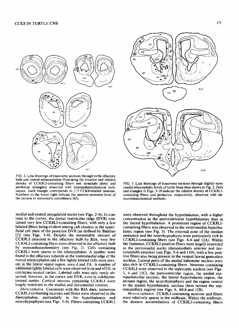
FIG. 2. Line drawings of transverse sections through turtle olfactory bulb and rostral telencephalon illustrating the location and relative density of CCKILI-containing fibers and terminals (dots) and perikarya (triangles) observed with immunohistochemical tech- niques. Each triangle corresponds to 1-5 CCKI-labeled neurons. Numbers to the lower right indicate the anterior-posterior level of the section in stereotaxic coordinates 1631.
medial and central amygdaloid nuclei (see Figs. 2-6). In con- trast to the cortex, the dorsal ventricular ridge (DVR) con- tained very few CCK8LI-confining fibers, with only a few labeled fibers being evident among cell clusters in the super- ficial cell plate of the posterior DVR (as defined by Balaban [2]) (see Figs. 3-6). Despite the measurable amount of CCKlLI detected in the olfactory bulb by RIA, very few CCK8LI-containing fibers were observed in the olfactory bulb by immunohistochemistry (see Fig. 2). Cells containing CCKSLI were sparse in the telencephalon. A number were found in the olfactory tubercle at the ventromedial edge of the rostra1 telencephalon and a few lightly labeled cells were pres- ent in the lateral septal region, area d and PA. A number of additional lightly labeled cells were observed in cp and nTOL in colchicine-treated turtles. Labeled cells were only rarely ob- served, however, in the cortex and DVR, even in colchicine- treated turtles. Corticat neurons containing CCKILI were largely restricted to the medial and dorsomedial cortices.
Diencephalon. Consistent with the RIA data, numerous CCK8LIcontaining neurons and fibers were observed in the diencephalon, particularly in the hypothalamus and neurohy~physis (see Figs. 3-6). Fibers containing CCK8LI
FIG. 3. Line drawings of transverse sections through slightly more caudal telencephalic levels of turtle brain than shown in Fig. 2. Dots and triangles in Figs. 3-10 indicate the relative density of CCKILI- containing fibers and perikarya, respectively, observed with im- munohistochemical methods.
were observed throughout the hypothalamus, with a higher concentration in the periventricular hypothalamus than in the lateral hypoth~~us. A prominent region of CCK8LI- containing fibers was observed in the ventromedial hypotha- lamic region (see Fig. 5). The external zone of the median eminence and the neurohypophysis were particularly rich in CCKSLI-containing fibers (see Figs. 4-6 and 12A). Within the thalamus, CCKSLI-positive fibers were largely restricted to the perirotundal nuclei (dorsomedialis anterior and dor- solateralis anterior) (see Figs. 5-6 and 1 lD), with a few posi- tive fibers also being present in the ventral lateral geniculate nucleus. Lateral parts of the medial habenular nucleus were also rich in CCKSLI-containing fibers. Neurons containing CCKSLI were observed in the supraoptic nucleus (see Figs. 3, 4 and llC), the paraventricular region, the medial sup- rapeduncular nucleus, the lateral hypothalamic region, the arcuate region, the neurohy~physis and the region ventral to the medial hypothalamic nucleus (here termed the sup- ramamillary region) (see Figs. 6, A0.8 and 12B).
Mcsencephalon. CCKgLI-containing neurons and fibers were relatively sparse in the midbrain. Within the midbrain, the densest accumulations of CCK8LI-containing fibers
172 REINER AND BEINFELD
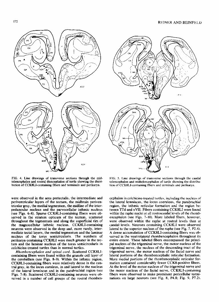
FIG. 4. Line drawings of transverse sections through the mid- telencephalon and rostral diencephalon of turtle showing the distri- bution of CCK8LI-containing fibers and terminals and perikarya.
were observed in the area pretectalis, the intermediate and periventricular layers of the tectum, the midbrain periven- tricular gray, the medial tegmentum, the midline of the inter- peduncular nucleus and the parvocellular isthmic nucleus (see Figs. 68). Sparse CCK8LI-containing fibers were ob- served in the stratum opticum of the tectum, scattered throughout the tegmentum and along the superficial rim of the magnocelhdar isthmic nucleus. CCK8LI-containing neurons were observed in the deep and, more rarely, inter- mediate tectal layers, the medial tegmentum and the laminar nucleus of the torus semicircularis. The numbers of perikarya containing CCKSLI were much greater in the tec- turn and the laminar nucleus of the torus semicircularis in colchicine-treated turtles than in normal turtles.
Rhombencephalon and spinal cord. Scattered CCKILI- containing fibers were found within the granule cell layer of the cerebellum (see Figs. 8-9). Within the isthmic region, CCKILI-containing fibers were relatively dense in the cen- tral gray, in the locus coruleus, in and lateral to the nucleus of the lateral lemniscus and in the parabrachial region (see Figs. 7-8). Scattered CCK8LI-containing neurons were ob- served in a number of cell groups of the rostra1 rhomben-
FIG. 5. Line drawings of transverse sections through the caudal telencephalon and midtelencephalon of turtle showing the distribu- tion of CCKILI-containing fibers and terminals and perikarya.
cephalon in colchicine-treated turtles, including the nucleus of the lateral lemniscus, the locus coeruleus, the parabrachial region, the isthmic reticular formation and the region be- tween TTd and nVI1. Fibers containing CCKSLI were found within the raphe nuclei at all rostrocaudal levels of the rhomb- encephalon (see Figs. 7-10). More labeled fibers, however, were observed within the raphe at rostra1 levels than at caudal levels. Neurons containing CCK8LI were observed lateral to the superior nucleus of the raphe (see Fig. 7, P2.4). A dense accumulation of CCKSLI-containing fibers was ob- served in the ventrolateral rhombencephalon throughout its entire extent. These labeled fibers encompassed the princi- pal nucleus of the trigeminal nerve, the motor nucleus of the trigeminal nerve, the nucleus of the descending tract of the trigeminal nerve, the motor nucleus of the facial nerve and lateral portions of the rhombencephalic reticular formation. More medial portions of the rhombencephalic reticular for- mation contained considerably fewer CCKS-positive fibers. At the level of the motor nucleus of the trigeminal nerve and the motor nucleus of the facial nerve, CCKILI-containing fibers were observed to make prominent pericellular termi- nations on large neurons (see Fig. 8, P4.8; Fig. 9, P7.2).

CCK8 IN TURTLE CNS 173
FIG. 6. Line drawings of transverse sections through the caudal telencephalon and the mesodiencephahc junctional region of turtle showing the distribution of CCKILI-containing fibers and terminals and perikarya.
FIG. 8. Line drawings of transverse sections through the rostra1 rhombencephalon of turtle illustrating the distribution of CCKILI- containing fibers and terminals and perikarya.
FIG. 7. Line drawings of transverse sections through the midbrain of turtle showing the distribution of CCKILI-containing fibers and terminals and perikarya.
FIG. 9. Line drawings of transverse sections through the caudal rhombencephalon of turtle illustrating the distribution of CCK8LI- containing fibers and terminals and perikarya.

174
nTS
P128
P136
FIG. 10. Line drawings of transverse sections through the spino- medulla~ junction and the spinal cord in turtle i~ust~t~~ the dis- tribution of CCKSLI-containing tibers and terminals and perikarya.
Numerous CCKSLI-containing neurons and fibers were ob- served in the nucleus of the solitary tract at all levels of the solitary tract (see Fig. 9, 10 and 12C). A number of CCK8LI-containing fibers were also observed in the motor nucleus of the vagus nerve and nucleus ambignus. Neurons confining CCKSLI were observed in the motor nucleus of the vagus and in and around nucleus ambiguus (see Fig. 10, P10.4 and P11.2). Within the spinal cord, CCKIBLI- containing fibers were observed in the zone (or tract) of Lis- sauer (see Fig. 12D), in the lateral funiculus, in the dorsal horn and in the lateral portions of the ventral horn (see Fig. 10). The CCKBLI-containing fibers of the zone of Lissauer were continuous rostrally with CCK8LI-containing fibers lateral to the nucleus of the descending tract of the trigeminal nerve. These CCK8LI-containing fibers may represent ter- minals of dorsal root fibers and terminals of trigeminal nerve fibers, respectively. In colchicine-treated turtles, CCKSLI- containing neurons were observed in the area of the tract of Lissauer and in the dorsal horn of the spinal cord.
DISCUSSION
In the present study, substantial amounts of a CCK-like peptide were found in the turtle nervous system and the bulk of this CGK-iike material was found to co-elute in gradient elution HPLC with CCKS-sulfate. Beinfeld and her co- workers have previously shown that a variety of HPLC sys-
FACING PAGE
REINER AND BEINFELD
terns, including ones similar to the present HPLC system, yield clear and consistent separation of CCK8-sulfate, CCK8-desulfate, CCK33, CCK4, Gastrinl7-I and Gastrinl7-I ([4,5, 7, 111 Beinfeld, unpublished observation). Other investigators 1531 have also used gradient elution HpLC systems similar to that used in the present study and found clear separation of the major CCK peptides. Thus. the present results indicate that: (1) the major form of CCK in turtle brain (representing 87% of the total CCK8LI measured by the RS antiserum) is distinct from CCKS-desulfate. CCK33, CCK4 and the gastrin peptides, and (2) the major CCK8-like peptide in the turtle brain is identical to CCK8 sulfate or differs from CCKS-sulfate in ways not detectible by our HPLC system. Although the elution profile of the CCKS-related peptide caerulein was not examined in our present HPLC system, it appears very likely that the major CCKS-like peptide in turtle brain is distinct from caerulein. Caerulein can be readily separated from CCKB-sulfate by a variety of procedures (e.g., fractionation on Sephadex col- umns, [ 18,851 or isocratic elution HPLC, [ 181 Beinfeld, un- published observation), including ones less sensitive than used here. Further, although caerulein was originally dis- covered in amphibian skin, caerulein does not appear to be present in the amphibian brain or retina (as further discussed below) [ 18,851.
Two minor peaks of CCKlLI (each representing 6.5% of the total CCKSLI eluted) were observed in the present HPLC study, one occurring shortly before the major CCK8LI peak (fraction 30) and one occurring shortly after the major CCKSLI peak (fraction 36). The latter minor peak may represent CCK&desulfate since this peptide has been shown to elute shortly after CCKI-sulfate in previous HPLC studies 14, 7, 111. Several lines of evidence suggest that the small peak at fraction 30 may indicate the presence of a CCK33-like peptide in the turtle nervous system. Based on previous HPLC studies using gradient elution systems simi- lar to that used in the present study (unpublished studies by M.C. Beinfeld using a Supelco column and a linear gradient of 20-X% acetonitrile). we would expect that CCK33 should elute in fraction 29-30 in the present HPLC system. Since the present extraction procedure (boiling followed by acidifi- cation) has previously been shown to effectively extract both high and low molecular weight forms of CCK [53,79] and since the antiserum used in the present study does crossreact completely with CCK33, the peak observed at fraction 30 in the present study could represent a high molecular weight peptide similar to CCK33. It would seem likely that this peptide differ in structure somewhat from mammalian CCK33 since previous immunochemicaf studies have shown that the high molecuiar weight CCK-like peptides of crocodilian gut differ from mammalian CCK33 [t31. The putative CCK3flike peptide of turtle nervous system may. nonetheless, be sufficiently similar to CCK33 to show a re- tention time in our HPLC system similar to that of CCK33.
We have also used the present HPLC system to Charac- terize the CCK-like materials present in the telencephalic cortex of turtle [68]. We found that 87% of the CCK8LI in
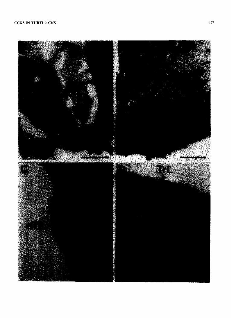
FIG. 11, Photomicrographs of CCKB-labeling in PAP-stained transverse sections through the turtle brain showing: (A) the CCK%containing fibers in the cellular layer of telencephalic cortex; (B) labeled terminals in the region of the lateral septal nucleus; (C) labeled neurons in the supraoptic nucleus; (D) labeled fibers and terminals in nucleus dorsomedialis anterior, immediately above the unlabeled nucleus rotundus. Medial is to the left in A and to the right in B-D. Scale bars: A=250 microns; B-D= 100 microns.

CCK8INTURTLECNS 175

f76
turtle cortex co-eluted with CCKI-sulfate and that 9.2% of the CCKSLI in turtle cortex eluted in fraction 30. Thus, gra- dient elution HPLC of whole turtle brain extract as well as extract of a specific region of turtle brain have shown that there consistently appear to be two CCK-related peptides in turtle brain, a predominant peptide (8% of the total) that is highly similar or identical to CCK&sulfate and a less abun- dant peptide (6-Y% of the total) that presumably represents a high molecular weight form of CCK similar to CCK33. No evidence was obtained in the study of the turtle cortex or in the present study for the presence of CCK4 in the turtle nervous system (CCKI should elute in fraction 21-22 in the present HPLC system, M. C. Beinfeld, unpublished obser- vation [53]). Since our anti-CCK8 antiserum is specific for the C-terminal ~ntapeptide of CCK8, it is possible, how- ever, that small amounts of CCK-related peptides, such as CCK4, are present in turtle nervous system but were not detected by our RIA analysis of the HPLC ffactions.
Previous studies of the reptilian gut have indicated that, as in mammals, CCK-like material is found in endocrine cells of the upper intestinal tract (while gastrin-like material is found in endocrine cells of the stomach) [13, 19, 431. Using several regionally specific antisera, Larsson and Rehfeld [43] have found that the sequence of the last nine amino acids of the C-terminus of the CCK-like peptide in turtle Gi tract is immunochemically indistinguishable from that of mamma- lian CCK. As noted above, Buchan et al. [13], however, have shown that the midportion of alligator CCK (corre- sponding to positions 9-20 of mammalian CCK) is im- munochemically distinct from the midportion of mammalian CCK. The present identification of a peptide in the turtle central nervous system that is highly similar or identical to mammalian CCK&sulfate and the previous discovery of a peptide in the upper intestinal tract of turtle that is highly similar (if not identical) in its C-terminus to mammaii~ CCK are not surprising in light of recent findings in amphibians. In frogs also, the bulk of the CCK-like material found in the nervous system and retina is indistinguishable from CCK& sulfate by HPLC or gel filtration chromatography [ 1 I, 1827, 851. Although caerulein is present in amphibian skin, neither caerulein nor a caerulein-like peptide have been detected in the amphibi~ brain [ IfI]. CCK-like peptides are also present in high concentration in the upper intestinal tract of frogs and one of these CCK-like peptides has been found to be indis- tinguishable from CCK&sulfate [ 181. The results in reptiles and frogs, thus, clearly suggest that a CCK-like peptide (which contains CCK&sulfate or a highly similar peptide at its C-terminus) has been present in intestinal cells and a pep- tide highly similar or identical to CC@sulfate has been pres- ent in neurons since the very beginnings of the evolution of land vertebrates (if not earlier). It seems likely that the phys- iological roles of gut CCK and brain CCKS in the first land vertebrates were similar to those in modern mammals. The fmdings on CCKS in the nervous system of frogs and turtles suggest that CCKB has had a considerably more conservative evolutionary history in terms of its chemical structure than
FACING PAGE
KEINER AND BEI~F~L~
have other neuropeptides such as LHRH [38,39]. neuroten- sin [14J, substance P 166. 68, 721, and vasopressin and oxytocin [ 11.
A number of lines of evidence indicate that CCK8 acts as a neurot~smitter or neuromodulator within the mammalian nervous system: it is localized in neurons and their terminals [25, 34,44,46,61,64,81], it is released in a calcium depend- ent fashion by depol~zation [25, 52, 621, it binds to specific receptor sites [35,77] and it has an excitatory effect on neurons [22, 36, 41, 601. The presence of CCKS in wide- spread areas of the turtle brain with regional concentrations largely similar to those found in mammals f9,10] suggests that CCKS also acts as a neurotransmitter/neuromodulator in reptilian brain. Further, the present immunohistochemical data suggests that CCK8 may act as a neuroactive agent in many of the same systems in reptiles as presumed to be the case in mammals. Fibers containing CCKSLI were observed in Lissauer’s zone of the spinal cord and along the margin of the rhombencephalon lateral to the nucleus of the descend- ing tract of the trigeminal nerve. Since these sites of CCKSLI-containing fibers are regions of termination of dor- sal root fibers and trigeminal nerve fivers, respectively. CCK8LI may be present in the terminals of dorsal root and trigeminal nerve primary afferent fibers, as reportedly the case in mammals ]48]. The dist~bution of CCK8LI in the rhombencephalon of turtle is also similar to that previously reported in mammals, with numerous CCKBLI-containing fibers being present in the parabrachial region, along the raphe, in more lateral portions of the rhombencephalic re- ticular formation and in the vagal motor nucleus [46]. In turtle, numerous CCKSLI-containing neurons and fibers are present in the nucleus of the solitary tract at all rostrocaudal levels. The presence of CCK8LI-containing neurons and fi- bers in the nucleus of the solitary tract has also been re- ported in mammals [40,58]. In turties, CCKSLI was also observed in neurons of the supraoptic nucleus and the paraventricular hypothalamic region and in fibers of the ex- ternal zone of the median eminence and neurohypophysis, as reportedly the case in mammals and amphibians [23, 46, 811. The extensive population of labeled cells spanning the medial suprapeduncular nucleus of the hypothalamus. the lateral hypothalamic region and the supramamillary region of the hypothalamus may be comparable to the CCKSLI- containing neurons reported in the supramamillary cell group and adjacent regions of the hypothalamus of mammals [15,34], In addition, numerous substance P-like im- munoreactivity (SPLI)-containing neurons are present in these same regions in both turtles and mammals [45,68.72]. In turtles, SPLI and CCK8LI co-occur in individual neurons of this hypothalamic cell field [68]. This SPLI/CCK8LI- containing cell field appears to give rise to the CCK8LI- containing fibers present in telencephalic cortex, fibers which also contain SPLI [ 17,681. Finally, in colchicine- treated turtles, a number of CCK8LI-containing neurons were evident in the ventral tegmental area (or medial teg- mentum~ of turtles. pa~i~ulariy at more caudal levels of the
FIG. 12 Photomicrographs of CCKS-labeled PAP stained transverse sections through the turtle brain showing: (A) labeled fibers and terminals in the external zone of the median eminence; (B) labeled neurons in the supramamillary region of the diencephalon: (C) labeled neurons in the rostral nucleus of the solitary tract; (D) labeled fibers and terminals in dorsolateral funiculus and Lissauer’s zone of the spinal cord. Medial is to the left in all photomicrogrphs. Scale Bars: A,C, and D= 100 microns; B= 150 microns. All abbreviations are found in the list of abbreviations.
CCKS IN TURTLE CNS

178 RElNER AND BEINFELD
tegmentum. Since this region of the turtle tegmentum is known to contain numerous dopaminergic neurons [ 121, it seems like1y that some of the dopaminergic neurons also contain CCK. In mammals, numerous neurons containing both dopamine and CCKIILI have been found in the ventral tegmental area [31,32]. Although the presence in turtles of CCKILI in the medial tegmental neurons and in fibers of the medial striatum (comparable to the nucleus accumbens of rn~rn~s) and olfactory tubercle suggests the existence of a CCK8LI-containing mesolimbic pathway to the basal telen- cephalon (comparable to that in mammals), it is of interest to note that the levels of CCKS measured in turtle basal telen- cephalon by RIA are only l/20-1/40 of those in the mamma- lian basal telencephalon.
Comparative neuroanatomical studies during the last two decades have considerably revised the unde~~nd~g of the evolution of the mammalian cerebral cortex by showing that large regions of the avian and reptilian telencephalon (in tur- tles, including the DVR and the nonolfactory cortex plus the pallial thickening) are histochemically and anatomically comparable to mammalian neocortex [54,X, 5’7,801. In this context, the Ievels of CCKlLI in cortex and DVR of turtle are of interest. Although moderate levels of CCKS were measured by RIA in cortex, very little CCK8 was found in the DVR. The immunochemicahy detectible CCK8LI pres- ent in turtle cortex, however, is contained in a fiber system that appears to originate from the hypothalamus [68]. Further, CCKSLI-containing neurons were rarely observed in either cortex or DVR in turtle. Thus, the cortex and DVR of turtles appear similar to one another in that both contain very little CCKLI of intrinsic origin. In contrast, in mammals CCK8 is found in its highest brain concentration in the cere- bral cortex [8,9], in which the bulk of the CCK8 appears to be contained in nonpyramidal local circuit (or intrinsic) neurons of (primarily) neocortical layers II-III [5 1,591. The relative scarcity of intrinsic CCKSLI in both the cortex and DVR of turtles is surprising since the telencephalic neocor- tex of mammais and the cortex and DVR of turtles are similar in terms of many other details of their connections and neurotransmitter-specific cell types [12, 24, 37, 54, 65, 67, 71, 721. preliminary immunohistochemical studies (Reiner, unpublished observation) indicate that intrinsic CCKIILI- containing neurons are also rare in the cortex and DVR of caiman (a crocodilian) and in the Wulst (comparable to repti- lian cortex) and DVR of pigeons. Although higher levels of a CCK-like substance have been measured by RIA in the tel- encephalic hemispheres (cortical-equivalent tissue plus basal telencephalon) of birds than in the telencephalic hemispheres of turtles [30,82], the levels in birds are nonetheless much lower than in the cerebral hemispheres of mammals. Further, previous studies in birds have not specifically measured the levels of CCK8 in the telencephalic cortical- equivalent tissue (Wulst and DVR); and thus, the concentra- tion of CCK8 in the Wulst and DVR in birds may be lower than in the teiencephalon as a whole. Even lower levels of CCKS have been found in the dorsal cerebrum of frogs by RIA than in turtle cortex (N. B. The frog telenceph~on lacks a DVR but does possess a dorsal pal&al region comparable to reptilian cortex) 111,571. As in turtles, the highest levels of CCKS in the frog CNS are found in the hypothalamus and neurohypophysis [ll]. Thus, although more information is needed on the levels of CCKI in the telencephalic cortical- equivalent regions in birds and nonchelonian reptiles, it seems likely that the telencephali~ ~o~i~~-equivalent re- gions of living nonmammalian terrestrial vertebrates largely
lack at least one major population of neurons characteristic of mammmalian neocortex, namely intrinsic CCK& containing neurons. These intrinsic CCK-containing cortical neurons may have not become characteristic of the cerebral hemispheres until the evolution of mammalian neocortex.
The observation that a population of neurons that is pre- dominantly restricted to layers II-III within mammalian neocortex is largely absent from cortical-equivalent regions of the reptilian telen~ephalon raises the question as to whether or not other populations of neurons predomiantly restricted to layers II-III of mammalian neocortex are also absent from or extremely rare in reptilian cortical-equivalent regions. Ebner [24] previously noted that the telencephalic cortex in turtles, although in many respects similar to mam- malian neocortex, appears to lack the afferent and efferent connections that uniquely characterize layers I-III of mam- malian neocortex. He consequentIy suggested that turtle cor- tex in its entire depth may be comparable to layers IV-VI of mammalian neocortex and that neurons in neocortical layers I-111 may largely be without correspondent in turtle cortex. The present data indicating that CCK8LI-containing neurons are rare in turtle cortex is consistent with this suggestion. The few ~CK8LI-confining neurons observed in turtle cor- tex and DVR in the present study may be comparable to the few CCKEiLI-containing neurons in layers IV-VI of mam- malian neocortex. The CCKSLI-containing neurons of layers II-III of mammalian neocortex may not be represented in turtle cortex and DVR. The results of several other im- munohistochemical studies are also consistent with Ebner’s suggestion. Prelimina~ results indicate that VIPergic neurons, which are predomin~tly found in layers ii-111 of mammalian neocortex [49], are also extremely rare in turtle cortex and DVR [66]. In contrast, peptidergic neurons that are abundant in layers IV-VI of the mammalian neocortex (regardless of whether or not they are also abundant in layers I-III), such as those containing somatostatin, a substance P-like peptide and an APP-like peptide 13, 28, SO], are ahun- dant in turtle cortex and DVR 166. 68. 72, 831. In order to more fully ascertain the extent of the changes that occurred during the evolution of neocortieal layers I-III in the mam- malian lineage, further studies are required to determine the extent to which the histochemically-defined and hodologi~ally-de~ned neuronal cell types predominantly found in layers II-III of the mammaIian neocortex are pres- ent or absent in living reptiles. Such studies will help deter- mine which of the cell types characteristic of layers II-111 of mammalian neocortex first appeared in the mammalian lineage and which were already present in the telencephalic cortical-equivalent tissue of the reptilian common ancestors of living mammals and reptiles. It is possible that the neurons predominantly found in layers II-III of mammalian neocor- tex may largely have been absent in ancestral reptiles and that the early evolution of mammals was characterized by an elaboration and proliferation of the neuronal populations making up layers II-III. The Wulst and DVR of birds, how- ever, clearly possess specific regions that show efferent pro- jections characteristic of layers II-III of mamma1ian neocor- tex [&9,74]. It is possible, however, that neuronal popula- tions with efferent projections similar to those of mammalian cortical layers II-III have evolved independently in birds.
In summary, CCK8 appears to be an evoiutionari1y stable neuropeptide whose chemical structure has been conserved during the evolution of terrestrial vertebrates. In addition, CCK8 appears to be present in many of the same neural systems of the dien~ephalon, brainstem and spinal cord in all
CCK8 IN TURTLE CNS 179
terrestrial vertebrates studied. The concentration (and presumably the role) of CCK8 in these brainstem regions also appears to have been largely conserved. During the evo- lution of mammals from earlier land vertebrates, however, CCK8 appears to have changed from a substance whose highest brain concentration -was within the basal di- encephalon to a peptide that has its highest concentration in the cerebral cortex. Although the functional significance of this specific change requires further elucidation, this change may have been one of the many related changes in cerebral
hemisphere organization that occurred during the evolution of mammalian neocortex.
ACKNOWLEDGEMENTS
We wish to gratefully thank Patricia A. Lindaman and Gary Henderson for excellent technical assistance and Theresa Gonzales and John Beckerman for assistance with photography and illustra- tions. This research was SuDDOIted bv NS-1%20 (A.R.). NS-18335 (M.C.B.), NS-18667 (M.C.B:) and a grant from the American Par- kinson Disease Association (M.C.B.).
REFERENCES
1. Acher, R. Evolution of neuropeptides. Trends Neurosci 4: 223-229, 1981.
2. Balaban, C. D. Structure of the anterior dorsal ventricular ridge in turtles (Pseudemys scriptcr elegans). J Morphol 158: 291-322, 1979.
3. Beach, T. G. and E. G. McGeer. Neocortical substance P neurons in the baboon: An immunohistochemical finding. Neurosci Lett 41: 265-270, 1983.
4. Beinfeld, M. C. An HPLC and RIA analysis of the cholecys- tokinin peptides in rat brain. Neuropeptides 1: 203-209, 1981
5. Beinfeld, M. C. Chromatographic characterization of gas- trin/cholecystokinin peptides in bovine and porcine pituitary. Peptides 3: 531-534, 1982.
6. Beinfeld, M. Cholecystokinin in the central nervous system: A minireview. Ncuropeptides 3: 41 l-427, 1983.
7. Beinfeld, M., R. T. Jensen and M. J. Brownstein. HPLC sep aration of cholecystokinin peptides, two systems. J Liquid Chromatography j: 1367-1361,- 1980. _
8. Beinfeld. M. C.. M. E. Lewis. L. E. Eiden. G. Nilaver. C. B.
9
10
11
12.
13
14.
15.
16.
Pert and’ A. Pert. The distribution of cholecystokinin and vas- oactive intestinal peptide in rhesus monkey brain as determined by radioimmunoassay. Neuropeptides 3: 337-344, 1983. Beinfeld, M. C., K. K. Meyer, R. L. Eskay, R. T. Jensen and M. J. Brownstein. The distribution of cholecystokinin im- munoreactivity in the central nervous system of the rat as de- termined by radioimmunoassay. Brain Res 212: 51-57, 1981. Beinfeld, M. C. and M. Palkovits. Distribution of cholecystoki- nin (CCK) in the rat lower brain stem nuclei. Brain Res 238: 260-265, 1982. Beinfeld, M. C., J. R. Trubatch and M. J. Brownstein. Cholecystokinin peptides in the brain and pituitary of the bullfrog Rana catesbiana: Distribution and characterization. Brain Res 268: 192-l%, 1983. Brauth, S. E., A. Reiner, C. A. Kitt and H. J. Karten. The substance P-containing striato-tegmental path in reptiles: An immunohistochemical study. J Comp Neural 219: 305-327, 1983. Buchan, A. M. J., V. Lance and J. M. Polak. Regulatory pep tides in the gastrointestinal tract of Alligator mississippiensis. An immunocytochemical study. Cell Tissue Rrs 231: 439-449, 1983. Carraway, R., S. E. Ruane and H. R. Kim. Distribution and immunochemical character of neurotensin-like material in representative vertebrates and invertebrates: Apparent conser- vation of the COOH-terminal region during evolution. Peptides 1: 115-123, 1982. Cho, H. J., Y. Shiotani, S. Shiosaka, S. Inagaki, Y. Kubota, H. Kiyama, K. Umegaki, K. Tateishi, E. Hashimura, T. Hamaoka and M. Tohyama. Ontogeny of cholecystokinin-&containing neuron system of the rat: An immunohistochemical analysis. I. Forebrain and upper brainstem. J Comp Neural 218: 25-41, .^__ lYS3. Coons, A. H. Fluorescent antibody methods. In: Genera/ Cytochemical Methods. edited by J. F. Danielli. New York:
18.
19.
20.
21.
22.
23.
24.
25.
26.
27.
28.
Dimaline, R. Is caerulein amphibian CCK? Peptides 4: 457-462, 1983. Dimaline, R., B. B. Rawdon, S. Brando, A. Andrew and J. P. Loveridge. Biologically active gastrin/CCK-related peptides in the stomach of a reptile, Crocodylus niloticus: Identified and characterized by immunochemical methods. Peptides 3: 977- 984, 1982. Dockray, G. J. The physiology of cholecystokinin in brain and gut. Br Med Bull 38: 253-258, 1982. Dockray, G. J., R. A. Gregory, J. B. Hutchison, J. I. Harris and M. J. Runswick. Isolation, structure and biological activity of two cholecystokinin octapeptides from sheep brain. Nature 274: 71 l-713, 1978. Dodd, J. and J. S. Kelly. The actions of cholecystokinin and related peptides on pyramidal neurons of the mammalian hip POC~~PUS. Brain Res 105: 337-350. 1981. Doer-Schott, J., J.-C. Garaud and R. 0. Claus. Immunohis- tochemical localization of a gastrin-like peptide in the brain of an amphibian, Xenopus laevis daud. Cell Tissue Res 203: 65-78, 1981. Ebner, F. F. The forebrain of reptiles and mammals. In: Evolu- tion of Brain and Behavior in Vertebrates. edited by R. B. Mas- terton. M. E. Bitterman. C. B. G. Camnbell and N. Hotton. New York: John Wiley and Sons, 1976, pp. 147-167. Emson, P. C., C. M. Lee and J. F. Rehfeld. Cholecystokinin octapeptide: Vesicular localization and calcium dependent re- lease from rat brain in vitro. Life Sci 26: 2157-2163, 1980. Engelhardt, R. P., N. Dhainaut-Courtois and G. Tramu. Im- munohistochemical demonstration of a CCK-like peptide in the nervous system of a marine annelid worm, Nereis diversicolor O.F. Muller. Cell Tissue Res 227: 401-411, 1982. Eskay, R. L. and M. C. Beinfeld. HPLC and RIA of the cholecystokinin peptides in the vertebrate neural retina. Brain Res 246: 315-318. Finlay, J. C. W., J. L. Maderdrut, L. J. Roger and P. Petrusz. The immunocytochemical localization of somatostatin- containing neurons in the rat central nervous system. Neurosci- ence 6: 2173-2192, 1981.
29. Frey, P. Cholecystokinin octapeptide (CCK 2633), nonsulfated octapeptide and tetrapeptide (CCK 30-33) in rat brain: Analysis by high pressure liquid chromatography (HPLC) and radioim- munoassay (RIA). Neurochem Int 5: 811-815, 1983.
30. Goldman, S. A. and B. S. Schneider. The ontogeny of cholecys- tokinin immunoreactivity in vertebrate brain. Sot Neurosci Abstr 7: 97, 1981.
31. Hokfelt, T., J. F. Rehfeld, L. Skirboll, B. Ivemark, M. Gold- stein and M. Markey. Evidence for co-existence of dopamine and CCK in mesolimbic neurons. Nature 285: 476-478, 1980.
32. Hokfelt, T., L. Skirboll, J. F. Rehfeld, M. Goldstein, M. Mar- key and 0. Dann. A subpopulation of mesencephalic do- pamine neurons projecting to limbic areas contains a cholecystokinin-like peptide: Evidence from immunohis- tochemistry combined with retrograde tracing. Neuroscience 5: 2093-2124, 1980.
Academic Press, 1958, pp. 399-422. 17. Desan, P. Connections of cerebral cortex in the turtle
(Pseudemys scripta elrgans). Sot Neurosci Abstr 7: 85, 1981.
180 REINER AND BEINFELD
33. Holmquist, A. L., G. J. Dockray, G. L. Rosenquist and J. H. Walsh. Immunochemical characterization of cholecystokinin- like peptides in lamprey gut and brain. Cm Contp ~nd~~~r~nl)l 37: 474-48 I ) 1979.
34. Innis, R. B., F. M. A. Correa, G. R. Uhl, B. Schneider and S. Snyder. Cholecystokinin octapeptide-like immunoreactivity: histochemical localization in rat brain. Proc Nat/ Arad Sri USA 76: 521-525, 1979.
35. Innis, R. B. and S. H. Snyder. Distinct cholecystokinin recep- tors in brain and pancreas. Proc~ Nat1 Acad .Sc+ USA 71: 6917- 6921, 1980.
36. Jeftinija, S., V. Miletic and M. Randic. Choiecystokinin oc- tapeptide excites dorsal horn neurons both in ~)ivo and in vitro. Brain Res 213: 231-236.
37.
38.
39.
40.
41.
42.
43.
44.
45.
46.
47.
48.
49.
SO.
51
Karten, H. J. The organization of the avian telencephalon and some speculations on the phylogeny of the amniote telencepha- Ion. A& NYAcad St+ 167: 1641179, 1969. Kin& J. A. and R. P. Millar. Structure of chicken hv~thalami~ lute&zing hormone-releasing hormone. I. Struct&I determi- nation on partially purified material. .I Biol Chem 257: 10722- 10728, 1982. King, J. A. and R. P. Millar. Structure of chicken hypothalamic luteinizing hormone-releasing hormone. II. Isolation and char- acterization. f Bioi them 257: 10729-10732, 1982. Kubota, Y., S. Inagaki, S. Shiosaka, H. J. Cho. K. Takeishi, E. Hashikawa, T. Hamaoka and M. Tohyama. The dist~bution of cholecystokinin octapeptide-like structures in the lower brain stem of the rat: An immunohistochemical analysis. N~~uvosci- ~rtce 9: 587-604, 1983. Lamour, Y., P. Dutar and A. Jobert. Effects of neuropeptides on rat cortical neurons: Laminar distribution and interaction with effect of acetylcholine. Neuroscienc~e 10~ 107-117, 1983. Larsen, B. A. and S. R. Vigna. Gast~~chole~ystokinin-like immunoreactive peptides in the Dungeness crab, Cancer magis- ter (Lkma): Immunohistochemical and biological characterization. Regul Pept 7: 155-170, 1983. Larsson, L. I. and J. F. Rehfeld. Evidence for a common ev- lutionary origin of gas&in and cholcystokinin. Nature London 269: 335-338, 1977. Larsson, L. I. and J. F. Rehfeld. Localization and molecular heterogeneity of cholecystokinin in the central and peripheral nervous system. Brain Res 265: 201-218, 1979. Ljungdahl, A., T. Hokfelt and G. Nilsson. Distribution of sub- stance P-like immunoreactivity in the central nervous system of the rat. 1. Cell bodies and nerve terminals. Neurosc?enc,c~ 3: 861-944, 1978. Loren, I., J. Aiumets, R. Hakanson and F. Sundler. Distribu- tion of gas&in and CCK-like peptides. ~i.~t~)~~e~fisfr~ 59: 249- 257, 1979. Lowry, 0. H., N. J. Rosebrough, A. L. Carr and R. J. Randall. Protein measurement with the Folin phenol reagent. J Biol Chem 193: 265-275, 1951. Maderdrut, J. L., T. L. Yaksh, P. Petrusz and V. L. W. Go. Origin and distribution of cholecystokinin-containing nerve terminals in the lumbar dorsal horn and nucleus caudalis of the cat. Brain Res 2431 363-368, 1982. McDonald, J. K., J. G. Pamavelas, A. N. Karamanlidis and N. Brecha. The morphology and distribution of peptide-containing neurons in the adult and developing visual cortex of the rat. II. Vasoactive intestinal nolvpeptide. J Neuroc~ytol 11: 825-837. . ._ - 1982. McDonaId, J. K., J. G. Pamavelas, A. N. Karamanlidis and N. Brecha. The rno~ho~o~y and dist~bution of ~ptide-con~ning neurons in the adult any-deveIoping cortex of the rat. IV. Avian pancreatic polypeptide. .I Neurucytol 11: 985-995, 1982. McDonald, J. K., J. G. Pamavelas, A. N. Karamanlidis, G. Rosenquist and N. Brecha. The morphology and distribution of peptide-containing neurons in the adult and developing visual cortex of the rat. III. Cholecystokinin. J Neurocytol 11: 88I- 89.5. 1982.
52.
53.
54.
55.
56.
57.
58.
59.
60.
61.
62.
63.
Micevych, P., V. C. W. Go and T. L. Yaksh. Simultaneouh measurement of VIP and CCK released from rat and cat cortical slices. SCX N~~~r~~s~~ Ahstr 7: 605, 1981. Miller, L. J., I. Jardine, E. Weissman, V. L. W. Go and D. Speicher. Characterization of cholecystokinin from human brain. J Neuroc,hem 43: 835-840, 1984. Nauta, W. J. H. and H. J. Karten. A general profile of the vertebrate brain with sidelights on the ancestry of the cerebral cortex. In: The Neuroscirtwes, Scwmd Study Program. edited by G. C. Quarton, T. Melnechek and F. 0. Schmitt. New York: Rockefeller University Press, 1970, pp. 7-26. Nicoll, R. A., C. Schenker and S. Leeman. Substance P as a transmitter candidate. Anna RP\~ Neurosci 3: 227-268, 1980. Northcutt, R. G. Forebrain and midbrain organization in lizards and its phylogenetic significance. In: Behavior and Neuro/og> of Lizards. edited by N. Greenberg and P. D. MacLean. Bethesda: NIMH, 1978, pp. 11-64. No~hcutt, R. G. Evolution of the telencephalon in nonmam- mals. Annu Ret, Nrr~orc~i 4: 301-350, 1981 Palkovits, M., J. 2. Kiss, M. C. Beinfeld and T. H. Williams. Cholecystokinin in the nucleus of the solitary tract of the rat: Evidence for its vagal origin. Bruin Res 252: 386390, 1982. Peters, A., M. Miller and L. M. Kimerer. Cholecystokinin-like immunoreactive neurons in rat cerebral cortex. Nr,Nn)sc.ic~nc,c, s: 431-448, 1983. Phillis, J. W. and J. R. Kirkpatrick. The actions of motilin, luteinizing hormone-releasing hormone, cholecystokinin, somatostatin, vasoactive intestinal peptide and other peptides on rat cerebral cortical neurons. c‘rr/t J Phqiol pharmcru~l 58: 612-623. 1980. Pinget, M., E. Straus and R. S. Yalow. Localization of cholecystokinin-like immunoreactivity in isolated nerve termi- nals. Prcx Nut! Arud SL.~ USA 75: 6324-6326, 1978. Pinget, M., E. Straus and R. S. Yalow. Release of cholecys- tokinin from a synaptosome-enriched fraction of rat cerebral cortex. L,if? Sci 25: 339-342, 1979. Powers, A. S. and A. Reiner. A stereotaxic atlas of the forebrain and midbrain of the eastern painted turtle (Chrvsemy~ picta pkra). J Hir&xhang 21: 125-159, 1980.
64. Rehfeld, J. F., N. Gotterman, L. I. Larsson, P. C. Emson and C. M. Lee. Gastrin and ~holecystokinin in the central and pe- ripheral neurons. Fed Proc 38: 2325-2329, 1979.
65. Reiner, A. Comparative studies of opioid peptides: enkephalin distribution in turtle central nervous system. Sot, NcJrrrosc,i Ahstr 9: 439. 1983.
66. Reiner, A. Neuropeptides in the reptilian nervous system. In: Biologv of Rez#i/ia, edited bv C. Gans and R. G. Northcutt. .._ New York: Academic Press, in press.
67. Reiner, A., S. E. Brauth and H. J. Karten. Evolution of the amniote basal eanalia. Trends Ncplrn>sci 7: 320-325, 1984.
68. Reiner, A., W.-D. ‘jEldred, M. C. Beinfeld and J. E. Krause. The co-occurrence of a substance P-like peptide and cholecystokinin-8 in a fiber system of turtle cortex. J Nc,rtr<~.sc.i 5: 1527-1544. 1985.
69. Reiner, A. and H. J. Karten. The Iaminar source of efferent projections from the avian Wulst. Brain Res 27% 349-354, 1983.
70. Reiner. A.. H. J. Karten and N. C. Brecha. Enkephalin- mediated basal ganglia influences over the optic tectum: Im- munohistochemistry of the tectum and the lateral spiriform nu- cleus in pigeons. J Camp New-o/ 208: 37-53, 1982.
71. Reiner, A., H. J. Karten and A. R. Solina. Substance P: Lo- calization within paleostriatal-tegmental pathways in the pi- geon. ~~t,u~~~,~(.~~n~,~, 9: 61-85, 1983.
72. Reiner, A., J. E. Krause, K. T. Keyser, W. D. Eldred and J. F. McKelvy. The distribution of substance P in the turtle nervous system: A radioimmunoassay and immunohistochemical study. .I Camp Nrurol 226: 50-75, 1984.
73. Riss, W., M. Halpern and F. Scalia. The quest for clues to forebrain evolution - the study of reptiles. Brain Erhav Ewl 2: I-50, 1969.

CCKS IN TURTLE CNS 181
74.
75.
76.
77.
78.
79.
Ritchie, T. C. and D. H. Cohen. The avian tectofugai visual pathway: Projections of its telencephalic target, the ectostriatal complex. Sot Neurosci Abstr 3: 94, 1977. Rogawski, M. A. Cholecystokinin octapeptide: Effects on the excitability of cultured spinal neurons. Peptides 3: 545-551, 1982. Rostene, W. H., C. Leranth, M. Maletti, E. Mezey, J. Besson, L. E. Eiden. G. Rosselin and M. Palkovits. Distribution of vas- oactive intestinal peptide (VIP) following various brain transec- tions in the rat by radioimmunoassay and electron microscopic immunocytochemistry. Neuropeptides 2: 337-350, 1982. Saito, A., H. Sankaran. I. D. Goldfine and J. A. Williams. Cholecystokinin receptors in the brain: Characterization and distribution. Science 208: 1155-l 156, 1980. Stemberger, L. Immunocytochemistry, second edition. New York: John Wiley and Sons, 1979. Strause, E., S. Ryder, J. Eng and R. S. Yalow. Nature of im- munoreactive CCK in rat and pig brain. Peptides 2: Suppl 2, 89-92, 1981.
80. Ulinski, P. S. Dorsal Ventriclar Ridge: A Treatise on Forebrain Organization in Reptiles and Birds. New York: John Wiley and Sons, 1983.
81. Vanderhaeghen, J. J., F. Lotstra, J. DeMey and C. Gilles. Im- munohistochemical localization of cholecystokinin and gastrin-like peptides in the brain and hypophysis of the rat. Proc Nat1 Acad Sci USA 71: 1184-1190, 1980.
82. Vanderhaeghen, J. J., J. C. Signeauand W. Gepts. New Peptide in the vertebrate CNS reacting with antigastrin antibodies. Na- ture 257: 604-605. 1975.
83. Weindl, A., J. Teipel and G. Kuchling. Somatostatin in the brain of the turtle Testudo Hermanni Gmelin. An immunohistochemi- cal study. Pepfides 5: 91-100, 1984.
84. Weller, T. H. and A. H. Coons. Fluorescent antibody studies with agents of variceUa and herpes zoster propagated in vitro. Proc Sot Exp Bioi Med 86: 789794, 1954.
85. Yamada, T., N. Brecha, G. Rosenquist and S. Basinger. Cholecystokinin-like immunoreactivity in frog retina: Localiza- tion, characterization, and biosynthesis. Peptides 2: 93-97, 1981.