mechanisms of cholecystokinin-induced calcium …...mechanisms of cholecystokinin-induced calcium...
TRANSCRIPT
ORIGINAL ARTICLE
Mechanisms of cholecystokinin-induced calcium mobilization in gastric antral interstitial cells of Cajal
Yao-Yao Gong, Xin-Min Si, Lin Lin, Jia Lu
Yao-Yao Gong, Xin-Min Si, Lin Lin, Jia Lu, Department of Gastroenterology, the First Affiliated Hospital of Nanjing Medi-cal University, Nanjing 210029, Jiangsu Province, ChinaAuthor contributions: Gong YY and Lu J performed the ma-jority of the experiments; Si XM designed the study, provided vital reagents and analytical tools and was involved in editing the manuscript; Lin L coordinated and provided financial sup-port for this work; and Gong YY analyzed the data and wrote the manuscript.Supported by The National Natural Science Foundation of China, No. 30900666; the Programs of Department of Health of Jiangsu Province, No. H201061Correspondence to: Xin-Min Si, Associate Chief Physician, Department of Gastroenterology, the First Affiliated Hospital of Nanjing Medical University, No. 300 of Guangzhou Road, Nan-jing 210029, Jiangsu Province, China. [email protected]: +86-25-68136920 Fax: +86-25-83674636Received: August 5, 2012 Revised: November 2, 2012Accepted: November 6, 2012Published online: December 28, 2012
AbstractAIM: To investigate the effect of sulfated cholecysto-kinin-8 (CCK-8S) on calcium mobilization in cultured murine gastric antral interstitial cells of Cajal (ICC) and its possible mechanisms.
METHODS: ICC were isolated from the gastric an-trum of mice and cultured. Immunofluorescence stain-ing with a monoclonal antibody for c-Kit was used to identify ICC. The responsiveness of ICC to CCK-8S was measured using Fluo-3/AM based digital microfluori-metric measurement of intracellular Ca2+ concentra-tion ([Ca2+]i). A confocal laser scanning microscope was used to monitor [Ca2+]i changes. The selective CCK1 receptor antagonist lorglumide, the intracellular Ca2+-ATPase inhibitor thapsigargin, the type Ⅲ inositol 1,4,5-triphosphate (InsP3) receptor blocker xestospon-gin C and the L-type voltage-operated Ca2+ channel inhibitor nifedipine were used to examine the mecha-
nisms of [Ca2+]i elevation caused by CCK-8S. Immuno-precipitation and Western blotting were used to deter-mine the regulatory effect of PKC on phosphorylation of type Ⅲ InsP3 receptor (InsP3R3) in ICC. Protein kinase C (PKC) activator phorbol 12-myristate 13-acetate (PMA) and inhibitor chelerythrine were used to assess the role of PKC in the CCK-8S-evoked [Ca2+]i increment of ICC.
RESULTS: ICC were successfully isolated from the gastric antrum of mice and cultured. Cultured ICC were identified by immunofluorescence staining. When given 80 nmol/L or more than 80 nmol/L CCK-8S, the [Ca2+]i in ICC increased and 100 nmol/L CCK-8S significantly increased the mean [Ca2+]i by 59.30% ± 4.85% (P < 0.01). Pretreatment of ICC with 5 μmol/L lorglumide inhibited 100 nmol/L CCK-8S-induced [Ca2+]i increment from 59.30% ± 4.85% to 14.97% ± 9.05% (P < 0.01), suggesting a CCK1R-mediated event. Emptying of in-tracellular calcium stores by thapsigargin (5 μmol/L) prevented CCK-8S (100 nmol/L) from inducing a [Ca2+]i increase. Moreover, pretreatment with xestospongin C (1 μmol/L) could also abolish the CCK-8S-induced ef-fect, indicating that Ca2+ release from InsP3R-operated stores appeared to be a major mechanism responsible for CCK-8S-induced calcium mobilization in ICC. On the other hand, by removing extracellular calcium or block-ing the L-type voltage-operated calcium channel with nifedipine, a smaller but significant rise in the [Ca2+]i could be still elicited by CCK-8S. These data suggest that the [Ca2+]i release is not stimulated or activated by the influx of extracellular Ca2+ in ICC, but the influx of extracellular Ca2+ can facilitate the [Ca2+]i increase evoked by CCK-8S. CCK-8S increased the phosphoryla-tion of InsP3R3, which could be prevented by chelery-thrine. Pretreatment with lorglumide (5 μmol/L) could significantly reduce the CCK-8S intensified phosphory-lation of InsP3R3. In the positive control group, treat-ment of cells with PMA also resulted in an enhanced phosphorylation of InsP3R3. Pretreatment with various concentrations of PMA (10 nmol/L-10 μmol/L) appar-ently inhibited the effect of CCK-8S and the effect of
World J Gastroenterol 2012 December 28; 18(48): 7184-7193 ISSN 1007-9327 (print) ISSN 2219-2840 (online)
© 2012 Baishideng. All rights reserved.
Online Submissions: http://www.wjgnet.com/esps/[email protected]:10.3748/wjg.v18.i48.7184
7184 December 28, 2012|Volume 18|Issue 48|WJG|www.wjgnet.com

Gong YY et al . Cholecystokinin and interstitial cells of Cajal
100 nmol/L PMA was most obvious. Likewise, the ef-fect of CCK-8S was augmented by the pretreatment with chelerythrine (10 nmol/L-10 μmol/L) and 100 nmol/L chelerythrine exhibited the maximum effect.
CONCLUSION: CCK-8S increases [Ca2+]i in ICC via the CCK1 receptor. This effect depends on the release of InsP3R-operated Ca2+ stores, which is negatively regu-lated by PKC-mediated phosphorylation of InsP3R3.
© 2012 Baishideng. All rights reserved.
Key words: Cholecystokinin octapeptide; Interstitial cells of Cajal; Calcium mobilization; Protein kinase C
Peer reviewers: Guanglin Cui, MD, PhD, Laboratory of Gastroenterology, Institute of Clinical Medicine, University of Tromso, N-9037 Tromso, Norway; Fabio Grizzi, PhD, Senior Scientist, Laboratory of Molecular Gastroenterology, Istituto Clinico Humanitas IRCCS, Viale Monza 325, 20126 Milan, Italy
Gong YY, Si XM, Lin L, Lu J. Mechanisms of cholecystokinin-induced calcium mobilization in gastric antral interstitial cells of Cajal. World J Gastroenterol 2012; 18(48): 7184-7193 Available from: URL: http://www.wjgnet.com/1007-9327/full/v18/i48/7184.htm DOI: http://dx.doi.org/10.3748/wjg.v18.i48.7184
INTRODUCTIONIn the gastrointestinal (GI) tract, phasic contractions are caused by electrical activity termed slow waves, which are generated and propagated by the interstitial cells of Cajal (ICC)[1,2]. The initiation of pacemaker activity in the ICC is caused by rhythmic cytoplasmic Ca2+ oscillation[3,4]. The ICC can regulate slow-wave-driven peristaltic activ-ity as well as mediate motor inputs from the GI nervous system[5,6]. ICC abnormalities are associated with many GI motility disorders, such as achalasia of cardia, slow transit constipation and irritable bowel syndrome[7,8]. Therefore, understanding the mechanisms underlying pacemaker activity and excitability of ICC is of crucial importance[9]. Although neurohumoral regulation of GI function has been studied in some detail, less is known about the mechanisms of how neurohumoral factors regulate pacemaker activities in the ICC.
Cholecystokinin (CCK) is a bioactive peptide that regulates a variety of physiological functions, acting as both hormone and neurotransmitter in the GI tract[10,11]. Sulfated CCK-8 (CCK-8S), which has frequently been used in research studies, is one of the main biologically active forms of CCK[12]. The regulatory actions of CCK are mediated by two receptor subtypes, CCK1 and CCK2 receptors. CCK receptors are distributed in enteric nerves and smooth muscles[13]. In addition, CCK1 receptors are also found in the ICC, suggesting a role for the ICC in the mediation of CCK effects[14].
CCK inhibits gastric emptying by relaxing the proxi-
mal part of the stomach and increasing pyloric pres-sure[10,12]. However, the mechanisms of the regulatory effects of CCK on gastrointestinal motility are not clear. Previous studies have shown that CCK can activate phospholipase C (PLC) through binding to its distinct receptors[15]. This activation leads to the production of diacylglycerol (DAG) and inositol 1,4,5- triphosphate (InsP3), which in turn activates protein kinase C (PKC) and mobilizes intracellular calcium ([Ca2+]i)[16]. Through this signaling pathway, CCK may participate in various physiological responses, such as secretion, neurotrans-mission and muscle contraction[17]. However, the cross-talk of the CCK-8S triggered PKC and Ca2+ signaling pathways is not been well understood. The aims of this study were to investigate the effect of CCK-8S on [Ca2+]i in the ICC and the respective contributions of InsP3R-sensitive intracellular Ca2+ stores and extracellular Ca2+ sources in those responses. Additionally, the role of PKC in regulation of CCK-8S-triggered calcium signal-ing pathway was also studied.
MATERIALS AND METHODSPreparation of cells and cell cultureAll experiments were performed according to the guid-ing principles for the care and use of animals approved by Institutional Animal Use and Care Committee of the First Affiliated Hospital of Nanjing Medical University. Every effort was made to minimize both the number of animals used and their suffering.
Balb/c mice (5-6 wk) of either sex were purchased from the laboratory of the First Affiliated Hospital of Nanjing Medical University. The animals were anesthe-tized by chloroform inhalation and killed by cervical dislocation. The stomach was excised and the contents were washed away with ice-cold Krebs-Ringer bicarbon-ate (KRB). The mucosa was removed by peeling. In Syl-gard dishes filled with Krebs solution, the tissues were washed three times and then cut into about 0.5 cm seg-ments. The segments were transferred into a centrifuge tube and dispersed with an enzyme solution containing collagenase 1.3 mg/mL, trypsin inhibitor 2 mg/mL and ATP 0.27 mg/mL. The centrifuge tube was incubated at 37 ℃ for 30 min and the tissue segments were blown for 30 s with pipette every 5 min during incubation. An equal volume Medium 199 containing 10% fetal bovine serum was added to stop digestion. The tube was cen-trifuged at 1000 rpm for 3 min and then the supernatant was removed. The sediment was suspended with Medium 199 and cells were collected by pouring the suspension through a 200-mesh sieve. After centrifugation at 1000 rpm for 3 min, the cells were dispersed with Medium 199 and then plated onto 35 mm glass-bottom culture dishes (NEST Biotechnology Co., Ltd, China) coated with rat-tail tendon collagen (5 mg/mL). The cells were then cul-tured at 37 ℃ in a 95% O2-5% CO2 incubator in Medium 199 supplemented with 2% Penicillin-Streptomycin liquid and murine stem cell factor (5 ng/mL).
7185 December 28, 2012|Volume 18|Issue 48|WJG|www.wjgnet.com

Labeling of cultured ICC by c-Kit immunofluorescenceCultured ICC were fixed in acetone (4 ℃, 8 min). Fol-lowing fixation, preparations were washed for 40 min in phosphate buffered saline (PBS; 0.01 mol/L, pH 7.4) and then incubated in 10% goat serum for 1 h to reduce nonspecific antibody binding. To examine the ICC, we incubated cultured ICC overnight at 4 ℃ with a rabbit anti-mouse monoclonal c-Kit antibody (1:300 in PBS). Immunoreactivity was detected using Alexa Fluor 488 (1:1000 in PBS, 60 min, room temperature), and nuclei were stained with Hoechst 33 258. Cells were examined under a confocal laser scanning microscope (LSM710, Zeiss, Germany) at an excitation wavelength appropriate for Alexa Fluor 488 (488 nm).
Measurement of intracellular Ca2+ concentrationChanges in [Ca2+]i were monitored using Fluo-3/AM, which was initially dissolved in dimethyl sulfoxide (DMSO) and stored at -20 ℃. The cultured ICC grown on glass-bottom dishes were rinsed twice with PBS and then incu-bated in Medium 199 containing 5 μmol/L Fluo-3/AM in the 95% O2-5% CO2 incubator for 40 min. Following rinsing for two more times, the dishes were scanned every 2 s with a confocal laser scanning microscope. Fluores-cence was excited at a wavelength of 488 nm and emitted light was observed at 515 nm. The variations of [Ca2+]i fluorescence emission intensity were expressed as F/F0, where F0 is the intensity of the first imaging.
Immunoprecipitation, electrophoresis and immunoblot-tingPrepared from gastric ICC using equal volumes of a cell suspension, isolated cells were assayed for protein con-centration. Following appropriate treatments, cells were pelleted and resuspended in 0.5 mL of ice-cold lysis buf-fer. Cell samples were sonicated and left on ice for 30 min to solubilize. Immunoprecipitation of InsP3R was performed using a 1:100 dilution of an InsP3R3-specific monoclonal antibody. Following 2 h of incubation with the InsP3R3 antibody at 4 ℃, immobilized protein A beads were added to each sample for 1 h at 4 ℃. As a control, samples were also prepared without immuno-precipitating antibody. Following immunoprecipitation of InsP3R3, proteins were separated by SDS-polyacryl-amide gel electrophoresis and transferred to nitrocellu-lose. Membranes were probed with a 1:1000 dilution of phospho-(Ser/Thr) substrate antibody that specifically detected phosphorylated Ser/Thr residues with Arg at the -2 or -3 position within the PKC substrate sequence. Immunoreactivity was visualized using a 1:1000 dilution of peroxidase-conjugated secondary antibody. Where indicated, the nitrocellulose membrane was stripped of primary and secondary antibodies at 50 ℃ for 30 min.
Drugs and solutions CCK-8S, collagenase, trypsin inhibitor, ATP, thapsigargin, chelerythrine, phorbol 12-myristate 13-acetate (PMA), nifedipine, Hoechst 33258, DMSO, HEPES were pur-
chased from Sigma (United States). SCF was purchased from Peprotech (United States). Rat-tail tendon collagen was purchased from Shengyou Biotechnology Co., Ltd (Hangzhou, China). C-Kit monoclonal antibody and phospho-(Ser/Thr) substrate antibody were from Cell Sig-naling Technology (United States). Type Ⅲ InsP3R-spe-cific monoclonal antibody, protein A beads were from BD Biosciences Transduction Laboratories (United States). Alexa Fluor 488 (goat anti-rabbit secondary antibody) and Fluo-3/AM were from Invitrogen (United States). Lor-glumide was purchased from Santa Cruz (United States). Xestospongin C was from Calbiochem (Germany). Me-dium 199, fetal bovine serum and Penicillin-Streptomycin liquid were purchased from Gibco (United States).
The KRB solution contains (mmol/L): NaCl 117, KCl 4.7, MgSO4 1.2, NaHCO3 25, KH2PO4 1.2, Glu-cose 11 and CaCl2 2.6; pH was 7.4. Lysis buffer contains (mmol/L): NaCl 150, NaF 100, Tris 50, EDTA 10, Triton X-100 1%, and 1 complete EDTA-free protease inhibitor mixture tablet; pH was 7.4.
Statistical analysisData were expressed as mean ± SE. Differences in the data were evaluated by ANOVA or by Student’s t test. Zeiss Zen 9.0 was used to analyze the calcium intensity data and GraphPad Prism 5.0 for charting. Differences between control and test values were considered signifi-cant when P < 0.05.
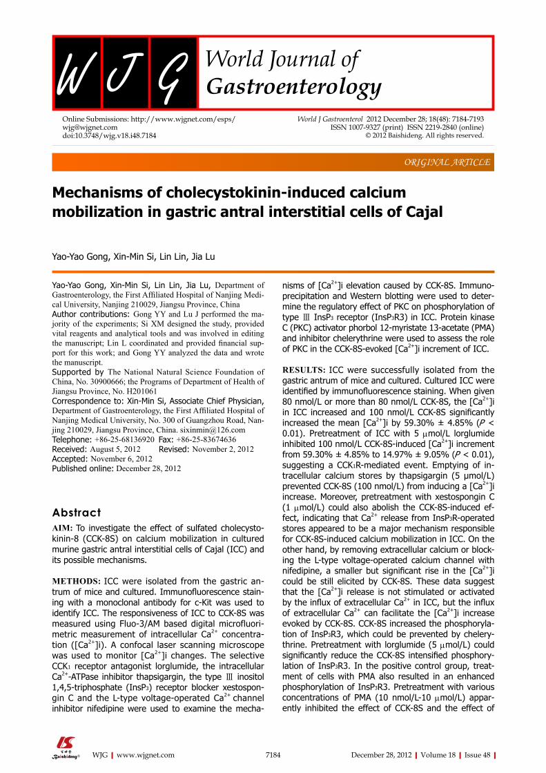
RESULTSIdentification of cultured ICC After the cells were isolated and plated onto culture dishes, it was initially difficult to identify the ICC. After prolonged culture (4-7 d), the cultured ICC, were identi-fied by c-Kit immunofluorescence and showed distinc-tive shapes, such as spindle, triangular or stellar-like with two to five long processes (Figure 1).
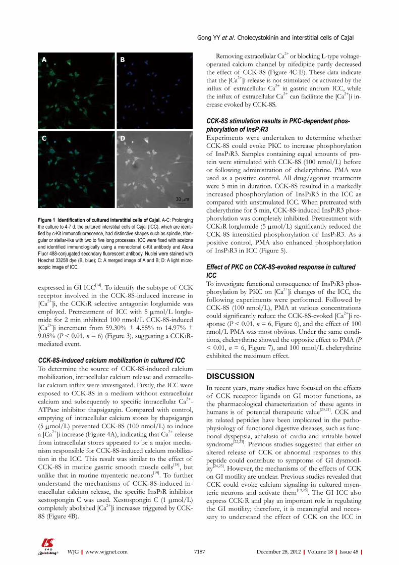
Effects of CCK-8S on intracellular Ca2+ intensity in cul-tured ICCAddition of CCK-8S produced substantial, dose-depen-dent elevations of Fluo-3/AM fluorescence in cytoplasm an nucleus of the ICC, indicating that free calcium level had increased compared with the control (Figure 2A). When given ≤ 50 nmol/L CCK-8S, the [Ca2+]i did not increase (Figure 2B). As shown in Figure 2D, CCK-8S (100 nmol/L) significantly increased the mean [Ca2+]i by 59.30% ± 4.85% (P < 0.01, n = 6) and CCK-8S (80 nmol/L and 500 nmol/L) also evoked [Ca2+]i increases in the percentage of cells responding (20.22% ± 5.48% and 39.32% ± 2.51%, respectively, Figure 2C, E and F). Group data for the [Ca2+]i changes in response to CCK-8S at different concentrations are shown in Figure 2F.
CCK-8S increases [Ca2+]i in cultured ICC via CCK1 recep-torThe CCK1 receptor (CCK1R) has been reported to be
7186 December 28, 2012|Volume 18|Issue 48|WJG|www.wjgnet.com
Gong YY et al . Cholecystokinin and interstitial cells of Cajal
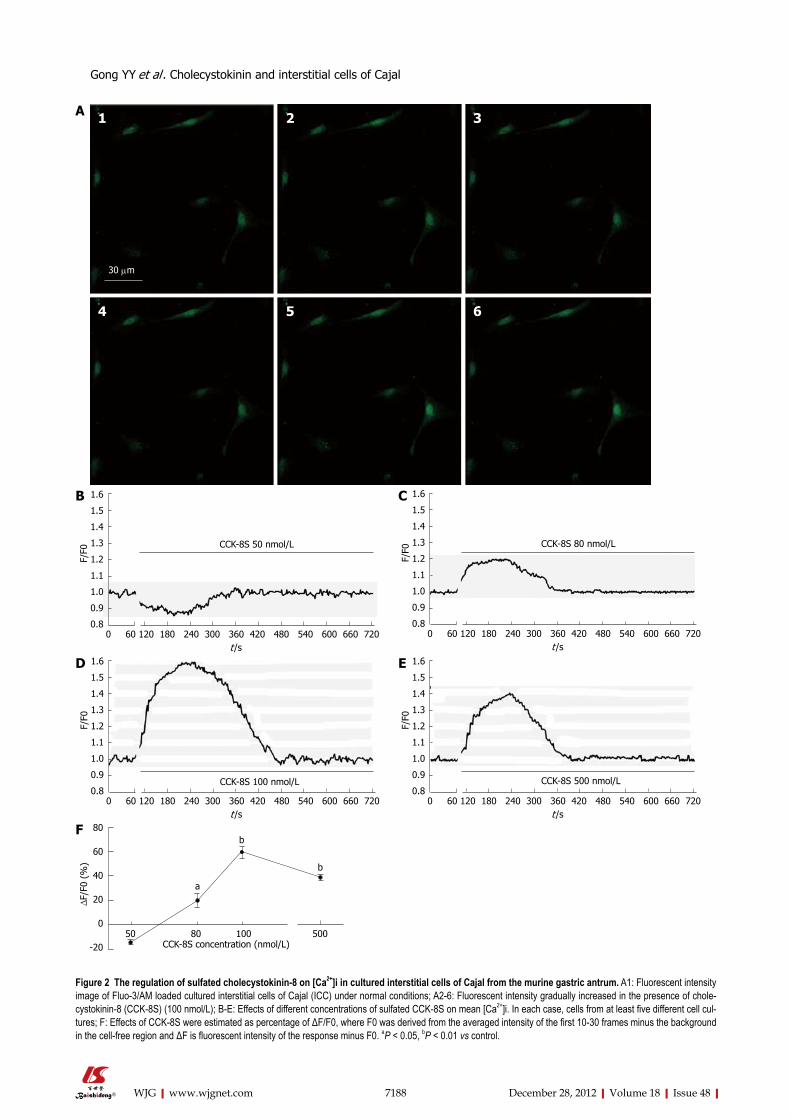
expressed in GI ICC[14]. To identify the subtype of CCK receptor involved in the CCK-8S-induced increase in [Ca2+]i, the CCK1R selective antagonist lorglumide was employed. Pretreatment of ICC with 5 μmol/L lorglu-mide for 2 min inhibited 100 nmol/L CCK-8S-induced [Ca2+]i increment from 59.30% ± 4.85% to 14.97% ± 9.05% (P < 0.01, n = 6) (Figure 3), suggesting a CCK1R-mediated event.
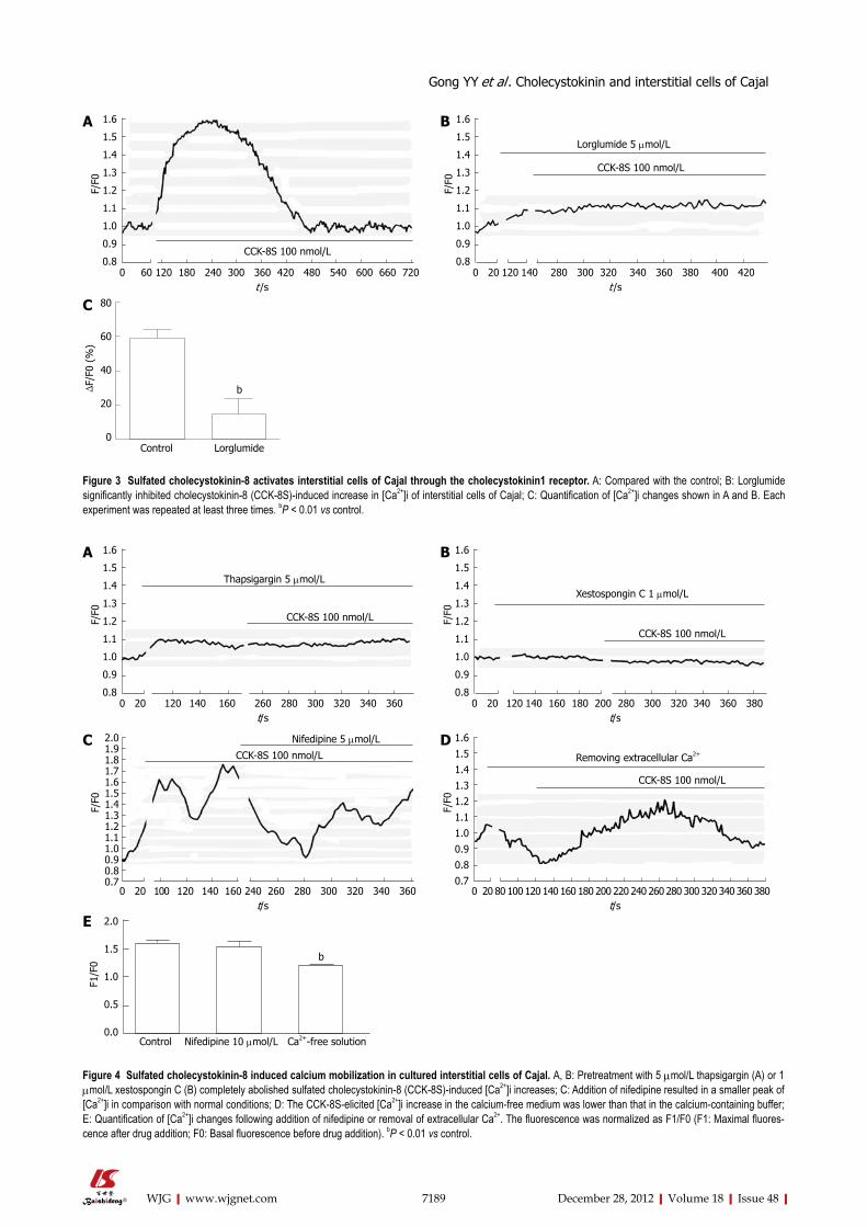
CCK-8S-induced calcium mobilization in cultured ICC To determine the source of CCK-8S-induced calcium mobilization, intracellular calcium release and extracellu-lar calcium influx were investigated. Firstly, the ICC were exposed to CCK-8S in a medium without extracellular calcium and subsequently to specific intracellular Ca2+-ATPase inhibitor thapsigargin. Compared with control, emptying of intracellular calcium stores by thapsigargin (5 μmol/L) prevented CCK-8S (100 nmol/L) to induce a [Ca2+]i increase (Figure 4A), indicating that Ca2+ release from intracellular stores appeared to be a major mecha-nism responsible for CCK-8S-induced calcium mobiliza-tion in the ICC. This result was similar to the effect of CCK-8S in murine gastric smooth muscle cells[18], but unlike that in murine myenteric neurons[19]. To further understand the mechanisms of CCK-8S-induced in-tracellular calcium release, the specific InsP3R inhibitor xestospongin C was used. Xestospongin C (1 μmol/L) completely abolished [Ca2+]i increases triggered by CCK-8S (Figure 4B).
Removing extracellular Ca2+ or blocking L-type voltage-operated calcium channel by nifedipine partly decreased the effect of CCK-8S (Figure 4C-E). These data indicate that the [Ca2+]i release is not stimulated or activated by the influx of extracellular Ca2+ in gastric antrum ICC, while the influx of extracellular Ca2+ can facilitate the [Ca2+]i in-crease evoked by CCK-8S.
CCK-8S stimulation results in PKC-dependent phos-phorylation of InsP3R3Experiments were undertaken to determine whether CCK-8S could evoke PKC to increase phosphorylation of InsP3R3. Samples containing equal amounts of pro-tein were stimulated with CCK-8S (100 nmol/L) before or following administration of chelerythrine. PMA was used as a positive control. All drug/agonist treatments were 5 min in duration. CCK-8S resulted in a markedly increased phosphorylation of InsP3R3 in the ICC as compared with unstimulated ICC. When pretreated with chelerythrine for 5 min, CCK-8S-induced InsP3R3 phos-phorylation was completely inhibited. Pretreatment with CCK1R lorglumide (5 μmol/L) significantly reduced the CCK-8S intensified phosphorylation of InsP3R3. As a positive control, PMA also enhanced phosphorylation of InsP3R3 in ICC (Figure 5).
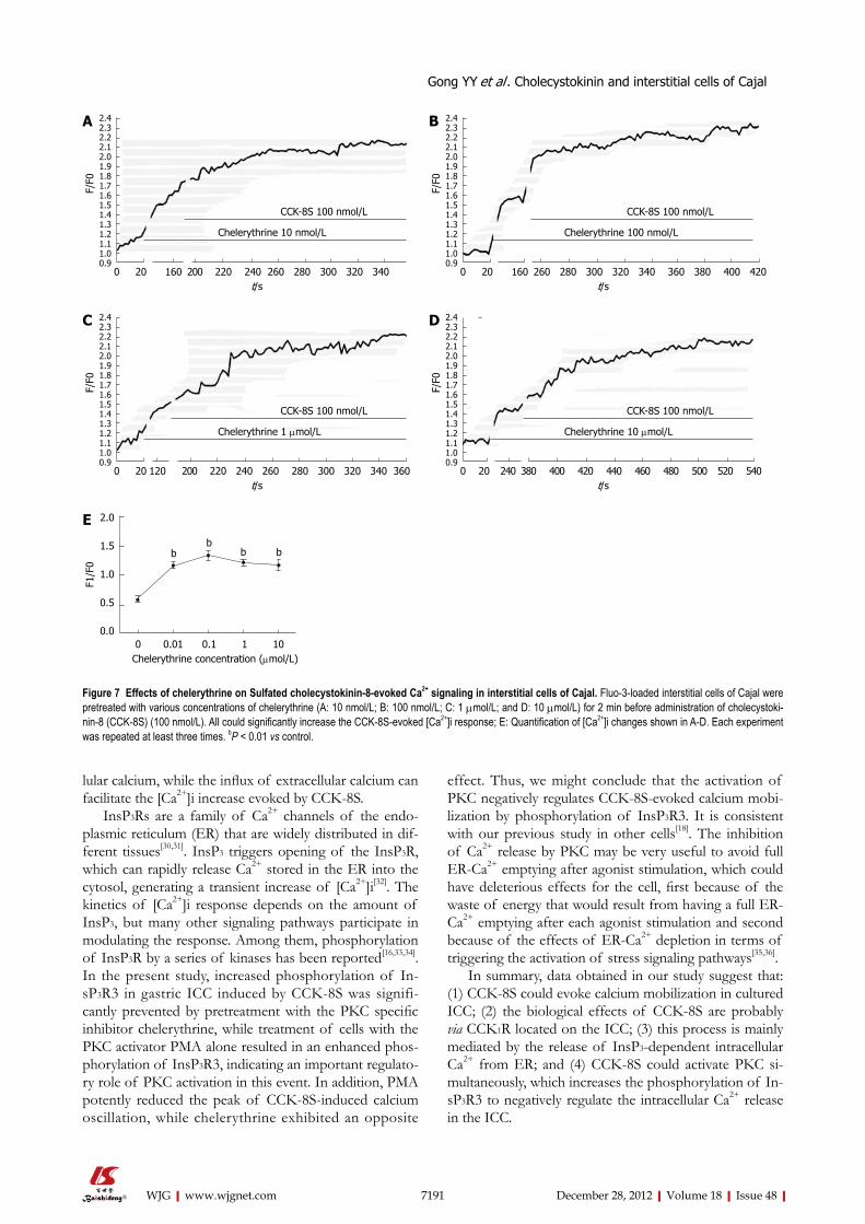
Effect of PKC on CCK-8S-evoked response in cultured ICCTo investigate functional consequence of InsP3R3 phos-phorylation by PKC on [Ca2+]i changes of the ICC, the following experiments were performed. Followed by CCK-8S (100 nmol/L), PMA at various concentrations could significantly reduce the CCK-8S-evoked [Ca2+]i re-sponse (P < 0.01, n = 6, Figure 6), and the effect of 100 nmol/L PMA was most obvious. Under the same condi-tions, chelerythrine showed the opposite effect to PMA (P < 0.01, n = 6, Figure 7), and 100 nmol/L chelerythrine exhibited the maximum effect.
DISCUSSIONIn recent years, many studies have focused on the effects of CCK receptor ligands on GI motor functions, as the pharmacological characterization of these agents in humans is of potential therapeutic value[20,21]. CCK and its related peptides have been implicated in the patho-physiology of functional digestive diseases, such as func-tional dyspepsia, achalasia of cardia and irritable bowel syndrome[22,23]. Previous studies suggested that either an altered release of CCK or abnormal responses to this peptide could contribute to symptoms of GI dysmotil-ity[24,25]. However, the mechanisms of the effects of CCK on GI motility are unclear. Previous studies revealed that CCK could evoke calcium signaling in cultured myen-teric neurons and activate them[19,26]. The GI ICC also express CCK1R and play an important role in regulating the GI motility; therefore, it is meaningful and neces-sary to understand the effect of CCK on the ICC in
7187 December 28, 2012|Volume 18|Issue 48|WJG|www.wjgnet.com
Figure 1 Identification of cultured interstitial cells of Cajal. A-C: Prolonging the culture to 4-7 d, the cultured interstitial cells of Cajal (ICC), which are identi-fied by c-Kit immunofluorescence, had distinctive shapes such as spindle, trian-gular or stellar-like with two to five long processes. ICC were fixed with acetone and identified immunologically using a monoclonal c-Kit antibody and Alexa Fluor 488-conjugated secondary fluorescent antibody. Nuclei were stained with Hoechst 33258 dye (B, blue); C: A merged image of A and B; D: A light micro-scopic image of ICC.
30 μm
A B
C D
Gong YY et al . Cholecystokinin and interstitial cells of Cajal
7188 December 28, 2012|Volume 18|Issue 48|WJG|www.wjgnet.com
30 μm
1 2 3
4 5 6
A
B
0 60 120 180 240 300 360 420 480 540 600 660 720 t /s
CCK-8S 50 nmol/L
1.6
1.5
1.4
1.3
1.2
1.1
1.0
0.9
0.8
F/F0
0 60 120 180 240 300 360 420 480 540 600 660 720 t /s
CCK-8S 80 nmol/L
1.6
1.5
1.4
1.3
1.2
1.1
1.0
0.9
0.8
F/F0
C
0 60 120 180 240 300 360 420 480 540 600 660 720 t /s
CCK-8S 100 nmol/L
1.6
1.5
1.4
1.3
1.2
1.1
1.0
0.9
0.8
F/F0
D
0 60 120 180 240 300 360 420 480 540 600 660 720 t /s
CCK-8S 500 nmol/L
1.6
1.5
1.4
1.3
1.2
1.1
1.0
0.9
0.8
F/F0
E
Figure 2 The regulation of sulfated cholecystokinin-8 on [Ca2+]i in cultured interstitial cells of Cajal from the murine gastric antrum. A1: Fluorescent intensity image of Fluo-3/AM loaded cultured interstitial cells of Cajal (ICC) under normal conditions; A2-6: Fluorescent intensity gradually increased in the presence of chole-cystokinin-8 (CCK-8S) (100 nmol/L); B-E: Effects of different concentrations of sulfated CCK-8S on mean [Ca2+]i. In each case, cells from at least five different cell cul-tures; F: Effects of CCK-8S were estimated as percentage of ΔF/F0, where F0 was derived from the averaged intensity of the first 10-30 frames minus the background in the cell-free region and ΔF is fluorescent intensity of the response minus F0. aP < 0.05, bP < 0.01 vs control.
F
a
b
b
50 80 100 500
80
60
40
20
0
-20
Δ F/F
0 (%
)
CCK-8S concentration (nmol/L)
Gong YY et al . Cholecystokinin and interstitial cells of Cajal
7189 December 28, 2012|Volume 18|Issue 48|WJG|www.wjgnet.com
0 60 120 180 240 300 360 420 480 540 600 660 720 t /s
CCK-8S 100 nmol/L
1.6
1.5
1.4
1.3
1.2
1.1
1.0
0.9
0.8
F/F0
A B
C
0 20 120 140 280 300 320 340 360 380 400 420 t /s
CCK-8S 100 nmol/L
1.6
1.5
1.4
1.3
1.2
1.1
1.0
0.9
0.8
F/F0
Lorglumide 5 μmol/L
b
80
60
40
20
0
Δ F/F
0 (%
)
Control Lorglumide
Figure 3 Sulfated cholecystokinin-8 activates interstitial cells of Cajal through the cholecystokinin1 receptor. A: Compared with the control; B: Lorglumide significantly inhibited cholecystokinin-8 (CCK-8S)-induced increase in [Ca2+]i of interstitial cells of Cajal; C: Quantification of [Ca2+]i changes shown in A and B. Each experiment was repeated at least three times. bP < 0.01 vs control.
0 20 120 140 160 260 280 300 320 340 360 t/s
CCK-8S 100 nmol/L
1.6
1.5
1.4
1.3
1.2
1.1
1.0
0.9
0.8
F/F0
Thapsigargin 5 μmol/L
A B
0 20 120 140 160 180 200 280 300 320 340 360 380 t/s
CCK-8S 100 nmol/L
1.6
1.5
1.4
1.3
1.2
1.1
1.0
0.9
0.8
F/F0
Xestospongin C 1 μmol/L
0 20 100 120 140 160 240 260 280 300 320 340 360 t/s
CCK-8S 100 nmol/L
2.01.91.81.71.61.51.41.31.21.11.00.90.80.7
F/F0
Nifedipine 5 μmol/LC
0 20 80 100 120 140 160 180 200 220 240 260 280 300 320 340 360 380 t/s
CCK-8S 100 nmol/L
1.6
1.5
1.4
1.3
1.2
1.1
1.0
0.9
0.8
0.7
F/F0
Removing extracellular Ca2+
D
Figure 4 Sulfated cholecystokinin-8 induced calcium mobilization in cultured interstitial cells of Cajal. A, B: Pretreatment with 5 μmol/L thapsigargin (A) or 1 μmol/L xestospongin C (B) completely abolished sulfated cholecystokinin-8 (CCK-8S)-induced [Ca2+]i increases; C: Addition of nifedipine resulted in a smaller peak of [Ca2+]i in comparison with normal conditions; D: The CCK-8S-elicited [Ca2+]i increase in the calcium-free medium was lower than that in the calcium-containing buffer; E: Quantification of [Ca2+]i changes following addition of nifedipine or removal of extracellular Ca2+. The fluorescence was normalized as F1/F0 (F1: Maximal fluores-cence after drug addition; F0: Basal fluorescence before drug addition). bP < 0.01 vs control.
b
2.0
1.5
1.0
0.5
0.0
F1/F
0
Control Nifedipine 10 μmol/L Ca2+-free solution
E
Gong YY et al . Cholecystokinin and interstitial cells of Cajal

7190 December 28, 2012|Volume 18|Issue 48|WJG|www.wjgnet.com
controlling GI motility. Our study indicated that CCK-evoked gastric contraction is probably mediated through direct action on CCK1R located on the ICC. However, with respect to GI motility, both the ICC network and the gastrointestinal nervous system play essential roles in producing highly coordinated peristalsis[27,28]. Additional studies are needed to investigate the role of interac-tions between the enteric neurons and the ICC in CCK-evoked effect.
In the GI ICC, the cytoplasmic Ca2+ oscillation is responsible for the pacemaker activity. The pacemaker activity is generated in the ICC and then transferred to smooth muscle cells through the gap junctions[1,29]. In this study, we proved that CCK-8S markedly increased [Ca2+]i in cultured gastric antrum ICC, and the biological effects of CCK-8S were mainly mediated via CCK1R located on the ICC. Two major mechanisms are involved in [Ca2+]i increment during the contraction: calcium release of endoplasmic reticulum Ca2+ store, and/or calcium influx from the extracellular space through activation of calcium channels[29]. We have shown that emptying of the intra-cellular calcium stores by thapsigargin completely blocked the enhancement effect of CCK-8S on the [Ca2+]i level of ICC. Removing extracellular calcium could also inhibit the effect of CCK-8S, but not abolish it. Therefore, both mechanisms mediate the CCK-8S action and Ca2+ release from intracellular stores appears to be the major mecha-nism. The CCK-8S-induced [Ca2+]i increment could be abolished by blockage of InsP3R, suggesting a predomi-nant role of InsP3R-operated stores in CCK-8S-induced intracellular Ca2+ release. However, the CCK-8S-evoked [Ca2+]i increment was persistent in the presence of L-type calcium channel blocker nifedipine, indicating that the [Ca2+]i release is not activated by the influx of extracel-
Probe: p-Ser/Thr PKC
Probe: InsP3R3
CCK-8S - + + + -Lorglumide - + - - -Chelerythrine - - + - -PMA - - - - +
A
B
InsP3R3
InsP3R3
Figure 5 Sulfated cholecystokinin-8 stimulation of interstitial cells of Cajal resulted in the protein kinase C-dependent phosphorylation of type Ⅲ inositol 1,4,5-triphosphate receptor. A: Western blots of proteins were immu-noprecipitated with type Ⅲ inositol 1,4,5-triphosphate receptor (InsP3R3)-specific antibody. The immunoprecipitated proteins were probed with antibody specific for phosphorylated Ser/Thr protein kinase C (PKC) substrate sequences. The sulfated cholecystokinin-8 (CCK-8S)-induced phosphorylation of InsP3R3 was apparently inhibited by pretreatment with chelerythine. Pretreatment with lorglu-mide (5 μmol/L) could significantly reduce the CCK-8S intensified phosphoryla-tion of InsP3R3. In the positive control group, treatment of cells with phorbol-12-myristate-13-acetate (PMA) also resulted in an enhanced phosphorylation of InsP3R3; B: The nitrocellulose membrane in A was stripped and reprobed with InsP3R3 (1:1000) to determine the levels of InsP3R3 immunoprecipitated. Each cell sample contained nearly equal amounts of InsP3R3.
Figure 6 Effects of phorbol-12-myristate-13-acetate on sulfated cholecys-tokinin-8-evoked Ca2+ signaling in interstitial cells of Cajal. Fluo-3-loaded interstitial cells of Cajal were pretreated with various concentrations of Phorbol-12-myristate-13-acetate (PMA) (A: 10 nmol/L; B: 100 nmol/L; C: 1 μmol/L; D: 10 μmol/L) for 4 min before addition of sulfated cholecystokinin-8 (CCK-8S) (100 nmol/L). All could significantly reduce the CCK-8S-evoked [Ca2+]i response; E: Quantification of [Ca2+]i changes shown in A-D. Each experiment was repeated at least three times. bP < 0.01 vs control.
0 20 200 220 260 280 300 320 340 360 380 400 420 t/s
CCK-8S 100 nmol/L
1.31.21.11.00.90.80.70.60.50.40.30.2
F/F0
PMA 10 nmol/LA
B
0 20 200 220 260 280 300 320 340 360 380 400 420 t/s
CCK-8S 100 nmol/L
1.31.21.11.00.90.80.70.60.50.40.30.2
F/F0
PMA 100 nmol/L
C
0 20 200 220 260 280 300 320 340 360 380 400 420 t/s
CCK-8S 100 nmol/L
1.31.21.11.00.90.80.70.60.50.40.30.2
F/F0
PMA 1 μmol/L
D
0 20 200 220 260 280 300 320 340 360 380 400 420 t/s
CCK-8S 100 nmol/L
1.31.21.11.00.90.80.70.60.50.40.30.2
F/F0
PMA 10 μmol/L
E 2.0
1.5
1.0
0.5
0.0
F1/F
0
0 0.01 0.1 1 10 PMA concentration (μmol/L)
b
bbb
Gong YY et al . Cholecystokinin and interstitial cells of Cajal
effect. Thus, we might conclude that the activation of PKC negatively regulates CCK-8S-evoked calcium mobi-lization by phosphorylation of InsP3R3. It is consistent with our previous study in other cells[18]. The inhibition of Ca2+ release by PKC may be very useful to avoid full ER-Ca2+ emptying after agonist stimulation, which could have deleterious effects for the cell, first because of the waste of energy that would result from having a full ER-Ca2+ emptying after each agonist stimulation and second because of the effects of ER-Ca2+ depletion in terms of triggering the activation of stress signaling pathways[35,36].
In summary, data obtained in our study suggest that: (1) CCK-8S could evoke calcium mobilization in cultured ICC; (2) the biological effects of CCK-8S are probably via CCK1R located on the ICC; (3) this process is mainly mediated by the release of InsP3-dependent intracellular Ca2+ from ER; and (4) CCK-8S could activate PKC si-multaneously, which increases the phosphorylation of In-sP3R3 to negatively regulate the intracellular Ca2+ release in the ICC.
7191 December 28, 2012|Volume 18|Issue 48|WJG|www.wjgnet.com
lular calcium, while the influx of extracellular calcium can facilitate the [Ca2+]i increase evoked by CCK-8S.
InsP3Rs are a family of Ca2+ channels of the endo-plasmic reticulum (ER) that are widely distributed in dif-ferent tissues[30,31]. InsP3 triggers opening of the InsP3R, which can rapidly release Ca2+ stored in the ER into the cytosol, generating a transient increase of [Ca2+]i[32]. The kinetics of [Ca2+]i response depends on the amount of InsP3, but many other signaling pathways participate in modulating the response. Among them, phosphorylation of InsP3R by a series of kinases has been reported[16,33,34]. In the present study, increased phosphorylation of In-sP3R3 in gastric ICC induced by CCK-8S was signifi-cantly prevented by pretreatment with the PKC specific inhibitor chelerythrine, while treatment of cells with the PKC activator PMA alone resulted in an enhanced phos-phorylation of InsP3R3, indicating an important regulato-ry role of PKC activation in this event. In addition, PMA potently reduced the peak of CCK-8S-induced calcium oscillation, while chelerythrine exhibited an opposite
Figure 7 Effects of chelerythrine on Sulfated cholecystokinin-8-evoked Ca2+ signaling in interstitial cells of Cajal. Fluo-3-loaded interstitial cells of Cajal were pretreated with various concentrations of chelerythrine (A: 10 nmol/L; B: 100 nmol/L; C: 1 μmol/L; and D: 10 μmol/L) for 2 min before administration of cholecystoki-nin-8 (CCK-8S) (100 nmol/L). All could significantly increase the CCK-8S-evoked [Ca2+]i response; E: Quantification of [Ca2+]i changes shown in A-D. Each experiment was repeated at least three times. bP < 0.01 vs control.
0 20 160 200 220 240 260 280 300 320 340 t/s
CCK-8S 100 nmol/L
2.42.32.22.12.01.91.81.71.61.51.41.31.21.11.00.9
F/F0
Chelerythrine 10 nmol/L
A B
C
0 20 160 260 280 300 320 340 360 380 400 420 t/s
CCK-8S 100 nmol/L
2.42.32.22.12.01.91.81.71.61.51.41.31.21.11.00.9
F/F0
Chelerythrine 100 nmol/L
0 20 120 200 220 240 260 280 300 320 340 360 t/s
CCK-8S 100 nmol/L
2.42.32.22.12.01.91.81.71.61.51.41.31.21.11.00.9
F/F0
Chelerythrine 1 μmol/L
D
E
0 20 240 380 400 420 440 460 480 500 520 540 t/s
CCK-8S 100 nmol/L
2.42.32.22.12.01.91.81.71.61.51.41.31.21.11.00.9
F/F0
Chelerythrine 10 μmol/L
2.0
1.5
1.0
0.5
0.0
F1/F
0
0 0.01 0.1 1 10Chelerythrine concentration (μmol/L)
b bb
b
Gong YY et al . Cholecystokinin and interstitial cells of Cajal
7192 December 28, 2012|Volume 18|Issue 48|WJG|www.wjgnet.com
COMMENTSBackgroundThe interstitial cells of Cajal (ICC) are pacemaker cells in the gastrointestinal (GI) tract; their pacemaker activity is mediated by rhythmic intracellular Ca2+ oscilla-tion. Cholecystokinin (CCK) contributes to the regulation of GI motility. To date, the mechanisms of the regulatory effects of CCK remain unclear. The finding that ICC express CCK1 receptor (also known as CCK-AR) suggests a role for ICC in the mediation of CCK effects. It is worth investigating the direct effect of CCK on intracellular Ca2+ concentration ([Ca2+]i) in the ICC. Research frontiersMany neurotransmitters, including circulating hormones, modulate the ICC pacemaker activity. However, the effect of CCK on GI ICC remains unclear.Innovations and breakthroughsIn the present study, the authors found that CCK evoked the calcium signaling in the ICC mainly via the Ca2+ release of InsP3R-operated stores and CCK acti-vated protein kinase C simultaneously to negatively regulate the release of Ca2+ stores. The findings provide a novel insight into the mechanisms of the regula-tory effects of CCK on GI motility.ApplicationsThe findings may provide clues to the mechanisms of the effects of CCK and its analogs in the treatment of GI motility disorders to some extent.Peer reviewThe authors investigated the effect of sulfated cholecystokinin octapeptide on calcium mobilization in cultured murine gastric antral interstitial cells of Cajal and its possible mechanisms. They found that cholecystokinin octapeptide could evoke the [Ca2+]i signaling in interstitial cells of Cajal through the Ca2+ release of InsP3R-operated stores. Additionally, CCK-8S activated PKC simul-taneously to negatively regulate the release of [Ca2+] stores. The manuscript is highly interesting.
REFERENCES1 Boddy G, Bong A, Cho W, Daniel EE. ICC pacing mecha-
nisms in intact mouse intestine differ from those in cultured or dissected intestine. Am J Physiol Gastrointest Liver Physiol 2004; 286: G653-G662
2 Sanders KM, Koh SD, Ward SM. Interstitial cells of cajal as pacemakers in the gastrointestinal tract. Annu Rev Physiol 2006; 68: 307-343
3 Ordög T, Ward SM, Sanders KM. Interstitial cells of cajal generate electrical slow waves in the murine stomach. J Physiol 1999; 518 (Pt 1): 257-269
4 van Helden DF, Imtiaz MS, Nurgaliyeva K, von der Weid P, Dosen PJ. Role of calcium stores and membrane voltage in the generation of slow wave action potentials in guinea-pig gastric pylorus. J Physiol 2000; 524 (Pt 1): 245-265
5 Ward SM, Beckett EA, Wang X, Baker F, Khoyi M, Sanders KM. Interstitial cells of Cajal mediate cholinergic neuro-transmission from enteric motor neurons. J Neurosci 2000; 20: 1393-1403
6 Ward SM, Morris G, Reese L, Wang XY, Sanders KM. Inter-stitial cells of Cajal mediate enteric inhibitory neurotrans-mission in the lower esophageal and pyloric sphincters. Gastroenterology 1998; 115: 314-329
7 Vanderwinden JM, Rumessen JJ, Liu H, Descamps D, De Laet MH, Vanderhaeghen JJ. Interstitial cells of Cajal in hu-man colon and in Hirschsprung’s disease. Gastroenterology 1996; 111: 901-910
8 Vanderwinden JM, Rumessen JJ. Interstitial cells of Cajal in human gut and gastrointestinal disease. Microsc Res Tech 1999; 47: 344-360
9 Sanders KM, Koh SD, Ordög T, Ward SM. Ionic conduc-tances involved in generation and propagation of electrical slow waves in phasic gastrointestinal muscles. Neurogastro-enterol Motil 2004; 16 Suppl 1: 100-105
10 Rehfeld JF. Clinical endocrinology and metabolism. Cho-
lecystokinin. Best Pract Res Clin Endocrinol Metab 2004; 18: 569-586
11 Lal S, McLaughlin J, Barlow J, D’Amato M, Giacovelli G, Varro A, Dockray GJ, Thompson DG. Cholecystokinin path-ways modulate sensations induced by gastric distension in humans. Am J Physiol Gastrointest Liver Physiol 2004; 287: G72-G79
12 Brennan IM, Little TJ, Feltrin KL, Smout AJ, Wishart JM, Horowitz M, Feinle-Bisset C. Dose-dependent effects of cholecystokinin-8 on antropyloroduodenal motility, gastro-intestinal hormones, appetite, and energy intake in healthy men. Am J Physiol Endocrinol Metab 2008; 295: E1487-E1494
13 Miyasaka K, Funakoshi A. Cholecystokinin and cholecysto-kinin receptors. J Gastroenterol 2003; 38: 1-13
14 Patterson LM, Zheng H, Ward SM, Berthoud HR. Immuno-histochemical identification of cholecystokinin A receptors on interstitial cells of Cajal, smooth muscle, and enteric neu-rons in rat pylorus. Cell Tissue Res 2001; 305: 11-23
15 Herranz R. Cholecystokinin antagonists: pharmacological and therapeutic potential. Med Res Rev 2003; 23: 559-605
16 Straub SV, Giovannucci DR, Bruce JI, Yule DI. A role for phosphorylation of inositol 1,4,5-trisphosphate receptors in defining calcium signals induced by Peptide agonists in pancreatic acinar cells. J Biol Chem 2002; 277: 31949-31956
17 MacIntosh CG, Andrews JM, Jones KL, Wishart JM, Mor-ris HA, Jansen JB, Morley JE, Horowitz M, Chapman IM. Effects of age on concentrations of plasma cholecystokinin, glucagon-like peptide 1, and peptide YY and their relation to appetite and pyloric motility. Am J Clin Nutr 1999; 69: 999-1006
18 Si XM, Huang L, Paul SC, An P, Luo HS. Signal transduc-tion pathways mediating CCK-8S-induced gastric antral smooth muscle contraction. Digestion 2006; 73: 249-258
19 Zhang W, Segura BJ, Mulholland MW. Cholecystokinin-8 induces intracellular calcium signaling in cultured myen-teric neurons from neonatal guinea pigs. Peptides 2002; 23: 1793-1801
20 Morton MF, Welsh NJ, Tavares IA, Shankley NP. Phar-macological characterization of cholecystokinin receptors mediating contraction of human gallbladder and ascending colon. Regul Pept 2002; 105: 59-64
21 Varga G, Bálint A, Burghardt B, D’Amato M. Involvement of endogenous CCK and CCK1 receptors in colonic motor function. Br J Pharmacol 2004; 141: 1275-1284
22 Lee KJ, Tack J. Duodenal implications in the pathophysiol-ogy of functional dyspepsia. J Neurogastroenterol Motil 2010; 16: 251-257
23 El-Salhy M, Vaali K, Dizdar V, Hausken T. Abnormal small-intestinal endocrine cells in patients with irritable bowel syndrome. Dig Dis Sci 2010; 55: 3508-3513
24 Fornai M, Colucci R, Antonioli L, Crema F, Buccianti P, Chiarugi M, Baschiera F, Ghisu N, Tuccori M, Blandizzi C, Del Tacca M. Cholecystokinin CCK2 receptors mediate the peptide’s inhibitory actions on the contractile activity of hu-man distal colon via the nitric oxide pathway. Br J Pharmacol 2007; 151: 1246-1253
25 Chey WY, Jin HO, Lee MH, Sun SW, Lee KY. Colonic motil-ity abnormality in patients with irritable bowel syndrome exhibiting abdominal pain and diarrhea. Am J Gastroenterol 2001; 96: 1499-1506
26 Washington MC, Murry CR, Raboin SJ, Roberson AE, Mansour MM, Williams CS, Sayegh AI. Cholecystokinin-8 activates myenteric neurons in 21- and 35-day old but not 4- and 14-day old rats. Peptides 2011; 32: 272-280
27 Takaki M, Suzuki H, Nakayama S. Recent advances in stud-ies of spontaneous activity in smooth muscle: ubiquitous pacemaker cells. Prog Biophys Mol Biol 2010; 102: 129-135
28 Mostafa RM, Moustafa YM, Hamdy H. Interstitial cells of Cajal, the Maestro in health and disease. World J Gastroen-
COMMENTS
Gong YY et al . Cholecystokinin and interstitial cells of Cajal
7193 December 28, 2012|Volume 18|Issue 48|WJG|www.wjgnet.com
terol 2010; 16: 3239-3248 29 Berridge MJ. Smooth muscle cell calcium activation mecha-
nisms. J Physiol 2008; 586: 5047-5061 30 Berridge MJ, Lipp P, Bootman MD. The versatility and uni-
versality of calcium signalling. Nat Rev Mol Cell Biol 2000; 1: 11-21
31 Janiak R, Wilson SM, Montague S, Hume JR. Heterogeneity of calcium stores and elementary release events in canine pulmonary arterial smooth muscle cells. Am J Physiol Cell Physiol 2001; 280: C22-C33
32 Parekh AB, Penner R. Store depletion and calcium influx. Physiol Rev 1997; 77: 901-930
33 Soulsby MD, Wojcikiewicz RJ. The type III inositol 1,4,5-tri-
sphosphate receptor is phosphorylated by cAMP-dependent protein kinase at three sites. Biochem J 2005; 392: 493-497
34 Murthy KS. cAMP inhibits IP(3)-dependent Ca(2+) release by preferential activation of cGMP-primed PKG. Am J Physi-ol Gastrointest Liver Physiol 2001; 281: G1238-G1245
35 Montero M, Lobatón CD, Gutierrez-Fernández S, Moreno A, Alvarez J. Modulation of histamine-induced Ca2+ release by protein kinase C. Effects on cytosolic and mitochondrial [Ca2+] peaks. J Biol Chem 2003; 278: 49972-49979
36 Hirose K, Kadowaki S, Tanabe M, Takeshima H, Iino M. Spatiotemporal dynamics of inositol 1,4,5-trisphosphate that underlies complex Ca2+ mobilization patterns. Science 1999; 284: 1527-1530
S- Editor Song XX L- Editor Stewart GJ E- Editor Li JY
Gong YY et al . Cholecystokinin and interstitial cells of Cajal