the digestive system and body metabolism 14
TRANSCRIPT

PowerPoint ® Lecture Slide Presentation by Patty Bostwick-Taylor, Florence-Darlington Technical College
Copyright © 2009 Pearson Education, Inc., publishin g as Benjamin Cummings
PART A14
The Digestive System and Body Metabolism

Copyright © 2009 Pearson Education, Inc., publishing as Benjamin Cummings
The Digestive System Functions
� Ingestion—taking in food
� Digestion—breaking food down both physically and chemically
� Absorption—movement of nutrients into the bloodstream
� Defecation—rids the body of indigestible waste
Copyright © 2009 Pearson Education, Inc., publishing as Benjamin Cummings
Organs of the Digestive System
� Two main groups
� Alimentary canal (gastrointestinal or GI tract)—continuous coiled hollow tube
� Accessory digestive organs

Copyright © 2009 Pearson Education, Inc., publishing as Benjamin Cummings
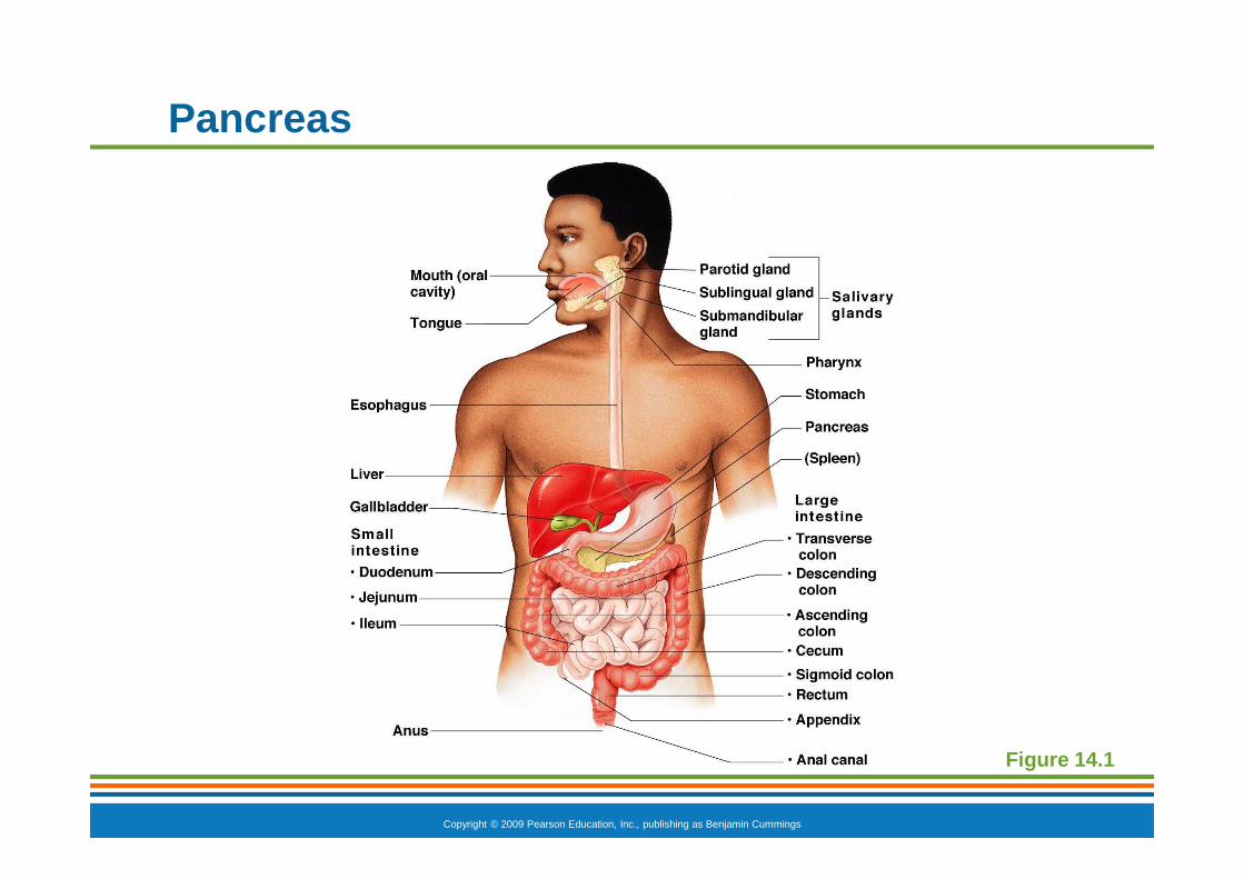
Organs of the Digestive System
Figure 14.1

Copyright © 2009 Pearson Education, Inc., publishing as Benjamin Cummings
Organs of the Alimentary Canal
� Mouth
� Pharynx
� Esophagus
� Stomach
� Small intestine
� Large intestine
� Anus

Copyright © 2009 Pearson Education, Inc., publishing as Benjamin Cummings
Mouth (Oral Cavity) Anatomy
� Lips (labia)—protect the anterior opening
� Cheeks—form the lateral walls
� Hard palate—forms the anterior roof
� Soft palate—forms the posterior roof
� Uvula—fleshy projection of the soft palate
Copyright © 2009 Pearson Education, Inc., publishing as Benjamin Cummings
Mouth (Oral Cavity) Anatomy
� Vestibule—space between lips externally and teeth and gums internally
� Oral cavity proper—area contained by the teeth
� Tongue—attached at hyoid bone and styloid processes of the skull, and by the lingual frenulum to the floor of the mouth
� Tonsils
� Palatine
� Lingual
Copyright © 2009 Pearson Education, Inc., publishing as Benjamin Cummings
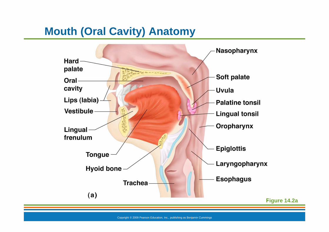
Mouth (Oral Cavity) Anatomy
Figure 14.2a

Copyright © 2009 Pearson Education, Inc., publishing as Benjamin Cummings
Mouth (Oral Cavity) Anatomy
Figure 14.2b

Copyright © 2009 Pearson Education, Inc., publishing as Benjamin Cummings
Mouth Physiology
� Mastication (chewing) of food
� Mixing masticated food with saliva
� Initiation of swallowing by the tongue
� Allows for the sense of taste
Copyright © 2009 Pearson Education, Inc., publishing as Benjamin Cummings
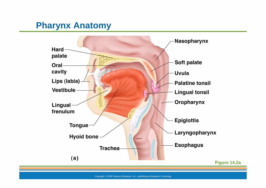
Pharynx Anatomy
� Nasopharynx—not part of the digestive system
� Oropharynx—posterior to oral cavity
� Laryngopharynx—below the oropharynx and connected to the esophagus
Copyright © 2009 Pearson Education, Inc., publishing as Benjamin Cummings
Pharynx Anatomy
Figure 14.2a

Copyright © 2009 Pearson Education, Inc., publishing as Benjamin Cummings
Pharynx Physiology
� Serves as a passageway for air and food
� Food is propelled to the esophagus by two muscle layers
� Longitudinal inner layer
� Circular outer layer
� Food movement is by alternating contractions of the muscle layers (peristalsis)

Copyright © 2009 Pearson Education, Inc., publishing as Benjamin Cummings
Esophagus Anatomy and Physiology
� Anatomy
� About 10 inches long
� Runs from pharynx to stomach through the diaphragm
� Physiology
� Conducts food by peristalsis (slow rhythmic squeezing)
� Passageway for food only (respiratory system branches off after the pharynx)

Copyright © 2009 Pearson Education, Inc., publishing as Benjamin Cummings
Layers of Alimentary Canal Organs
� Four layers
� Mucosa
� Submucosa
� Muscularis externa
� Serosa
Copyright © 2009 Pearson Education, Inc., publishing as Benjamin Cummings
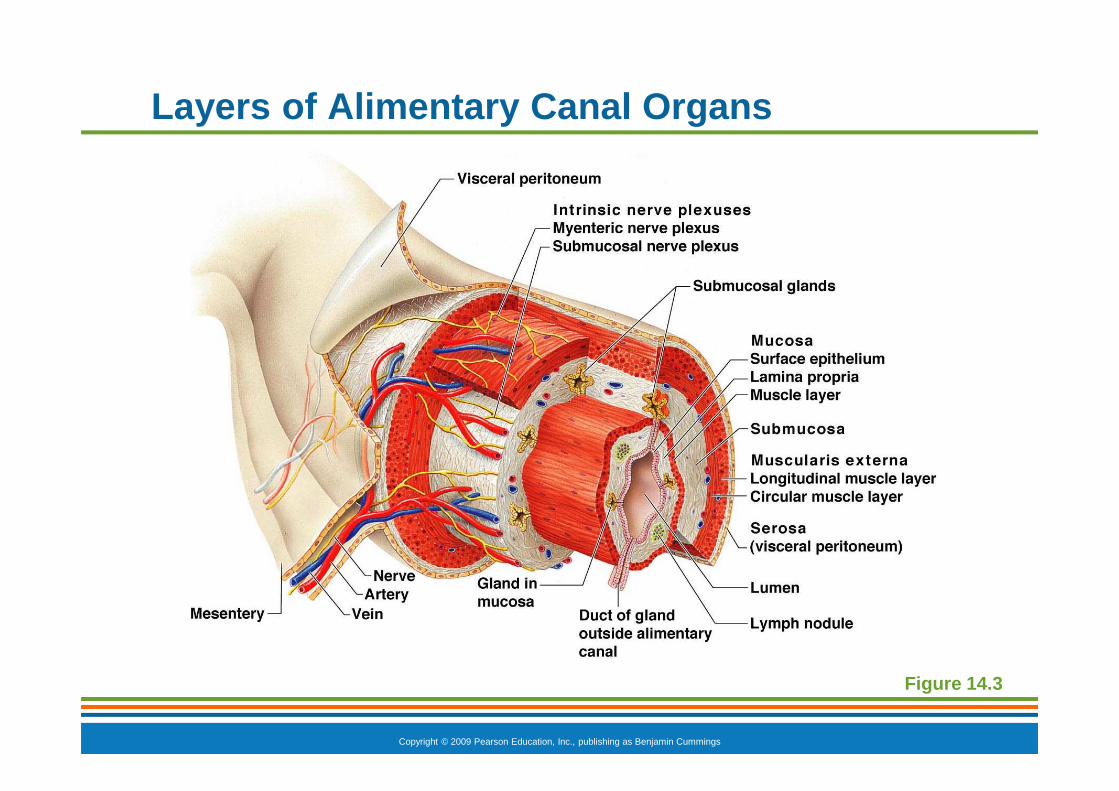
Layers of Alimentary Canal Organs
� Mucosa
� Innermost, moist membrane consisting of
� Surface epithelium
� Small amount of connective tissue (lamina propria)
� Small smooth muscle layer

Copyright © 2009 Pearson Education, Inc., publishing as Benjamin Cummings
Layers of Alimentary Canal Organs
Figure 14.3

Copyright © 2009 Pearson Education, Inc., publishing as Benjamin Cummings
Layers of Alimentary Canal Organs
� Submucosa
� Just beneath the mucosa
� Soft connective tissue with blood vessels, nerve endings, and lymphatics

Copyright © 2009 Pearson Education, Inc., publishing as Benjamin Cummings
Layers of Alimentary Canal Organs
Figure 14.3

Copyright © 2009 Pearson Education, Inc., publishing as Benjamin Cummings
Layers of Alimentary Canal Organs
� Muscularis externa—smooth muscle
� Inner circular layer
� Outer longitudinal layer
� Serosa—outermost layer of the wall contains fluid-producing cells
� Visceral peritoneum —outermost layer that is continuous with the innermost layer
� Parietal peritoneum —innermost layer that lines the abdominopelvic cavity
Copyright © 2009 Pearson Education, Inc., publishing as Benjamin Cummings
Layers of Alimentary Canal Organs
Figure 14.3

Copyright © 2009 Pearson Education, Inc., publishing as Benjamin Cummings
Alimentary Canal Nerve Plexuses
� Two important nerve plexuses serve the alimentary canal
� Both are part of the autonomic nervous system
� Submucosal nerve plexus
� Myenteric nerve plexus
� Function is to regulate mobility and secretory activity of the GI tract organs
PowerPoint ® Lecture Slide Presentation by Patty Bostwick-Taylor, Florence-Darlington Technical College
Copyright © 2009 Pearson Education, Inc., publishin g as Benjamin Cummings
PART A14
The Digestive System and Body Metabolism

Copyright © 2009 Pearson Education, Inc., publishing as Benjamin Cummings
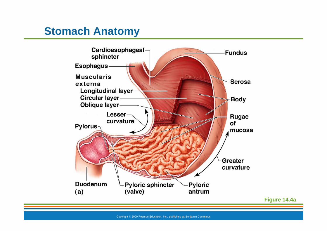
Stomach Anatomy
� Located on the left side of the abdominal cavity
� Food enters at the cardioesophageal sphincter
� Food empties into the small intestine at the pyloric sphincter (valve)

Copyright © 2009 Pearson Education, Inc., publishing as Benjamin Cummings
Stomach Anatomy
� Regions of the stomach
� Cardiac region—near the heart
� Fundus—expanded portion lateral to the cardiac region
� Body—midportion
� Pylorus—funnel-shaped terminal end
Copyright © 2009 Pearson Education, Inc., publishing as Benjamin Cummings
Stomach Anatomy
� Rugae—internal folds of the mucosa
� External regions
� Lesser curvature—concave medial surface
� Greater curvature—convex lateral surface
Copyright © 2009 Pearson Education, Inc., publishing as Benjamin Cummings
Stomach Anatomy
Figure 14.4a

Copyright © 2009 Pearson Education, Inc., publishing as Benjamin Cummings
Stomach Anatomy
Figure 14.4b

Copyright © 2009 Pearson Education, Inc., publishing as Benjamin Cummings
Stomach Anatomy
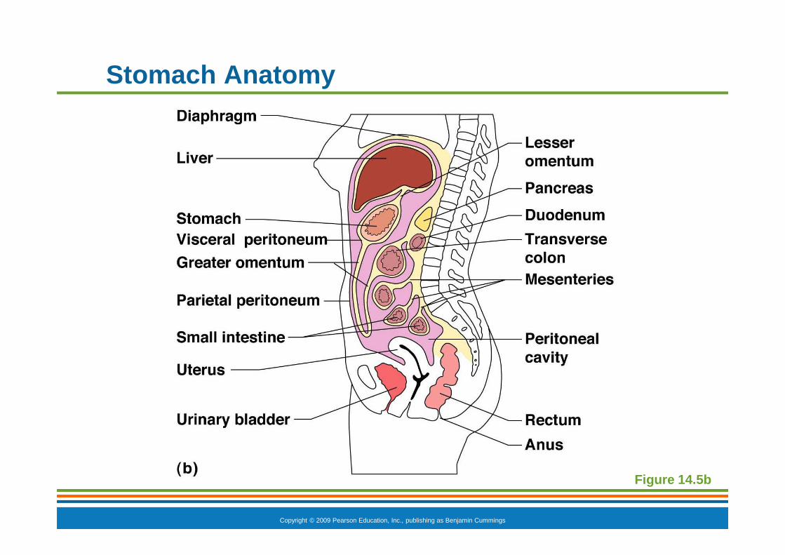
� Layers of peritoneum attached to the stomach
� Lesser omentum —attaches the liver to the lesser curvature
� Greater omentum —attaches the greater curvature to the posterior body wall
� Contains fat to insulate, cushion, and protect abdominal organs
� Has lymph nodules containing macrophages

Copyright © 2009 Pearson Education, Inc., publishing as Benjamin Cummings
Stomach Anatomy
Figure 14.5a
Copyright © 2009 Pearson Education, Inc., publishing as Benjamin Cummings
Stomach Anatomy
Figure 14.5b
Copyright © 2009 Pearson Education, Inc., publishing as Benjamin Cummings
Stomach Physiology
� Temporary storage tank for food
� Site of food breakdown
� Chemical breakdown of protein begins
� Delivers chyme (processed food) to the small intestine
Copyright © 2009 Pearson Education, Inc., publishing as Benjamin Cummings
Structure of the Stomach Mucosa
� Mucosa is simple columnar epithelium
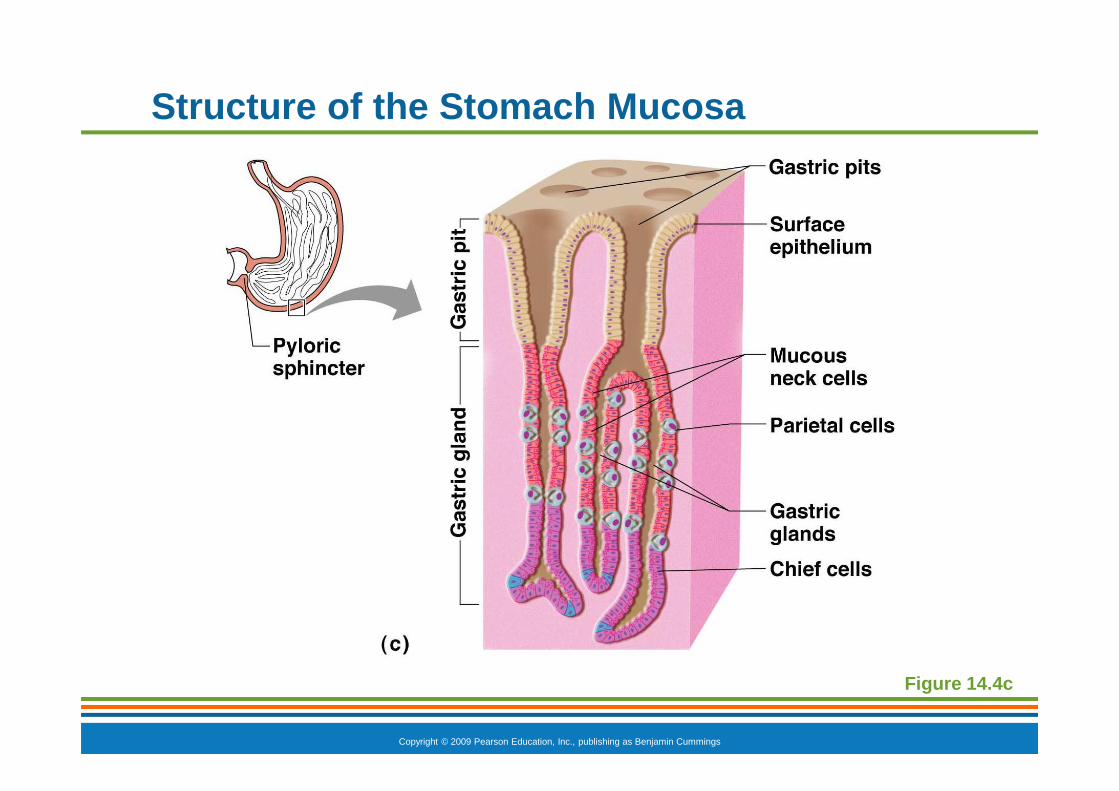
� Mucous neck cells—produce a sticky alkaline mucus
� Gastric glands—situated in gastric pits and secrete gastric juice
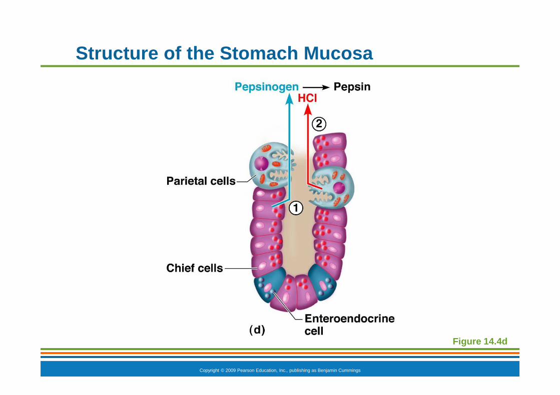
� Chief cells—produce protein-digesting enzymes (pepsinogens)
� Parietal cells—produce hydrochloric acid
� Enteroendocrine cells—produce gastrin
Copyright © 2009 Pearson Education, Inc., publishing as Benjamin Cummings
Structure of the Stomach Mucosa
Figure 14.4c
Copyright © 2009 Pearson Education, Inc., publishing as Benjamin Cummings
Structure of the Stomach Mucosa
Figure 14.4d

Copyright © 2009 Pearson Education, Inc., publishing as Benjamin Cummings
Small Intestine
� The body’s major digestive organ
� Site of nutrient absorption into the blood
� Muscular tube extending from the pyloric sphincter to the ileocecal valve
� Suspended from the posterior abdominal wall by the mesentery
Copyright © 2009 Pearson Education, Inc., publishing as Benjamin Cummings
Subdivisions of the Small Intestine
� Duodenum
� Attached to the stomach
� Curves around the head of the pancreas
� Jejunum
� Attaches anteriorly to the duodenum
� Ileum
� Extends from jejunum to large intestine
Copyright © 2009 Pearson Education, Inc., publishing as Benjamin Cummings
Chemical Digestion in the Small Intestine
� Chemical digestion begins in the small intestine
� Enzymes are produced by
� Intestinal cells
� Pancreas
� Pancreatic ducts carry enzymes to the small intestine
� Bile, formed by the liver, enters via the bile duct

Copyright © 2009 Pearson Education, Inc., publishing as Benjamin Cummings
Chemical Digestion in the Small Intestine
Figure 14.6
Copyright © 2009 Pearson Education, Inc., publishing as Benjamin Cummings
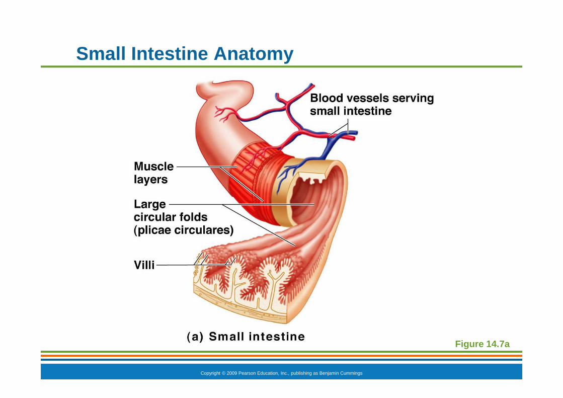
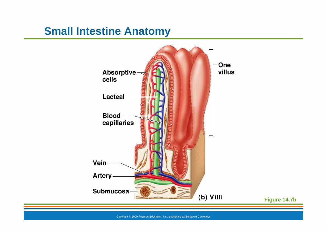
Small Intestine Anatomy
� Three structural modifications that increase surface area
� Microvilli—tiny projections of the plasma membrane (create a brush border appearance)
� Villi—fingerlike structures formed by the mucosa
� Circular folds (plicae circulares)—deep folds of mucosa and submucosa
Copyright © 2009 Pearson Education, Inc., publishing as Benjamin Cummings
Small Intestine Anatomy
Figure 14.7a
Copyright © 2009 Pearson Education, Inc., publishing as Benjamin Cummings
Small Intestine Anatomy
Figure 14.7b

Copyright © 2009 Pearson Education, Inc., publishing as Benjamin Cummings
Figure 14.7c
Small Intestine Anatomy
Copyright © 2009 Pearson Education, Inc., publishing as Benjamin Cummings
Large Intestine
� Larger in diameter, but shorter in length, than the small intestine
� Frames the internal abdomen

Copyright © 2009 Pearson Education, Inc., publishing as Benjamin Cummings
Large Intestine Anatomy
� Cecum —saclike first part of the large intestine
� Appendix
� Accumulation of lymphatic tissue that sometimes becomes inflamed (appendicitis)
� Hangs from the cecum
Copyright © 2009 Pearson Education, Inc., publishing as Benjamin Cummings
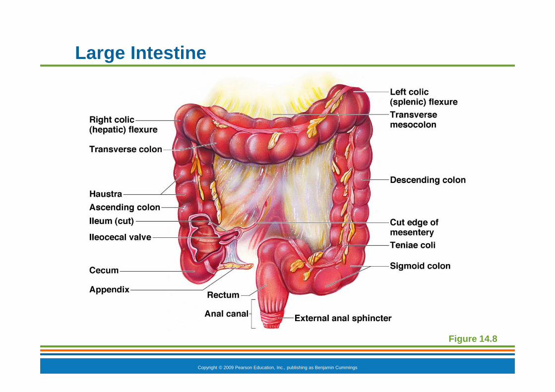
Large Intestine
Figure 14.8
Copyright © 2009 Pearson Education, Inc., publishing as Benjamin Cummings
Large Intestine Anatomy
� Colon
� Ascending—travels up right side of abdomen
� Transverse—travels across the abdominal cavity
� Descending—travels down the left side
� Sigmoid—enters the pelvis
� Rectum and anal canal—also in pelvis

Copyright © 2009 Pearson Education, Inc., publishing as Benjamin Cummings
Large Intestine
Figure 14.8

Copyright © 2009 Pearson Education, Inc., publishing as Benjamin Cummings
Large Intestine Anatomy
� Anus—opening of the large intestine
� External anal sphincter—formed by skeletal muscle and under voluntary control
� Internal involuntary sphincter—formed by smooth muscle
� These sphincters are normally closed except during defecation

Copyright © 2009 Pearson Education, Inc., publishing as Benjamin Cummings
Large Intestine
Figure 14.8

Copyright © 2009 Pearson Education, Inc., publishing as Benjamin Cummings
Large Intestine Anatomy
� No villi present
� Goblet cells produce alkaline mucus which lubricates the passage of feces
� Muscularis externa layer is reduced to three bands of muscle called teniae coli
� These bands cause the wall to pucker into haustra (pocketlike sacs)

PowerPoint ® Lecture Slide Presentation by Patty Bostwick-Taylor, Florence-Darlington Technical College
Copyright © 2009 Pearson Education, Inc., publishin g as Benjamin Cummings
PART A14
The Digestive System and Body Metabolism

Copyright © 2009 Pearson Education, Inc., publishing as Benjamin Cummings
Accessory Digestive Organs
� Teeth
� Salivary glands
� Pancreas
� Liver
� Gallbladder
Copyright © 2009 Pearson Education, Inc., publishing as Benjamin Cummings
Teeth
� Function is to masticate (chew) food
� Humans have two sets of teeth
� Deciduous (baby or “milk”) teeth
� 20 teeth are fully formed by age two
Copyright © 2009 Pearson Education, Inc., publishing as Benjamin Cummings
Teeth
� Permanent teeth
� Replace deciduous teeth between the ages of 6 and 12
� A full set is 32 teeth, but some people do not have wisdom teeth (third molars)
� If they do emerge, the wisdom teeth appear between ages of 17 and 25
Copyright © 2009 Pearson Education, Inc., publishing as Benjamin Cummings
Classification of Teeth
� Incisors—cutting
� Canines—tearing or piercing
� Premolars—grinding
� Molars—grinding

Copyright © 2009 Pearson Education, Inc., publishing as Benjamin Cummings
Human Deciduous and Permanent Teeth
Figure 14.9
Copyright © 2009 Pearson Education, Inc., publishing as Benjamin Cummings
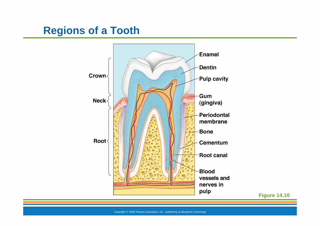
Regions of a Tooth
� Crown—exposed part
� Enamel—hardest substance in the body
� Dentin—found deep to the enamel and forms the bulk of the tooth
� Pulp cavity—contains connective tissue, blood vessels, and nerve fibers
� Root canal—where the pulp cavity extends into the root
Copyright © 2009 Pearson Education, Inc., publishing as Benjamin Cummings
Regions of a Tooth
� Neck
� Region in contact with the gum
� Connects crown to root
� Root
� Cementum —covers outer surface and attaches the tooth to the periodontal membrane
Copyright © 2009 Pearson Education, Inc., publishing as Benjamin Cummings
Regions of a Tooth
Figure 14.10
Copyright © 2009 Pearson Education, Inc., publishing as Benjamin Cummings
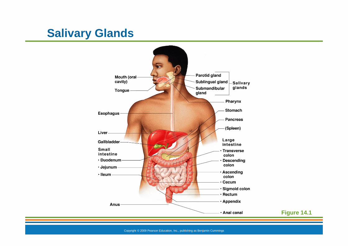
Salivary Glands
� Three pairs of salivary glands empty secretions into the mouth
� Parotid glands
� Submandibular glands
� Sublingual glands
Copyright © 2009 Pearson Education, Inc., publishing as Benjamin Cummings
Figure 14.1
Salivary Glands
Copyright © 2009 Pearson Education, Inc., publishing as Benjamin Cummings
Saliva
� Mixture of mucus and serous fluids
� Helps to form a food bolus
� Contains salivary amylase to begin starch digestion
� Dissolves chemicals so they can be tasted
Copyright © 2009 Pearson Education, Inc., publishing as Benjamin Cummings
Pancreas
� Found posterior to the parietal peritoneum
� Extends across the abdomen from spleen to duodenum
Copyright © 2009 Pearson Education, Inc., publishing as Benjamin Cummings
Pancreas
� Produces a wide spectrum of digestive enzymes that break down all categories of food
� Enzymes are secreted into the duodenum
� Alkaline fluid introduced with enzymes neutralizes acidic chyme coming from stomach
� Hormones produced by the pancreas
� Insulin
� Glucagon
Copyright © 2009 Pearson Education, Inc., publishing as Benjamin Cummings
Pancreas
Figure 14.1

Copyright © 2009 Pearson Education, Inc., publishing as Benjamin Cummings
Pancreas
Figure 14.6
Copyright © 2009 Pearson Education, Inc., publishing as Benjamin Cummings
Liver
� Largest gland in the body
� Located on the right side of the body under the diaphragm
� Consists of four lobes suspended from the diaphragm and abdominal wall by the falciform ligament
� Connected to the gallbladder via the common hepatic duct

Copyright © 2009 Pearson Education, Inc., publishing as Benjamin Cummings
Liver
Figure 14.1

Copyright © 2009 Pearson Education, Inc., publishing as Benjamin Cummings
Figure 14.5
Liver
Copyright © 2009 Pearson Education, Inc., publishing as Benjamin Cummings
Bile
� Produced by cells in the liver
� Composition is
� Bile salts
� Bile pigments (mostly bilirubin from the breakdown of hemoglobin)
� Cholesterol
� Phospholipids
� Electrolytes

Copyright © 2009 Pearson Education, Inc., publishing as Benjamin Cummings
Bile
� Function—emulsify fats by physically breaking large fat globules into smaller ones
Copyright © 2009 Pearson Education, Inc., publishing as Benjamin Cummings
Gallbladder
� Sac found in hollow fossa of liver
� When no digestion is occurring, bile backs up the cystic duct for storage in the gallbladder
� When digestion of fatty food is occurring, bile is introduced into the duodenum from the gallbladder
� Gallstones are crystallized cholesterol which can cause blockages
Copyright © 2009 Pearson Education, Inc., publishing as Benjamin Cummings
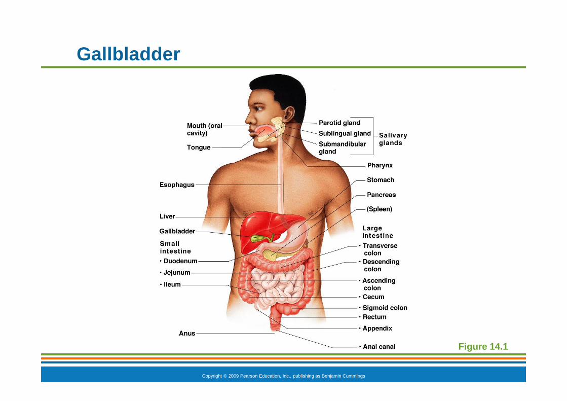
Gallbladder
Figure 14.1
Copyright © 2009 Pearson Education, Inc., publishing as Benjamin Cummings
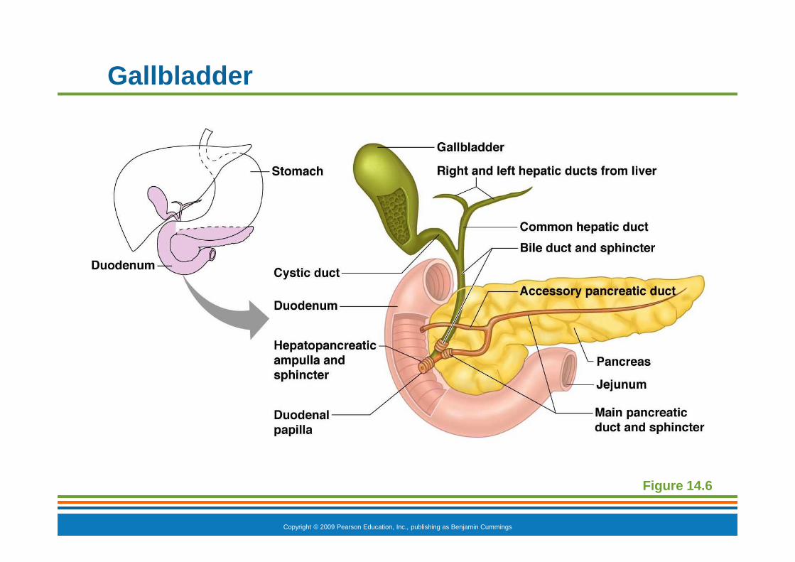
Gallbladder
Figure 14.6

Copyright © 2009 Pearson Education, Inc., publishing as Benjamin Cummings
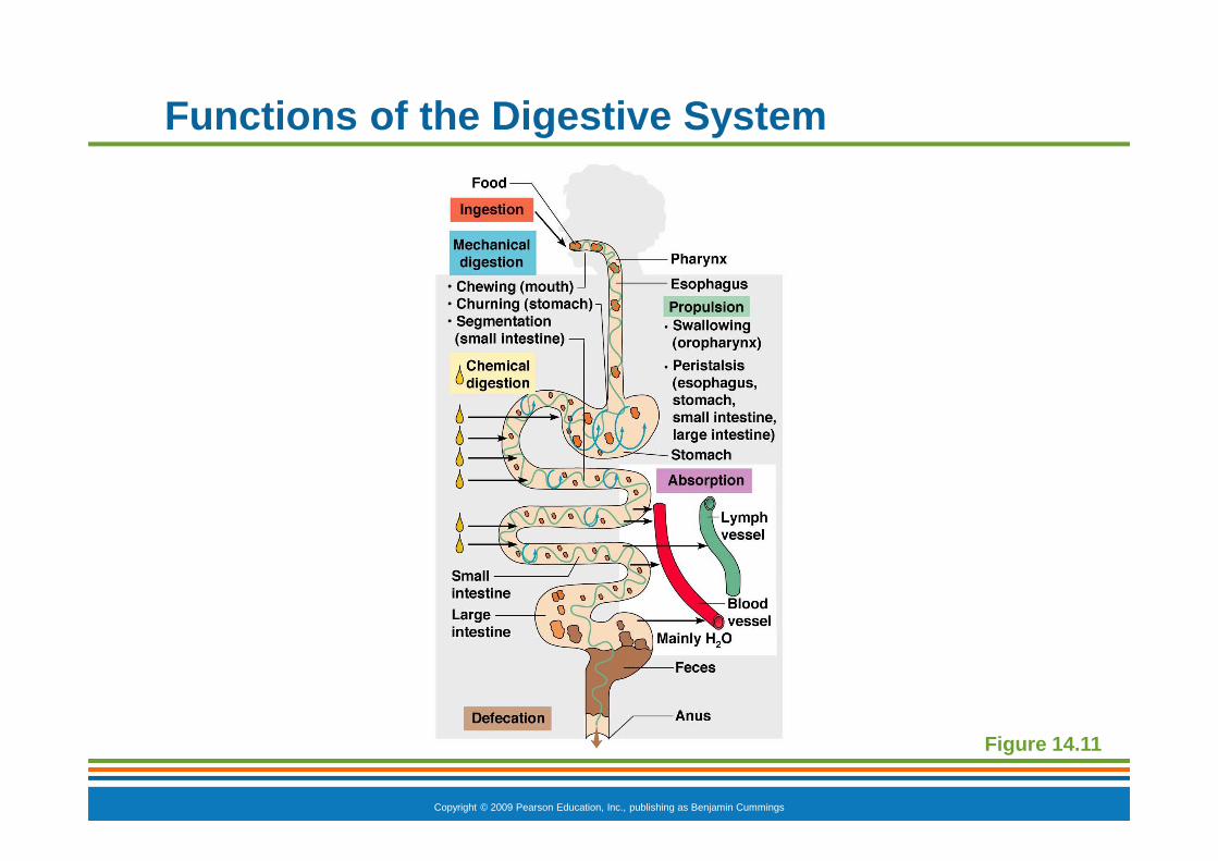
Functions of the Digestive System
� Ingestion—getting food into the mouth
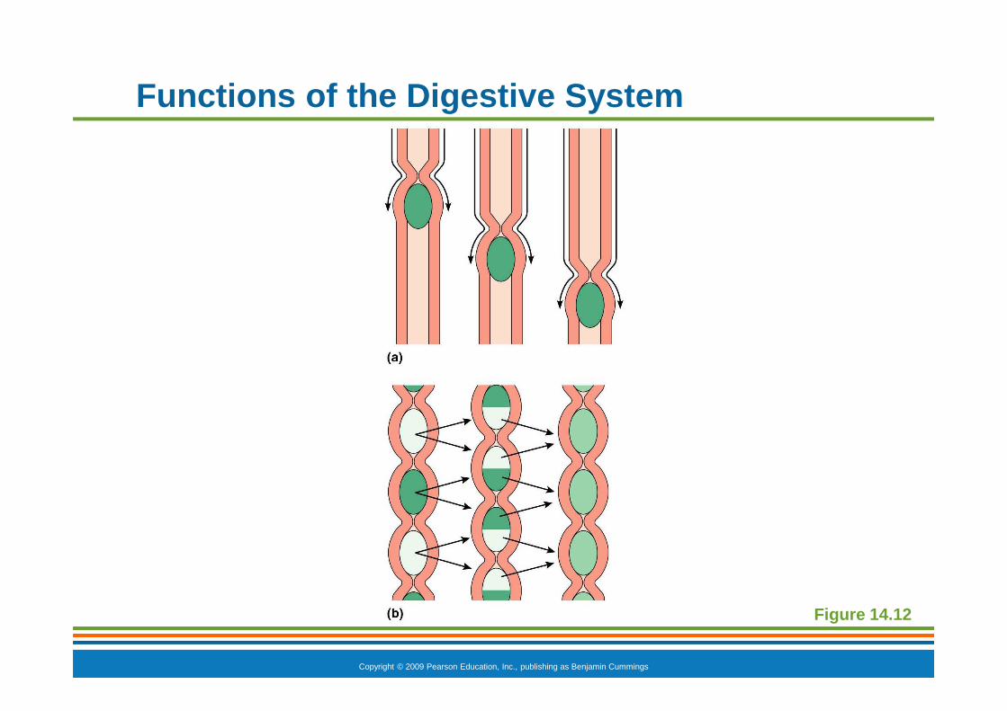
� Propulsion—moving foods from one region of the digestive system to another
� Peristalsis—alternating waves of contraction and relaxation that squeezes food along the GI tract
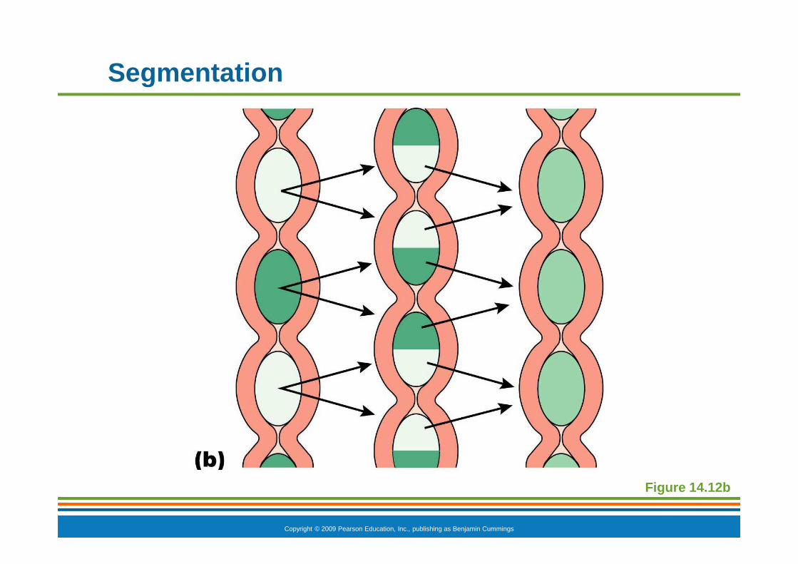
� Segmentation—moving materials back and forth to aid with mixing in the small intestine
Copyright © 2009 Pearson Education, Inc., publishing as Benjamin Cummings
Functions of the Digestive System
Figure 14.12

Copyright © 2009 Pearson Education, Inc., publishing as Benjamin Cummings
Functions of the Digestive System
� Food breakdown as mechanical digestion
� Examples:
� Mixing food in the mouth by the tongue
� Churning food in the stomach
� Segmentation in the small intestine
� Mechanical digestion prepares food for further degradation by enzymes
Copyright © 2009 Pearson Education, Inc., publishing as Benjamin Cummings
Functions of the Digestive System
� Food breakdown as chemical digestion
� Enzymes break down food molecules into their building blocks
� Each major food group uses different enzymes
� Carbohydrates are broken to simple sugars
� Proteins are broken to amino acids
� Fats are broken to fatty acids and alcohols
Copyright © 2009 Pearson Education, Inc., publishing as Benjamin Cummings
Functions of the Digestive System
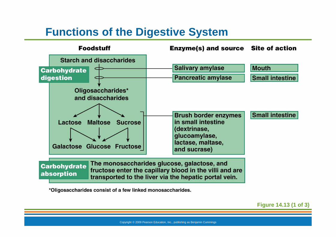
Figure 14.13 (1 of 3)
Copyright © 2009 Pearson Education, Inc., publishing as Benjamin Cummings
Functions of the Digestive System
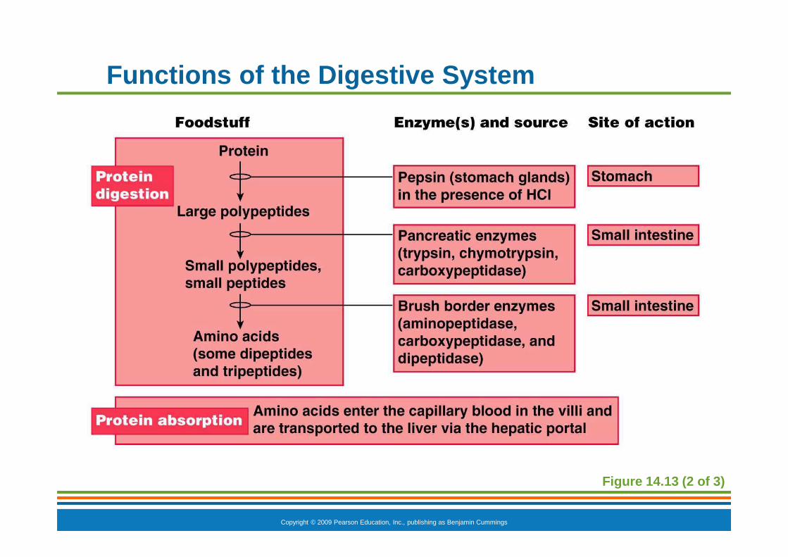
Figure 14.13 (2 of 3)

Copyright © 2009 Pearson Education, Inc., publishing as Benjamin Cummings
Functions of the Digestive System
Figure 14.13 (3 of 3)
Copyright © 2009 Pearson Education, Inc., publishing as Benjamin Cummings
Functions of the Digestive System
� Absorption
� End products of digestion are absorbed in the blood or lymph
� Food must enter mucosal cells and then into blood or lymph capillaries
� Defecation
� Elimination of indigestible substances from the GI tract in the form of feces
Copyright © 2009 Pearson Education, Inc., publishing as Benjamin Cummings
Functions of the Digestive System
Figure 14.11
Copyright © 2009 Pearson Education, Inc., publishing as Benjamin Cummings
Control of Digestive Activity
� Mostly controlled by reflexes via the parasympathetic division
� Chemical and mechanical receptors are located in organ walls that trigger reflexes
Copyright © 2009 Pearson Education, Inc., publishing as Benjamin Cummings
Control of Digestive Activity
� Stimuli include
� Stretch of the organ
� pH of the contents
� Presence of breakdown products
� Reflexes include
� Activation or inhibition of glandular secretions
� Smooth muscle activity

Copyright © 2009 Pearson Education, Inc., publishing as Benjamin Cummings
Digestive Activities of the Mouth
� Mechanical breakdown
� Food is physically broken down by chewing
� Chemical digestion
� Food is mixed with saliva
� Starch is broken down into maltose by salivary amylase
Copyright © 2009 Pearson Education, Inc., publishing as Benjamin Cummings
Activities of the Pharynx and Esophagus
� These organs have no digestive function
� Serve as passageways to the stomach
Copyright © 2009 Pearson Education, Inc., publishing as Benjamin Cummings
Deglutition (Swallowing)
� Buccal phase
� Voluntary
� Occurs in the mouth
� Food is formed into a bolus
� The bolus is forced into the pharynx by the tongue
Copyright © 2009 Pearson Education, Inc., publishing as Benjamin Cummings
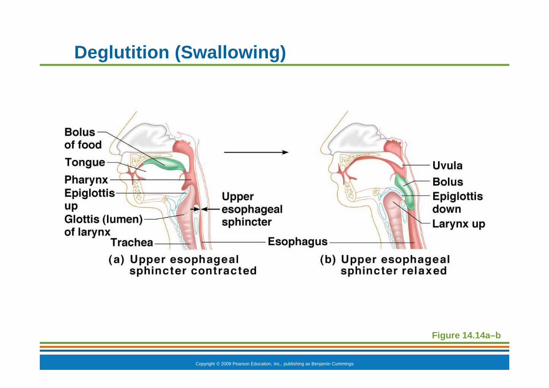
Deglutition (Swallowing)
� Pharyngeal-esophageal phase
� Involuntary transport of the bolus
� All passageways except to the stomach are blocked
� Tongue blocks off the mouth
� Soft palate (uvula) blocks the nasopharynx
� Epiglottis blocks the larynx
Copyright © 2009 Pearson Education, Inc., publishing as Benjamin Cummings
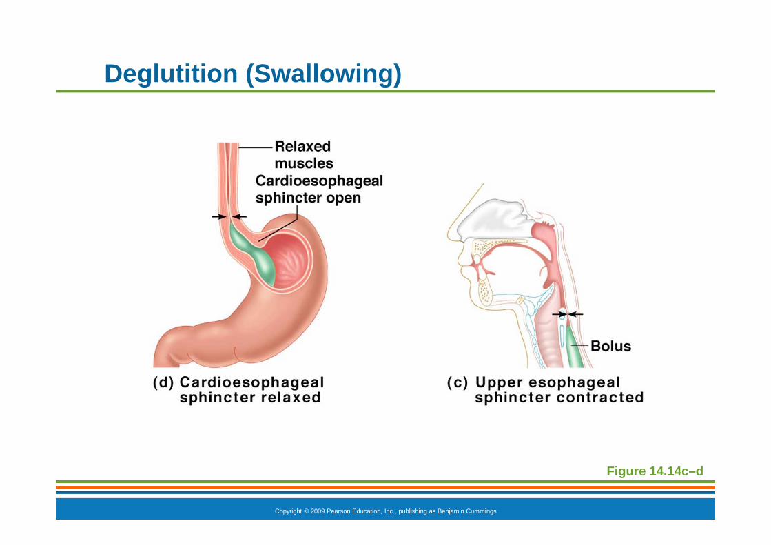
Deglutition (Swallowing)
� Pharyngeal-esophogeal phase (continued)
� Peristalsis moves the bolus toward the stomach
� The cardioesophageal sphincter is opened when food presses against it
Copyright © 2009 Pearson Education, Inc., publishing as Benjamin Cummings
Deglutition (Swallowing)
Figure 14.14a–b
Copyright © 2009 Pearson Education, Inc., publishing as Benjamin Cummings
Deglutition (Swallowing)
Figure 14.14c–d
Copyright © 2009 Pearson Education, Inc., publishing as Benjamin Cummings
Food Breakdown in the Stomach
� Gastric juice is regulated by neural and hormonal factors
� Presence of food or rising pH causes the release of the hormone gastrin
� Gastrin causes stomach glands to produce
� Protein-digesting enzymes
� Mucus
� Hydrochloric acid
Copyright © 2009 Pearson Education, Inc., publishing as Benjamin Cummings
Food Breakdown in the Stomach
� Hydrochloric acid makes the stomach contents very acidic
� Acidic pH
� Activates pepsinogen to pepsin for protein digestion
� Provides a hostile environment for microorganisms
Copyright © 2009 Pearson Education, Inc., publishing as Benjamin Cummings
Digestion and Absorption in the Stomach
� Protein digestion enzymes
� Pepsin—an active protein-digesting enzyme
� Rennin—works on digesting milk protein in infants, not adults
� Alcohol and aspirin are the only items absorbed in the stomach

Copyright © 2009 Pearson Education, Inc., publishing as Benjamin Cummings
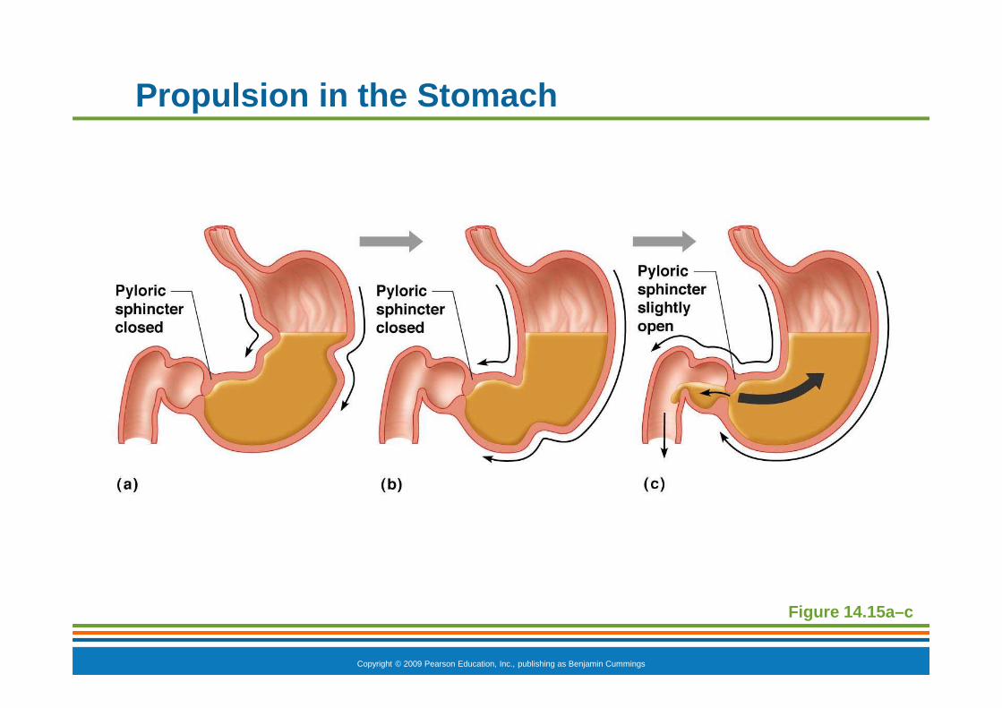
Propulsion in the Stomach
� Food must first be well mixed
� Rippling peristalsis occurs in the lower stomach
� The pylorus meters out chyme into the small intestine (30 mL at a time)
� The stomach empties in 4–6 hours
Copyright © 2009 Pearson Education, Inc., publishing as Benjamin Cummings
Propulsion in the Stomach
Figure 14.15a–c

Copyright © 2009 Pearson Education, Inc., publishing as Benjamin Cummings
Digestion in the Small Intestine
� Enzymes from the brush border function to
� Break double sugars into simple sugars
� Complete some protein digestion

Copyright © 2009 Pearson Education, Inc., publishing as Benjamin Cummings
Digestion in the Small Intestine
� Pancreatic enzymes play the major digestive function
� Help complete digestion of starch (pancreatic amylase)
� Carry out about half of all protein digestion
� Digest fats using lipases from the pancreas
� Digest nucleic acids using nucleases
� Alkaline content neutralizes acidic chyme
Copyright © 2009 Pearson Education, Inc., publishing as Benjamin Cummings
Regulation of Pancreatic Juice Secretion
� Release of pancreatic juice into the duodenum is stimulated by
� Vagus nerve
� Local hormones
� Secretin
� Cholecystokinin (CCK)
� Hormones travel the blood to stimulate the pancreas to release enzyme- and bicarbonate-rich product
Copyright © 2009 Pearson Education, Inc., publishing as Benjamin Cummings
Regulation of Pancreatic Juice Secretion
� Secretin causes the liver to increase bile output
� CCK causes the gallbladder to release stored bile
� Bile is necessary for fat absorption and absorption of fat-soluble vitamins (K, D, A)

Copyright © 2009 Pearson Education, Inc., publishing as Benjamin Cummings
Figure 14.16
Regulation of Pancreatic Juice Secretion
Copyright © 2009 Pearson Education, Inc., publishing as Benjamin Cummings
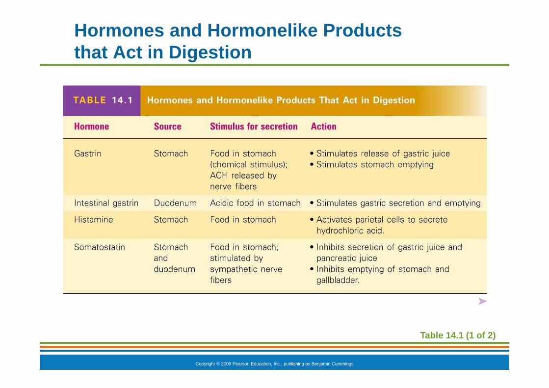
Hormones and Hormonelike Products that Act in Digestion
Table 14.1 (1 of 2)
Copyright © 2009 Pearson Education, Inc., publishing as Benjamin Cummings
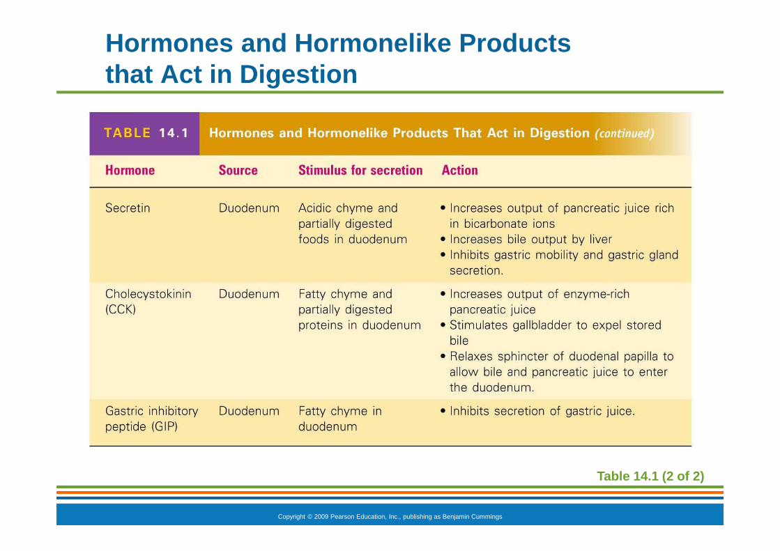
Hormones and Hormonelike Products that Act in Digestion
Table 14.1 (2 of 2)

Copyright © 2009 Pearson Education, Inc., publishing as Benjamin Cummings
Absorption in the Small Intestine
� Water is absorbed along the length of the small intestine
� End products of digestion
� Most substances are absorbed by active transport through cell membranes
� Lipids are absorbed by diffusion
� Substances are transported to the liver by the hepatic portal vein or lymph
Copyright © 2009 Pearson Education, Inc., publishing as Benjamin Cummings
Propulsion in the Small Intestine
� Peristalsis is the major means of moving food
� Segmental movements
� Mix chyme with digestive juices
� Aid in propelling food
Copyright © 2009 Pearson Education, Inc., publishing as Benjamin Cummings
Segmentation
Figure 14.12b
Copyright © 2009 Pearson Education, Inc., publishing as Benjamin Cummings
Food Breakdown and Absorption in the Large Intestine
� No digestive enzymes are produced
� Resident bacteria digest remaining nutrients
� Produce some vitamin K and B
� Release gases
� Water and vitamins K and B are absorbed
� Remaining materials are eliminated via feces

Copyright © 2009 Pearson Education, Inc., publishing as Benjamin Cummings
Food Breakdown and Absorption in the Large Intestine
� Feces contains
� Undigested food residues
� Mucus
� Bacteria
� Water

Copyright © 2009 Pearson Education, Inc., publishing as Benjamin Cummings
Propulsion in the Large Intestine
� Sluggish peristalsis
� Mass movements
� Slow, powerful movements
� Occur three to four times per day
� Presence of feces in the rectum causes a defecation reflex
� Internal anal sphincter is relaxed
� Defecation occurs with relaxation of the voluntary (external) anal sphincter

Copyright © 2009 Pearson Education, Inc., publishing as Benjamin Cummings
Nutrition
� Nutrient—substance used by the body for growth, maintenance, and repair
� Major nutrients
� Carbohydrates
� Lipids
� Proteins
� Water
� Minor nutrients
� Vitamins
� Minerals

Copyright © 2009 Pearson Education, Inc., publishing as Benjamin Cummings
Five Basic Food Groups and Some of Their Major Nutrients
Table 14.2 (1 of 2)

Copyright © 2009 Pearson Education, Inc., publishing as Benjamin Cummings
Five Basic Food Groups and Some of Their Major Nutrients
Table 14.2 (2 of 2)
Copyright © 2009 Pearson Education, Inc., publishing as Benjamin Cummings
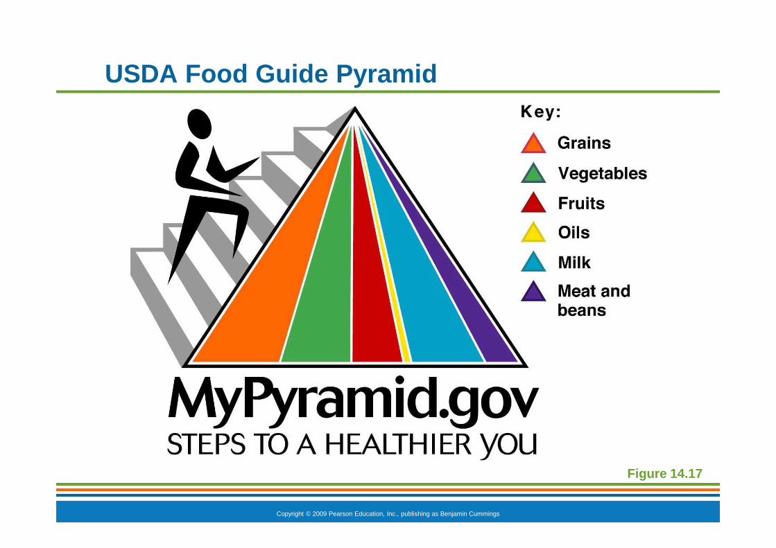
USDA Food Guide Pyramid
Figure 14.17
Copyright © 2009 Pearson Education, Inc., publishing as Benjamin Cummings
Dietary Sources of Major Nutrients
� Carbohydrates
� Most are derived from plants
� Exceptions: lactose from milk and small amounts of glycogens from meats
� Lipids
� Saturated fats from animal products
� Unsaturated fats from nuts, seeds, and vegetable oils
� Cholesterol from egg yolk, meats, and milk products
Copyright © 2009 Pearson Education, Inc., publishing as Benjamin Cummings
Dietary Sources of Major Nutrients
� Proteins
� Complete proteins—contain all essential amino acids
� Most are from animal products
� Legumes and beans also have proteins, but are incomplete
� Vitamins
� Most vitamins are used as coenzymes
� Found in all major food groups

Copyright © 2009 Pearson Education, Inc., publishing as Benjamin Cummings
Dietary Sources of Major Nutrients
� Minerals
� Play many roles in the body
� Most mineral-rich foods are vegetables, legumes, milk, and some meats