chapters 24 & 25 the digestive system, nutrition & body metabolism
TRANSCRIPT
Chapters 24 & 25
The Digestive System, Nutrition & Body Metabolism

The Digestive System & Body MetabolismThe Digestive System & Body Metabolism
Digestion
Breakdown of ingested food
Absorption Passage of nutrients into the blood
Metabolism Production of cellular energy (ATP)
Elimination
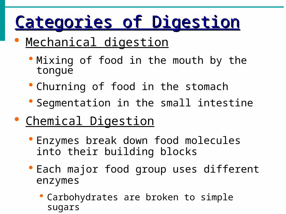
Categories of DigestionCategories of Digestion Mechanical digestion
Mixing of food in the mouth by the tongue
Churning of food in the stomach
Segmentation in the small intestine
Chemical Digestion
Enzymes break down food molecules into their building blocks
Each major food group uses different enzymes
Carbohydrates are broken to simple sugars
Proteins are broken to amino acids
Fats are broken to fatty acids and alcohols
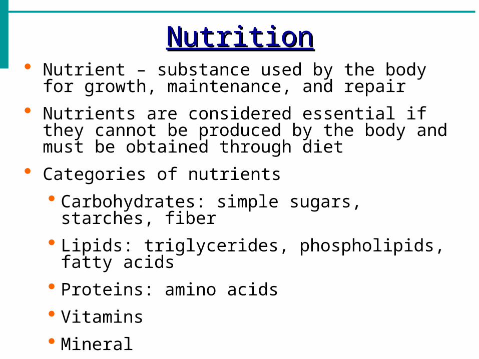
NutritionNutrition Nutrient – substance used by the body for growth,
maintenance, and repair
Nutrients are considered essential if they cannot be produced by the body and must be obtained through diet
Categories of nutrients
Carbohydrates: simple sugars, starches, fiber
Lipids: triglycerides, phospholipids, fatty acids
Proteins: amino acids
Vitamins
Mineral
Water

Organs of the Digestive SystemOrgans of the Digestive System
Two main groups
Alimentary canal: continuous coiled hollow tube; pathway that food takes from mouth to anus
Accessory digestive organs: help the organs of the alimentary canal carry out their digestive role

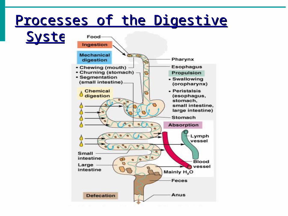
Processes of the Digestive SystemProcesses of the Digestive System Ingestion: getting food into the mouth
Propulsion: moving foods from one region of the digestive system to another
Peristalsis: alternating waves of contraction
Segmentation: moving materials back and forth to aid in mixing
Mechanical Digestion: physically preparing food for breakdown
Chemical Digestion: nutrient molecules are broken down into smaller absorbable building blocks (protein to AA)
Absorption
End products of digestion are absorbed in the blood or lymph
Food must enter mucosal cells and then into blood or lymph capillaries
Defecation
Elimination of indigestible substances as feces
Processes of the Digestive SystemProcesses of the Digestive System
Control of Digestive ActivityControl of Digestive Activity Mostly controlled by reflexes via the
parasympathetic division
Chemical and mechanical receptors are located in organ walls that trigger reflexes
Stimuli include: Stretch of the organ
pH of the contents
Presence of breakdown products
Reflexes include: Activation or inhibition of glandular secretions
Smooth muscle activity
Body Energy BalanceBody Energy Balance
Energy intake = total energy output
(heat + work + energy storage)
Energy intake is liberated during food oxidation
Energy output
Heat is usually about 60%
Storage energy is in the form of fat or glycogen

Organs of the Digestive SystemOrgans of the Digestive System
Layers of Alimentary Canal Organs ILayers of Alimentary Canal Organs I
Mucosa
Innermost layer
Moist membrane
Surface epithelium
Small amount of connective tissue (lamina propria)
Small smooth muscle layer

Layers of Alimentary Canal Organs IILayers of Alimentary Canal Organs II Submucosa
Just beneath the mucosa
Soft connective tissue with blood vessels, nerve endings, and lymphatics
Muscularis externa – smooth muscle
Inner circular layer
Outer longitudinal layer
Serosa
Outermost layer – visceral peritoneum
Layer of serous fluid-producing cells

Layers of Alimentary Canal OrgansLayers of Alimentary Canal Organs
Organs of the Alimentary CanalOrgans of the Alimentary Canal
Mouth
Pharynx
Esophagus
Stomach
Small intestine
Large intestine
AnusDigestion Animation
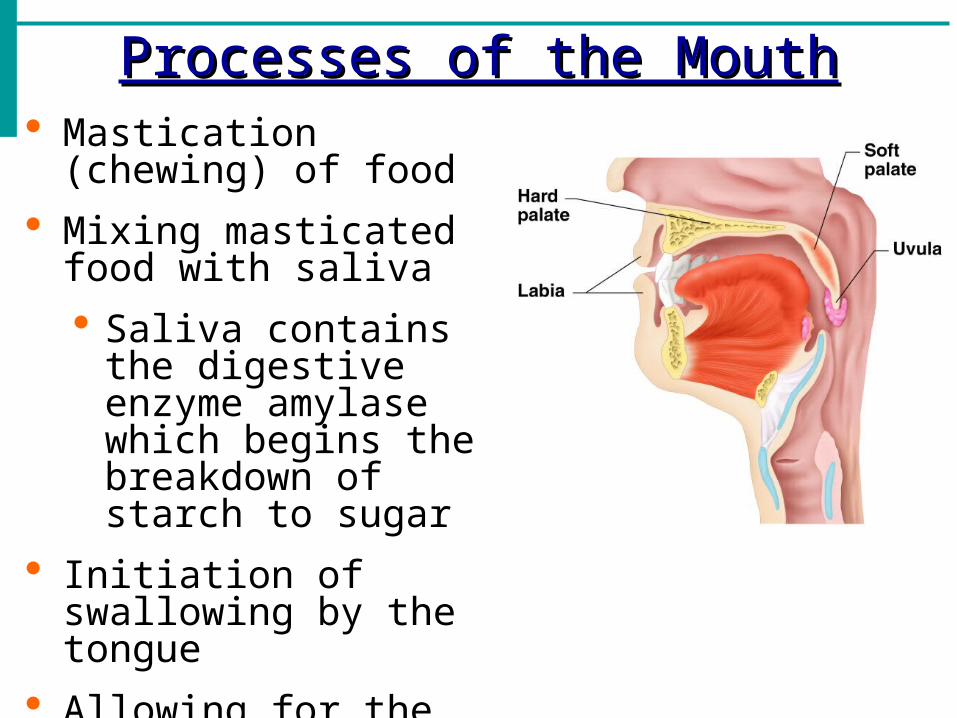
Processes of the MouthProcesses of the Mouth Mastication (chewing) of
food
Mixing masticated food with saliva
Saliva contains the digestive enzyme amylase which begins the breakdown of starch to sugar
Initiation of swallowing by the tongue
Allowing for the sense of taste
Pharynx FunctionPharynx Function
Serves as a passageway for air and food
Food is propelled to the esophagus by two muscle layers Longitudinal outer layer
Circular inner layer
Food movement is by alternating contractions of the muscle layers (peristalsis)

EsophagusEsophagus Runs from pharynx to
stomach through the diaphragm
Conducts food by peristalsis (slow rhythmic squeezing)
Passageway for food only (respiratory system branches off after the pharynx)

Stomach AnatomyStomach Anatomy Located on the left side of the abdominal cavity
Food enters at the cardioesophageal sphincter
Regions of the stomach Cardiac region – near the heart
Fundus
Body
Phylorus – funnel-shaped terminal end
Food empties into the small intestine at the pyloric sphincter

Stomach AnatomyStomach Anatomy Rugae: internal folds of the mucosa
External regions
Lesser curvature
Greater curvature
Layers of peritoneum attached to the stomach
Lesser omentum: attaches the liver to the lesser curvature
Greater omentum: attaches the greater curvature to the posterior body wall
Contains fat to insulate, cushion, and protect abdominal organs
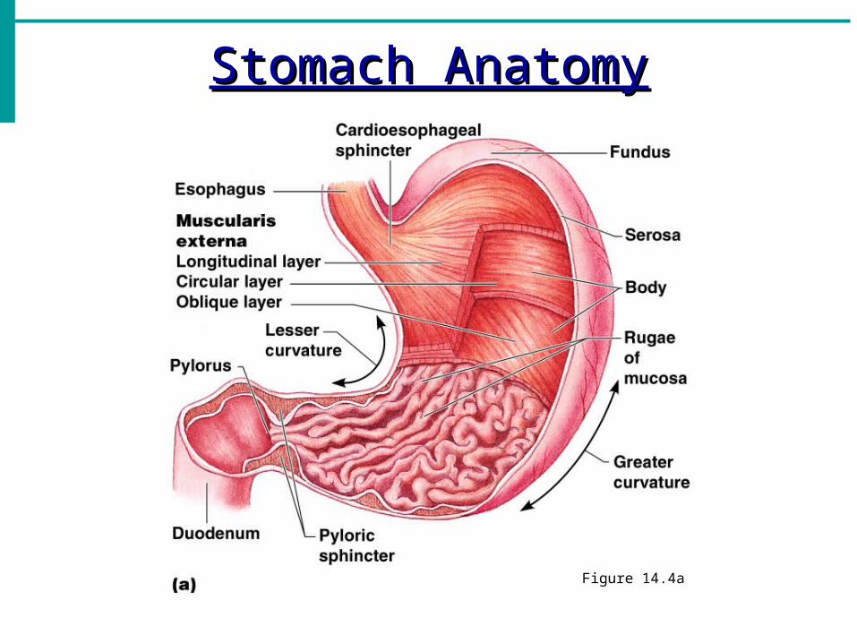
Stomach AnatomyStomach Anatomy
Figure 14.4a
Stomach FunctionsStomach Functions
Acts as a storage tank for food
Site of food breakdown
Chemical breakdown of protein begins
Delivers chyme (processed food) to the small intestine
Specialized Mucosa of the StomachSpecialized Mucosa of the Stomach Simple columnar epithelium, composed entirely of goblet
cells, which produce a protective coat of alkaline mucus
Mucous neck cells: produce a sticky acidic mucus different from that of goblet cells. Exact function unknown.
Chief cells: produce inactive protein-digesting enzymes called pepsinogens
Parietal cells: produce hydrochloric acid that activate and provide optimal conditions for pespin
Endocrine cells: release a variety of hormones/hormone-like products including gastrin, which stimulates HCl secretion
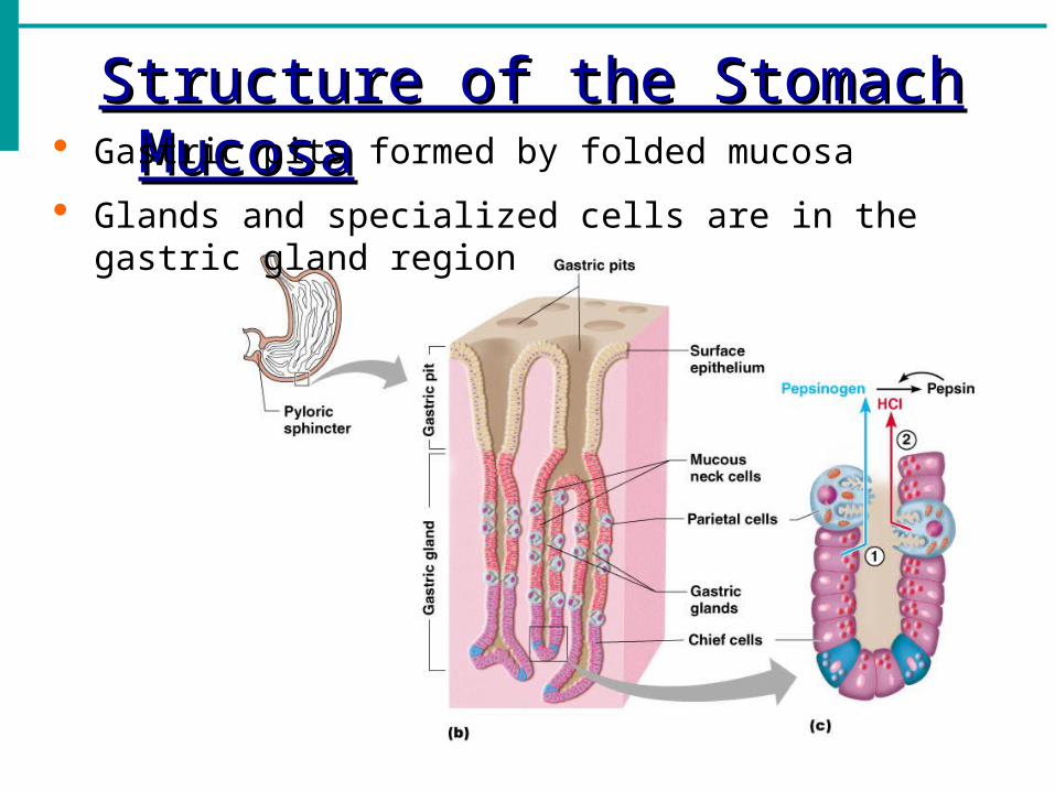
Structure of the Stomach MucosaStructure of the Stomach Mucosa Gastric pits formed by folded mucosa
Glands and specialized cells are in the gastric gland region

Small IntestineSmall Intestine
The body’s major digestive organ
Site of nutrient absorption into the blood
Muscular tube extending form the pyloric sphincter to the ileocecal valve
Suspended from the posterior abdominal wall by the mesentery

Subdivisions of the Small IntestineSubdivisions of the Small Intestine
Duodenum Attached to the stomach
Curves around the head of the pancreas
Jejunum Attaches anteriorly to the
duodenum
Ileum Extends from jejunum to large
intestine
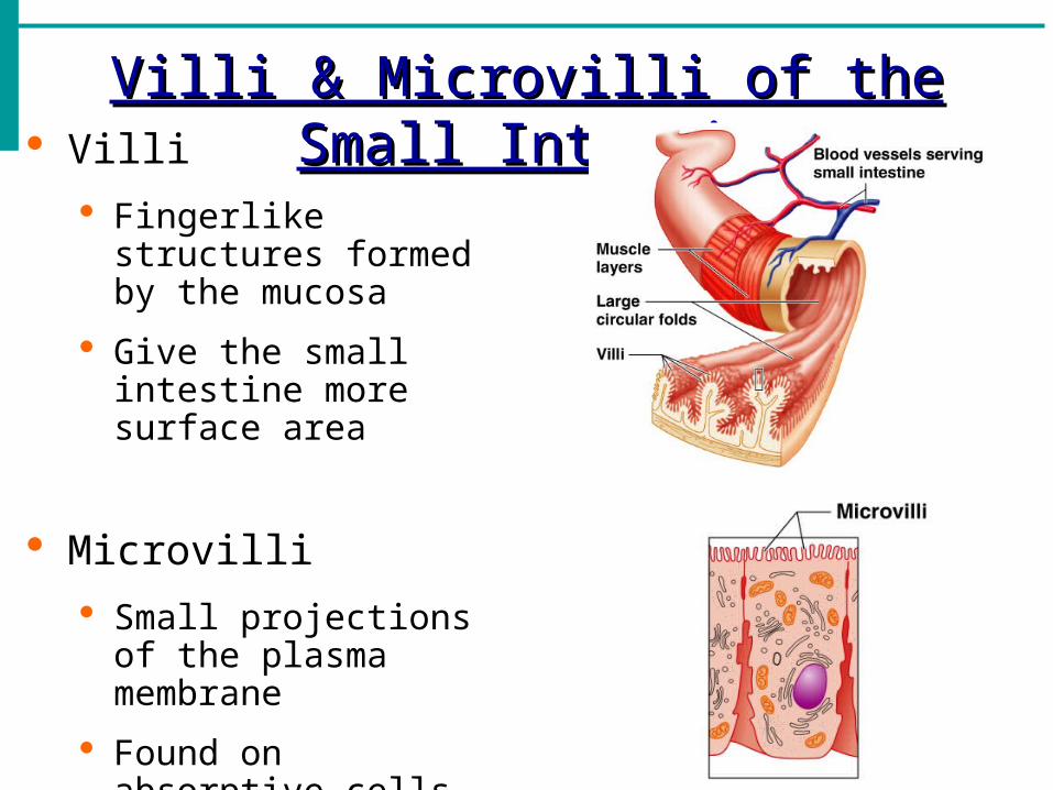
Villi & Microvilli of the Small IntestineVilli & Microvilli of the Small Intestine Villi
Fingerlike structures formed by the mucosa
Give the small intestine more surface area
Microvilli
Small projections of the plasma membrane
Found on absorptive cells

Structures Involved in Absorption of NutrientsStructures Involved in Absorption of Nutrients
Absorptive cells
Blood capillaries
Lacteals (specialized lymphatic capillaries)
Figure 14.7b

Folds of the Small IntestineFolds of the Small Intestine Called circular folds or
plicae circulares
Deep folds of the mucosa and submucosa
Do not disappear when filled with food
The submucosa has Peyer’s patches (collections of lymphatic tissue)

Digestion in the Small Intestine IDigestion in the Small Intestine I Source of enzymes that are mixed with chyme
Intestinal cells
Pancreas
Bile enters from the gall bladder – helps emulsify fats
Digestion in the Small Intestine IIDigestion in the Small Intestine II Enzymes from the brush border
Break double sugars into simple sugars
Complete some protein digestion
Pancreatic enzymes play the major digestive function Help complete digestion of starch (pancreatic
amylase)
Carry out about half of all protein digestion (trypsin, pepsin etc.)
Responsible for fat digestion (lipase)
Digest nucleic acids (nucleases)
Alkaline content neutralizes acidic chyme

Propulsion in the Small IntestinePropulsion in the Small Intestine Peristalsis is the major means of moving food
Segmental movements
Mix chyme with digestive juices
Aid in propelling food
Absorption in the Small IntestineAbsorption in the Small Intestine
Water is absorbed along the length of the small intestine
End products of digestion
Most substances are absorbed by active transport through cell membranes
Lipids are absorbed by diffusion
Substances are transported to the liver by the hepatic portal vein or lymph

Large IntestineLarge Intestine Larger in diameter, but shorter than the small intestine
Frames the internal abdomen
Functions of the Large IntestineFunctions of the Large Intestine
Absorption of water
Eliminates indigestible food from the body as feces
Does not participate in digestion of food
Goblet cells produce mucus to act as a lubricant

Food Breakdown and Absorption in the L. I.Food Breakdown and Absorption in the L. I.
No digestive enzymes are produced
Resident bacteria digest remaining nutrients Produce some vitamin K and B
Release gases
Water and vitamins K and B are absorbed
Remaining materials are eliminated via feces
Structures of the Large IntestineStructures of the Large Intestine
Cecum – saclike first part of the large intestine
Appendix
Accumulation of lymphatic tissue that sometimes becomes inflamed (appendicitis)
Hangs from the cecum
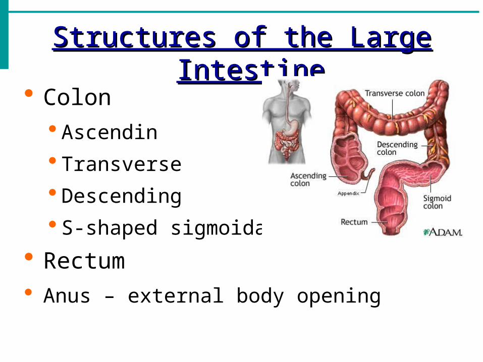
Structures of the Large IntestineStructures of the Large Intestine
Colon Ascendin
Transverse
Descending
S-shaped sigmoidal
Rectum Anus – external body opening

Propulsion in the Large IntestinePropulsion in the Large Intestine Sluggish peristalsis
Mass movements Slow, powerful movements
Occur three to four times per day
Presence of feces in the rectum causes a defecation reflex Internal anal sphincter is relaxed
Defecation occurs with relaxation of the voluntary (external) anal sphincter
Accessory Digestive OrgansAccessory Digestive Organs
Salivary glands
Teeth
Pancreas
Liver
Gall bladder
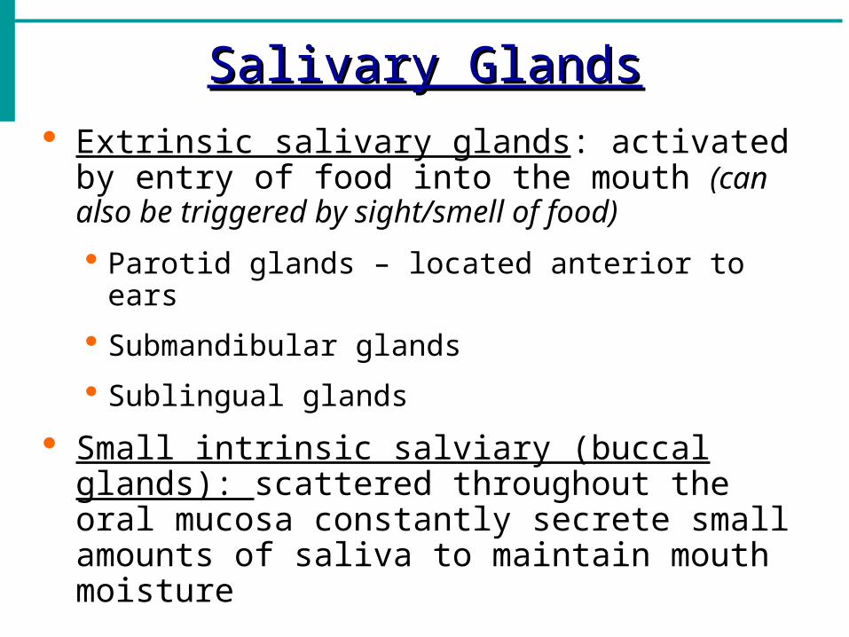
Salivary GlandsSalivary Glands Extrinsic salivary glands: activated by entry of
food into the mouth (can also be triggered by sight/smell of food)
Parotid glands – located anterior to ears
Submandibular glands
Sublingual glands
Small intrinsic salviary (buccal glands): scattered throughout the oral mucosa constantly secrete small amounts of saliva to maintain mouth moisture

SalivaSaliva
Mixture of mucus and serous fluids
Cleanses the mouth
Moistens food and helps compact it to form a food bolus
Contains salivary amylase to begin starch digestion
Dissolves chemicals in food so they can be tasted

TeethTeeth The role is to masticate (chew) food
Humans have two sets of teeth
Deciduous (baby or milk) teeth
20 teeth are fully formed by age two
Permanent teeth
Replace deciduous teeth beginning between the ages of 6 to 12
A full set is 32 teeth, but some people do not have wisdom teeth
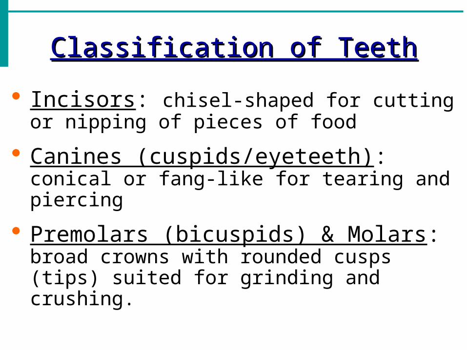
Classification of TeethClassification of Teeth
Incisors: chisel-shaped for cutting or nipping of pieces of food
Canines (cuspids/eyeteeth): conical or fang-like for tearing and piercing
Premolars (bicuspids) & Molars: broad crowns with rounded cusps (tips) suited for grinding and crushing.

Classification of TeethClassification of Teeth
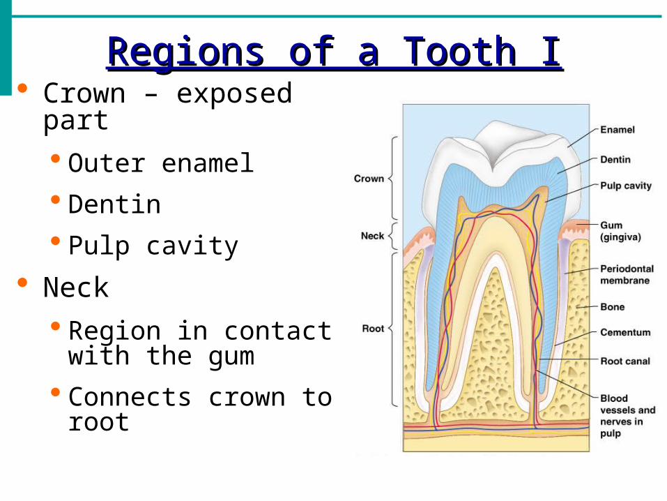
Regions of a Tooth IRegions of a Tooth I Crown – exposed part
Outer enamel
Dentin
Pulp cavity
Neck Region in contact with
the gum
Connects crown to root

Regions of a Tooth IIRegions of a Tooth II
Root
Periodontal membrane attached to the bone
Root canal carrying blood vessels and nerves

PancreasPancreas Produces a wide spectrum of digestive enzymes
that break down all categories of food
Enzymes are secreted into the duodenum
Alkaline fluid introduced with enzymes neutralizes acidic chyme
Acini: clusters of secretory cells around ducts
Endocrine products of pancreas secreted from pancreatics Isles of Langerhans
Insulin – lower blood glucose by conversion to glycogen
Glucagon – raise blood sugar by breakdown of glycogen
LiverLiver
Largest gland in the body
Located on the right side of the body under the diaphragm
Consists of four lobes suspended from the diaphragm and abdominal wall by the falciform ligament
Connected to the gall bladder via the common hepatic duct

Role of the Liver in MetabolismRole of the Liver in Metabolism
Several roles in digestion – primarily to produce bile
Detoxifies drugs and alcohol
Degrades hormones
Produce cholesterol and blood proteins (albumin and clotting proteins)

BileBile Yellow-green alkaline solution produced by cells
in the liver
Emulsifies fats into millions of small fatty droplets to provide large surface area for fat-digesting enzymes (lipase) to work on
Composition Bile salts – primary emulsifying agents
Bile pigment (mostly bilirubin waste product of hemoglobin from breakdown of worn erythrocytes)
Cholesterol
Phospholipids
Electrolytes

Gall BladderGall Bladder Sac found in hollow fossa of liver
Stores bile from the liver by way of the cystic duct
Concentrates bile to 10x that of secretion from liver by absorbing some of its water and ions
Bile is introduced into the duodenum in the presence of fatty food
Gallstones can cause blockages
Crystallization of cholesterol due to overabundance or too little bile salts
Sharp crystals cause pain when gallbladder contracts