the biogenesis and metabolism of cyanogenic … · nutritionally and industrially useful sources of...
TRANSCRIPT

THE BIOGENESIS AND METABOLISM OF CYANOGENIC GLUCOSIDES IN GERMINATING CASSAVA SEED AND SEEDLINGS
F. Nartey*
SUMMARY
Analyses of the storage lipids (47%) and proteins (34%) of cassava seeds indicate that these could be nutritionally and industrially useful sources of vegetable fats and proteins. Seedlings have high lipolytic and proteolytic activities. In seedlings storage lipids are converted to carbohydrates. Valine and Isoleucine are incorporated respectively into the cyanogenic glucosides linamarin and lotaustralin. Seedlings of both bitter and sweet cultivars contain large amounts of cyanogenic glucosides. Electronmicroscope and tracer studies showed that the biosynthesis and metabolism of cyanogens, cyanide, proteins and lipids are associated with specific organelles and microbodies which become apparent after 10 days active seed germination.
RESUME
Les analyses du contenu de lipides (47%) et de proteines reservees (34%) ont montre que les graines de la cassave forment une source de graisses et de proteines vegthales utilisables en industrie. Les germes portent une grande activite de lipolyse et de proteolyse, reforment les lip ides reservees en carbohydrates, et ils peuvent incorporer la valine et I'isoleucine dans les glycosides cyanogenes linamarine et lotaustraline, respectivement. La microscopie electronique et I'utilisation des substrats radioactifs ont montre que la biosynthese et la metabolisme de substances cyanogenes, du cyanure, des proteines et des lip ides sont liees a des organelles specifiques et a des 'microbodies' qui se developpent clairement au cour de 10 jours de germination active de graines.
RESUMEN
Los analisis de las reservas de lipidos (47%) y de proteinas (34%) en semillas de yuca, indican que podrian ser nutricional e industrialmente fuentes (!tiles de grasas vegetales y proteinas. Las plantas tienen actividades lipoliticas y proteoliticas elevadas; en elias las reservas de iipidos son convertidas en carbohi· dratos. La Valina y la Isoleucina se incorporan respectivament a los glucosidos cianogenicos linamarina y if}tfH!f:1:ralina, I as nlantu!as tanto de cultivos 8gri0s COITIO dulces, conti~r'~'n qrandes cantidades de glucosidos cianogenicos. La microscopia electronica y los estudios con trazadores demo~nraron que la biosrntesis y metabolismo de cianogenos, ciaminas proteinas y Irpidos seasodan con organelos y corpusculos especrficos que se hacen aparentes a los 10 dras de la germinacion de la semilla act iva.
INTROOUCTIOt\
The potential, and ultimate usefulness of cassava and all its products are expressions of genetic, biochemical and physiological processes and mechanisms we are investigating in Denmark.
CO NSTITU E NTS 0 F CASSAVA SE ED
Localization of lipids and proteins
The cassava seed kernel contains 47% lipids and 34% protE, s as major reserves, and 0.13% soluble nitrogenous compounds, 0.3% starch and 3.8% soluble carbohydrates as minor constitutents'7. Thus the major constituents of the cassava seed compare favourably with those of typical useful oil seeds such as those of Ricinus, Sesamum, Elaeis and Glycine3 .
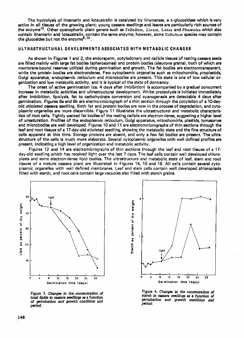
The bulk of cassava seed storage lipids and proteins are localized in the endosperm, although cotyledonary and radicle tissues show a similar distribution of storage materials. Figures 1 and 2 are electronmicrographs of thin sections through the endosperm and radicle tissues of a dry cassava seed. They show
·University of Copenhagen, Institute of Plant Physiology, (b. Farimagsgade 2A, 1353 Copenhagen K. Denmark.
145
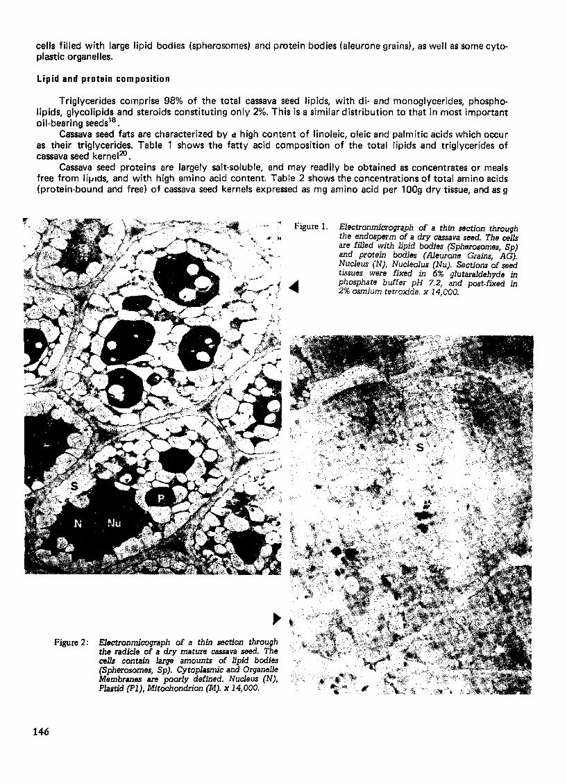
cells filled with large lipid bodies (spherosomes) and protein bodies (aleurone grains), as well as some cytoplastic organelles.
Lipid and protein composition
Triglycerides comprise 98% of the total cassava seed lipids, with di- and monoglycerides, phospholipids, glycolipids and steroids constituting only 2%. This is a similar distribution to that in most important oil-bearing seeds18 •
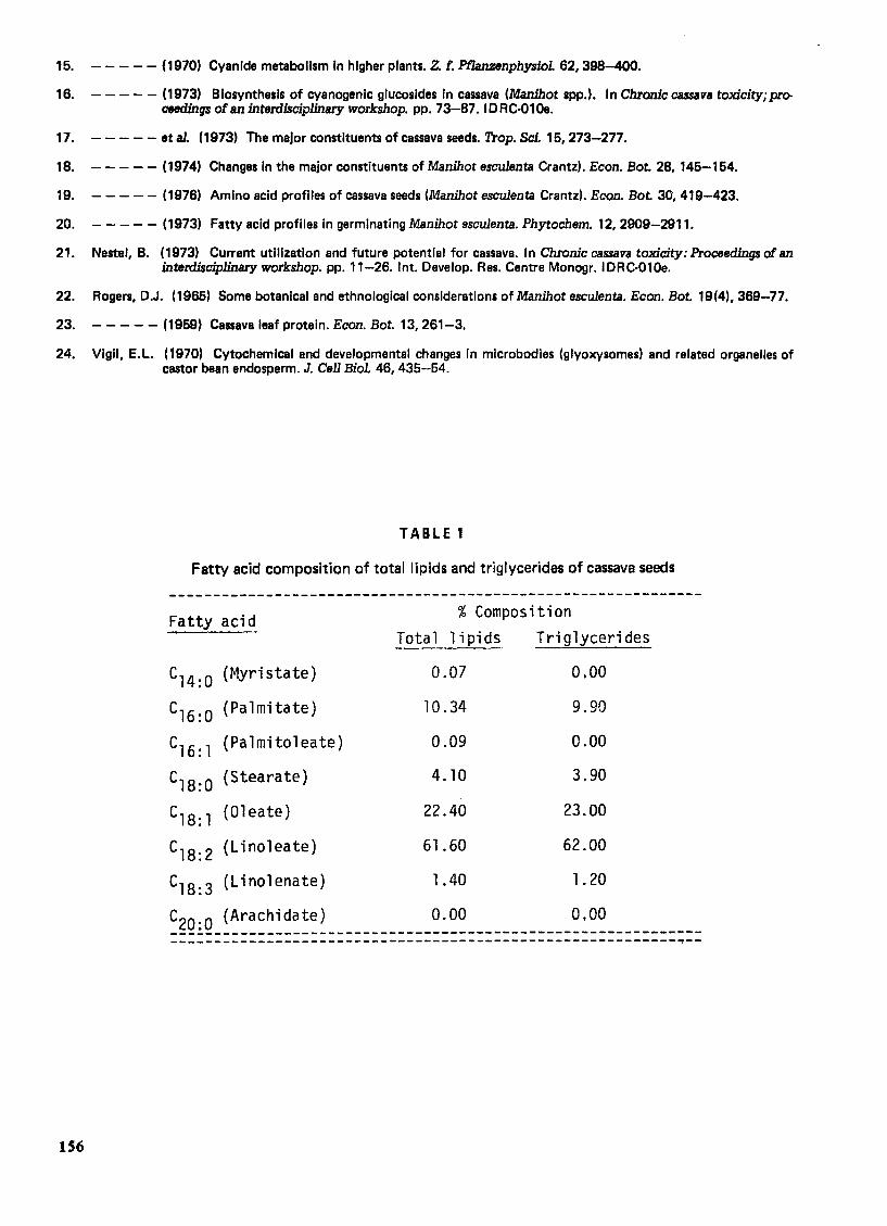
Ca'ssava seed fats are characterized by d high content of linoleic, oleic and palmitic acids which occur as their triglycerides. Table 1 shows the fatty acid composition of the total lipids and triglycerides of cassava seed kernel20.
Cassava seed proteins are largely salt-soluble, and may readily be obtained as concentrates or meals free from lifJldS, and with high amino acid content. Table 2 shows the concentrations of total amino acids (protein-bound and free) of cassava seed kernels expressed as mg amino acid per 100g dry tissue, and as 9
Figure 2:
146
Electronmicograph of a thin section through the radicle of a dry mature cassava seed. The cells contain large amounts of lipid bodies (Spherosomes, Sp). Cytoplasmic and Organelle Membranes are poorly defined. Nucleus (N), Plastid (PI), Mitochondrion (M). x 14,000.
Figure 1. Electronmicrograph of a thin section through the endosperm of a dry cassava seed. The cells are filled with lipid bodies (Spherosomes, Sp) and protein bodies (Aleurone Grains, AG). Nucleus (N), Nucleolus (Nu). Sections of seed tissues were fixed in 6% glutaraldehyde in phosphate buffer pH 7.2, and post-fixed in 2% osmium tetroxide. x 14,000.

amino acid per 16g nitrogen. A comparison of the amino acid profiles of cassava seeds, leaves and tubers19 ,23 shows that seeds contain higher levels of both essential and non-essential amino acids, and therefore better quality proteins. Cassava seed proteins may therefore be of potential nutritional use in cassavagrowing countries.
The low starch content of the cassava seed kernel is accompanied by a relatively high level of soluble sugars almost entirely composed of sucrose.
Cyanogenic glucosides
Generally, variations in the concentration of cyanide in cassava root tubers, as well as the morphological characteristics of the plants, form the basis of a taxonomic differentiation between the bitter (high cyanide content) and the sweet (low cyanide content) cultivars22. This basis for delineation does not offer an adequate means for differentiating all cassava cultivars; it implies that tic;sues of all cassava cultivars are potentially cyanophoric. Seeds of cassava cultivars generally characterized as sweet do not contain cyanogenic glucosides, whereas those of bitter cultivars contain low levels of cyanogenic materials. Table 3 shows the concentrations of cyanogenic glucosides in seeds and other tissues of sweet and bitter cu Itivars of cassava. 16
CHANGES IN THE MAJOR CONSTITUENTS DURING GERMINATION AND GROWTH
Conversion of lipids ti) carbohydrates
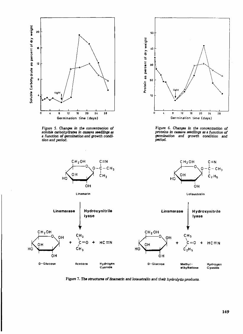
In seeds beginning to germinate in the dark, total cassava seed storage lipids remains largely unchanged. After about 4 days of germination, total lipids decrease gradually, and then rather steeply. Dormant cassava seeds have weak lipase activity which increases several fold during active germination, with the resultant formation of free fatty acids. As free fatty acids accumulate, the activities of enzymes involved with the B-oxidation of the fatty acids also increase several fold. The over-all results are that large amounts of storage lipids are mobilized and converted to non-lipid materials. The rate of lipid degradation is remarkably higher in light-grown seedlings than in etiolated seedlings18 . Figure 3 shows the changes in the concentration of total lipids as a function of growth condition and period. These changes are accompanied by both qualitative and quantitative variations in the composition of fatty acids (Table 4). From Table 4 it can be seen that although etiolated and light-grown seedlings degrade storage lipids at different rates that no specific fatty acids are preferentially metabol ized20 .
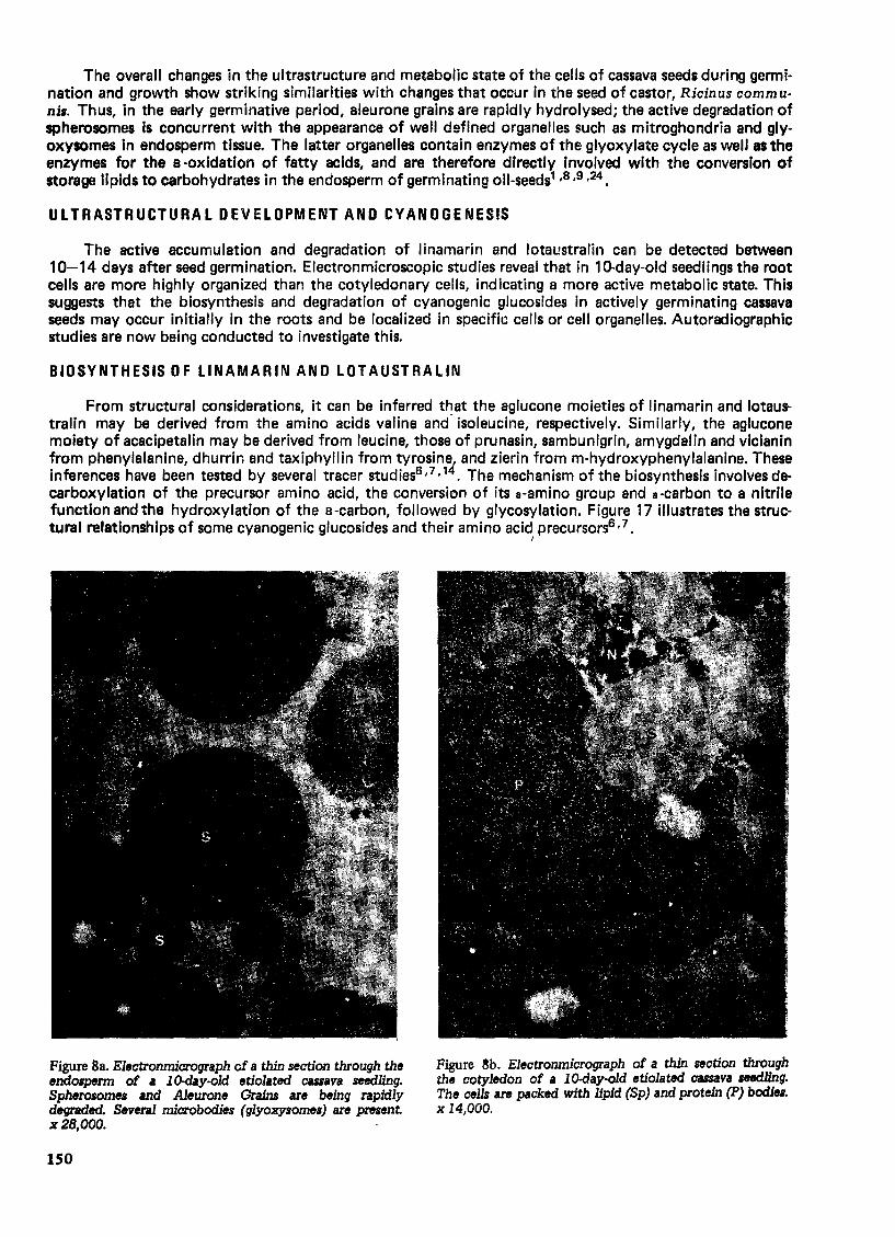
As the lipid concentration falls, the carbohydrate levels undergo striking changes. Figures 4 and 5 show variations in the concentrations of starch and soluble sugars as a function of growth condition and period. The variations in the concentrations of starch and soluble sugars are more pronounced in etiolated seedlings, where sucrose is the major carbohydrate. This provides evidence for the operation of a fat to carbohydrate conversion in cassava through sucrose. A similar mechanism operates in some other oil seeds such as Ricinus through the glyoxylate cycle in which cassava seedlings rapidly convert storage lipids to sucrose, which is readily channelled into starch synthesis1 ,8,9.
Mobilization of amino acids from storage proteins
One of the earliest metabolic events in germinating cassava seeds involves the degradation of storage proteins. Electronmicroscopic studies indicate that reserve proteins disappear at a faster rate than lipids. This is illustrated in Figure 7 which shows variations in the concentration of proteins in dark and lightgrown seedlings. Data in Figure 6 show that imbibed seeds rapidly lose proteins within 8 days of germination. During this period, there is a high increase in the activities of proteolytic enzymes, as shown by experiments with seedling extracts. Except for a transient period, light-grown seedlings generally contain higher levels of proteins.
CYANOGENSIS IN CASSAVA
As shown in Table 3, seeds of sweet cassava cultivars do not contain cyanogenic glucosides, and are therefore non-cyanophoric. On the other hand, seeds of bitter cassava cultivars contain low levels of cyanogenic glucosides, and are therefore cyanophoric. However, 10-day-old seedlings of both bitter and sweet cultivars synthesize and accumulate large amounts of cyanogenic glucosides, namely, linamarin and lotaustralln (methyllinamarin). Linarmarin, 2(B -D-glucopyranosyloxy) isobutryronitrile, accounts for 93%, while lotaustralin, 2(B-D-glucopyranosyloxy) 2-methylbutyronitrile, accounts for 7% of the total cyanogens in cassava. Cyanogenesis in cassava therefore involves enzyme-catalysed hydrolysis of linamarin and lotaustralin with the resultant release of hydrogen cyanide from these glucosides. The structures and hydrolytic products of linamarin and lotaustralin are illustrated in Figure 7.
147
The hydrolysis of linamarin and lotaustralin is catalysed by linamarase, a B-glucosidase which is very active in all tissues of the growing plant; young cassava seedlings and leaves are particularly rich sources of the enzyme13 • Other cyanophoric plant genera such as Trifolium, Linum, Lotus and Phaseolus which also contain linamarin and lotaustralina contain the same enzyme; however, some Trifolium species may contain the glucosides but not the enzyme ,10.
ULTRASTRUCTURAL DEVELOPMENTS ASSOCIATED WITH METABOLIC CHANGES
As shown in Figures 1 and 2, the endorsperm, cotyledonary and radicle tissues of resting cassava seeds are filled mainly with large fat bodies (spherosomes) and protein bodies (aleurone grains), both of which are membrane-bound reserves utilized during germination and growth. The fat bodies are electrontransparent, while the protein bodies are electrondense. Few cytoplasmic organelles such as mitochondria, proplastids, Golgi apparatus, endoplasmic reticulum and microbodies are present. This state is one of low cellular organization and low metabolic activity, and it is typical of the state of dormancy.
The onset of active germination (ca. 4 days after imbibition) is accompanied by a gradual concurrent increase in metabolic activities and ultrastructural development. Whilst proteolysis is initiated immediately after imbibition, lipolysis, fat to carbohydrate conversion and cyanogenesis are detectable 4 days after germination. Figures 8a and 8b are electronmicrograph of a thin section through the cotyledon of a 10-dayold etiolated cassava seedling. Both fat and protein bodies are now in the process of degradation, and cytoplasmic organelles are more discernible. Figure 11 illustrates the ultrastructural and metabolic characteristics of root cells. Tightly packed fat bodies of the resting radicle are electron-dense, suggesting a higher level of unsaturation. Profiles of the endoplasmic reticulum, Golgi apparatus, mitochondria, plastids, lomasomes and microbodies are well developed. Figures 10 and 11 are electronmicrographs of thin sections through the leaf and root tissues of a 17-day-old etiolated seedling, showing the metabolic state and the fine structure of cells apparent at this time. Storage proteins are absent, and only a few fat bodies are present. The ultrastructure of the cells is much more elaborate. Several cytoplasmic organelles with well defined profiles are present, indicating a high level of organization and metabolic activity.
Figures 12 and 14 are electromicrographs of thin sections through the leaf and root tissues of a 17-day-old seedling which has received light over the last 7 days. The leaf cerls contain well developed chloroplasts and some electron-dense lipid bodies. The ultrastructure and metabolic. state of leaf, stem and root tissues of a mature cassava plant are illustrated in Figures 14, 15 and 16. All cells contain several cytoplasmic organelles with well defined membranes. Leaf and stem cells contain well developed chloroplasts filled with starCh, and root cells contain large vacuoles also filled with starch grains.
50 • if '.0. light
C 30 .. u
1/1 It 20
10
148
\. + -.\:'. \ "". 6\ ~.
•
o 4 8 12 16 20 24 28
Germination time (days)
Figure 3. Changes in the concentration of tota11ipidJ in cassava seedlings as a function of germination and growth condition and period.
-c: II U
~ Q.
III .. ~ u ~ .. -If)
2
•
.-. jy--'
o 4 8 12 16 20 24 28
Germination time (days)
Figure 4. Changes in the concentration of starch in cassava seedlings as a function of germination and growth condition and period.
~ 01 'ij 20 ~
-c:
" u ... " a.
16
= 12
o
•
4 8 12 16 20 24 28
Germination time (days)
Figure 5, Changes in the concentration of soluble carbohydrate. in cassava seedlings as a function of germination and growth condition and period.
QCH20HO ?=N
O-y-CH 3
OH CH 3 HO
linamarase
OH
Linamarin
Hydroxynitrile lyase
OH CH 3 \
OH + C=O + HC=N /
HO CH 3
OH
D-Glucose Acetone Hydrogen Cyanide
50
o 8 12 16 20 24 28
Germination time (days)
Figure 6. Change. in the concentration of proteins in cassava seedlings as a function of germination and growth condition and period,
Linamarase
Lotaustralin
H ydroxynitrile lyase
OH CH3 \
OH + C=O + HC=N /
HO C2HS
OH
D-Glucose Methyl- Hydrogen ethylketone Cyanide
Figure 7. The .tructures of linamarin and lotaustralin and their hydrolytic products.
149
The overall changes in the ultrastructure and metabolic state of the cells of cassava seeds during germination and growth show striking similarities with changes that occur in the seed of castor, Ricinus communu. Thus, in the early germinative period, aleurone grains are rapidly hydrolysed; the active degradation of spherosomes is concurrent with the appearance of well defined organelles such as mitroghondria and glyoxysomes in endosperm tissue. The latter organelles contain enzymes of the glyoxylate cycle as well as the enzymes for the B -oxidation of fatty acids, and are therefore directly involved with the conversion of storage lipids to carbohydrates in the endosperm of germinating oil-seeds1 ,8,9,24.
ULTRASTRUCTURAL DEVELOPMENT AND CYANOGENESIS
The active accumulation and degradation of linamarin and lotal!stralin can be detected between 10-14 days after seed germination. Electronmicroscopic studies reveal that in 10-day-old seedlings the root cells are more highly organized than the cotyledonary cells, indicating a more active metabolic state_ This suggests that the biosynthesis and degradation of cyanogenic glucosides in actively germinating cassava seeds may occur initially in the roots and be localized in specific cells or cell organelles. Autoradiographic studies are now being conducted to investigate this.
BIOSYNTHESIS OF LlNAMARIN AND LOTAUSTRALIN
From structural considerations, it can be inferred that the aglucone moieties of linamarin and lotau. tralin may be derived from the amino acids valine and- isoleucine, respectively. Similarly, the aglucone moiety of acacipetalin may be derived from leucine, those of prunasin, sambunigrin, amygdalin and vicianin from phenylalanine, dhurrin and taxiphyllin from tyrosine, and zierin from m-hydroxyphenylalanine. These inferences have been tested by several tracer studies6 , 7 ,14. The mechanism of the biosynthesis involves decarboxylation of the precursor amino acid, the conversion of its a-amino group and a-carbon to a nitrile function and the hydroxylation of the a-carbon, followed by glycosylation. Figure 17 illustrates the structural relationships of some cyanogenic glucosides and their amino acid precursors6 ,7.
Figure 8a. Electronmicrograph cf a thin section through the endo6pm1l of I 10-day-old etiolated CUiava seedling. Spherosomes and Aleurone Grains are being rapidly degraded. Several microbodies (glyoxysomes) are present x2B,OOO.
ISO
/
Figure ~b. Electronmicrograph of a thin section through the cotyledon of a lO-day-old etiolated cassava seedling. The cells are packed with lipid (Sp) and protein (P) bodies. x 14,000.
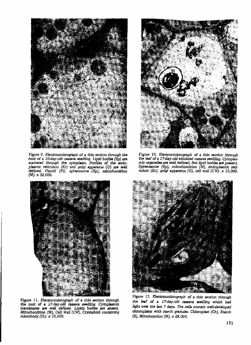
Figure 9. Electronmicrograph of a thin section through the root of a 10-day-old cassava seedling. Lipid bodies (Sp) are scattered through the cytoplasm Profiles of the endoplasmic reticulum (Er) and golgi apparatus (G) are well defined. Plastid (PI), spherosome (Sp), mitochondrion (M). x 32,000.
Figure 11. Electronmicrograph of a thin section through the root of a 17-day-old cassava seedling. Cytop1asrn1c membranes are well defined. Lipids bodies are absent. Mitochondrion (M), Cell Wall (CW), Crystalloid containing microbody (Cr). x 10,000.
Figure 10. Electronmicrograph of a thin section through the leaf of a 17-day-old etiolated cassava seedling. Cytoplasmic organelles are well defined, few lipid bodies are present. Spherosome (Sp), mitochondrion (M), endoplasmic reticulum (Er), golgi apparatus (G), cell wall (CW). x 10,000.
Figure 12. Electronmicrograph of a thin section through the leaf of a 17-day-old cassava seedling which had light over the last 7 days. The cells contain well-developed chloroplasts with starch granules. Chloroplast (Ch), Starch (S), Mitochondion (M). x 28.000.
151

Seeds of both sweet and bitter cassava cultivars contain low levels of free valine and isoleucine14 , 19. However, on germination, the activation of proteolytic enzymes leads to the accumulation of high levels of free amino acids, notably valine and isoleucine. Concurrent with this accumulation, the concentrations of Iinamarin and lotaustralin increase sharply. When uniformly labelled L-valine- 14 C and L-isoleucine- 14 C are administered to seedlings during the period of active cyanogen synthesis (10-14 days after germination), large amounts of radioactivity are incorporated in the aglucone moieties of linamarin and lotaustralin, respectively 13,14. Table 5 illustrates the incorporation of radioactive valine and isoleucine into the aglucone moieties of cassava cyanogens, but also into asparagine; the latter is significant with respect to the mechanism for cyanide detoxification and metabolism in cassava.
The biosynthesis and accumulatfon of cassava cyanogens are influenced by such factors as protein degradation and synthesis, and photosynthesis 14. Other factors such as the activities of the enzymes involved in the biosynthesis of cyanogens, the concentrations and availability of substrates, as well as the rates of transiJort, storage and degradation of cyanogens in specific tissues, influence variations in the concentrations of cyanogenic glucosides in tissues of different cultivars21 .
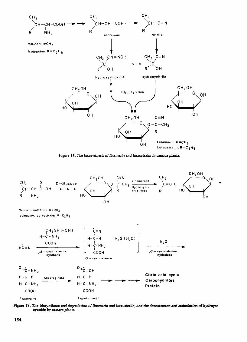
The concentrations of cyanogens in cassava plants generally fluctuate, indicating enzyme cetalysed degradation and resynthesis, and therefore, the dynamic nature of these compounds. The biosynthesis of Iinamarin and lotaustral in is illustrated in Figure 18. Th is pathway is operative in a variety of cyanophoric plant species which accumulate linamarin and lotaustralin during germination and growth7 •
The concentration of cyanogenic glucosides in cassava plants fluctuates with growth period and condition. If cassava is grown in a closed container, decreases in cyanogen content do not lead to the release of HCN into the surrounding air. Furthermore, cassava plants kept in closed systems containing high levels of HCN appear to grow normally, in spite of the fact that cyanide normally inhibits plant respiration. Investigations on the fate of HCN released intracellu!arly from linamarin and lotaustralin show that cassava plants possess two enzyme catalysed systems for the detoxication of cyanide.
DETOXIFICATION AND METABOLISM OF HeN
Cassava seedlin~s metabolize 14C-labelled HCN, CO2 and acetate equally efficiently. However, the labelling patterns found in metabolites from seedlings fed these compounds are different. Whereas small frac-
Figure 13. Electronmicrograph of a thin section through the root of a 17-day-old cassava seedling which had received light over the last 7 days. Cytoplasmic membranes are well defined. Lipid bodies are absent. x 32,000.
152
Figure 14. Electronmicrograph of a thin section through the leaf of a 5-month-old cassava plant. The qelh contain wen developed chloroplasts and other cytoplasmic organenes. x 5,000.

Figure 15. Electronmicrograph of a thin section through the stem tissue of a 5-month-old cassava plant, showing the presence of a chloroplast containing starch, mitochondrion and other cytoplasmic organelles. x 10,000.
Figure 16. Elecuonmicrograph of a thin section through the root of a 5-month-old CUBa va plant. Many ce1J.s are vacuolated. Several lomuomes and other cytoplasmic organelles are present. x 5,000.
L - Valine Llnamarln L-Isoleucine Lotaustralln
--L - LeUCine 01 hydroacacipeta lin Acacipetalin "Proacaclpetalin
H COOH H
o-~ t-t-NH2 -+o-~ t-C=N - I I - I
H H 0
H eOOH H
HO-Q-" t-t-NH2 --+ HO-Q-~ t-C::N - I I - I
H H ° I I C6 H" 05 C6 Hll 0 s
L - Phenylalanine Prunasin L-Tyroslne Dhurdn
Figure 17. The structural relationships between some cyanogenic glucosides and their preCW'Bor amino acids.
153
C H3 "-
CH 3 "-CH-CH-COOH ---. ---+ CH-CH=NOH --I ..... / I
R NH2 /
R
Valine: R =CH 3
Isoleucine: R = C 2 H 5
OH
Aldoxime
~ CH3 CN=NOH "'-'I
C ---R/ 'OH
Nitrile
~ Hydroxyaldoxime Hydroxynitrile
Glycosylation
OH
C=N I
O-C- CH 3 I R
OH
Linamarln: R=CH 3
Lotaustralin: R=C2HS
Figure 1~. The biosynthesis of linamarin and lotaUSItralin in cassava phn~
CH 3 0 \ II
D-Glucose
CH-CH-C-OH ---/ I R NH2
Valine, Linamarin: R=CH3
Isoleucine, lotaustralln: R=C2H S
CHzSH!-OH) I
H-C-NH2 I COOH
t"3 - cyanoalanine synthase
o • ':::'C-NH 2 I
H -C - H Asparaginase I ~
H-C-NHZ I COOH
Asparagine
CH 20H C=N
8' O,\O-~-CH3 OH R
HO
OH
I * C=N I
H-C-H
H-t-NH L I 2
COOH ;3 - cyanoalanine
o * ~C-OH I
H-C-H
CH 3 \ -----I.... C= 0 +
llnamarase
Hydroxynltrile lyase
/ R
j3 - cyanoalanine hydrolase
I --ta-- --ta-- --.......
Citric acid cycle
Carbohydrates
Protein H-C-NH2 I COOH
Aspartic acid
Figure 19. The biosynthem and degradation of linamarin and lotaUSItralin, and the detoxication and assimilation of hydrogen cyanide by ca.rsava plants.
154
+

tlons of radioactivity from 14C02 and 14C-acetate are found in the free amino acid pools, large amounts of radioactivity from H14 CN are incorporated in the free amino acid pools. A most striking feature of the labelling patterns is that although 49% of the total radioactivity from H14 CN is located in the free amino acid fraction of seedling extracts, over 95% of the total radioactivity in this fraction is located in asparagine, aspartic acid, glutamine and glutamic acid. Table 6 shows that asparagine is the major labelled product of cyanide metabolism in cassava. Furthermore, wetfover 97% of the total radioactivity in asparagine is located in its amide-carbon 14. This mechanism for cyanide detoxification operates in a variety of cyanophorlc plant speclesl, 16, and involves the enzyme ,catalyzed reaction of cyanide with serine or cysteine, with the resultant formation of B-cyanoalanine' as the primary reaction product"], 14, 1'.
Quantitative data from in vivo and in vitro studies on cyanide metabolism by cassava seedlings and seedling extracts show that the natural acceptor of cyanide in cassava is serine 14. B -cyanoaline is not accumulated in cassava tissues, which contain highly active B-cyanoalanine synthase, B-cyanoalanine hydrolase and asparaginase 16. The collective operation of these enzyme systems ensures that HCN evolved from cassava cyanogens by the action of linamarase and 2-hydroxynitrile lyase (or by non-enzyme catalysed dissociation of the hydroxynitrile moieties) is rapidly converted to amino acids, proteins, carbohydrates, lipids and other cellular materials. Figure 19 illustrates the detoxification and assimilation of hydrogen cyanide by cassava plants 14.
Cassava plants possess also rhodanase activity. Rhodanase catalyses the reaction of cyanide with inorganic and organic sulphur compounds with the resultant formation of thiocyanate4,5, 16. Cassava rhodanase activity is inhibited by cysteine, while B-cyanoalanine synthase activity is inhibited by thiosulphate. Thus only one cyanide detoxification system in cassava may operate at any particular time. Both enzyme systems are localized in the metochondria, and their functions may well be the reduction of high intracellular concentrations of cyanide for the preservation of electron transport and oxidative phosphorylation in cassava plants16 .
ACKNOWL EDG EME NTS
This investigation was supported by DAN I DA, The Danish Foreign Ministry. The Danish Natural Science Research Council provided travel funds to the author.
REFERENCES
1. Beevers, H. (1961) Metabolic production ofsucrose from fat. Nature 191,384-6.
2. Blumenthal-Goldschmidt, S. et al. (1965) Incorporation of hydrocyanic acid labelled with carbon-14 into asparagine in seedlings. Phytochem. 4, 127-31.
3. Butt, V.S. et al. (1966) The plant lipids. In. Plant Physiology, a treatise. (Ed. F.C. Steward) 1VB, 226-414. Academic Press, New York.
4. Chew, M.Y. et al. (1972) Rhodanase in tapioca leaf. Phytochem. 11,127-31.
6. - - - - - (1973) Rhodanase in higher plantsPhytochem. 12,2365-7.
6. Conn, E.E. (1973) Cyanogenic glycosides: their occurrence, biosynthesis, and function. In Chronic cassava toxicity: proceeding of an interdisciplinary workshop. pp. 55-63. Int. Develop. Res. Centre Monogr. IORC-OlOe.
7. Conn, E.E. et a1 (1969) The biosynthesis of cyanogenic glycosides and other simple nitrogen compounds. In Perspectives in Phytochemistry. p. 47-74. J.B. Harbourne and T. Swain (Ed). Academic Press, London.
8. Cooper, T.G. et al. (1969) Mitochondria and glyoxysomes from cartor bean endosperm. J. Biol. Chern. 244,3507-13.
9. - - - - - (1969) Oxidation in glyoxysomes from castor bean endosperm. J. BioL Chem. 244,3514-20.
10. Corkill, L (1940) Cyanogenesis in white clover (Trifolium repens L) 1. Cyanogenesis in single plants. N.Z.J. Sci. 22B, 66-7.
11. Fowden, Let a1 (1965) Cyanide metabolism by seedlings. Nature 206,110-12.
12. Hitchcock, C. et al. (1971) Plant Lipid Biochemistry. Academic Press, London.
13. Nartey, F. (1968) Studies on cassava, Manihot utilissima Pohl. 1. Cyanogenesis: the biosynthesis of linamarin and lotaustralin in etiolated seedlings. Phytochem. 7,1307-12.
14. Nartey, F. (1969) Studies on cassava, Manihot utilissima. 11. Biosynthesis of asparagine _1 4 C from '4C~labelied hydrogen cyanide and its relations with cyanogenesis. PhysioL Plant 22, 1085-96.
ISS
15. - - - - - (1970) Cyanide metabolism in higher plants. Z. f. Pi1anzenphysioL 62,398-400.
16. - - - - - (1973) Biosynthesis of cyanogenic glucosides in cassava (Manihot spp.). In Chronic cassava toxicity; pro-ceedings of an interdisciplinary workshop. pp. 73-87. IORC·Ol0e.
17. - - - - - et al. (1973) The major constituents of cassava seeds. Trop. Sci 15, 273-217.
18. - - - - - (1974) Changes in the major constituents of Manihot esculenta Crantz). Econ. Bot. 28, 145-154.
19. - - - - - (1976) Amino acid profiles of cassava seeds (Manihot esculenta Crantz). Econ. Bot. 30,419-423.
20. - - - - - (1973) Fatty acid profiles in germinating Manihot esculenta. Phytochem. 12, 2909-2911.
21. Nestel, B. (1973) Current utilization and future potential for cassava. In Chronic cassava toxicity: Proceedings of an interdisciplinary workshop. pp. 11-26. Int. Develop. Res. Centre Monogr. IORC·Ol0e.
22. Rogers, O.J. (1965) Some botanical and ethnological considerations of Manihot esculenta. Econ. Bot. 19(4),369-71.
23. - - - - - (1959) Cassava leaf protein. Econ. Bot. 13,261-3.
24. Vigil, E.l. (1970) Cytochemical and developmental changes In microbodies (glyoxysomes) and related organelles of castor bean endosperm. J. Cell BioL 46,435-54.
TABLE 1
Fatty acid composition of total lipids and triglycerides of cassava seeds
Fatty acid % Composition Total 1 i pi ds Trig1ycerides --
C14 :0 (Myristate) 0.07 0.00
C16 :0 (Palmitate) 10.34 9.90
C16 : 1 (Palmitoleate) 0.09 0.00
C18:0 (Stearate) 4.10 3,90
C18: 1 (Oleate) 22.40 23.00
C18:2 (Linoleate) 61.60 62.00
C18 :3 (Linolenate) 1.40 1. 20
C20 :0 (Arach;date) 0.00 0,00 -------------------------------------------------------------
156
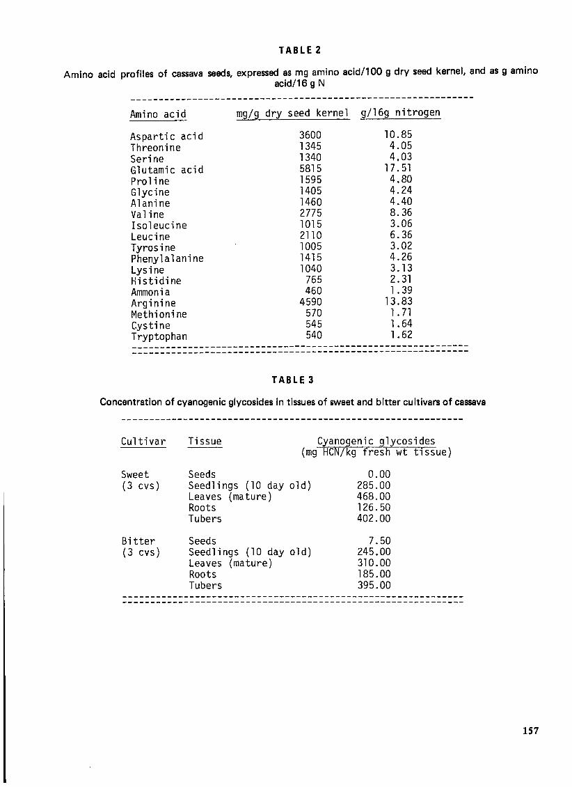
TABLE 2
Amino acid profiles of cassava seeds, expressed as mg amino acid/100 9 dry seed kernel, and as 9 amino acid/16 9 N
-------------------------------------------------------------Amino acid
Aspartic acid Threonine Serine Glutamic acid Proline Glycine Alanine Valine Isoleucine Leucine Tyrosine Phenylalanine Lysine Histidine Ammonia Arginine Methionine Cystine Tryptophan
mg/g dry seed kernel
3600 1345 1340 5815 1595 1405 1460 2775 1015 2110 1005 1415 1040
765 460
4590 570 545 540
g/16g nitrogen
10.85 4.05 4,03
17.51 4.80 4.24 4.40 8.36 3.06 6.36 3.02 4.26 3.13 2.31 1. 39
13.83 1. 71 1.64 1.62
------------------------------------------------------------------------------------------------------------------------
TAB L E 3
Concentration of cyanogenic glycosides in tissues of sweet and bitter cultivars of cassava
Cultivar
Sweet (3 cvs)
Bitter (3 cvs)
Tissue Cyanogenic glycosides (mg HCN/kg fresh wt tissue)
Seeds Seedlings (10 day old) Leaves (mature) Roots Tubers
Seeds Seedlings (10 day old) Leaves (mature) Roots Tubers
0.00 285.00 468.00 126.50 402.00
7.50 245.00 310.00 185.00 395.00
--------------------------------------------------------------------------------------------------------------------------
157

TABLE 4
Net changes in the fatty acid composition of cassava seedlings as a function of growth condition and period
--------------------------------------------------------------------% comeosition
Age (days) 3 9 13 13 17 17 Dark Dark Dark Light Dark Light
Fatty acid (49.2) (43.B) (41.0) (35.7) (37.2) (2B.5)
C14 :0 (Myristate) 0.04 0.10 trace 0.00 0.00 trace
C16 :0 (Palmitate) 10.20 11.60 11.BO 10.20 11.00 12.50
C16 : 1 (Palmitoleate) 0.05 0.11 trace trace trace trace
C1B .0 (Stearate) 4.10 5.10 4.60 3.40 4.10 4.60
C1B : 1 (Oleate) 21.BO 22.70 23.00 21.60 21.60 22.60
C1B :2 (Linoleate) 62.20 59.20 5B.70 63.30 60.20 5B.70
C1B :3 (Linolenate) 1.60 1.30 2.00 1.60 3.00 1.60
C20 .0 (Arachidate) 0.11 trace 0.00 0.00 0.00 0.00
----------------------------------------------------------------------------------------------------------------------------------------TABLE5
Incorporation of radioactivity from L-Valine'4 -C(U) and L-lsoleucine'4C (U) into the aglucone moieties of Linamarin and Lotaustralin, and into Asparagine, by cassava seedlings
Label administered
L-Valine14-C(U)
L-Isoleucine14-C(U)
Incorporation (%) into Linamarin, Lotaustrali~,Asparagin~
13.20
2.40
1.10
0.53
-----------------------------------------------------------TABLE 6
Incorporation of radioactivity from H'4CN into free amino acids by 10g cassava seedlings exposed to H'4CN released from 2.3 mole Na'4CN, 125 Ci. 8.38 x 10⁶ cpm. in a closed system for the periods specified
Period of Radioactivity (cpm x 10-5) incorporated in feeding(min) Asparagine Aspartic Glutamine Glutamic
-ac.,-o- acid
10 0.B6 O. 11 0.16 0.07
60 24.91 5.73 0.B9 O.lB
lBO 42.03 6.6B 0.65 0.43
158