the 5! untranslated region of rhopalosiphum padi virus contains an internal ribosome entry
TRANSCRIPT

JOURNAL OF VIROLOGY,0022-538X/01/$04.00�0 DOI: 10.1128/JVI.75.21.10244–10249.2001
Nov. 2001, p. 10244–10249 Vol. 75, No. 21
Copyright © 2001, American Society for Microbiology. All Rights Reserved.
The 5� Untranslated Region of Rhopalosiphum padi VirusContains an Internal Ribosome Entry Site Which Functions
Efficiently in Mammalian, Plant, and Insect Translation SystemsKATHRYN E. WOOLAWAY,1 KONSTANTINOS LAZARIDIS,1† GRAHAM J. BELSHAM,2
MICHAEL J. CARTER,1 AND LISA O. ROBERTS1*
School of Biomedical and Life Sciences, University of Surrey, Guildford GU2 7XH,1 andInstitute for Animal Health, Pirbright, Woking, Surrey GU24 ONF,2 United Kingdom
Received 18 August 2000/Accepted 31 July 2001
Rhopalosiphum padi virus (RhPV) is one of several picorna-like viruses that infect insects; sequence analysishas revealed distinct differences between these agents and mammalian picornaviruses. RhPV has a single-stranded positive-sense RNA genome of about 10 kb; unlike the genomes of Picornaviridae, however, thisgenome contains two long open reading frames (ORFs). ORF1 encodes the virus nonstructural proteins, whilethe downstream ORF, ORF2, specifies the structural proteins. Both ORFs are preceded by long untranslatedregions (UTRs). The intergenic UTR is known to contain an internal ribosome entry site (IRES) which directsnon-AUG-initiated translation of ORF2. We have examined the 5� UTR of RhPV for IRES activity by trans-lating synthetic dicistronic mRNAs containing this sequence in a variety of systems. We now report that the 5�UTR contains an element which directs internal initiation of protein synthesis from an AUG codon inmammalian, plant, and Drosophila in vitro translation systems. In contrast, the encephalomyocarditis virusIRES functions only in the mammalian system. The RhPV 5� IRES element has features in common withpicornavirus IRES elements, in that no coding sequence is required for IRES function, but also with cellularIRES elements, as deletion analysis indicates that this IRES element does not have sharply defined boundaries.
Rhopalosiphum padi virus (RhPV) is an insect virus with anarrow host range, infecting aphids of the Rhopalosiphum andSchizaphis families (6, 11). Virus infection reduces both the lifespan and the reproductive capacity of the insects (6). RhPVwas initially classified within the Picornaviridae, based largelyon its physicochemical properties. However, sequence analysishas prompted a reevaluation of this attribution, and RhPV isnow considered to belong to a group of insect viruses (thecricket paralysis-like viruses) with a picornavirus-like capsidstructure but a distinct genome organization (15, 16). Othermembers of this group include Drosophila C virus, Plautia staliintestinal virus (PSIV), and cricket paralysis virus (CrPV) it-self. The RNA genome of each of these viruses, includingRhPV, encodes two polyproteins in separate open readingframes (ORFs) (15). ORF1 encodes nonstructural proteinsthat possess sequence similarity to both mammalian picorna-virus and plant comovirus proteins. ORF2 encodes the threestructural proteins that also show similarity to picornaviruscapsid proteins. Both ORFs are preceded by long untranslatedregions (UTRs) about 500 nucleotides (nt) long. In contrast,mammalian picornaviruses encode one long polyprotein withthe structural proteins at the N-terminal region and the non-structural proteins at the C terminus. The genome organiza-tion of the cricket paralysis-like viruses resembles that of thecaliciviruses (23). However, there is no evidence for the pro-
duction of a subgenomic RNA by these insect viruses, andcalicivirus ORFs are not preceded by long UTRs.
It is well established that the initiation of translation onpicornavirus RNA occurs by a cap-independent mechanismthat is directed by an internal ribosome entry site (IRES)element within the 5� UTR of the genome (reviewed in refer-ences 2 and 3). Picornavirus IRES elements are grouped intotwo major classes according to their predicted secondary struc-ture and their activity in vitro; there is little sequence identitybetween the two classes (reviewed in references 2 and 14). Oneclass contains IRES elements from the enteroviruses and rhi-noviruses, while the second class contains the cardiovirus andaphthovirus IRES elements. The latter IRES elements func-tion efficiently in the rabbit reticulocyte lysate (RRL) transla-tion system. In contrast, the poliovirus and rhinovirus IRESelements are inefficient in this system until the reaction issupplemented with HeLa cell extracts (5, 8). The hepatitis Avirus IRES is distinct from those listed above and forms aminor class on its own; it can function in the RRL system, butits activity is stimulated in this system by liver cell and notHeLa cell extracts (12). These findings highlight the impor-tance of cellular trans-acting factors in the mechanism of IRESaction and could provide some explanation for the cellulartropism of picornaviruses. Indeed, it has been demonstratedthat the intracellular activities of different picornavirus IRESelements vary in different cell types (4, 18).
The intergenic regions (IGRs) of both PSIV and RhPV haverecently been shown to contain IRES elements. These directthe translation of the second ORFs, and the initiation of trans-lation on both virus IGRs occurs at non-AUG start codons: onPSIV RNA, a CAA codon is used (21), while a CCU codon isprobably used on RhPV RNA (7). Both the IGR and the 5�
* Corresponding author. Mailing address: School of Biomedical andLife Sciences, University of Surrey, Guildford GU2 7XH, UnitedKingdom. Phone: (44) 1483 686499. Fax: (44) 1483 300374. E-mail: [email protected].
† Present address: School of Biological Sciences, University of EastAnglia, Norwich NR4 7TJ, United Kingdom.
10244
Dow
nloa
ded
from
http
s://j
ourn
als.
asm
.org
/jour
nal/j
vi o
n 12
Oct
ober
202
1 by
37.
115.
86.1
97.
UTR of CrPV have very recently been shown to contain IRESelements (25). In common with the situation for the relatedPSIV and RhPV, the initiation of CrPV ORF2 takes place ata non-AUG codon (CCU) (25). Both the 5� UTR and the IGRsequences of CrPV were reported to function as IRES ele-ments in insect cells and in the RRL in vitro translation system.Furthermore, the IGR IRES but not the 5� UTR IRES ofCrPV functioned in the wheat germ in vitro translation system(25).
The 5� UTR of RhPV has many features in common with the5� ends of mammalian picornavirus RNAs. The RhPV 5� ter-minus is uncapped and is predicted to form extensive second-ary structure (15). The 5� UTR of RhPV is predicted to be 580nt long and is highly A and U rich. It has been suggested, butnot proven, that the initiation of protein synthesis occurs at thethird AUG (15). The RhPV 5� UTR contains two AUG codonsupstream of ORF1 but out of frame with the coding sequenceof this gene; both are followed quickly by termination codonsand are therefore unlikely to be used. However, there is an-other AUG codon, 6 nt downstream of the proposed ORF1initiation site, which could also function as an initiation codon.Many of these features are also found in picornavirus IRESelements and prompted us to examine the 5� UTR of RhPVfor the presence of an IRES element which could direct thecap-independent initiation of translation of ORF1. To date,most IRES elements isolated from virus or mammalian mR-NAs have been shown to be functional only in mammalian cellsand translation systems derived from them. Here, we reportthat the 5� UTR of RhPV contains an IRES element whichfunctions in the RRL, wheat germ, and Drosophila in vitrotranslation systems.
MATERIALS AND METHODS
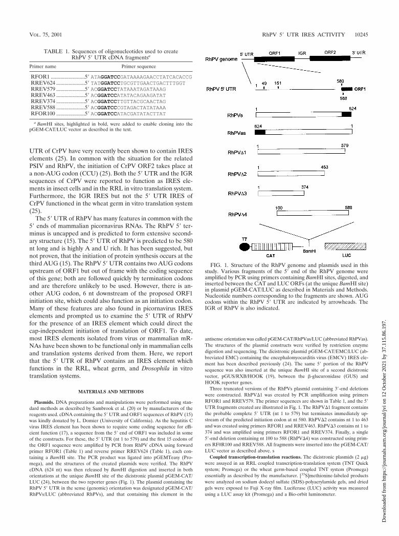
Plasmids. DNA preparations and manipulations were performed using stan-dard methods as described by Sambrook et al. (20) or by manufacturers of thereagents used. cDNA containing the 5� UTR and ORF1 sequences of RhPV (15)was kindly donated by L. Domier (University of California). As the hepatitis Cvirus IRES element has been shown to require some coding sequence for effi-cient function (17), a sequence from the 5� end of ORF1 was included in someof the constructs. For these, the 5� UTR (nt 1 to 579) and the first 15 codons ofthe ORF1 sequence were amplified by PCR from RhPV cDNA using forwardprimer RFOR1 (Table 1) and reverse primer RREV624 (Table 1), each con-taining a BamHI site. The PCR product was ligated into pGEMTeasy (Pro-mega), and the structures of the created plasmids were verified. The RhPVcDNA (624 nt) was then released by BamHI digestion and inserted in bothorientations at the unique BamHI site of the dicistronic plasmid pGEM-CAT/LUC (24), between the two reporter genes (Fig. 1). The plasmid containing theRhPV 5� UTR in the sense (genomic) orientation was designated pGEM-CAT/RhPVs/LUC (abbreviated RhPVs), and that containing this element in the
antisense orientation was called pGEM-CAT/RhPVas/LUC (abbreviated RhPVas).The structures of the plasmid constructs were verified by restriction enzymedigestion and sequencing. The dicistronic plasmid pGEM-CAT/EMC/LUC (ab-breviated EMC) containing the encephalomyocarditis virus (EMCV) IRES ele-ment has been described previously (24). The same 5� portion of the RhPVsequence was also inserted at the unique BamHI site of a second dicistronicvector, pGUS/RXB/HOOK (19), between the �-glucuronidase (GUS) andHOOK reporter genes.
Three truncated versions of the RhPVs plasmid containing 3�-end deletionswere constructed. RhPV�1 was created by PCR amplification using primersRFOR1 and RREV579. The primer sequences are shown in Table 1, and the 5�UTR fragments created are illustrated in Fig. 1. The RhPV�1 fragment containsthe probable complete 5� UTR (nt 1 to 579) but terminates immediately up-stream of the predicted initiation codon at nt 580. RhPV�2 contains nt 1 to 463and was created using primers RFOR1 and RREV463. RhPV�3 contains nt 1 to374 and was amplified using primers RFOR1 and RREV374. Finally, a single5�-end deletion containing nt 100 to 588 (RhPV�4) was constructed using prim-ers RF0R100 and RREV588. All fragments were inserted into the pGEM-CAT/LUC vector as described above. s
Coupled transcription-translation reactions. The dicistronic plasmids (2 �g)were assayed in an RRL coupled transcription-translation system (TNT Quicksystem; Promega) or the wheat germ-based coupled TNT system (Promega)essentially as described by the manufacturer. [35S]methionine-labeled productswere analyzed on sodium dodecyl sulfate (SDS)-polyacrylamide gels, and driedgels were exposed to Fuji X-ray film. Luciferase (LUC) activity was measuredusing a LUC assay kit (Promega) and a Bio-orbit luminometer.
FIG. 1. Structure of the RhPV genome and plasmids used in thisstudy. Various fragments of the 5� end of the RhPV genome wereamplified by PCR using primers containing BamHI sites, digested, andinserted between the CAT and LUC ORFs (at the unique BamHI site)in plasmid pGEM-CAT/LUC as described in Materials and Methods.Nucleotide numbers corresponding to the fragments are shown. AUGcodons within the RhPV 5� UTR are indicated by arrowheads. TheIGR of RhPV is also indicated.
TABLE 1. Sequences of oligonucleotides used to createRhPV 5� UTR cDNA fragmentsa
Primer name Primer sequence
RFOR1 .........................5� ATAGGATCCGATAAAAGAACCTATCACACCGRREV624 .....................5� TATGGATCCTGCGTTGAACTGACTTTGGTRREV579 .....................5� ACGGATCCTATAAATAGATAAAGRREV463 .....................5� ACGGATCCATATACAGAAGATATRREV374 .....................5� ACGGATCCTTGTTACGCAACTAGRREV588 .....................5� ACGGATCCCGTAGACTATATAAARFOR100 .....................5� ACGGATCCATACGATATACTTAT
a BamHI sites, highlighted in bold, were added to enable cloning into thepGEM-CAT/LUC vector as described in the text.
VOL. 75, 2001 RhPV 5� UTR IRES ACTIVITY 10245
Dow
nloa
ded
from
http
s://j
ourn
als.
asm
.org
/jour
nal/j
vi o
n 12
Oct
ober
202
1 by
37.
115.
86.1
97.

In vitro transcription reactions. The pGEM-CAT/LUC-based plasmids werelinearized with XhoI, and transcripts were made using T7 RNA polymerase(Epicentre). For the Drosophila extract translation reactions, capped RNA tran-scripts were prepared using a CapScribe system (Boehringer Mannheim).
Northern blot analysis. RNA transcript size and integrity were analyzed byNorthern blot analysis using a probe specific for the LUC sequence. AHindIII-SacI fragment from pLUC (Promega) was labeled with [�-32P]dCTPusing Ready-To-Go labeling beads (Amersham Pharmacia Biotech) and purifiedon a ProbeQuant G50 purification column (Amersham Pharmacia Biotech). Acontrol transcript corresponding to the LUC sequence was made from XhoI-linearized pGEM-LUC (Promega) using SP6 RNA polymerase (Promega).Transcripts were analyzed by agarose gel electrophoresis, transferred to a nylonmembrane (Boehringer Mannheim), and probed with the LUC probe. Mem-branes were exposed to film and visualized by autoradiography.
In vitro translation reactions. RRL (25 �l; Promega) was programmed with 25ng of RNA as described in the manufacturer’s protocol. Samples were analyzedby SDS-polyacrylamide gel electrophoresis (PAGE) and autoradiography. Al-ternatively, aliquots were assayed for LUC activity as described above. Drosoph-ila embryo extracts (12 �l; kind gifts from Fatima Gebauer, European MolecularBiology Laboratory) were programmed with 15 ng of capped RNA transcripts asdescribed previously (10) but without spermidine and dithiothreitol. Sampleswere assayed for LUC activity as described above.
RESULTS
The 5� UTR of RhPV contains an efficient IRES. A dicis-tronic reporter plasmid (RhPVs) was constructed in which the5� terminus of the RhPV genome (all 579 bases of the 5� UTRplus the first 15 codons of RhPV ORF1) was inserted betweenthe coding sequences for chloramphenicol acetyltransferase(CAT) and LUC (Fig. 1). A second dicistronic construct wasprepared which contained the identical RhPV sequence butinserted in the opposite (antisense) orientation (RhPVas).When the RNA transcripts were translated, the presence of anactive IRES element led to the expression of the second ORF(LUC), whereas cap-dependent translation was monitored by
CAT expression. The activities of each plasmid were assessedinitially with a coupled transcription-translation system basedon RRL. Plasmid pGEM-CAT/EMC/LUC (EMC), containingthe well-characterized EMCV IRES, was used as a positivecontrol, and plasmid pGEM-CAT/LUC, which lacks any IRESsequences, was used as a negative control.
All plasmids induced efficient expression of CAT (Fig. 2A).Reactions containing the RhPVs and EMC constructs alsoproduced high levels of LUC expression (Fig. 2A). LUC en-zyme activity from plasmid RhPVs was measured at about 20to 30% that observed with the EMCV IRES (data not shown).Little LUC expression was detected from plasmid RhPVas orfrom pGEM-CAT/LUC, which contains no IRES element(Fig. 2A). The ability of the RhPV sequence to promote in-ternal initiation was also tested in a different context using aGUS-HOOK dicistronic construct as described previously(19). With this construct, too, efficient expression of the secondORF was achieved only when the RhPV sequence was insertedbetween the GUS and HOOK ORFs in the sense orientation(data not shown).
To confirm the IRES activity of the RhPV 5� UTR, theRhPV sequence was also tested for IRES activity in the RRLin vitro translation system programmed with in vitro-derivedtranscripts (Fig. 2B). The RhPV sequence in RhPVs directedtranslation of the LUC sequence, consistent with the resultobtained in the TNT system (Fig. 2A). Thus, we conclude thatthe RhPV 5� UTR contains an IRES element that is active inthe RRL system.
The RhPV IRES functions efficiently in the wheat germtranslation system. RhPV is believed to make use of plantsonly as passive vehicles for the transmission of infection to
FIG. 2. The RhPV 5� UTR displays IRES activity in vitro. Plasmids encoding dicistronic mRNAs containing the indicated virus sequences wereanalyzed using in vitro transcription-translation systems or in vitro-derived RNA transcripts were analyzed in the RRL translation system asdescribed in Materials and Methods. Samples were analyzed by SDS-PAGE and autoradiography. (A) Transcription-translation in RRL system.(B) Translation in RRL system with in vitro-derived transcripts. (C) Transcription-translation in wheat germ translation system. IRES-containingplasmids are indicated by the name of the IRES insert. Results shown are representative of three separate experiments.
10246 WOOLAWAY ET AL. J. VIROL.
Dow
nloa
ded
from
http
s://j
ourn
als.
asm
.org
/jour
nal/j
vi o
n 12
Oct
ober
202
1 by
37.
115.
86.1
97.

other aphids, but the virus genome does have some similarityto that of the comoviruses, which actively replicate in plants(11). Thus, it was possible that the reported lack of RhPVreplication in plant cells might be due to a failure of IRESfunction in this environment. We therefore examined the abil-ity of the RhPV IRES to direct translation initiation in thewheat germ translation system. Reporter plasmids EMC,RhPVs, and RhPVas were analyzed in a wheat germ-basedcoupled T7 transcription-translation system. As expected, ef-ficient expression of CAT was observed for all plasmids. TheEMCV IRES was totally inactive in this plant system andshowed less LUC expression than the pGEM-CAT/LUC con-trol, lacking any IRES element. However, LUC was very effi-ciently expressed from the RhPVs construct (17-fold abovethat in the control), and once again this expression was abro-gated when the 5� UTR was present in the antisense form (Fig.2C). The CrPV IGR has also recently been reported to func-tion in the wheat germ translation system (25). However, ourdata contrast with the inactivity of the CrPV 5� UTR in thewheat germ translation system reported by these authors.
The RhPV IRES functions in Drosophila extracts. RhPVinfects only a narrow range of aphid species; host cell-depen-dent restriction of IRES function could be a possible contrib-utor to the determination of host range, and we thereforeexamined the ability of the RhPV IRES to function in a Dro-sophila-based in vitro translation system (10). Capped tran-scripts were made in vitro using T7 RNA polymerase andtranslated in Drosophila lysates. On analysis of LUC expres-sion, the EMCV IRES was found to be very inefficient in thissystem (Fig. 3). In contrast, the RhPV 5� UTR directed LUCexpression nearly 30-fold above the background expressionobtained from the negative control plasmid (pGEM-CAT/LUC) and the EMCV IRES (Fig. 3). It should be noted thatthe level of LUC expression was about 10-fold lower in theDrosophila system than in the RRL translation system (theDrosophila extract is not treated with nuclease and hence con-tains cellular mRNAs).
Integrity of RNA transcripts containing the RhPV 5� UTR.In order to confirm the size and integrity of the dicistronicRNA transcripts generated by T7 RNA polymerase, theseRNAs were analyzed by electrophoresis on an agarose gel,transferred to a nylon membrane, and probed with a 32P-labeled probe specific for the LUC sequence. A single speciesof RNA of the expected size was detected in each instance(Fig. 4), indicating that the RhPV sequence did not contain acryptic T7 promoter or induce RNA cleavage which could havegenerated monocistronic LUC transcripts.
Mapping the 5� and 3� boundaries of the RhPV IRES. At-tempts to delineate the 5� and 3� boundaries of the RhPV 5�UTR IRES were made by construction of truncated versions inwhich sequences were removed from either end of the insertcontained in the RhPVs construct. Truncated versions ofRhPVs were generated by PCR and cloned into the pGEM-CAT/LUC dicistronic vector as described above (Fig. 1). Eachplasmid was tested in the RRL and wheat germ-based coupledtranscription-translation systems. Plasmid RhPV�1 efficientlyexpressed both CAT and LUC (Fig. 2B and 5A); deletion ofthe coding sequence from the RhPVs construct had no nega-tive effect on the amount of LUC expressed in the RRL sys-tem. Note the faster migration of the LUC protein producedfrom this construct than of the fusion protein generated by theinclusion of 15 amino acids of the virus coding sequence in theRhPVs construct (Fig. 5A). Quantitation of LUC expressionby phosphorimager analysis showed that expression from theRhPV�1 construct was greater than that from the RhPVsconstruct. Similarly, the activity of the RhPV IRES measuredby protein synthesis in the wheat germ translation system andin the Drosophila translation system was not inhibited by de-letion of these codons (Fig. 3 and 5B). A modest increase inLUC expression was observed by the removal of the viruscoding sequence from the constructs (1.5-fold increase; Fig.5B). Thus, the RhPV IRES does not extend into the viruscoding sequence. Deletion of the 3� end of the RhPV 5� UTR(removal of nt 464 to 579) generated construct RhPV�2. Thisdeletion significantly reduced the ability of the 5� UTR to
FIG. 3. The RhPV 5� UTR displays IRES activity in a Drosophilatranslation system. Drosophila translation extracts were programmedwith RNA transcripts of the form CAT-IRES-LUC. LUC activitieswere measured using a Promega LUC assay kit as described in Mate-rials and Methods. LUC activity observed from the EMCV IRES wasset at 100. IRES-containing plasmids are indicated by the name of theIRES insert. Similar results were obtained in two separate experi-ments.
FIG. 4. Integrity of RNA transcripts containing the RhPV se-quence. In vitro-derived RNA transcripts were analyzed by agarose gelelectrophoresis, transferred to a nylon membrane, and probed with a32P-labeled probe specific for the LUC sequence. An autoradiograph isshown. The IRES-containing dicistronic transcripts are referred to bythe name of the IRES. Note the smaller product obtained from mono-cistronic pGEM-LUC (lane LUC).
VOL. 75, 2001 RhPV 5� UTR IRES ACTIVITY 10247
Dow
nloa
ded
from
http
s://j
ourn
als.
asm
.org
/jour
nal/j
vi o
n 12
Oct
ober
202
1 by
37.
115.
86.1
97.
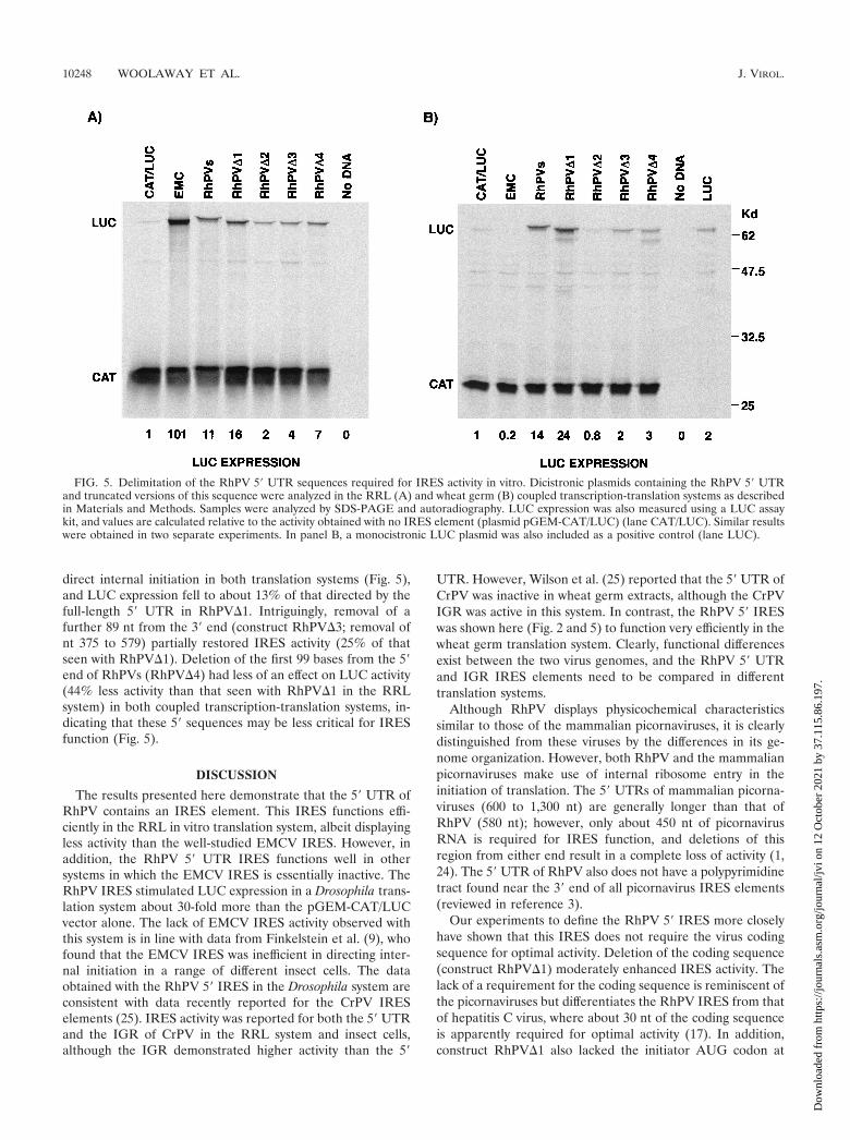
direct internal initiation in both translation systems (Fig. 5),and LUC expression fell to about 13% of that directed by thefull-length 5� UTR in RhPV�1. Intriguingly, removal of afurther 89 nt from the 3� end (construct RhPV�3; removal ofnt 375 to 579) partially restored IRES activity (25% of thatseen with RhPV�1). Deletion of the first 99 bases from the 5�end of RhPVs (RhPV�4) had less of an effect on LUC activity(44% less activity than that seen with RhPV�1 in the RRLsystem) in both coupled transcription-translation systems, in-dicating that these 5� sequences may be less critical for IRESfunction (Fig. 5).
DISCUSSION
The results presented here demonstrate that the 5� UTR ofRhPV contains an IRES element. This IRES functions effi-ciently in the RRL in vitro translation system, albeit displayingless activity than the well-studied EMCV IRES. However, inaddition, the RhPV 5� UTR IRES functions well in othersystems in which the EMCV IRES is essentially inactive. TheRhPV IRES stimulated LUC expression in a Drosophila trans-lation system about 30-fold more than the pGEM-CAT/LUCvector alone. The lack of EMCV IRES activity observed withthis system is in line with data from Finkelstein et al. (9), whofound that the EMCV IRES was inefficient in directing inter-nal initiation in a range of different insect cells. The dataobtained with the RhPV 5� IRES in the Drosophila system areconsistent with data recently reported for the CrPV IRESelements (25). IRES activity was reported for both the 5� UTRand the IGR of CrPV in the RRL system and insect cells,although the IGR demonstrated higher activity than the 5�
UTR. However, Wilson et al. (25) reported that the 5� UTR ofCrPV was inactive in wheat germ extracts, although the CrPVIGR was active in this system. In contrast, the RhPV 5� IRESwas shown here (Fig. 2 and 5) to function very efficiently in thewheat germ translation system. Clearly, functional differencesexist between the two virus genomes, and the RhPV 5� UTRand IGR IRES elements need to be compared in differenttranslation systems.
Although RhPV displays physicochemical characteristicssimilar to those of the mammalian picornaviruses, it is clearlydistinguished from these viruses by the differences in its ge-nome organization. However, both RhPV and the mammalianpicornaviruses make use of internal ribosome entry in theinitiation of translation. The 5� UTRs of mammalian picorna-viruses (600 to 1,300 nt) are generally longer than that ofRhPV (580 nt); however, only about 450 nt of picornavirusRNA is required for IRES function, and deletions of thisregion from either end result in a complete loss of activity (1,24). The 5� UTR of RhPV also does not have a polypyrimidinetract found near the 3� end of all picornavirus IRES elements(reviewed in reference 3).
Our experiments to define the RhPV 5� IRES more closelyhave shown that this IRES does not require the virus codingsequence for optimal activity. Deletion of the coding sequence(construct RhPV�1) moderately enhanced IRES activity. Thelack of a requirement for the coding sequence is reminiscent ofthe picornaviruses but differentiates the RhPV IRES from thatof hepatitis C virus, where about 30 nt of the coding sequenceis apparently required for optimal activity (17). In addition,construct RhPV�1 also lacked the initiator AUG codon at
FIG. 5. Delimitation of the RhPV 5� UTR sequences required for IRES activity in vitro. Dicistronic plasmids containing the RhPV 5� UTRand truncated versions of this sequence were analyzed in the RRL (A) and wheat germ (B) coupled transcription-translation systems as describedin Materials and Methods. Samples were analyzed by SDS-PAGE and autoradiography. LUC expression was also measured using a LUC assaykit, and values are calculated relative to the activity obtained with no IRES element (plasmid pGEM-CAT/LUC) (lane CAT/LUC). Similar resultswere obtained in two separate experiments. In panel B, a monocistronic LUC plasmid was also included as a positive control (lane LUC).
10248 WOOLAWAY ET AL. J. VIROL.
Dow
nloa
ded
from
http
s://j
ourn
als.
asm
.org
/jour
nal/j
vi o
n 12
Oct
ober
202
1 by
37.
115.
86.1
97.

position 588, and this construct was the most efficient in di-recting internal initiation. This finding supports the suggestionof Moon et al. (15) that AUG 580 is the functional initiationcodon; however, it does not exclude occasional use of AUG588.
The RhPV IRES continued to function, albeit at a reducedefficiency, when almost 100 bases were removed from eitherend of the 5� UTR. Deletion of 200 nt from the 3� end of theRhPV 5� UTR had less effect on IRES activity than deletion ofonly 100 nt from the same end of the UTR. This was a sur-prising result, and more work is required to fully explain thisfinding; however, it may indicate that the boundaries of the 5�IRES are not sharply defined, a property that is shared withcertain IRES elements of cellular origin. For example, a vari-ety of fragments of the immunoglobulin heavy-chain bindingprotein IRES element are known to retain IRES function (26),but this and certain other cellular IRES elements (e.g., c-myc)do not function in the RRL system or when expressed in thecytoplasm of mammalian cells (13, 22). Studies on the IGRIRES of RhPV (7) showed that deletion of sequences from the3� end of the 186-nt IRES inhibited IRES activity in vitro,although no quantitation of this effect was reported. However,the RhPV 5� UTR IRES is clearly different from the IGRIRES, which initiates at a non-AUG codon. Folding analysissuggests that the IGR IRES elements from both RhPV andPSIV should form a pseudoknot at the 3� end (7, 21). Thisfeature is thought to be crucial in directing non-AUG initiationof translation. Clearly, further comparison of the 5� UTR andIGR IRES elements of these viruses is required to determinethe structural features responsible for these functional prop-erties.
The ability of the RhPV 5� UTR to function not only in theRRL system, but also in both plant and insect translationsystems suggests potential utility of this IRES in both insectand plant expression systems.
ACKNOWLEDGMENTS
This work was funded by the Royal Society (support given toL.O.R.) and the BBSRC (support given to L.O.R. and G.J.B.). ABBSRC studentship award given to K.E.W. is also gratefully acknowl-edged.
REFERENCES
1. Belsham, G. J., and J. K. Brangwyn. 1990. A region of the 5� noncodingregion of foot-and-mouth disease virus RNA directs efficient internal initi-ation of protein synthesis within cells: involvement with the role of L pro-tease in translational control. J. Virol. 64:5389–5395.
2. Belsham, G. J., and R. J. Jackson. 2000. Translation initiation on picorna-virus RNA, p. 869–900. In N. Sonenberg, J. W. B. Hershey, and M. B.Mathews (ed.), Translational control of gene expression. Monograph 39.Cold Spring Harbor Laboratory Press, Cold Spring Harbor, N.Y.
3. Belsham, G. J., and N. Sonenberg. 1996. RNA-protein interactions in picor-navirus translational regulation. Microbiol. Rev. 60:499–511.
4. Borman, A. M., R. Kirchweger, E. Ziegler, R. E. Rhoads, T. Skern, and K. M.
Kean. 1997. eIF4G and its proteolytic cleavage products: effect on initiationof protein synthesis from capped, uncapped, and IRES-containing mRNAs.RNA 3:186–196.
5. Brown, B. A., and E. Ehrenfeld. 1979. Translation of poliovirus RNA in vitro:changes in cleavage pattern and initiation sites by ribosomal salt wash.Virology 97:396–405.
6. D’Arcy, C. J., P. A. Burnett, and A. D. Hewings. 1981. Detection, biologicaleffects, and transmission of a virus from the aphid Rhopalosiphum padi.Virology 114:268–272.
7. Domier, L. L., N. K. McCoppin, and C. J. D’Arcy. 2000. Sequence require-ments for translation initiation of Rhopalosiphum padi virus ORF2. Virology268:264–271.
8. Dorner, A. J., B. L. Semler, R. J. Jackson, R. Hanecak, E. Duprey, and E.Wimmer. 1984. In vitro translation of poliovirus RNA: utilization of internalinitiation sites in reticulocyte lysate. J. Virol. 50:507–514.
9. Finkelstein, Y., O. Faktor, O. Elroy-Stein, and B.-Z. Levi. 1999. The use ofbi-cistronic transfer vectors for the baculovirus expression system. J. Bio-technol. 75:33–44.
10. Gebauer, F., D. F. V. Corona, T. Preiss, P. B. Becker, and M. W. Hentze.1999. Translational control of dosage compensation in Drosophila by Sex-lethal: cooperative silencing via the 5� and 3� UTRs of msl-2 mRNA isindependent of the poly(A) tail. EMBO J. 18:6146–6154.
11. Gildow, F. E., and C. J. D’Arcy. 1990. Cytopathology and experimental hostrange of Rhopalosiphum padi virus, a small isometric RNA virus infectingcereal grain aphids. J. Invertebr. Pathol. 55:245–257.
12. Glass, M. J., and D. F. Summers. 1993. Identification of a trans-actingactivity from liver that stimulates hepatitis A virus translation in vitro. Vi-rology 193:1047–1050.
13. Izuka, N., C. Chen, Q. Yang, G. Johannes, and P. Sarnow. 1995. Cap-independent translation and internal initiation of translation in eukaryoticcellular mRNAs. Curr. Top. Microbiol. Immunol. 203:155–177.
14. Jackson, R. J., S. L. Hunt, C. L. Gibbs, and A. Kaminski. 1994. Internalinitiation of translation of picornavirus RNAs. Mol. Biol. Rep. 19:147–159.
15. Moon, J. S., L. L. Domier, N. K. McCoppin, C. J. D’Arcy, and H. Jin. 1998.Nucleotide sequence analysis shows that Rhopalosiphum padi virus is a mem-ber of a novel group of insect-infecting RNA viruses. Virology 243:54–65.
16. Pringle, C. R. 1999. Virus taxonomy—1999. Arch. Virol. 144:421–429.17. Reynolds, J. E., A. Kaminski, H. J. Kettinen, K. Grace, B. E. Clarke, A. R.
Carroll, D. J. Rowlands, and R. J. Jackson. 1995. Unique features of internalinitiation of hepatitis C virus RNA translation. EMBO J. 14:6010–6020.
18. Roberts, L. O., R. A. Seamons, and G. J. Belsham. 1998. Recognition ofpicornavirus internal ribosome entry sites within cells; influence of cellularand viral proteins. RNA 4:520–529.
19. Robertson, M. E. M., R. A. Seamons, and G. J. Belsham. 1999. A selectionsystem for functional internal ribosome entry site (IRES) elements: analysisof the requirement for a conserved GNRA tetraloop in the encephalomyo-carditis virus IRES. RNA 5:1167–1179.
20. Sambrook, J., E. F. Fritsch, and T. Maniatis. 1989. Molecular cloning: alaboratory manual, 2nd ed. Cold Spring Harbor Laboratory Press, ColdSpring Harbor, N.Y.
21. Sasaki, J., and N. Nakashima. 1999. Translation initiation at the CUU codonis mediated by the internal ribosome entry site of an insect picorna-like virusin vitro. J. Virol. 73:1219–1226.
22. Stoneley, M., T. Soubkhankulova, J. P. C. Le Quesne, M. Coldwell, C. L.Jopling, G. J. Belsham, and A. E. Willis. 2000. Analysis of the c-myc IRES;a potential role for cell-type specific trans-acting factors and the nuclearcompartment. Nucleic Acids Res. 28:687–694.
23. Thiel, H.-J., and M. Konig. 1999. Caliciviruses: an overview. Vet. Microbiol.69:55–62.
24. van der Velden, A., A. Kaminski, R. J. Jackson, and G. J. Belsham. 1995.Defective point mutants of the encephalomyocarditis virus internal ribosomeentry site can be complemented in trans. Virology 214:82–90.
25. Wilson, J. E., M. J. Powell, S. E. Hoover, and P. Sarnow. 2000. Naturallyoccurring dicistronic cricket paralysis virus RNA is regulated by two internalribosome entry sites. Mol. Cell. Biol. 20:4990–4999.
26. Yang, Q., and P. Sarnow. 1997. Location of the internal ribosome entry sitein the 5� non-coding region of the immunoglobulin heavy-chain bindingprotein (BiP) mRNA: evidence for specific RNA-protein interactions. Nu-cleic Acids Res. 25:2800–2807.
VOL. 75, 2001 RhPV 5� UTR IRES ACTIVITY 10249
Dow
nloa
ded
from
http
s://j
ourn
als.
asm
.org
/jour
nal/j
vi o
n 12
Oct
ober
202
1 by
37.
115.
86.1
97.