novel 5# untranslated region directed blockers of iron
TRANSCRIPT

Novel 5# Untranslated Region DirectedBlockers of Iron-Regulatory Protein-1Dependent Amyloid Precursor Protein
Translation: Implications for DownSyndrome and Alzheimer's Disease
The Harvard community has made thisarticle openly available. Please share howthis access benefits you. Your story matters
Citation Bandyopadhyay, Sanghamitra, Catherine Cahill, AmelieBalleidier, Conan Huang, Debomoy K. Lahiri, Xudong Huang,and Jack T. Rogers. 2013. “Novel 5# Untranslated RegionDirected Blockers of Iron-Regulatory Protein-1 DependentAmyloid Precursor Protein Translation: Implications for DownSyndrome and Alzheimer's Disease.” PLoS ONE 8 (7): e65978.doi:10.1371/journal.pone.0065978. http://dx.doi.org/10.1371/journal.pone.0065978.
Published Version doi:10.1371/journal.pone.0065978
Citable link http://nrs.harvard.edu/urn-3:HUL.InstRepos:11855917
Terms of Use This article was downloaded from Harvard University’s DASHrepository, and is made available under the terms and conditionsapplicable to Other Posted Material, as set forth at http://nrs.harvard.edu/urn-3:HUL.InstRepos:dash.current.terms-of-use#LAA
Novel 59 Untranslated Region Directed Blockers of Iron-Regulatory Protein-1 Dependent Amyloid PrecursorProtein Translation: Implications for Down Syndromeand Alzheimer’s DiseaseSanghamitra Bandyopadhyay1¤, Catherine Cahill1,2, Amelie Balleidier1, Conan Huang1,
Debomoy K. Lahiri3, Xudong Huang1, Jack T. Rogers1*
1 Neurochemistry Laboratory, Department of Psychiatry, Massachusetts General Hospital and Harvard Medical School, Charlestown, Massachusetts, United States of
America, 2 Department of Pediatrics, Massachusetts General Hospital and Harvard Medical School, Charlestown, Massachusetts, United States of America, 3 Laboratory of
Molecular Neurogenetics, Department of Psychiatry, Institute of Psychiatric Research, Indiana University School of Medicine, Indianapolis, Indiana, United States of
America
Abstract
We reported that iron influx drives the translational expression of the neuronal amyloid precursor protein (APP), which has arole in iron efflux. This is via a classic release of repressor interaction of APP mRNA with iron-regulatory protein-1 (IRP1)whereas IRP2 controls the mRNAs encoding the L- and H-subunits of the iron storage protein, ferritin. Here, we identifiedthirteen potent APP translation blockers that acted selectively towards the uniquely configured iron-responsive element(IRE) RNA stem loop in the 59 untranslated region (UTR) of APP mRNA. These agents were 10-fold less inhibitory of 59UTRsequences of the related prion protein (PrP) mRNA. Western blotting confirmed that the ‘ninth’ small molecule in the seriesselectively reduced neural APP production in SH-SY5Y cells at picomolar concentrations without affecting viability or theexpression of a-synuclein and ferritin. APP blocker-9 (JTR-009), a benzimidazole, reduced the production of toxic Ab in SH-SY5Y neuronal cells to a greater extent than other well tolerated APP 59UTR-directed translation blockers, includingposiphen, that were shown to limit amyloid burden in mouse models of Alzheimer’s disease (AD). RNA binding assaysdemonstrated that JTR-009 operated by preventing IRP1 from binding to the IRE in APP mRNA, while maintaining IRP1interaction with the H-ferritin IRE RNA stem loop. Thus, JTR-009 constitutively repressed translation driven by APP 59UTRsequences. Calcein staining showed that JTR-009 did not indirectly change iron uptake in neuronal cells suggesting a directinteraction with the APP 59UTR. These studies provide key data to develop small molecules that selectively reduce neuralAPP and Ab production at 10-fold lower concentrations than related previously characterized translation blockers. Our dataevidenced a novel therapeutic strategy of potential impact for people with trisomy of the APP gene on chromosome 21,which is a phenotype long associated with Down syndrome (DS) that can also cause familial Alzheimer’s disease.
Citation: Bandyopadhyay S, Cahill C, Balleidier A, Huang C, Lahiri DK, et al. (2013) Novel 59 Untranslated Region Directed Blockers of Iron-Regulatory Protein-1Dependent Amyloid Precursor Protein Translation: Implications for Down Syndrome and Alzheimer’s Disease. PLoS ONE 8(7): e65978. doi:10.1371/journal.pone.0065978
Editor: Koichi M. Iijima, Thomas Jefferson University, United States of America
Received February 2, 2013; Accepted April 30, 2013; Published July 31, 2013
Copyright: � 2013 Bandyopadhyay et al. This is an open-access article distributed under the terms of the Creative Commons Attribution License, which permitsunrestricted use, distribution, and reproduction in any medium, provided the original author and source are credited.
Funding: JR was funded from an R21 NS077079-01A1 (Post Transcriptional Control of hemorrhagic iron damage). JR is a recipient of the Alzheimer’s AssociationZenith Award, and of a Michael JFOX NADD grant. CC was supported by an R01 Supplement from the NICH. The funders had no role in study design, datacollection and analysis, decision to publish, or preparation of the manuscript.
Competing Interests: The authors have declared that no competing interests exist.
* E-mail: [email protected]
¤ Current address: Developmental Toxicology Division, Council of Scientific and Industrial Research-Indian Institute of Toxicology Research, Lucknow, India
Introduction
Many RNA-binding protein interactions are closely associated
with neurological and psychiatric disease processes such as
amyotrophic lateral sclerosis (ALS) [1] and autism [2]. In this
report, we sought proof that the use of APP translation blockers
can reduce amyloid expression pertinent to providing therapy for
individuals afflicted with Alzheimer’s disease (AD) and Down
syndrome (DS).
Increased levels of the metals iron, copper, zinc in the brain are
associated with increased risk to accelerate the course of AD [3].
To safely store excess iron, canonical iron-responsive elements
(IREs) are the 59UTR-specific RNA stem loops that control
translation of L- and H-ferritin mRNAs (iron storage) so that the
L- and H chains can assemble into this iron storage multimer. The
iron-regulatory proteins (IRP1 (90 kDa) and IRP2 (105 kDa)) are
the two known RNA-binding proteins that are key gatekeepers for
cellular iron homeostasis because of their inducible interaction
with IREs to control ferritin mRNA translation and transferrin
receptor (TfR) mRNA stability (iron uptake) [4].
Consistent with our report that APP is an iron export
ferroxidase [5], RNAi knockout studies showed that IRP1 binds
strongly to 59UTR sequences in the APP transcript to repress
expression of the precursor [6]. In fact, the APP mRNA encodes
an active IRE that binds with a different RNA-binding specificity
PLOS ONE | www.plosone.org 1 July 2013 | Volume 8 | Issue 7 | e65978
to IRP1 relative to the IRE of ferritin mRNA (which interacts with
IRP1 & IRP2) [6]. Thus the APP 59UTR is a unique, highly
specific drug target to identify APP (and Ab) repressors. This
model is consistent with a recent report that IRP1 outcompetes
IRP2 in regulating cellular iron homeostasis in response to nitric
oxide [7].
The concept of repressing APP translation as a therapeutic
strategy in DS and AD was proven as a novel anti-amyloid strategy
as exemplified by our use of the APP 59UTR-directed FDA drug
N-acetyl-cysteine (NAC) in the TgCRND8 APP(Swe) mouse
model of AD [8]. An additional benefit of limiting the APP levels
may be to restore perturbations to iron homeostasis during DS
since APP is over-expressed by one third on the DS trisomy
chromosome 21 [9]. Increased APP may well alter brain iron
homeostasis based on its capacity to bind ferroportin and export
iron [5]. In this regard, mice that are trisomic for chromosome 16,
the orthologue of human chromosome 21, over-express APP and
are genetically shown to develop the DS phenotype because of a
triplicated expression of the APP gene [9,10]. The progression of
familial Alzheimer’s disease (FAD) can be the result of a genetically
inherited over-expression of the APP gene or by somatically
induced non-disjunction events that cause APP to be over-
expressed [11,12,13].
Thus, in addition to the altered processing of APP and other risk
factors (e.g., inflammation, metal-catalyzed oxidative stress
[3,14,15,16], and the increased levels of apolipoprotein-E
[17,18] and a-1 anti-chymotrypsin (ACT) [19]), simple elevation
of APP levels is a sufficient genetic cause of DS and AD [12,20].
This report centers on our RNA targeting strategy as a starting
point to develop drugs that can limit APP expression by a novel
therapeutic mechanism for offsetting APP mRNA translation rates
and reducing severe amyloidosis during the progression of DS and
AD.
The proven in vivo efficacy of APP 59UTR-acting FDA drugs,
including NAC and the APP translation blocker posiphen,
encouraged us to pursue a high-throughput screening campaign
against APP 59UTR in search of potent and selective APP
translation blockers [21].
We identified and characterized a novel APP 59UTR-specific
translation blocker of neuronal APP and Ab that operates at
nanomolar concentrations while maintaining b-actin expression
and cell viability [9]. JTR-009 is a benzimidazole that was found
to reduce intracellular APP and toxic Ab production in both SH-
SY5Y neural cell lines and primary mouse neurons. Here we have
shed light on the mechanism of action of JTR-009, which is
consistent with the drug intercalating into RNA sequences folded
from the APP 59UTR and irreversibly replacing IRP1 as the
repressor of APP translation. These findings supported our
pharmacological goal, as described in this report, to reduce APP
expression with therapeutic implications particularly for DS and
AD.
Materials and Methods
1. AntibodiesRabbit anti-human IRP1 antibody (Alpha Diagnostics Interna-
tional, San Antonio, TX) and anti-IRP1 (kind gift from Dr.
Sharon Cooperman and Dr. Tracey Rouault, National Institutes
of Health) each generated the same results in the assays shown;
mouse anti-human IRP2 (Santa Cruz Biotechnology, Santa Cruz,
CA) detected the H-ferritin IRE-IRP2 interaction, and a second
antibody to IRP2 was also utilized to confirm the selectivity of
IRP2 binding as detected with the Santa Cruz Biotechnology
antibody. Anti-b-actin, anti-a-tubulin, rabbit anti-APP C-terminal
antibody (A8717) were from (Sigma, St. Louis, MO), and the APP
N-terminal antibody (22C11) was from Chemicon (Temecula,
CA).
2. Cell Culture and Preparation of LysatesHuman SH-SY5Y neuroblastoma cells were cultured in
DMEM supplemented with 10% FBS (Invitrogen, Carlsbad,
CA) and penicillin/streptomycin (Bio-Whittaker, Walkersville,
MD). Cells were exposed to JTR-009 (0–100 mM, Calbiochem)
and iron (50 mM, National Institute of Standards and Technology
(NIST), Gaithersburg, MD), provided to cells as ferrous ammo-
nium sulfate. Cytoplasmic protein lysates were prepared by
homogenizing the cells in ribonucleoprotein immunoprecipitation
buffer (25 mM Tris, pH 7.4, 1%Nonidet P-40, 0.5% sodium
deoxycholate, 15 mM NaCl, protease inhibitors, RNase inhibitor,
and 10 mM DTT). For preparation of conditioned medium for Aband LDH measurements, cells were treated for 48–72 hours with
each compound as described in the legends. 1 mL was used for
total Ab determination by ELISA as described by Biosource
International. according to maufacturers conditions (see Ref [8]).
Primary cortical neurons from wild type mice and from the
PAC-Tg(SNCA(wt) human SNCA genomic mice [22] were cultured
as outlined by the method of Ray et al., 2009 [23]. We recovered
the embryonic (E15-18) pups after sacrificing pregnant females,
separated out the brain, and removed the meninges and blood
vessels. We then dissected out the cortices and placed them in
separate Eppendorf tubes containing 500 mL of HBSS without
Ca+2/Mg+2 salts supplemented with 1 mM sodium pyruvate and
10 mM HEPES, pH 7.4. On ice, individual cells were isolated by
titrating 10 times using a glass pasture pipette with the tip barely
fire polished. We adjusted the volume to 1.5 mL by adding 1 mL
of HBSS with Ca+2/Mg+2 salts+Na. pyruvate+HEPES, restoring
the divalent cations by adding HBSS so that the non-dispersed
tissue could settle for 5 min, on ice. In the tissue culture laminar
hood, we transferred the supernatant into a new 15 mL tube and
centrifuged for 1 min at 900 rpm, 4uC. We gently re-suspended
the pellet in 2 mL of HBSS with Ca+2/Mg+2 salts+Na pyruva-
te+HEPES and took an aliquot for counting (2 mL for approx 5
embryos). We then plated ,16105 cells/well of a 24 well or
26105/in 12 well plates. Each set of plates was coated with poly
D-lysine containing poly L-lysine coverslips for micro immuncy-
tochemical confirmation of neuronal integrity.
3. Methodology of Molecular ScreensWe screened the 110,000 compounds of the molecular library of
LDDN at Harvard to identify novel and more potent APP
59UTR-directed inhibitors [21]. The LDDN library had already
yielded small molecules that inhibit mesangial cell proliferation
[24], following three-dimensional pharmacophore modeling and
screening. A second Molecular Libraries Screening Centers
Network HTS was conducted at the Columbia University
Genome Center to generate hits as listed on PUBCHEM (AID:
1285), from which our dose-response assays identified 50 lead APP
59UTR-directed luciferase reporter inhibitors. Two classes of APP
59UTR-directed translation blockers from the second screen
exhibited a potent IC50 in the 1028 M range. We pooled a
shortlist of the thirteen most selective APP 59UTR inhibitors from
both screens. These thirteen leads were tricyclic aromatic
compounds that included two major classes of hits: compounds
with a benzimidazole backbone, i.e. APP blockers -2, -7, and -9
(JTR-009) and compounds with a benzothiazole backbone, i.e.
APP blockers -8 and -13. The compounds with a benzothiazole
backbone were also identified to be similar to PFTa, another
benzothiazole, and P53 inhibitor, by showing protection against
APP Translation Blocker Limits Abeta Production
PLOS ONE | www.plosone.org 2 July 2013 | Volume 8 | Issue 7 | e65978
oxidative injuries in synaptosomes from wild-type mice and
preserving presynaptic terminals in cultured hippocampal neurons
exposed to etoposide [25] [26]. The anti-APP 59UTR efficacy of
the 13 top inhibitors was directly compared with their anti-APP
efficacy by Western blotting of lysates prepared from SH-SY5Y
cells.
4. Western BlottingAfter cells were exposed to increasing concentrations of the
compounds as outlined in each figure legend, cytoplasmic protein
lysates were prepared by homogenizing the cells in midRIPA
buffer (25 mM Tris pH 7.4, 1% NP40, 0.5% sodium deoxycho-
late, 15 mM NaCl, protease inhibitors, RNase inhibitor and
10 mM DTT). Western blotting for APP was performed using the
N-terminal 22C11 antibody (Millipore, inc) and the A8717 C-
terminal specific APP antibody (Sigma, inc), while asyn was
detected using mouse monoclonal anti-asyn (BD Transduction
Laboratories) and anti-b-actin was from Chemicon. The blots
were developed using chemiluminescence (PIERCE, Rockford, IL)
and visualized with a Phosphoimager (BioRad, Hercules, CA).
The bands were quantified using QuantityOneH software
(BioRad).
5. RNA QuantitationWe conducted qRT-PCR to measure the capacity of JTR-009
to change steady state levels of APP mRNA levels, as was
previously described (see Ref [6]). Desferrioxamine treatment for
48 h was used for a positive control to assess changes to the steady
state levels of both APP mRNA and transferrin receptor mRNAs.
Primers for b-actin were employed as a control for an mRNA
previously shown to be unchanged by desferrioxamine and other
inducers [6]. Experiments were carried out on the ABI Prism 7000
sequence detection system (Applied Biosystems). Total RNA was
isolated using TRIzol reagent (Sigma) according to the manufac-
turer’s instructions. cDNA was synthesized with SuperScript III
first-strand qPCR supermix (Invitrogen) according to the manu-
facturer’s instructions. The primers to b-actin, TfR1) were
designed and ordered from Invitrogen. The APP primer set was
purchased from Qiagen and has been benchmarked on several
reports for accurate measurement of APP mRNA levels.
6. Transfections and Luciferase Reporter Assays forCounterscreens
APP 59UTR-Luciferase inhibitor compounds obtained from the
preliminary HTS of pIRES-APP-59UTR transfectants were
picked, and the dose-response assays were conducted at 0.1, 1.0,
and 5.0 mM (based on the exact molecular weights of the
compounds). For the purpose of counter-screening, pIRES-PrP-
59UTR)-transfected SH-SY5Y cells were plated in 384-well black
plates, and the identified compound hits that were not cytotoxic
were manually added to the cells. Each hit was added in 5 wells,
and this was repeated twice on 2 different days. There was a
positive control and negative control column of cells as previously
described [21]. The inhibition of luciferase was calculated, and the
average of the values obtained was considered (see data shown in
Table 1).
7. Biotinylated RNA Pulldown AssayBiotinylated RNA oligonucleotides: H-ferritin IRE (biotin-59-
GGG UUU CCU GCU UCA ACAGUG CUU GGA CGG AAC
CCG G-39) and APP IRE (59-biotin-GC GGU GGC GGC GCG
GGC AGA GCA AGG ACG CGG CGG AU-39) were purchased
from Invitrogen. Cell lysates (100 mg) were incubated with100 nM
biotinylated oligonucleotide for each of the IREs, for 3 h at room
temperature. Paramagnetic streptavidin-conjugated Dynabeads
(Invitrogen) were washed with ribonucleoprotein immunoprecip-
itation buffer, added into lysates to bind IRP(1/2)-biotinylated-
RNA complexes, and incubated for 1 hour at room temperature.
After five washes, the proteins that bound to the beads were
analyzed by Western blotting for IRP1, IRP2, and biotin. The
blots were developed with chemiluminescence (Pierce) and
visualized with a 4000 MP VersaDocTM Imaging System (Bio-
Rad). The IREs-bound IRPs were quantified by Quantity OneHsoftware (Bio-Rad).
8. Calcein AssayCells were loaded with calcein after incubation with 0.1 mM of
Calcein-AM for 10 min in 0.15 M NaCl-20 mM HEPES buffer,
pH 7.4, with 0.1% BSA at 37uC, an action followed by extensive
washing with NaCl-HEPES buffer to remove extracellular bound
calcein. The cells were aliquoted at 56104–16105 cells/well in 96-
well plates containing test compounds at 10 mM and incubated for
30 min in a humidified 37uC incubator with 5% CO2 before
baseline fluorescence was obtained at 485/520 nm (excitation/
emission) with 0.1% DMSO, as the vehicle control, and DTPA as
a strong iron chelator control to block all iron uptake. Using Using
a SpectraMax M5e plate reader and SoftMax Pro software
(Molecular Devices, Sunnyvale, CA), the fluorescence was then
measured 30 min after the addition of 10 mM ferrous ammonium
sulfate in 500 mM ascorbic acid (AA). The percentage of
fluorescence quench was calculated relative to 200 mM DTPA,
which was added as a blocking control, and DMSO as a vehicle
control, as follows:
DF~(F0-Ff )
F0ð1Þ
where D F is the change in fluorescence, or fluorescence quench
observed in any well. F0 represents the fluorescence after 30 min
of the compound, and Ff represents the fluorescence 30 min after
addition of Fe. These results were normalized to the blocking and
vehicle controls as follows:
DFn~(Fcompound-Fmin)
(Fmax-Fmin)ð2Þ
where DFn is the normalized quench observed after addition of
iron. Fcompound is the D F observed with the compound, Fmin is the
average D F of the DMSO control, and Fmax is the average DF of
the DTPA control.
With this normalization, 100% indicates that the test compound
is as potent as DTPA in blocking iron-induced quenching, and 0%
indicates no inhibition of iron quenching by the test compound or
the same quench as observed with the DMSO vehicle control.
Compounds with DFn between 0% and 100% are defined as
inhibitors of iron uptake. Negative values for DF represents
compounds that facilitate iron uptake into cells. Our criteria for
active compounds to be further investigated were arbitrarily set as
DFn = 50–100% quenching for iron uptake inhibitors and
,250% quenching for iron uptake facilitators.
9. ELISA Measurement of Secreted Ab levels and LactaseDehydrogenase (LDH)
After reaching 80% confluence, SH-SY5Y cells were 1:3 split
onto two 12-well plates. After allowing the cells to settle for
24 hours, the medium was switched to a 1% FBS DMEM
APP Translation Blocker Limits Abeta Production
PLOS ONE | www.plosone.org 3 July 2013 | Volume 8 | Issue 7 | e65978
(Dulbecco’s modified essential medium supplemented with 1%
FBS and penicillin/streptomycin).
Ab Assays. Total Ab amyloid levels were assessed as we
previously described (8) and Ab-42 levels were measured by use of
ELISA according to manufacturers instructions (Covance Chemi-
luminescent BetaMark x-42 ELISA).
LDH assay. The 1% FBS medium was recommended by the
LDH Cytotoxicity Kit (Cayman Chemical, Ann Harbor, MI) to
reduce interference as FBS also contains LDH. Cells were exposed
to 10-fold increases in concentrations of JTR-009 reconstituted in
16 PBS (0.1 nM-100 mM) compared to PBS as a control for
48 hours. Thus, eight wells on each 12-well were treated for 48-
hour after which time 100 mL of supernatant was extracted from
each of the treated wells and transferred to a 96-well plate. A LDH
standard from the kit was also added to the plate. Using the
reaction mixture in the kit, LDH absorbance values were obtained
with a SpectraMax M5e plate reader and SoftMax Pro software
((Molecular Devices, Sunnyvale, CA)).
10. MTS Assay for Neuronal ViabilityCell viability was determined using MTT (thiazolyl blue
tetrazolium) viability assays. Cells were grown in 96 well plates
and treated as indicated above. After treatment, they were
incubated with 20 mL of 5 mg MTT (Sigma)/1 mL PBS solution
for 3.5 hours. The media was aspirated from the cells and 150 mL
of solvent (0.1% Nondet P-40, 4 mM HCl in isopropanol) was
added to each well and the plate shaken for 15 minutes. The
absorbance was then read at 590 nm using a SpectraMax M5e
plate reader and SoftMax Pro software (Molecular Devices,
Sunnyvale, CA).
Results
A: Selectivity of APP 59UTR translation blockers from PrP59UTR-based counter-screen
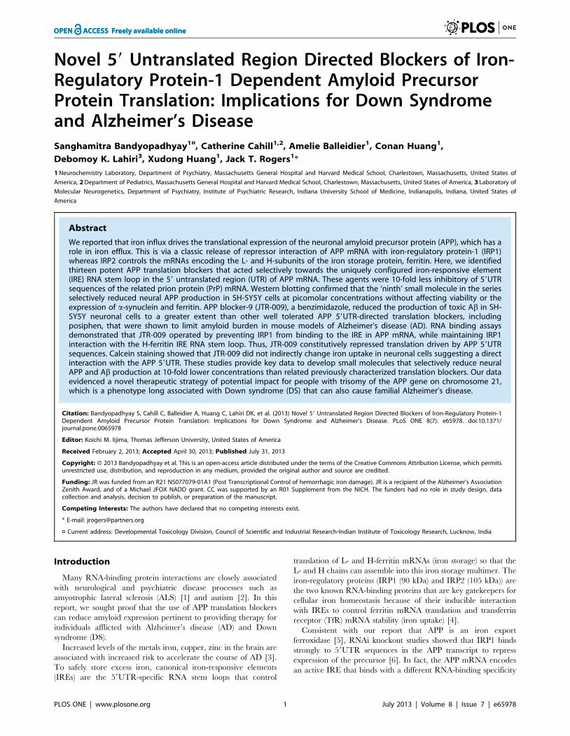
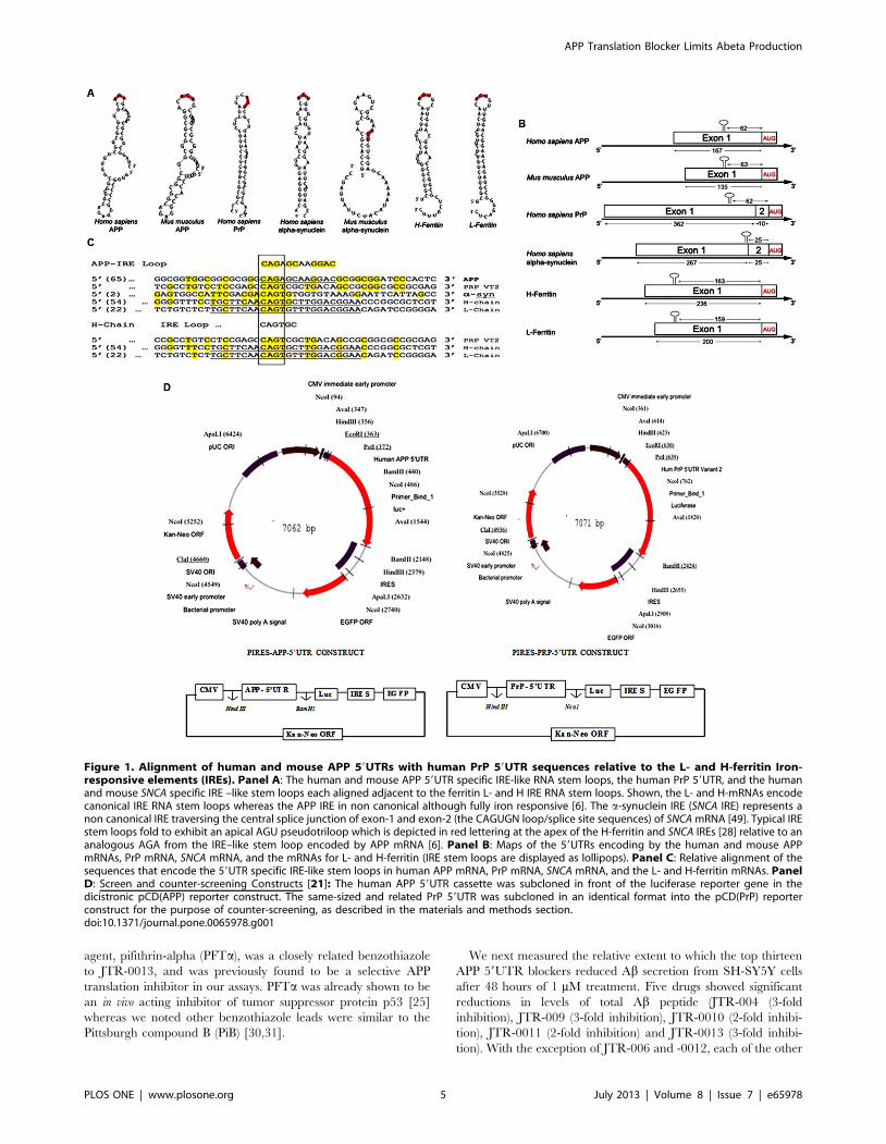
In Figure 1, Panel A shows the specific RNA stem-loops
encoded by the 59UTRs of several neurodegenerative disease
transcripts, specifically those for APP, PrP, and a-synuclein
(SNCA). Each mRNA encodes uniquely configured variations of
an IRE RNA stem loop that potentially bind to the IRP
translational repressors in their 59UTRs. The prion PrP 59UTR
was chosen as a stringent counter control for ensuring that APP
59UTR directed compounds would be sufficiently specific not to
inhibit luciferase reporter gene expression in matched PrP
59UTR-driven transfectants. Panels B and C of Figure 1 shows
the maps and alignments of the 59UTRs encoding IRE stem loops
in the neurodegenerative transcripts for APP and asyn relative to
the canonical ferritin L- and H- chain IRE stem loops [21].
Figure 1C presents that this homology extends to that of the PrP
59UTR, which encodes an IRE-like sequence, although diverged
from the proven APP IRE (NCBI, Clustal software, [27]).
Alignments elucidated a 56% similarity between this region of
the 59UTR of PrP mRNA (splice variant-2) and APP IRE
sequences. This homology is centered around the CAGUGN loop
domain of the canonical ferritin IRE and the projected IRP1
binding AGU/AGA tri-loops that were shown to be key for IRP1
and IRP2 binding and translation repression [6,28]. The PrP(Vt2)
59UTR was therefore deemed a stringent screening control to
ensure specificity of our APP 59UTR-directed translation blockers.
Figure 1D shows the complete coordinates of the screening
constructs, pIRES-APP-59UTR and pIRES-PrP-59UTR, which
were matched for insertion of equal length 59UTRS for screen/
counter-screen comparisons in transfection based assays (Bandyo-
padhyay et al., 2006).
We conducted a screening campaign of library of 110,000
compounds with the stable transfected SH-SY5Y cells expressing
the constructs shown in figure 1D. To identify APP 59UTR-
specific translation blockers from LDDN Harvard (see ref [21])
and from the Columbia University Genome center, we then
counter-screened against the PrP 59UTR and shortlisted thirteen
potent inhibitors to be further characterized. In Figure 1D, we
employed the listed constructs to conduct these transfection based
assays to ensure that the 13 APP specific leads were not also PrP
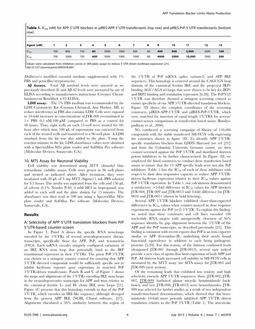
inhibitors. Table 1 lists the IC50 of each of these inhibitors with
respect to their dose-responsive capacity to reduce APP 59UTR-
driven luciferase expression relative to their IC50 values against
PrP 59UTR expression. In Table 1, our calculations demonstrated
a satisfactory .5-fold difference in IC50 values for APP blockers
JTR-004, JTR-009 and JTR-0013 and 3-fold difference for JTR-
0010 and JTR-0011 (shown in bold lettering).
Several APP 59UTR blockers exhibited closer-than-expected
differences in IC50 values when counter-assayed in dose response
experiments against the PrP (vt-2) 59UTR. To explain this finding,
we noted that these constructs and cell lines encoded 100
nucleotide RNA targets with unexpectedly closeness of 56%
sequence identity by gap alignment between the 59UTR of the
APP and the PrP transcripts, as described previously [21]. This
finding is consistent with recent reports that PrP is an iron exporter
similar to APP (ferroxidase-II), underlying their newly found
functional equivalence in addition to each being pathogenic
proteins [5,29]. For this reason, of the thirteen confirmed leads
(designated JTR-001 through JTR-0013), several may indeed
provide a new class of agents that limit expression of both APP and
PrP. All thirteen leads increased cell viability in SH-SY5Y cells as
measured by the MTT assay (see MTS assays for JTR-005 and
JTR-009 (next section).
Of the remaining leads that exhibited low toxicity and high
selectivity towards APP 59UTR sequences, three (JTR-002, JTR-
007, JTR-009) harbored planar tricyclic benzimidazole back-
bones, and two (JTR-008, JTR-0013) were benzothiazoles. JTR-
009 was selected for further studies as a result of two independent
transfection-based determinations, which showed that this benz-
imidazole 10-fold more potently inhibited APP 59UTR driven
translation relative to the PrP 59UTR (Table 1). The anti-stroke
Table 1. IC50 (nM) for APP 59UTR blockers in pIRES-APP-59UTR transfectants (top row) and pIRES-PrP-59UTR transfectants (bottomrow).
Agent (nM) 1 2 3 4 5 6 7 8 9 10 11 12 13
IC50 700 800 750 80 5000 1000 500 80 400 900 3,000 3000 100
IC50 700 5000 750 400 5000 1000 1000 80 4000 2000 10,000 7000 500
Values were calculated from inhibiton curves in 384-plate assays to reduce 59UTR driven luciferase expression [21].doi:10.1371/journal.pone.0065978.t001
APP Translation Blocker Limits Abeta Production
PLOS ONE | www.plosone.org 4 July 2013 | Volume 8 | Issue 7 | e65978
agent, pifithrin-alpha (PFTa), was a closely related benzothiazole
to JTR-0013, and was previously found to be a selective APP
translation inhibitor in our assays. PFTa was already shown to be
an in vivo acting inhibitor of tumor suppressor protein p53 [25]
whereas we noted other benzothiazole leads were similar to the
Pittsburgh compound B (PiB) [30,31].
We next measured the relative extent to which the top thirteen
APP 59UTR blockers reduced Ab secretion from SH-SY5Y cells
after 48 hours of 1 mM treatment. Five drugs showed significant
reductions in levels of total Ab peptide (JTR-004 (3-fold
inhibition), JTR-009 (3-fold inhibition), JTR-0010 (2-fold inhibi-
tion), JTR-0011 (2-fold inhibition) and JTR-0013 (3-fold inhibi-
tion). With the exception of JTR-006 and -0012, each of the other
Figure 1. Alignment of human and mouse APP 59UTRs with human PrP 59UTR sequences relative to the L- and H-ferritin Iron-responsive elements (IREs). Panel A: The human and mouse APP 59UTR specific IRE-like RNA stem loops, the human PrP 59UTR, and the humanand mouse SNCA specific IRE –like stem loops each aligned adjacent to the ferritin L- and H IRE RNA stem loops. Shown, the L- and H-mRNAs encodecanonical IRE RNA stem loops whereas the APP IRE in non canonical although fully iron responsive [6]. The a-synuclein IRE (SNCA IRE) represents anon canonical IRE traversing the central splice junction of exon-1 and exon-2 (the CAGUGN loop/splice site sequences) of SNCA mRNA [49]. Typical IREstem loops fold to exhibit an apical AGU pseudotriloop which is depicted in red lettering at the apex of the H-ferritin and SNCA IREs [28] relative to ananalogous AGA from the IRE–like stem loop encoded by APP mRNA [6]. Panel B: Maps of the 59UTRs encoding by the human and mouse APPmRNAs, PrP mRNA, SNCA mRNA, and the mRNAs for L- and H-ferritin (IRE stem loops are displayed as lollipops). Panel C: Relative alignment of thesequences that encode the 59UTR specific IRE-like stem loops in human APP mRNA, PrP mRNA, SNCA mRNA, and the L- and H-ferritin mRNAs. PanelD: Screen and counter-screening Constructs [21]: The human APP 59UTR cassette was subcloned in front of the luciferase reporter gene in thedicistronic pCD(APP) reporter construct. The same-sized and related PrP 59UTR was subcloned in an identical format into the pCD(PrP) reporterconstruct for the purpose of counter-screening, as described in the materials and methods section.doi:10.1371/journal.pone.0065978.g001
APP Translation Blocker Limits Abeta Production
PLOS ONE | www.plosone.org 5 July 2013 | Volume 8 | Issue 7 | e65978

APP 59UTR directed inhibitors (1 mM dose) modestly reduced Ablevels as measured by use of an ELISA (Biosource Int.) (See
Figure 2), a finding that confirmed which APP translation blockers
reduced levels of cellular APP template sufficiently enough to
reduce Ab peptide output from SH-SY5Y cells.
Of these, JTR-009 has consistently provided maximal cell
viability (MTT assays see Fig. 3D). Thus JTR-009, as a translation
blocker of APP mRNA, was sufficiently specific towards APP
59UTR sequences and was considered to be a bonafide anti-
amyloid agent (inhibition of Ab by JTR-009 was 3-fold, ANOVA:
p = 0.0046, N = 5 by pair-wise comparison of groups). JTR-009
was advanced for further analysis of the mechanism of the APP
59UTR as a regulatory domain for APP gene expression at the
level of message translation and as a candidate for future analog-
based drug development as an anti-APP and anti-Ab blocker for
potential DS and AD therapy.
B. JTR-009: The most selective and potent of the thirteentop APP 59UTR-directed translation blockers
Consistently, JTR-009 was a highly specific APP 59UTR
translation blocker of luciferase reporter gene expression and also
of steady state levels of APP (see Table 1 and Figure 3).
(Significantly, JTR-009 was an equally potent suppressor of Abpeptide levels (Figures 2 and 3) [21]. Pifithrin (PFT-a), a well
tolerated anti-apoptotic drug that has a benzothiazole structure
similar to JTR-0013, was employed for the purpose of comparison
with JTR-009. Therefore, as proven APP 59UTR inhibitors, the
benzimidazole JTR-009 and the benzothiazole pifithrin were
compared for their relative capacities to limit APP expression in
SH-SY5Y cells. In Figure 3A, a representative Western blotting
experiment demonstrated that both JTR-009 and PFTa dose
dependently reduced APP translation. In Panel B densitometry
quantified from five separate experiments, including the one
shown, demonstrated a 50% reduction of APP at 20 mM
concentrations (48 h treatment) after standardization for b-actin.
JTR-009 reduced APP levels to 30% of control levels at 30 mM,
while maintaining both b-actin and a-synuclein (SNCA) levels
(N = 5, p = 0.003). Several similar western blots experiments
showed that JTR-009, but not PFTa, had sufficient specificity to
limit APP while also maintaining b-actin and asyn levels (48 h).
We consistently found that PFTa, which has a benzothiazole
backbone like JTR-0013, inhibited neural APP with the same
potency as JTR-009 but was less specific since PFTa co-reduced
asyn levels (another IRE encoding mRNA [32] as well as b-actin
(Figure 3A, B).
Dose-responsive comparisons of JTR-009 with another APP
59UTR-screened inhibitor, JTR005, were then assessed at
equimolar concentrations to demonstrate the differential capacities
of these two agents to limit Ab secretion from SH-SY5Y cells
(Figure 3C). JTR-009 consistently inhibited secreted Ab at
concentrations as low as 10 nM (Figure 3C). The fifth inhibitor
in the series, JTR-005, was a typical comparative control
compound since it is also a tricyclic planar compound but without
a benzimidazole backbone. JTR-005 also targeted the APP
59UTR (.PrP 59UTR), although at 10-fold less potency than
JTR-009. Consistent with this fact, JTR-009 reduced Ab levels at
lower concentrations than JTR-005 without causing any signifi-
cant cell death as measured by an LDH cytotoxicity assay at
concentrations up to 10 mM (Figure 3C).
The relative cellular toxicity of the APP 59UTR inhibitors JTR-
005 and JTR-009 was determined by the MTT assay for cellular
mitochondrial activity. Figure 3D shows a representative exper-
iment where the mean values for MTS absorbance was a reflection
of viability after treatment of the cells with JTR-009 compared to
JTR-005 at 0.01 mM (Percent of maximal viability for each
treatment 6 SEM (N = 3)). These results consistently showed that
mitochondrial staining was compromised by increased doses of
JTR-005 whereas JTR-009 sustained cellular viability at 80%
compared to controls, for concentrations of the drug as high as
Figure 2. Relative capacity of thirteen APP 59UTR translation blockers to reduce Ab levels in the conditioned medium of SH-SY5Ycells. Following 48 h treatment (1 mM) for each inhibitor, the histogram shows reduction of total Ab levels confirmed after averaging fiveindependent samplings from the following:- JTR-009 treated,control, p,0.01). Total Ab levels were also documented for the APP blockers JTR-004,JTR-10, JTR-0011 JTR-0013 (N = 5). Data are means 6 SEM, N = 5, * = p,0.01, ** = p,0.01, *** = p,0.0013, p,0.01, **** p,0.011, *****, where eachtreatment was analyzed by ANOVA+Dunnett’s post hoc test compared to untreated samples. JTR-009 was the ninth and JTR-005 was fifth in theseries of 13 APP translation blockers.doi:10.1371/journal.pone.0065978.g002
APP Translation Blocker Limits Abeta Production
PLOS ONE | www.plosone.org 6 July 2013 | Volume 8 | Issue 7 | e65978
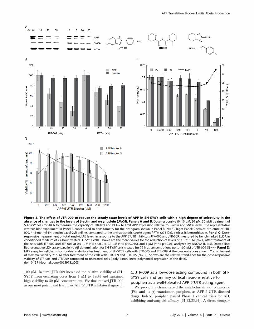
100 mM. In sum, JTR-009 increased the relative viability of SH-
SY5Y from escalating doses from 1 nM to 1 mM and sustained
high viability to 30 mM concentrations. We thus ranked JTR-009
as our most potent and least toxic APP 59UTR inhibitor (Figure 3).
C. JTR-009 as a low-dose acting compound in both SH-SY5Y cells and primary cortical neurons relative toposiphen as a well-tolerated APP 59UTR acting agent
We previously characterized the anticholinesterase, phenserine
(PS), and its (+)-enantiomer, posiphen, as APP 59UTR-directed
drugs. Indeed, posiphen passed Phase 1 clinical trials for AD,
exhibiting anti-amyloid efficacy [31,32,33,34]. A direct compar-
Figure 3. The effect of JTR-009 to reduce the steady state levels of APP in SH-SY5Y cells with a high degree of selectivity in theabsence of changes to the levels of b-actin and a-synuclein (SNCA). Panels A and B: Dose-responsive (0, 10 mM, 20 mM, 30 mM) treatment ofSH-SY5Y cells for 48 h to measure the capacity of JTR-009 and PFT-a to limit APP expression relative to b-actin and SNCA levels. The representativewestern blot experiment in Panel A contributed to densitometry for the histogram shown in Panel B (N = 3). Right Panel: Chemical structure of JTR-009, 4-(5-methyl-1H-benzimidazol-2yl) aniline, compared to the anti-apoptotic stroke agent PFTa, (275 Da), a tricyclic benzothiazole. Panel C: Dose-responsive measurement of total amyloid Ab levels in response to the APP 59UTR inhibitors JTR-005 and JTR-009, measured by benchmarked ELISA inconditioned medium of 72-hour treated SH-SY5Y cells. Shown are the mean values for the reduction of levels of Ab 6 SEM (N = 4) after treatment ofthe cells with JTR-009 and JTR-005 at 0.01 mM (* = p,0.01), 0.1 mM (** = p,0.015), and 1 mM (*** = p,0.01) analyzed by ANOVA (N = 5). Dotted line:Representative LDH assay parallel to Ab determination for SH-SY5Y cells treated for 72 h at concentrations up to 100 mM of JTR-009 (N = 4). Panel D:MTS assay for cellular mitochondrial viability after treatment of SH-SY5Y cells with JTR-005 and JTR-009 at the concentrations shown. Y axis: Percentof maximal viability 6 SEM after treatment of the cells with JTR-009 and JTR-005 (N = 3)). Shown are the relative trend-lines for the dose-responsiveviability of JTR-005 and JTR-009 compared to untreated cells (‘poly’ = non linear polynomial regression of the data).doi:10.1371/journal.pone.0065978.g003
APP Translation Blocker Limits Abeta Production
PLOS ONE | www.plosone.org 7 July 2013 | Volume 8 | Issue 7 | e65978
ison of the inhibitory potency of JTR-009 and posiphen is shown
in Figure 4 (Panel A). Here, the comparative IC50 of posiphen to
reduce APP 59UTR-luciferase expression was 5 mM whereas JTR-
009 was maximally 50-fold more potent (see also Table 2). At
0.1 mM drug concentrations, JTR-009 treatment reduced APP
59UTR activity two-fold (N = 4, p,00015) whereas posiphen
increased APP 59UTR activity by 15% (p,0.0299 under matched
conditions). These experiments were highly reproducible and
confirmed the potency of the action of JTR-009 compared to
posiphen as a well-tolerated APP 59UTR-directed translation
blocker that had previously been reported to display anti-amyloid
efficacy in vivo [33][34].
In the experiments represented by Panels B and C of Figure 4,
at 80% confluence, SH-SY5Y cells were tested with JTR-009 at
the 0.1 mM, 0.5 mM and 1 mM concentrations indicated. After
48 hours of treatment, the cells were collected in lysis buffer and
analyzed by multiple western blots. We consistently observed a low
dose efficacy of JTR-009 to limit APP expression in SH-SY5Y cells
whereas, even at higher doses, the compound maintained cell
viability (N = 7). These western blot data demonstrated that JTR-
009 consistently reduced APP levels (b-actin standardized in SH-
SY5Y neural cells) (Figures 4B,C) at equivalent concentrations.
Here, both A8717 (APP C-terminal specific in Panel C) and
22C11 (APP N-terminal specific in Panel B) antibodies were used
to detect APP whereas b-actin was used as a loading standard in
two separate experiments. In sum, JTR-009 effectively limited
APP production on SH-SY5Y cells at doses as low as 100 nM.
Shown in Figure 4D, JTR-009 reduced APP levels by 60% at
concentrations as low as 10 nM in primary mouse cortical neurons
while a-synuclein (SNCA) levels were unchanged and cell viability
was maintained. The histogram shows the measured levels of APP
as assessed with the 22C11 APP specific N- terminal antibody for
western blots after standardization with b-actin. The average pair-
wise reduction of SNCA levels after control/JTR-009-treatment
was at a 50% threshold for 0.001 mM drug exposure to cells for
48 hours. These same treatment conditions left a-synuclein
expression unchanged (p,0.01, analyzed by ANOVA).
Of significance, APP blocker-9 did not reduce APP mRNA
levels to account for the reduction of precursor protein as judged
by qRT-PCR analysis (N = 4). In fact, APP mRNA levels were
unchanged at increasing doses from 0.1 nM to 10 mM drug
(Figure 4E). Thus, at concentrations that ablated APP protein
expression by .75%, APP mRNA levels were unchanged. Indeed
the steady state levels of APP mRNA were found increased at
concentrations of JTR-009 that were greater than 10 mM (48 h
treatment). Specifically, exposure of SH-SY5Y cells to 100 mM
JTR-009 increased APP mRNA levels by ,10% whereas APP
protein expression was nearly completely blocked (Figure 4 Panels
Figure 4. Evaluation of the potency and selectivity of APP blocker-9. Panel A: Dose responsive measurement of the capacity of JTR-009 tolimit APP 59UTR-luciferase expression relative to posiphen, a known APP translation blocker (JTR-009: IC50 = 0.1 mM; posiphen: IC50 = 5 mM, N = 4).Panel B: Dose-responsive reduction APP levels in SH-SY5Y cells treated 48 hours at 0.1 mM, 0.5 mM and 1 mM JTR-009. Western blot for APP levelsusing N- terminal 22C11 antibody (standardization with b-actin as loading control). Bottom Panel: Histogram quantitation of the relative expression ofAPP/b-actin in SH-SY5Y cells. Panel C: Lysates from the experiment in Panel B was analyzed by Western blotting using APP the C-terminal specific(A8717) antibody and b-actin antibody. Bottom Panel: histogram quantitation of the relative expression of APP/b-actin in SH-SY5Y cells fromautoradiographic film subjected to densitometry (N = 3). Panel D: Dose-responsive capacity of JTR-009 to limit APP expression in primary E-18 mouseneurons (1 nM). The relative a-synuclein (SNCA) expression was calculated. Shown, the combined data was graphed into a histogram where meanvalues from separate assays were calculated from densitometry of Western blots (N = 5). Panel E: Real-time qPCR measurement of the dose-responsive measurement of the levels of APP mRNA in SH-SY5Y cells treated with escalating concentrations of JTR-009 for 48 hours. Panel F:Equivalent real-time qRT-PCR analysis to measure APP mRNA and TfR mRNA levels in SH-SY5Y cells after 48 h treatment with 25 mM desferrioxamine(DFO) (Positive control for qRT-PCR analysis shown in Panel E).doi:10.1371/journal.pone.0065978.g004
APP Translation Blocker Limits Abeta Production
PLOS ONE | www.plosone.org 8 July 2013 | Volume 8 | Issue 7 | e65978

A–D compared to Panel E). These data underscore that JTR-009
blocks APP expression at the level of APP mRNA translation and
not at the level of APP transcription.
As a positive control, transferrin receptor mRNA levels were 2-
fold increased in the presence of 48 h iron chelation with
desferrioxamine (Fig. 4F). By contrast, APP mRNA was
unchanged by iron chelation with desferrioxamine, as we
previously reported [6].
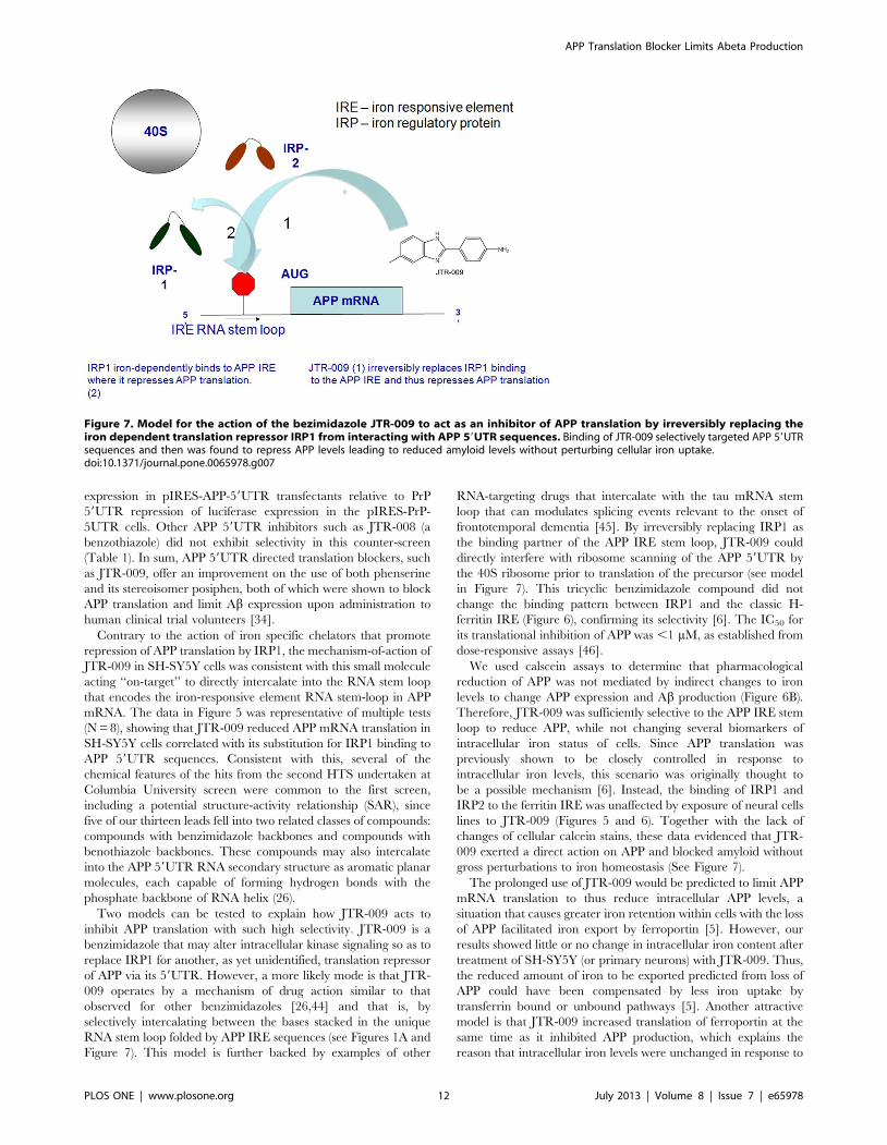
D. Mechanism of action: JTR-009 is a benzimidazole andirreversibly replaces IRP1 from binding to the APP5utranslated region
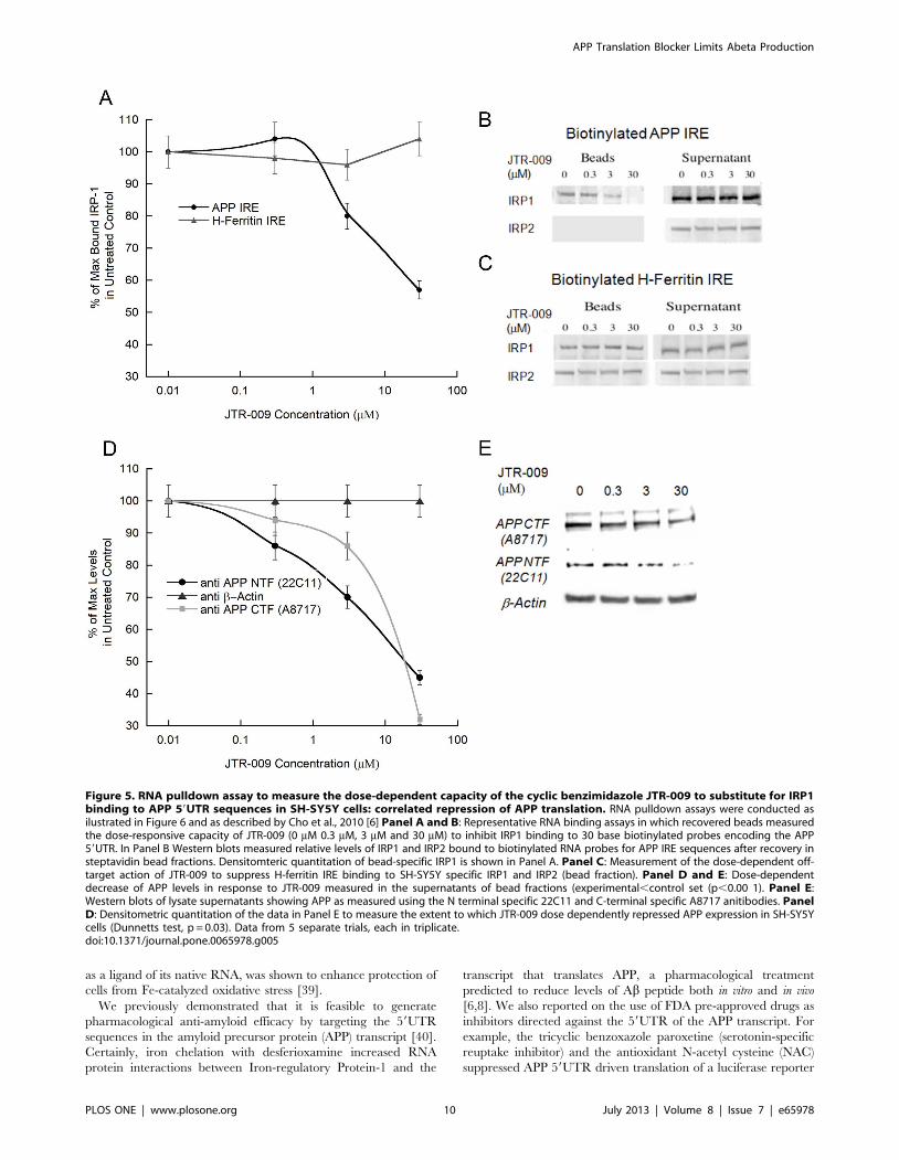
When evaluating the mechanism of JTR-009, we noted that a
low molecular weight RNA intercalator had been previously
reported from the same molecular library source [35], and this
agent had been shown to prevent a tau-mRNA splicing event that
can cause frontotemporal dementia [35]. Therefore, using SH-
SY5Y cells, we employed biotinylated RNA pulldown assays to
measure the effect of JTR-009 on the binding of IRP1 to the APP
5UTR (Figures 5 and 6). Multiple biotinylated RNA pulldown
assays provided data to confirm that this benzimidazole-based
molecule acted ‘‘on-target’’ to substitute for IRP1 interaction as a
repressor of APP translation at the site of the APP iron-responsive
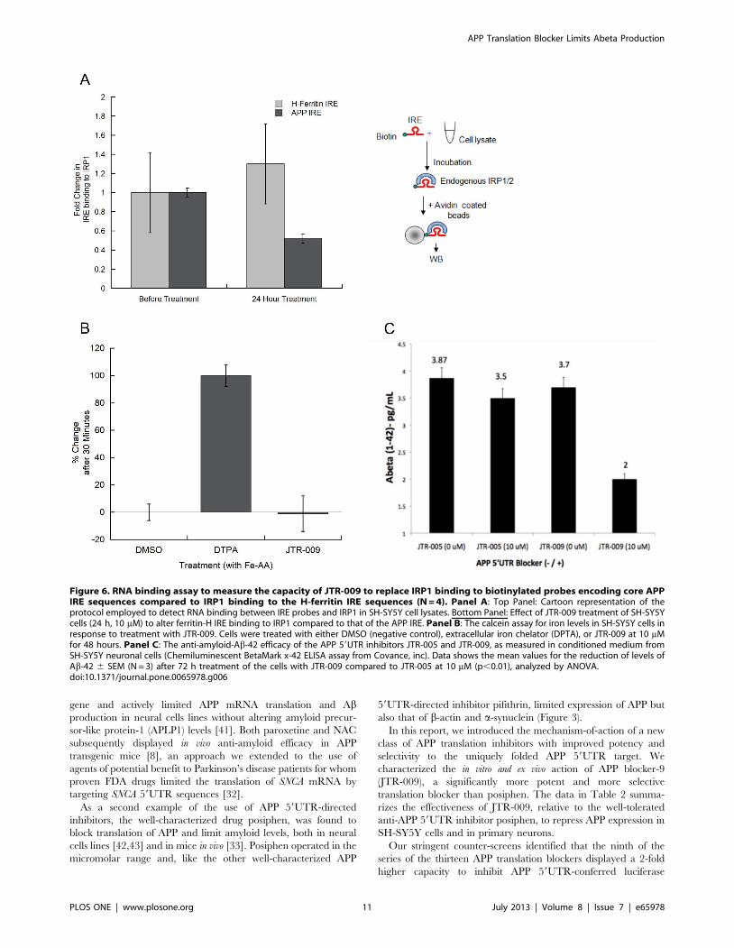
element RNA stem loop. In Panel B of Figure 5 we showed that
administration of of JTR-009 to SH-SY5Y cells dose dependently
diminished the percent of IRP1 bound to biotinylated APP IRE
RNA probes. Densitometry, as shown in Panel B, quantitated that
IRP1 binding was reduced by 20% (62%) at 0.3 mM, by 50% (+/
25%) at 3 mM, and was completely inhibited at 30 mM (61%)
JTR-009 (N = 7, p = 0.003). Confirming specificity, full interaction
between IRP1 and IRP2 and the H-ferritin for IRE probes were
always maintained during conditions of induction with JTR-009
(Densitometry in Panel A is shown to reflect a representative
Western blot in Panel C)).
The data in Figure 5E provides a representative Western blot
experiment from 7 independent experiments when using JTR-009
at 0.3 mM, 3 mM and 30 mM concentrations to inhibit APP
expression in the lysate supernatant fractions of SH-SY5Y cells
subjected to RNA pulldown analysis (Figure 6A). Consistently, we
observed that JTR-009 blocked APP expression as shown by the
decreased levels of the precursor when detected with both C- and
N-terminal specific antibodies. This reduction of APP levels
directly correlated to the elimination of IRP1 binding to APP
59UTR sequences. Figure 5D shows densitometry to obtain the
average reductions of APP levels from multiple Western blots as
represented by Panel E (N = 6 for each set). We observed a 70%+/
25% reduction of APP at 30 mM (N = 4, p = 0.02) and 35% at
3 mM JTR-009 (p = 0.01, Dunnetts post-hoc test).
E. JTR-009 reduced APP expression via its IRE in an ironindependent manner
To confirm an ‘‘on-target’’ mechanism for JTR-009 and its
relative iron-independence when acting via APP 59UTR, we had
previously performed a molecular determination of the iron-
dependent, reversible binding of IRP1 to the APP IRE stem loop
(Kd = 30 pM) [6]. Our data shown in Figure 5 is consistent with
the model that JTR-009 substituted for IRP1 binding to APP
59UTR sequences. In Figure 6A, we compared the extent to which
JTR-009 decreased IRP1 binding of APP-IRE in SH-SY5Y cells
compared to H-Ferritin-IRE RNA probes. Here, the results
consistently showed that 3 mM JTR-009 (48 h exposure) reduced
IRP1 binding to the APP IRE by 2-fold whereas under the same
conditions this benzimidazole displayed no inhibitory change in
binding to H-ferritin IRE probes. These data were consistent with
the conclusion that JTR-009 bound selectively to the APP IRE
sequences and not to related RNA probes encoding the ferritin-H
IRE, an observation consistent with the proposed mechanism of
action of JTR-009 as shown in Figure 7.
We directly measured the effect of JTR-009 on iron homeostasis
in a calcein uptake assay in SH-SY5Y cells (Figure 6B). The drug
(DTPA) was the positive control as an extracellular chelator that
completely blocks iron uptake. Normalized results were graphed
and these data showed that DMSO caused 066% inhibition of the
amount of calcein stain (proportional to iron levels) and DTPA
caused 10068% inhibition. Of note, JTR-009 induced a
21613% inhibition of calcein staining. Thus under these
conditions, we systematically observed that JTR-009 had little or
no effect on iron uptake (TfR dependent and independent uptake
pathways).
Aß-42 peptide is the critical APP derived peptide to trigger the
aggregation of amyloid in both AD and DS and is critically linked
to tau induced neurotoxicity [36]. Thus, we tested and reproduc-
ibly demonstrated that JTR-009 limited Ab-42 secretion from SH-
SY5Y cells by more than two fold. In this representative
experiment, a chemiluminescent BetaMark x-42 ELISA assay
for Ab-42 measurement was employed (Covance, inc). The assays
were carried out according to the manufactures conditions such
that the standard curve was linear and the measured points were
within the ‘standards’ range. JTR-005 exhibited only a 20%
reduction of Ab-42 output after the same 3-day treatment as that
of JTR-009 (72 hour treatment).
In sum, these experiments demonstrated that JTR-009 operated
by direct pathways to reduce APP translation an Ab-42 output and
the compound did not act via indirect pathways as a secondary
iron chelator in which case the drug would be expected to activate
binding of IRP1 as a translational repressor. Our working model
shown in Figure 7 evidenced that JTR-009 interacts directly with
the APP IRE RNA stem loop.
Discussion
RNA-directed drugs have long been used to treat infectious
diseases, e.g. antibiotic aminoglycosides, and small RNA-directed
molecules have been used to control gene expression in cell culture
models [37] (e.g. therapeutic control of viral Hepatitis C/HIV
gene expression [38]). In mammals, endogenous up-regulation of
the translation of the iron storage protein ferritin by ‘yohimbine’,
Table 2. Comparative IC50 of JTR-009 relative to posiphen to inhibit APP 59UTR driven luciferase expression relative to suppressionof APP and Ab levels in SH-SY5Y cells and primary neurons.
Drug APP 59UTR Inhibition (IC50) APP and amyloid-beta Inhibition (IC50) Specificity (610 of IC50) Toxicity (MTT)
JTR-009 100 nM 100 nM(max 10 nM) b-actin (620), asyn (620) 100 mM
Posiphen 5 mM 1 mM (Refs 27, 33) b-actin (620),asyn (61) 100 mM
doi:10.1371/journal.pone.0065978.t002
APP Translation Blocker Limits Abeta Production
PLOS ONE | www.plosone.org 9 July 2013 | Volume 8 | Issue 7 | e65978
as a ligand of its native RNA, was shown to enhance protection of
cells from Fe-catalyzed oxidative stress [39].
We previously demonstrated that it is feasible to generate
pharmacological anti-amyloid efficacy by targeting the 59UTR
sequences in the amyloid precursor protein (APP) transcript [40].
Certainly, iron chelation with desferioxamine increased RNA
protein interactions between Iron-regulatory Protein-1 and the
transcript that translates APP, a pharmacological treatment
predicted to reduce levels of Ab peptide both in vitro and in vivo
[6,8]. We also reported on the use of FDA pre-approved drugs as
inhibitors directed against the 59UTR of the APP transcript. For
example, the tricyclic benzoxazole paroxetine (serotonin-specific
reuptake inhibitor) and the antioxidant N-acetyl cysteine (NAC)
suppressed APP 59UTR driven translation of a luciferase reporter
Figure 5. RNA pulldown assay to measure the dose-dependent capacity of the cyclic benzimidazole JTR-009 to substitute for IRP1binding to APP 59UTR sequences in SH-SY5Y cells: correlated repression of APP translation. RNA pulldown assays were conducted asilustrated in Figure 6 and as described by Cho et al., 2010 [6] Panel A and B: Representative RNA binding assays in which recovered beads measuredthe dose-responsive capacity of JTR-009 (0 mM 0.3 mM, 3 mM and 30 mM) to inhibit IRP1 binding to 30 base biotinylated probes encoding the APP59UTR. In Panel B Western blots measured relative levels of IRP1 and IRP2 bound to biotinylated RNA probes for APP IRE sequences after recovery insteptavidin bead fractions. Densitomteric quantitation of bead-specific IRP1 is shown in Panel A. Panel C: Measurement of the dose-dependent off-target action of JTR-009 to suppress H-ferritin IRE binding to SH-SY5Y specific IRP1 and IRP2 (bead fraction). Panel D and E: Dose-dependentdecrease of APP levels in response to JTR-009 measured in the supernatants of bead fractions (experimental,control set (p,0.00 1). Panel E:Western blots of lysate supernatants showing APP as measured using the N terminal specific 22C11 and C-terminal specific A8717 anitibodies. PanelD: Densitometric quantitation of the data in Panel E to measure the extent to which JTR-009 dose dependently repressed APP expression in SH-SY5Ycells (Dunnetts test, p = 0.03). Data from 5 separate trials, each in triplicate.doi:10.1371/journal.pone.0065978.g005
APP Translation Blocker Limits Abeta Production
PLOS ONE | www.plosone.org 10 July 2013 | Volume 8 | Issue 7 | e65978
gene and actively limited APP mRNA translation and Abproduction in neural cells lines without altering amyloid precur-
sor-like protein-1 (APLP1) levels [41]. Both paroxetine and NAC
subsequently displayed in vivo anti-amyloid efficacy in APP
transgenic mice [8], an approach we extended to the use of
agents of potential benefit to Parkinson’s disease patients for whom
proven FDA drugs limited the translation of SNCA mRNA by
targeting SNCA 59UTR sequences [32].
As a second example of the use of APP 59UTR-directed
inhibitors, the well-characterized drug posiphen, was found to
block translation of APP and limit amyloid levels, both in neural
cells lines [42,43] and in mice in vivo [33]. Posiphen operated in the
micromolar range and, like the other well-characterized APP
59UTR-directed inhibitor pifithrin, limited expression of APP but
also that of b-actin and a-synuclein (Figure 3).
In this report, we introduced the mechanism-of-action of a new
class of APP translation inhibitors with improved potency and
selectivity to the uniquely folded APP 59UTR target. We
characterized the in vitro and ex vivo action of APP blocker-9
(JTR-009), a significantly more potent and more selective
translation blocker than posiphen. The data in Table 2 summa-
rizes the effectiveness of JTR-009, relative to the well-tolerated
anti-APP 59UTR inhibitor posiphen, to repress APP expression in
SH-SY5Y cells and in primary neurons.
Our stringent counter-screens identified that the ninth of the
series of the thirteen APP translation blockers displayed a 2-fold
higher capacity to inhibit APP 59UTR-conferred luciferase
Figure 6. RNA binding assay to measure the capacity of JTR-009 to replace IRP1 binding to biotinylated probes encoding core APPIRE sequences compared to IRP1 binding to the H-ferritin IRE sequences (N = 4). Panel A: Top Panel: Cartoon representation of theprotocol employed to detect RNA binding between IRE probes and IRP1 in SH-SY5Y cell lysates. Bottom Panel: Effect of JTR-009 treatment of SH-SY5Ycells (24 h, 10 mM) to alter ferritin-H IRE binding to IRP1 compared to that of the APP IRE. Panel B: The calcein assay for iron levels in SH-SY5Y cells inresponse to treatment with JTR-009. Cells were treated with either DMSO (negative control), extracellular iron chelator (DPTA), or JTR-009 at 10 mMfor 48 hours. Panel C: The anti-amyloid-Ab-42 efficacy of the APP 59UTR inhibitors JTR-005 and JTR-009, as measured in conditioned medium fromSH-SY5Y neuronal cells (Chemiluminescent BetaMark x-42 ELISA assay from Covance, inc). Data shows the mean values for the reduction of levels ofAb-42 6 SEM (N = 3) after 72 h treatment of the cells with JTR-009 compared to JTR-005 at 10 mM (p,0.01), analyzed by ANOVA.doi:10.1371/journal.pone.0065978.g006
APP Translation Blocker Limits Abeta Production
PLOS ONE | www.plosone.org 11 July 2013 | Volume 8 | Issue 7 | e65978
expression in pIRES-APP-59UTR transfectants relative to PrP
59UTR repression of luciferase expression in the pIRES-PrP-
5UTR cells. Other APP 59UTR inhibitors such as JTR-008 (a
benzothiazole) did not exhibit selectivity in this counter-screen
(Table 1). In sum, APP 59UTR directed translation blockers, such
as JTR-009, offer an improvement on the use of both phenserine
and its stereoisomer posiphen, both of which were shown to block
APP translation and limit Ab expression upon administration to
human clinical trial volunteers [34].
Contrary to the action of iron specific chelators that promote
repression of APP translation by IRP1, the mechanism-of-action of
JTR-009 in SH-SY5Y cells was consistent with this small molecule
acting ‘‘on-target’’ to directly intercalate into the RNA stem loop
that encodes the iron-responsive element RNA stem-loop in APP
mRNA. The data in Figure 5 was representative of multiple tests
(N = 8), showing that JTR-009 reduced APP mRNA translation in
SH-SY5Y cells correlated with its substitution for IRP1 binding to
APP 59UTR sequences. Consistent with this, several of the
chemical features of the hits from the second HTS undertaken at
Columbia University screen were common to the first screen,
including a potential structure-activity relationship (SAR), since
five of our thirteen leads fell into two related classes of compounds:
compounds with benzimidazole backbones and compounds with
benothiazole backbones. These compounds may also intercalate
into the APP 59UTR RNA secondary structure as aromatic planar
molecules, each capable of forming hydrogen bonds with the
phosphate backbone of RNA helix (26).
Two models can be tested to explain how JTR-009 acts to
inhibit APP translation with such high selectivity. JTR-009 is a
benzimidazole that may alter intracellular kinase signaling so as to
replace IRP1 for another, as yet unidentified, translation repressor
of APP via its 59UTR. However, a more likely mode is that JTR-
009 operates by a mechanism of drug action similar to that
observed for other benzimidazoles [26,44] and that is, by
selectively intercalating between the bases stacked in the unique
RNA stem loop folded by APP IRE sequences (see Figures 1A and
Figure 7). This model is further backed by examples of other
RNA-targeting drugs that intercalate with the tau mRNA stem
loop that can modulates splicing events relevant to the onset of
frontotemporal dementia [45]. By irreversibly replacing IRP1 as
the binding partner of the APP IRE stem loop, JTR-009 could
directly interfere with ribosome scanning of the APP 59UTR by
the 40S ribosome prior to translation of the precursor (see model
in Figure 7). This tricyclic benzimidazole compound did not
change the binding pattern between IRP1 and the classic H-
ferritin IRE (Figure 6), confirming its selectivity [6]. The IC50 for
its translational inhibition of APP was ,1 mM, as established from
dose-responsive assays [46].
We used calscein assays to determine that pharmacological
reduction of APP was not mediated by indirect changes to iron
levels to change APP expression and Ab production (Figure 6B).
Therefore, JTR-009 was sufficiently selective to the APP IRE stem
loop to reduce APP, while not changing several biomarkers of
intracellular iron status of cells. Since APP translation was
previously shown to be closely controlled in response to
intracellular iron levels, this scenario was originally thought to
be a possible mechanism [6]. Instead, the binding of IRP1 and
IRP2 to the ferritin IRE was unaffected by exposure of neural cells
lines to JTR-009 (Figures 5 and 6). Together with the lack of
changes of cellular calcein stains, these data evidenced that JTR-
009 exerted a direct action on APP and blocked amyloid without
gross perturbations to iron homeostasis (See Figure 7).
The prolonged use of JTR-009 would be predicted to limit APP
mRNA translation to thus reduce intracellular APP levels, a
situation that causes greater iron retention within cells with the loss
of APP facilitated iron export by ferroportin [5]. However, our
results showed little or no change in intracellular iron content after
treatment of SH-SY5Y (or primary neurons) with JTR-009. Thus,
the reduced amount of iron to be exported predicted from loss of
APP could have been compensated by less iron uptake by
transferrin bound or unbound pathways [5]. Another attractive
model is that JTR-009 increased translation of ferroportin at the
same time as it inhibited APP production, which explains the
reason that intracellular iron levels were unchanged in response to
Figure 7. Model for the action of the bezimidazole JTR-009 to act as an inhibitor of APP translation by irreversibly replacing theiron dependent translation repressor IRP1 from interacting with APP 59UTR sequences. Binding of JTR-009 selectively targeted APP 59UTRsequences and then was found to repress APP levels leading to reduced amyloid levels without perturbing cellular iron uptake.doi:10.1371/journal.pone.0065978.g007
APP Translation Blocker Limits Abeta Production
PLOS ONE | www.plosone.org 12 July 2013 | Volume 8 | Issue 7 | e65978
JTR-009. For example, JTR-009 could have altered IRP1 binding
to the IRE in DMT1 mRNA and/or ferroportin mRNA to
account for the predicted compensatory increase in cellular iron
expected by loss of APP expression since APP is an iron export
ferroxidase [5].
Consistent with the capacity of JTR-009 to maintain correct
iron balance and to operate as a potent anti-amyloid agent, we
observed this benzimidazole-enhanced cell viability by use of MTS
and LDH assays. In fact, JTR0-009 was pharmacologically non-
toxic in SH-SY5Y cells as measured by MTS assay (Figure 3D).
Dose-responsive measurements confirmed the IC50 against the
APP 59UTR to be in the 1 nM to 100 nM range while the APP
59UTR-directed JTR-009, only displayed cell toxiciy at concen-
trations .30 mM in SH-SY5Y cells and in primary neurons.
Significantly, JTR-009 exhibited a similar toxicity at 100 mM in
SH-SY5Y cells as was previously demonstrated for posiphen (+ve
control) [21].
Certainly JTR-009 was a highly selective APP inhibitor that
operated at very low concentrations in the nanomolar range, as
has been evident in other kinase inhibitors. The series of
experiments shown in Figure 4 revealed that low doses of JTR-
009 typically reduced APP expression. Intracellular APP inhibition
was quantified by densitometry such that both the N-terminus
22C11 antibody and the C-terminus A8717 antibody of APP were
cross-referenced. We found that each of the APP 59UTR
inhibitors lowered Ab secretion from SH-SY5Y neuroblastoma
cells (Figure 2), albeit at levels near the limits of detection for Ab(pg/ml range).
Of note, the potency of JTR-009 to inhibit APP 59UTR
conferred translation was greater than posiphen (Figure 4A). In
primary mouse E-18 neurons, JTR-009 inhibited APP levels with
no reduction of b-actin at doses as low as 1 nM and, dose-
responsively, as high as 100 nM (Figure 4D). The same
concentrations of JTR-009 demonstrated no significant changes
to a-synuclein levels (the western blots were standardized to b-
actin) (Figure 4D).
We sought to more stringently identify novel agents as
translation blockers specifically of APP 59UTR sequences by
counter-screening APP inhibitor leads against the PrP (Vt-2)
59UTR. To this purpose, we employed SH-SH5Y cells stably
transfected with the pIRES-APP-59UTR and pIRES-PrP-59UTR
constructs, which expressed a luciferase reporter driven respec-
tively by the APP and PrP (V2) 59UTRs (Figure 1D). While
undertaking the counter-screens, we identified an unexpectedly
close 56% identity between PrP and APP mRNAs as well as a
putative IRP1 binding site in the 59untranslated regions in both
mRNAs. These similarities provided an explanation as to why our
dose-responsive counter-screen identified similar IC50 values for
both APP and PrP 59UTR dependent inhibition of luciferase
reporter expression (Table 1).
Consistent with these observations, recent reports linked the
function of both APP [5,6] and PrP to a common role in iron
transport where both these neurodegenerative gene products are
differentially increased by iron [29,47]. APP mRNA encodes a
fully functional IRE [6] and our bioinformatic alignments showed
that secondary structures of the 59UTRs of several neurodegen-
erative transcripts also appear to encode IRE RNA stem loops, for
instance in the related 59UTR of PrP mRNAs (Figure 1). This
unexpected close sequence similarity between the PrP 59UTR to
that of the APP 59UTR may explain why many of the top APP
specific inhibitors were found to also repressed prion (PrP)
expression. Certainly these two neurodegenerative proteins are
linked with a common role in iron transport and metabolism [29]
where recent discoveries also show an in vivo interaction of the Abamyloid protein and the PrP prion during the progression of
neurodegenerative disease [48]. However, these findings also
underscored the specificity of JTR-009, which inhibited APP
expression 10-fold more actively than that of the prion PrP
protein.
Conclusions
The single major conclusion from this work is that APP 59UTR
sequences indeed are a significant regulator of Ab precursor
expression in neural cells. In this report, it was via APP 59UTR-
dependent pathways that we pharmacologically limited the steady
state levels of APP not only in SH-SY5Y cells but also in primary
cortical neurons. These APP 59UTRs-directed inhibitors are
currently being tested as representatives of a class of intercalators
that exhibit high selectivity in reducing APP production in SH-
SY5Y neuroblastoma cells. They also limited Ab production with
little or no perturbation of cellular iron homeostasis while
maintaining neuronal viability. Such capabilities are the require-
ments for future drugs with therapeutic potential for AD and
Down syndrome. On a technical level, the optimal compound was
shown to be a benzimidazole, JTR-009, that limits APP translation
at less than one nanomolar concentrations with little evidence of
neurotoxicity. This tricyclic compound inhibits APP translation by
directly interacting with the IRE in the 59UTR of APP mRNA,
irreversibly replacing IRP1 as a repressor of translation. JTR-009
was 10-fold more effective to limit APP translation than posiphen,
a well tolerated APP 59UTR-directed translation blocker [33].
Acknowledgments
Dr. Jack Rogers is grateful Dr. Marcie Glicksman and Dr. Lars Branden,
respectively the directors of the LDDN, Brigham and Women’s Hospital,
Cambridge, MA (MG) and of the Columbia University Screening Center,
NY, NY (LB).
Author Contributions
Conceived and designed the experiments: JTR SB. Performed the
experiments: CH XH CC JTR SB AB DKL. Analyzed the data: CH
JTR SB. Contributed reagents/materials/analysis tools: JTR. Wrote the
paper: SB JTR. Wrote and edited the paper using microsoft word for text
files and photoshop graphics for figure production: CH JTR.
References
1. Mackenzie IR, Rademakers R, Neumann M (2010) TDP-43 and FUS in
amyotrophic lateral sclerosis and frontotemporal dementia. Lancet Neurol 9:
995–1007.
2. Darnell JC, Van Driesche SJ, Zhang C, Hung KY, Mele A, et al. (2011) FMRP
stalls ribosomal translocation on mRNAs linked to synaptic function and autism.
Cell 146: 247–261.
3. Lovell MA, Robertson JD, Teesdale WJ, Campbell JL, Markesbery WR (1998)
Copper, iron and zinc in Alzheimer’s disease senile plaques. J Neurol Sci 158:
47–52.
4. Bandyopadhyay S, Huang X, Lahiri DK, Rogers JT (2011) Novel drug targets
based on metallobiology of Alzheimer’s disease. Expert Opin Ther Targets 14:
1177–1197.
5. Duce JA, Tsatsanis A, Cater MA, James SA, Robb E, et al. (2010) Iron-export
ferroxidase activity of beta-amyloid precursor protein is inhibited by zinc in
Alzheimer’s disease. Cell 142: 857–867.
6. Cho HH, Cahill CM, Vanderburg CR, Scherzer CR, Wang B, et al. (2010)
Selective translational control of the Alzheimer amyloid precursor protein
transcript by iron regulatory protein-1. J Biol Chem 285: 31217–31232.
APP Translation Blocker Limits Abeta Production
PLOS ONE | www.plosone.org 13 July 2013 | Volume 8 | Issue 7 | e65978
7. Stys A, Galy B, Starzynski RR, Smuda E, Drapier JC, et al. (2011) Iron
regulatory protein 1 outcompetes iron regulatory protein 2 in regulating cellulariron homeostasis in response to nitric oxide. J Biol Chem 286: 22846–22854.
8. Tucker S, Ahl M, Cho HH, Bandyopadhyay S, Cuny GD, et al. (2006) RNA
Therapeutics Directed to the Non Coding Regions of APP mRNA, In VivoAnti-Amyloid Efficacy of Paroxetine, Erythromycin, and N-acetyl cysteine. Curr
Alzheimer Res 3: 221–227.9. Salehi A, Delcroix JD, Belichenko PV, Zhan K, Wu C, et al. (2006) Increased
App Expression in a Mouse Model of Down’s Syndrome Disrupts NGF
Transport and Causes Cholinergic Neuron Degeneration. Neuron 51: 29–42.10. Salehi A, Faizi M, Colas D, Valletta J, Laguna J, et al. (2009) Restoration of
Norepinephrine-Modulated Contextrual memory in a Mouse Model of DownSyndrome. Science Translational Medicine 1: 1–9.
11. Granic A, Padmanabhan J, Norden M, Potter H (2012) Alzheimer Abetapeptide induces chromosome mis-segregation and aneuploidy, including trisomy
21: requirement for tau and APP. Mol Biol Cell 21: 511–520.
12. Hooli BV, Mohapatra G, Mattheisen M, Parrado AR, Roehr JT, et al. (2012)Role of common and rare APP DNA sequence variants in Alzheimer disease.
Neurology 78: 1250–1257.13. Rovelet-Lecrux A, Hannequin D, Raux G, Meur NL, Laquerriere A, et al.
(2006) APP locus duplication causes autosomal dominant early-onset Alzheimer
disease with cerebral amyloid angiopathy. Nat Genet 38: 24–26.14. Perry G, Nunomura A, Hirai K, Zhu X, Perez M, et al. (2002) Is oxidative
damage the fundamental pathogenic mechanism of Alzheimer’s and otherneurodegenerative diseases? Free Radic Biol Med 33: 1475–1479.
15. Smith CD, Carney JM, Starke-Reed PE, Oliver CN, Stadtman ER, et al. (1991)Excess brain protein oxidation and enzyme dysfunction in normal aging and in
Alzheimer disease. Proc Natl Acad Sci U S A 88: 10540–10543.
16. Butterfield DA (2012) Oxidative stress in Alzheimer disease: synergy between theButterfield and Markesbery laboratories. Neuromolecular Med 13: 19–22.
17. Kwon OD, Khaleeq A, Chan W, Pavlik VN, Doody RS (2012) ApolipoproteinE polymorphism and age at onset of Alzheimer’s disease in a quadriethnic
sample. Dement Geriatr Cogn Disord 30: 486–491.
18. Wisniewski T, Castano EM, Golabek A, Vogel T, Frangione B (1994)Acceleration of Alzheimer’s fibril formation by apolipoprotein E in vitro.
Am J Pathol 145: 1030–1035.19. Nilsson LN, Arendash GW, Leighty RE, Costa DA, Low MA, et al. (2004)
Cognitive impairment in PDAPP mice depends on ApoE and ACT-catalyzedamyloid formation. Neurobiol Aging 25: 1153–1167.
20. McNaughton D, Knight W, Guerreiro R, Ryan N, Lowe J, et al. (2012)
Duplication of amyloid precursor protein (APP), but not prion protein (PRNP)gene is a significant cause of early onset dementia in a large UK series.
Neurobiol Aging 33: 426 e413–421.21. Bandyopadhyay S, Ni J, Ruggiero A, Walshe K, Rogers MS, et al. (2006) A
high-throughput drug screen targeted to the 59untranslated region of Alzheimer
amyloid precursor protein mRNA. J Biomol Screen 11: 469–480.22. Kuo YM, Li Z, Jiao Y, Gaborit N, Pani AK, et al. (2010) Extensive enteric
nervous system abnormalities in mice transgenic for artificial chromosomescontaining Parkinson disease-associated alpha-synuclein gene mutations precede
central nervous system changes. Hum Mol Genet 19: 1633–1650.23. Ray B, Bailey JA, Sarkar S, Lahiri DK (2009) Molecular and immunocyto-
chemical characterization of primary neuronal cultures from adult rat brain:
Differential expression of neuronal and glial protein markers. J NeurosciMethods 184: 294–302.
24. Kurogi Y, Miyata K, Okamura T, Hashimoto K, Tsutsumi K, et al. (2001)Discovery of novel mesangial cell proliferation inhibitors using a three-
dimensional database searching method. J Med Chem 44: 2304–2307.
25. Gilman CP, Chan SL, Guo Z, Zhu X, Greig N, et al. (2003) p53 is present insynapses where it mediates mitochondrial dysfunction and synaptic degeneration
in response to DNA damage, and oxidative and excitotoxic insults. Neuromo-lecular Med 3: 159–172.
26. Schulz WG, Nieman RA, Skibo EB (1995) Evidence for DNA phosphate
backbone alkylation and cleavage by pyrrolo[1,2-a]benzimidazoles: smallmolecules capable of causing base-pair-specific phosphodiester bond hydrolysis.
Proc Natl Acad Sci U S A 92: 11854–11858.27. Mikkilineni S, Cantuti-Castelvetri I, Cahill CM, Balliedier A, Greig NH, et al.
(2012) The anticholinesterase phenserine and its enantiomer posiphen as59untranslated-region-directed translation blockers of the Parkinson’s alpha
synuclein expression. Parkinsons Dis 2012: 142372.
28. Goforth JB, Anderson SA, Nizzi CP, Eisenstein RS (2010) Multiple determinantswithin iron-responsive elements dictate iron regulatory protein binding and
regulatory hierarchy. Rna 16: 154–169.
29. Singh A, Kong Q, Luo X, Petersen RB, Meyerson H, et al. (2009) Prion protein(PrP) knock-out mice show altered iron metabolism: a functional role for PrP in
iron uptake and transport. PLoS One 4: e6115.
30. Wolk DA, Klunk W (2009) Update on amyloid imaging: from healthy aging to
Alzheimer’s disease. Curr Neurol Neurosci Rep 9: 345–352.
31. Mathis CA, Wang Y, Holt DP, Huang GF, Debnath ML, et al. (2003) Synthesisand evaluation of 11C-labeled 6-substituted 2-arylbenzothiazoles as amyloid
imaging agents. J Med Chem 46: 2740–2754.
32. Rogers JT, Mikkilineni S, Cantuti-Castelvetri I, Smith DH, et al. (2011) The
alpha-synuclein 59untranslated region targeted translation blockers: anti-alphasynuclein efficacy of cardiac glycosides and Posiphen. J Neural Transm 118:
493–507.
33. Lahiri DK, Chen D, Maloney B, Holloway HW, Yu QS, et al. (2007) The
experimental Alzheimer’s disease drug posiphen [(+)-phenserine] lowersamyloid-beta peptide levels in cell culture and mice. J Pharmacol Exp Ther
320: 386–396.
34. Maccecchini M Roffman M, Greig NH (2009) Posiphen lowers amyloidprecursor protein and amyloid b as well as acetylcholinesterase levels in culture,
animals and humans Alzheimer’s & Dementia 5(4S): 47–48.
35. Donahue CP, Ni J, Rozners E, Glicksman MA, Wolfe MS (2007) Identification
of tau stem loop RNA stabilizers. J Biomol Screen 12: 789–799.
36. Iijima K, Gatt A, Iijima-Ando K (2013) Tau Ser262 phosphorylation is criticalfor Abeta42-induced tau toxicity in a transgenic Drosophila model of
Alzheimer’s disease. Hum Mol Genet 19: 2947–2957.
37. Werstuck G, Green MR (1998) Controlling gene expression in living cells
through small molecule-RNA interactions. Science 282: 296–298.
38. Lancaster AM, Jan E, Sarnow P (2006) Initiation factor-independent translationmediated by the hepatitis C virus internal ribosome entry site. Rna 12: 894–902.
39. Tibodeau JD, Fox PM, Ropp PA, Theil EC, Thorp HH (2006) The up-
regulation of ferritin expression using a small-molecule ligand to the native
mRNA. Proc Natl Acad Sci U S A 103: 253–257.
40. Venti A, Giordano T, Eder P, Lahiri DK, Greig NH, et al. (2004) TheIntegrated Role of Desferrioxamine and Phenserine Targeted to an Iron-
Responsive Element in the APP-mRNA 59-Untranslated Region. Ann N Y Acad
Sci 1035: 34–48.
41. Morse LJ, Payton SM, Cuny GD, Rogers JT (2004) FDA-Preapproved DrugsTargeted to the Translational Regulation and Processing of the Amyloid
Precursor Protein. J Mol Neurosci 24: 129–136.
42. Shaw KT, Utsuki T, Rogers J, Yu QS, Sambamurti K, et al. (2001) Phenserine
regulates translation of beta -amyloid precursor protein mRNA by a putativeinterleukin-1 responsive element, a target for drug development. Proc Natl Acad
Sci U S A 98: 7605–7610.
43. Utsuki T, Yu QS, Davidson D, Chen D, Holloway HW, et al. (2006)Identification of novel small molecule inhibitors of amyloid precursor protein
synthesis as a route to lower Alzheimer’s disease amyloid-beta peptide.
J Pharmacol Exp Ther 318: 855–862.
44. Skibo EB, Islam I, Heileman MJ, Schulz WG (1994) Structure-activity studies ofbenzimidazole-based DNA-cleaving agents. Comparison of benzimidazole,
pyrrolobenzimidazole, and tetrahydropyridobenzimidazole analogues. J MedChem 37: 78–92.
45. Donahue CP, Muratore C, Wu JY, Kosik KS, Wolfe MS (2006) Stabilization ofthe tau exon 10 stem loop alters pre-mRNA splicing. J Biol Chem 281: 23302–
23306.
46. Hamy F, Felder ER, Heizmann G, Lazdins J, Aboul-ela F, et al. (1997) Aninhibitor of the Tat/TAR RNA interaction that effectively suppresses HIV-1
replication. Proc Natl Acad Sci U S A 94: 3548–3553.
47. Singh A, Isaac AO, Luo X, Mohan ML, Cohen ML, et al. (2009) Abnormal
brain iron homeostasis in human and animal prion disorders. PLoS Pathog 5:e1000336.
48. Morales R, Estrada LD, Diaz-Espinoza R, Morales-Scheihing D, Jara MC, et al.
(2012) Molecular cross talk between misfolded proteins in animal models of
Alzheimer’s and prion diseases. J Neurosci 30: 4528–4535.
49. Friedlich AL, Tanzi RE, Rogers JT (2007) The 59-untranslated region ofParkinson’s disease alpha-synuclein messengerRNA contains a predicted iron
responsive element. Mol Psychiatry 12: 222–223.
APP Translation Blocker Limits Abeta Production
PLOS ONE | www.plosone.org 14 July 2013 | Volume 8 | Issue 7 | e65978