tgfβ signaling inhibits goblet cell differentiation via...
TRANSCRIPT

RESEARCH ARTICLE
TGFβ signaling inhibits goblet cell differentiation via SPDEF inconjunctival epitheliumHeather A. McCauley1, Chia-Yang Liu2, Aria C. Attia3, Kathryn A. Wikenheiser-Brokamp3,4, Yujin Zhang2,Jeffrey A. Whitsett3 and Geraldine Guasch1,*
ABSTRACTThe ocular surface epithelia, including the stratified but non-keratinized corneal, limbal and conjunctival epithelium, in concertwith the epidermal keratinized eyelid epithelium, function together tomaintain eye health and vision. Abnormalities in cellular proliferationor differentiation in any of these surface epithelia are central in thepathogenesis of many ocular surface disorders. Goblet cells areimportant secretory cell components of various epithelia, includingthe conjunctiva; however, mechanisms that regulate goblet celldifferentiation in the conjunctiva are not well understood. Herein, wereport that conditional deletion of transforming growth factor βreceptor II (Tgfbr2) in keratin 14-positive stratified epithelia causesocular surface epithelial hyperplasia and conjunctival goblet cellexpansion that invaginates into the subconjunctival stroma in themouse eye. We found that, in the absence of an external phenotype,the ocular surface epithelium develops properly, but young micedisplayed conjunctival goblet cell expansion, demonstrating thatTGFβ signaling is required for normal restriction of goblet cells withinthe conjunctiva. We observed increased expression of SAM-pointeddomain containing ETS transcription factor (SPDEF) in stratifiedconjunctival epithelial cells in Tgfbr2 cKOmice, suggesting that TGFβrestricted goblet cell differentiation directly by repressing Spdeftranscription. Gain of function of Spdef in keratin 14-positive epitheliaresulted in the ectopic formation of goblet cells in the eyelid andperipheral cornea in adult mice. We found that Smad3 bound twodistinct sites on the Spdef promoter and that treatment of keratin14-positive cells with TGFβ inhibited SPDEF activation, therebyidentifying a novel mechanistic role for TGFβ in regulating goblet celldifferentiation.
KEY WORDS: Conjunctiva, Differentiation, Goblet cells, SPDEF,TGFβ signaling, Mouse
INTRODUCTIONThe ocular surface epithelia, including the stratified but non-keratinized corneal, limbal, and conjunctival epithelium, in concertwith the epidermal, keratinized eyelid epithelium, function together toenable eye health and vision (Swamynathan, 2013). The transparentcornea is essential for vision, and increases in cellular stratification and
keratinization of the cornea result in vision loss or blindness and areassociated with many severe ocular surface diseases (Li et al., 2007).The limbus exists at the transition between corneal and conjunctivalepithelium, and is a stem cell niche containing limbal stem cells thatmigrate centripetally to maintain the cornea (Pellegrini et al., 1999;Swamynathan, 2013). Conjunctival epithelium, which encompassesthe palpebral conjunctiva, the fornix and the bulbar conjunctiva, linesthe inner surface of the eyelid, is composed of stratified epitheliuminterspersed with goblet cells, and adjoins the cornea at the limbus(Wei et al., 1993, 1997; Pellegrini et al., 1999). Goblet cells areimportant secretory cell components of various epithelia, includingthe lung (Park et al., 2007; Chen et al., 2009; Tompkins et al., 2009),the intestine (Radtke and Clevers, 2005) and the conjunctiva (Weiet al., 1993, 1995, 1997; Mantelli and Argueso, 2008). Within theconjunctiva, goblet cells secrete aqueous mucins that protect thesurface of the eye by contributing to the tear film to maintainlubrication, clear molecules and maintain mucosal barrier integrity(Wei et al., 1993; Mantelli and Argueso, 2008). Abnormalities ingoblet cell function are associated with dry eye syndrome and otherdrying diseases (Mantelli and Argueso, 2008; Marko et al., 2013;Zhang et al., 2013), and lead to perturbations of the ocular surfaceepithelium that negatively affect eye health and vision. Mechanismsthat regulate goblet cell differentiation in the conjunctiva are not wellunderstood, thereby limiting therapeutic options for ocular surfacedisorders, such as dry eye syndrome, largely to analgesic measures,such as artificial tears (Cornec et al., 2014).
Although the precise regulatory networks governing goblet celldifferentiation in the conjunctiva are unknown, conjunctivalphenotypes in murine models have identified Krüppel-like family4 (KLF4) (Swamynathan et al., 2007; Gupta et al., 2011) and5 (KLF5) (Kenchegowda et al., 2011), SAM pointed domain-containing ETS transcription factor (SPDEF) (Marko et al., 2013)and the Notch signaling pathway (Zhang et al., 2013) as essentialfactors required for goblet cell differentiation in the mouseconjunctiva, as deletion of these genes, or inhibition of Notchsignaling, results in conjunctiva that lack goblet cells. SPDEF playsa crucial role in goblet cell differentiation in multiple organs,including the lung (Park et al., 2007), the intestine (Gregorieff et al.,2009; Noah et al., 2010) and the conjunctiva (Marko et al., 2013).Although upstream regulators of SPDEF have been reported in thelung (Ren et al., 2013) and the intestine (Aronson et al., 2014), lessis known about its upstream regulators in the conjunctiva.
Transforming growth factor β (TGFβ) has an established role incontrolling epithelial cell proliferation and differentiation(Derynck et al., 1998; Feng and Derynck, 2005). In the presenceof TGFβ ligand, the transmembrane serine/threonine kinasesTGFβ receptor I (TGFβRI) and receptor II (TGFβRII)heterodimerize to phosphorylate Smad2 (Derynck et al., 1998;Massague and Wotton, 2000). Phosphorylated Smad2 and Smad3form a heteromeric complex in the cytoplasm with Smad4, theReceived 12 September 2014; Accepted 6 October 2014
1Division of Developmental Biology, Cincinnati Children’s Hospital Medical Center,3333 Burnett Avenue, Cincinnati, OH 45229, USA. 2Department of Ophthalmology,Edith J. Crawley Vision Research Center, College of Medicine, University ofCincinnati, Cincinnati, OH 45267, USA. 3Division of Pulmonary Biology, CincinnatiChildren’s Hospital Medical Center, 3333 Burnett Avenue, Cincinnati, OH 45229,USA. 4Pathology and Laboratory Medicine, Cincinnati Children’s Hospital MedicalCenter and University of Cincinnati, 3333 Burnett Avenue, Cincinnati, OH 45229,USA.
*Author for correspondence ([email protected])
4628
© 2014. Published by The Company of Biologists Ltd | Development (2014) 141, 4628-4639 doi:10.1242/dev.117804
DEVELO
PM
ENT
common mediator of TGFβ and bone morphogenetic protein(BMP) signaling, which translocates to the nucleus and directlybinds DNA to effect transcription of target genes (Derynck et al.,1998; Massague and Wotton, 2000; McNairn et al., 2013a). BMPsignaling, via Smad4, has been implicated in eyelid developmentand differentiation of the mouse conjunctiva at early stages, butloss of components of canonical TGFβ signaling failed to producea perinatal phenotype using Le-Cre (Huang et al., 2009). AlthoughTGFβ signaling is important for corneal epithelial wound healing(Terai et al., 2011), and loss of Tgfbr2 in CD4+ T cells induces animmune response in the eye (DePaiva et al., 2011), a cell-autonomous function for TGFβ signaling in conjunctivalepithelial cell fate or goblet cell differentiation has not beenidentified.Here, we report that conditional deletion of Tgfbr2 in keratin 14
(K14)-positive stratified epithelia causes ocular surface epithelialhyperplasia and conjunctival goblet cell expansion that invaginatesinto the subconjunctival stroma in the mouse eye. We found that theocular surface epithelium develops properly in the absence of TGFβsignaling, but young asymptomatic mice displayed conjunctivalgoblet cell expansion, demonstrating that TGFβ signaling isrequired for restriction of goblet cells differentiation within theconjunctiva. The adult hyperplastic Tgfbr2-deficient palpebralconjunctiva retained its conjunctival identity but displayedincreased SPDEF expression, not only in goblet cells but also in
stratified conjunctival epithelial cells. Overexpression of SPDEF inK14-positive epithelia resulted in the formation of ectopic gobletcells in the eyelid and peripheral cornea in adult mice. Wehypothesized that TGFβ restricted goblet cell differentiation directlyby repressing Spdef transcription. We found that Smad3 bound twodistinct sites on the Spdef promoter and that treatment of K14-positive cells with TGFβ inhibited SPDEF activation, therebyidentifying a novel mechanistic role for TGFβ in the regulation ofgoblet cell differentiation.
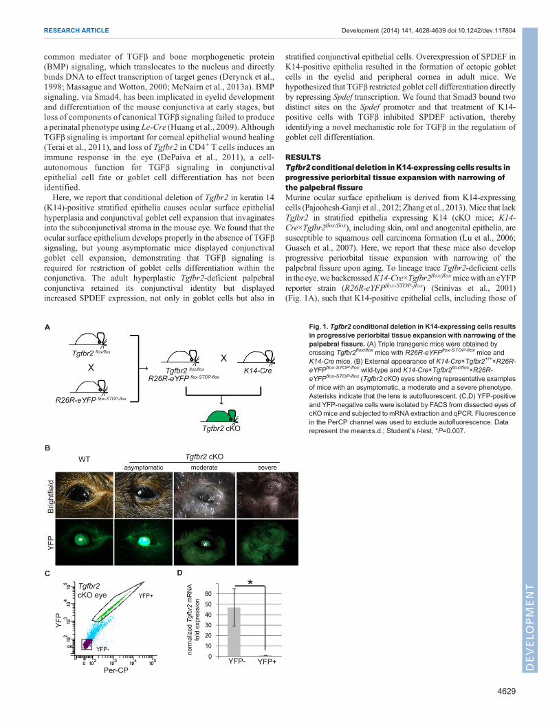
RESULTSTgfbr2 conditional deletion inK14-expressing cells results inprogressive periorbital tissue expansion with narrowing ofthe palpebral fissureMurine ocular surface epithelium is derived from K14-expressingcells (Pajoohesh-Ganji et al., 2012; Zhang et al., 2013).Mice that lackTgfbr2 in stratified epithelia expressing K14 (cKO mice; K14-Cre×Tgfbr2flox/flox), including skin, oral and anogenital epithelia, aresusceptible to squamous cell carcinoma formation (Lu et al., 2006;Guasch et al., 2007). Here, we report that these mice also developprogressive periorbital tissue expansion with narrowing of thepalpebral fissure upon aging. To lineage trace Tgfbr2-deficient cellsin the eye, we backcrossedK14-Cre×Tgfbr2flox/floxmicewith an eYFPreporter strain (R26R-eYFPflox-STOP-flox) (Srinivas et al., 2001)(Fig. 1A), such that K14-positive epithelial cells, including those of
Fig. 1. Tgfbr2 conditional deletion in K14-expressing cells resultsin progressive periorbital tissue expansion with narrowing of thepalpebral fissure. (A) Triple transgenic mice were obtained bycrossing Tgfbr2flox/flox mice with R26R-eYFPflox-STOP-flox mice andK14-Cremice. (B) External appearance of K14-Cre×Tgfbr2+/+×R26R-eYFPflox-STOP-flox wild-type and K14-Cre×Tgfbr2flox/flox×R26R-eYFPflox-STOP-flox (Tgfbr2 cKO) eyes showing representative examplesof mice with an asymptomatic, a moderate and a severe phenotype.Asterisks indicate that the lens is autofluorescent. (C,D) YFP-positiveand YFP-negative cells were isolated by FACS from dissected eyes ofcKOmice and subjected to mRNA extraction and qPCR. Fluorescencein the PerCP channel was used to exclude autofluorescence. Datarepresent the mean±s.d.; Student’s t-test, *P=0.007.
4629
RESEARCH ARTICLE Development (2014) 141, 4628-4639 doi:10.1242/dev.117804
DEVELO
PM
ENT
the ocular surface epithelium, lacked Tgfbr2 and expressed YFP(McCauley and Guasch, 2013). The external appearance of juvenileTgfbr2 cKO eyes, between birth and 8 months of age, appearedindistinguishable from the eyes of age-matched wild-type mice;however, by ∼9 months of age, the periocular tissue of Tgfbr2 cKOmice became grossly swollen and enlarged, with excessive mucousdischarge andmarked narrowing of the palpebral fissure (Table 1 andFig. 1B). YFP fluorescence was detected in both wild-type(K14-Cre×Tgfbr2+/+×R26R-eYFPflox-STOP-flox) and Tgfbr2 cKO skinand eyelid epithelium, demonstrating efficient targeting by K14-Cre(Fig. 1B). We confirmed expression of YFP in the ocular surfaceepithelium of adult wild-type mice, and verified the normalcell-surface expression pattern of TGFβRII in the basal layer ofeyelid, conjunctival and corneal epithelia (supplementary materialFig. S1A-C). Tgfbr2 cKO ocular surface epithelium also expressedYFP, indicating its derivation from K14-expressing cells, but lackedexpression of TGFβRII in eyelid, conjunctival and corneal epithelia(supplementarymaterial Fig. S1D-F). Additionally, the loss ofTgfbr2was directly demonstrated at the mRNA level in YFP-positive cellsisolated from Tgfbr2 cKO eyes (Fig. 1C,D), providing evidence thatthe loss of Tgfbr2 in the ocular surface epithelium caused ocularpathology in these mice.
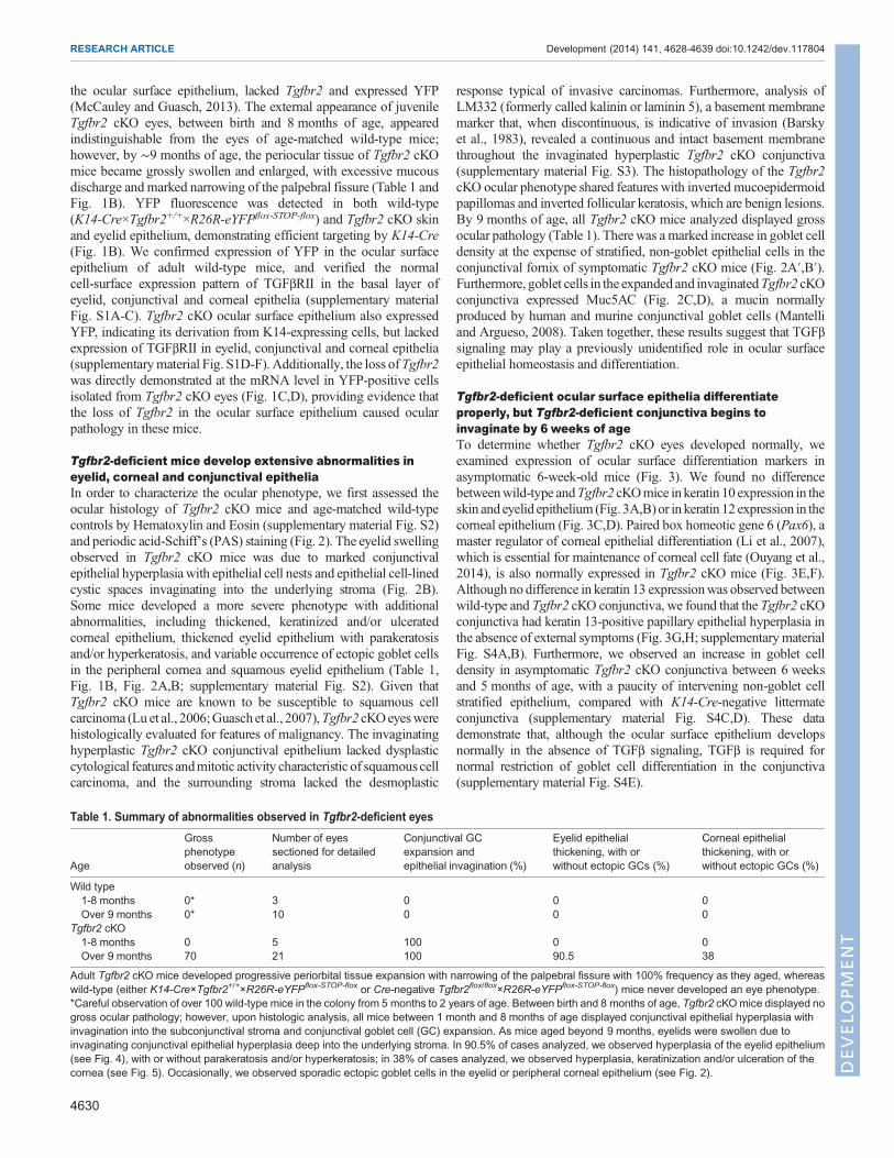
Tgfbr2-deficient mice develop extensive abnormalities ineyelid, corneal and conjunctival epitheliaIn order to characterize the ocular phenotype, we first assessed theocular histology of Tgfbr2 cKO mice and age-matched wild-typecontrols by Hematoxylin and Eosin (supplementary material Fig. S2)and periodic acid-Schiff’s (PAS) staining (Fig. 2). The eyelid swellingobserved in Tgfbr2 cKO mice was due to marked conjunctivalepithelial hyperplasiawith epithelial cell nests and epithelial cell-linedcystic spaces invaginating into the underlying stroma (Fig. 2B).Some mice developed a more severe phenotype with additionalabnormalities, including thickened, keratinized and/or ulceratedcorneal epithelium, thickened eyelid epithelium with parakeratosisand/or hyperkeratosis, and variable occurrence of ectopic goblet cellsin the peripheral cornea and squamous eyelid epithelium (Table 1,Fig. 1B, Fig. 2A,B; supplementary material Fig. S2). Given thatTgfbr2 cKO mice are known to be susceptible to squamous cellcarcinoma (Lu et al., 2006;Guasch et al., 2007),Tgfbr2 cKOeyeswerehistologically evaluated for features of malignancy. The invaginatinghyperplastic Tgfbr2 cKO conjunctival epithelium lacked dysplasticcytological features andmitotic activity characteristic of squamous cellcarcinoma, and the surrounding stroma lacked the desmoplastic
response typical of invasive carcinomas. Furthermore, analysis ofLM332 (formerly called kalinin or laminin 5), a basement membranemarker that, when discontinuous, is indicative of invasion (Barskyet al., 1983), revealed a continuous and intact basement membranethroughout the invaginated hyperplastic Tgfbr2 cKO conjunctiva(supplementary material Fig. S3). The histopathology of the Tgfbr2cKO ocular phenotype shared features with inverted mucoepidermoidpapillomas and inverted follicular keratosis, which are benign lesions.By 9 months of age, all Tgfbr2 cKO mice analyzed displayed grossocular pathology (Table 1). Therewas a marked increase in goblet celldensity at the expense of stratified, non-goblet epithelial cells in theconjunctival fornix of symptomatic Tgfbr2 cKO mice (Fig. 2A′,B′).Furthermore, goblet cells in the expanded and invaginatedTgfbr2 cKOconjunctiva expressed Muc5AC (Fig. 2C,D), a mucin normallyproduced by human and murine conjunctival goblet cells (Mantelliand Argueso, 2008). Taken together, these results suggest that TGFβsignaling may play a previously unidentified role in ocular surfaceepithelial homeostasis and differentiation.
Tgfbr2-deficient ocular surface epithelia differentiateproperly, but Tgfbr2-deficient conjunctiva begins toinvaginate by 6 weeks of ageTo determine whether Tgfbr2 cKO eyes developed normally, weexamined expression of ocular surface differentiation markers inasymptomatic 6-week-old mice (Fig. 3). We found no differencebetweenwild-type andTgfbr2 cKOmice in keratin 10 expression in theskin and eyelid epithelium (Fig. 3A,B)or in keratin 12 expression in thecorneal epithelium (Fig. 3C,D). Paired box homeotic gene 6 (Pax6), amaster regulator of corneal epithelial differentiation (Li et al., 2007),which is essential for maintenance of corneal cell fate (Ouyang et al.,2014), is also normally expressed in Tgfbr2 cKO mice (Fig. 3E,F).Although no difference in keratin 13 expressionwas observed betweenwild-type and Tgfbr2 cKO conjunctiva, we found that the Tgfbr2 cKOconjunctiva had keratin 13-positive papillary epithelial hyperplasia inthe absence of external symptoms (Fig. 3G,H; supplementarymaterialFig. S4A,B). Furthermore, we observed an increase in goblet celldensity in asymptomatic Tgfbr2 cKO conjunctiva between 6 weeksand 5 months of age, with a paucity of intervening non-goblet cellstratified epithelium, compared with K14-Cre-negative littermateconjunctiva (supplementary material Fig. S4C,D). These datademonstrate that, although the ocular surface epithelium developsnormally in the absence of TGFβ signaling, TGFβ is required fornormal restriction of goblet cell differentiation in the conjunctiva(supplementary material Fig. S4E).
Table 1. Summary of abnormalities observed in Tgfbr2-deficient eyes
Age
Grossphenotypeobserved (n)
Number of eyessectioned for detailedanalysis
Conjunctival GCexpansion andepithelial invagination (%)
Eyelid epithelialthickening, with orwithout ectopic GCs (%)
Corneal epithelialthickening, with orwithout ectopic GCs (%)
Wild type1-8 months 0* 3 0 0 0Over 9 months 0* 10 0 0 0
Tgfbr2 cKO1-8 months 0 5 100 0 0Over 9 months 70 21 100 90.5 38
Adult Tgfbr2 cKO mice developed progressive periorbital tissue expansion with narrowing of the palpebral fissure with 100% frequency as they aged, whereaswild-type (either K14-Cre×Tgfbr2+/+×R26R-eYFPflox-STOP-flox or Cre-negative Tgfbr2flox/flox×R26R-eYFPflox-STOP-flox) mice never developed an eye phenotype.*Careful observation of over 100 wild-type mice in the colony from 5 months to 2 years of age. Between birth and 8 months of age, Tgfbr2 cKOmice displayed nogross ocular pathology; however, upon histologic analysis, all mice between 1 month and 8 months of age displayed conjunctival epithelial hyperplasia withinvagination into the subconjunctival stroma and conjunctival goblet cell (GC) expansion. As mice aged beyond 9 months, eyelids were swollen due toinvaginating conjunctival epithelial hyperplasia deep into the underlying stroma. In 90.5% of cases analyzed, we observed hyperplasia of the eyelid epithelium(see Fig. 4), with or without parakeratosis and/or hyperkeratosis; in 38% of cases analyzed, we observed hyperplasia, keratinization and/or ulceration of thecornea (see Fig. 5). Occasionally, we observed sporadic ectopic goblet cells in the eyelid or peripheral corneal epithelium (see Fig. 2).
4630
RESEARCH ARTICLE Development (2014) 141, 4628-4639 doi:10.1242/dev.117804
DEVELO
PM
ENT
Adult Tgfbr2-deficient eyelid epithelium is hyperplasticAlthough Tgfbr2-deficient eyes differentiated properly at early stages,we observed marked changes in eyelid, corneal and conjunctivalepithelium in adult Tgfbr2 cKOmice (Table 1, Fig. 2; supplementarymaterial Fig. S2). Because TGFβ is known to regulate epithelialcellular proliferation and differentiation (McNairn et al., 2013a,b), wequestioned whether the K14-positive ocular surface epithelia in adultTgfbr2 cKO mice was hyperproliferative. The eyelid epithelium of
adult Tgfbr2 cKO mice was expanded, containing three to fiveadditional p63-positive basal layers and three to five additional keratin10-positive suprabasal layers compared with wild-type eyelidepithelium (Fig. 4A-D). Quantification of 5-bromo-2-deoxyuridine(BrdU) staining revealed that Tgfbr2 cKO eyelid epithelium washyperproliferative, compared with wild type (Fig. 4E-G).
The hyperplastic Tgfbr2-deficient adult cornea becomeskeratinizedOf all adult Tgfbr2 cKO mice analyzed, ∼38% displayed severelyaffected corneal epithelium (Table 1).AdultTgfbr2 cKOmicewithoutsevere corneal pathology displayed a reduction in the number ofstratified corneal epithelial layers and aweakening of expression of thecorneal epithelial marker keratin 12 (Fig. 5A,B). However, in Tgfbr2cKO mice with severely affected corneal pathology, keratin 12expression was completely lost in the corneal epithelium, which wasassociated with an increase in epithelial stratification (Fig. 5C). Lossof keratin 12 expression occurred progressively, beginning with lossof expression in the central cornea as the number of corneal epithelial
Fig. 2. Tgfbr2-deficient mice develop eyelid, corneal and conjunctivalepithelial hyperplasia. (A-B′) Combined PAS and Hematoxylin and Eosinstaining demonstrated extensive squamous andmucous epithelial hyperplasiawith invaginations into the underlying stroma, involving the palpebralconjunctiva, fornix and eyelids of symptomatic Tgfbr2 cKOmice compared withsections of comparable regions from age-matched wild-type mice. Ectopicgoblet cells (magenta) were found within Tgfbr2 cKO hyperplastic eyelidepithelium and peripheral corneal epithelium (B, asterisks). Highermagnification of the boxed areas are shown in A′ and B′. White arrows indicatenon-goblet cell stratified conjunctival epithelial cells interspersed betweengoblet cells. (C,D) Goblet cells within the expanded and invaginated Tgfbr2cKO conjunctiva expressed Muc5AC. Dotted lines indicate the basal layer.DAPI counterstains nuclei in blue. co, cornea; le, lens; f, fornix; p, palpebralconjunctiva; M, Meibomian gland; el, eyelid; f, fornix. Scale bars: 100 µm inA,B,C,D; 20 µm in A′,B′.
Fig. 3. Tgfbr2-deficient ocular surface epithelia differentiate properly.(A-F) Immunofluorescent staining with antibodies against keratin 10 (A,B),keratin 12 (C,D), Pax6 (E,F) and keratin 13 (G,H) on eyes dissected from6-week-old mice indicated that the eyelid (A,B), cornea (C-F) and conjunctiva(G,H) developed normally in Tgfbr2 cKO mice. In the absence of externalsymptoms, the Tgfbr2 cKO conjunctiva was noted to invaginate into thesubepithelial stroma by 6 weeks of age (H). Dotted lines indicate the basallayer. DAPI counterstains nuclei in blue. el, eyelid; hf, hair follicle; f, fornix.Scale bars: 50 µm in A,B,G,H; 20 µm in C-F.
4631
RESEARCH ARTICLE Development (2014) 141, 4628-4639 doi:10.1242/dev.117804
DEVELO
PM
ENT

layers increased (supplementarymaterial Fig. S5).Wewere interestedto see whether the expression of other corneal markers was lost oraffected in Tgfbr2 cKO mice. We observed no change in Pax6expression between wild-type and Tgfbr2 cKO corneal epithelium(Fig. 5D-F), suggesting that Pax6 functions either upstream orindependently of the TGFβ signaling pathway in this context. Themurine cornea normally expresses p63 in the basal layer; p63 stainingrevealed an expansion of the corneal basal layer in severely affectedTgfbr2 cKO mice (Fig. 5G-I). The severely affected Tgfbr2 cKOcornea became keratinized, as the expanded suprabasal layersexpressed keratin 10 (Fig. 5L). We did not find any keratin 10expression in adult wild-type or adult asymptomatic Tgfbr2 cKOcorneal epithelium (Fig. 5J,K). Quantification of BrdU stainingrevealed that severely affected Tgfbr2 cKO corneal epithelia washyperproliferative compared with wild-type or asymptomatic adultTgfbr2 cKO (Fig. 5M-P).
The hyperplastic Tgfbr2-deficient palpebral conjunctiva ishyperproliferativeThe hyperplastic Tgfbr2 cKO conjunctiva retained keratin 13expression, a marker of normal conjunctival epithelium (Fig. 6A-C).Quantification of BrdU staining indicated that the Tgfbr2 cKOpalpebral conjunctival epithelium was hyperproliferative comparedwith wild-type palpebral conjunctiva (Fig. 6D-F), but there was nosignificant difference in proliferation in the conjunctival fornix(Fig. 6G-I) between wild-type and Tgfbr2 cKO mice. Quantificationof the ratio of BrdU-positive cells to total basal epithelial cellsdemonstrated that the stratified conjunctival epithelial cells, but not
Fig. 4. The Tgfbr2-deficient eyelid becomes hyperplastic with age.Immunofluorescence staining with antibodies against the suprabasal markerkeratin 10 (A,B), the basal marker p63 (C,D) and BrdU (E,F) indicated that theTgfbr2 cKO eyelid epithelium (B) was hyperplastic with increased epitheliallayers (brackets) compared with wild type. (E-G) Quantification (G) of BrdUimmunofluorescence staining (E,F, arrows) indicated that the Tgfbr2 cKOeyelid epithelium was hyperproliferative. Data represent the mean number ofBrdU-positive cells in relation to total basal epithelial cells±s.d.; Student’st-test; *P=0.003. hf, hair follicle. Asterisks in C,D indicate autofluorescenceoriginating from the hair shaft. Scale bars: 50 µm in A-D; 20 µm in E,F.
Fig. 5. The hyperplastic Tgfbr2-deficient adult cornea becomeskeratinized. (A-C) Immunofluorescence staining with antibodies againstkeratin 12 (A-C), Pax6 (D-F), p63 (G-I), keratin 10 (J-L) and BrdU (M-O)indicated that the corneal epithelium of adult Tgfbr2 cKO mice becomeskeratinized with increasing phenotypic severity. (M-P) Quantification (P) ofBrdU immunofluorescence staining (M-O, arrows) indicated that the severelyaffected Tgfbr2 cKO corneal epithelium was hyperproliferative compared withwild-type and asymptomatic Tgfbr2 cKO adult corneal epithelium. Datarepresent the mean number of BrdU-positive cells in relation to total basalepithelial cells ±s.d.; Student’s t-test; *P=0.04. Dotted lines indicate the basallayer. Asterisk in C denotes autofluorescence in the stroma. DAPIcounterstains nuclei in blue. Scale bars: 20 µm.
4632
RESEARCH ARTICLE Development (2014) 141, 4628-4639 doi:10.1242/dev.117804
DEVELO
PM
ENT
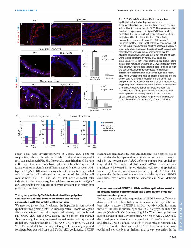
goblet cells, were hyperproliferative in Tgfbr2 cKO palpebralconjunctiva, whereas the ratio of stratified epithelial cells to gobletcells was unchanged (Fig. 6J). Conversely, quantification of the ratioof BrdU-positive cells to total basal epithelial cells in the conjunctivalfornix revealed no significant difference inproliferationbetweenwild-type and Tgfbr2 cKO mice, whereas the ratio of stratified epithelialcells to goblet cells reflected an expansion of the goblet cellcompartment (Fig. 6K). The lack of BrdU-positive goblet cellsindicated that the increase in goblet cell density observed in theTgfbr2cKO conjunctiva was a result of aberrant differentiation rather thangoblet cell proliferation.
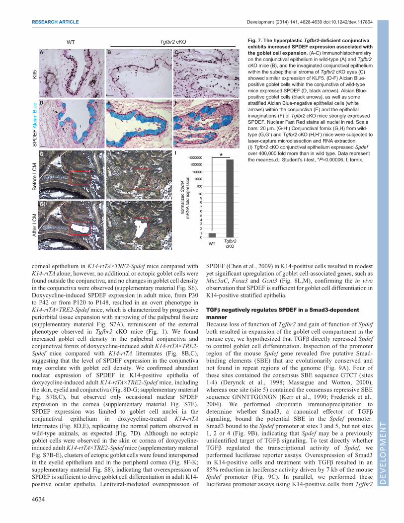
The hyperplastic Tgfbr2-deficient stratified palpebralconjunctiva exhibits increased SPDEF expressionassociated with the goblet cell expansionWe next sought to identify whether the hyperplastic conjunctivalepithelium invaginating into the subconjunctival stroma of Tgfbr2cKO mice retained normal conjunctival identity. We confirmedthat Tgfbr2 cKO conjunctiva, despite the expansion and markedabundance of goblet cells, expressed normal markers of conjunctivalepithelium, including keratin 13 (Fig. 6A-C), KLF5 (Fig. 7A-C) andSPDEF (Fig. 7D-F). Interestingly, although KLF5 staining appearedconsistent between wild-type and Tgfbr2 cKO conjunctiva, SPDEF
staining appeared markedly increased in the nuclei of goblet cells, aswell as abundantly expressed in the nuclei of interspersed stratifiedcells in the hyperplastic Tgfbr2-deficient conjunctival epithelium(Fig. 7D-F). We confirmed that Spdef mRNA expression wassignificantly increased in Tgfbr2-deficient conjunctival epitheliumisolated by laser-capture microdissection (Fig. 7G-I). These datasuggest that the increased conjunctival stratified epithelial SPDEFexpression may promote goblet cell expansion in Tgfbr2-deficientmice.
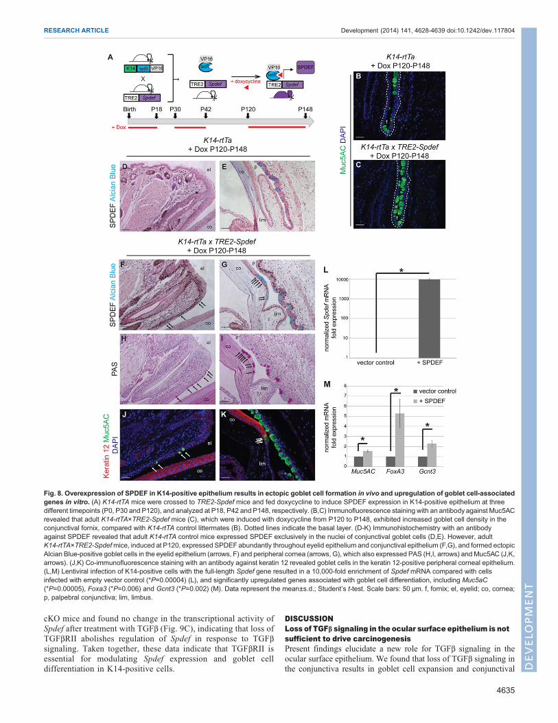
Overexpression of SPDEF in K14-positive epithelium resultsin ectopic goblet cell formation and upregulation of gobletcell-associated genesTo test whether epithelial expression of SPDEF was sufficient todrive goblet cell differentiation in the ocular surface epithelia, webred mice to express SPDEF in all K14-positive cells, includingthose of the ocular surface epithelia, in a doxycycline-induciblemanner (K14-rtTA×TRE2-Spdef ) (Fig. 8A). When doxycycline wasadministered continuously from birth, K14-rtTA×TRE2-Spdef micedisplayed growth retardation compared with K14-rtTA littermates,and died by 3 weeks of age. Analysis of these mice at postnatal day18 (P18) revealed abundant nuclear SPDEF expression in skin,eyelid and conjunctival epithelium, and patchy expression in the
Fig. 6. Tgfbr2-deficient stratified conjunctivalepithelial cells, but not goblet cells, arehyperproliferative. (A-I) Immunofluorescence stainingwith antibodies against keratin 13 (A-C) revealed positivekeratin 13 expression in the Tgfbr2 cKO conjunctivalepithelium (B), including the hyperplastic conjunctivalepithelium (C). (D-I) Quantification (F,I) of BrdUimmunofluorescence staining (D,E,G,H, arrows)indicated that the Tgfbr2 cKO palpebral conjunctiva, butnot the fornix, was hyperproliferative compared with wildtype. (J,K) Quantification of the ratio of BrdU-positive cellsto total basal epithelial cells demonstrated that thestratified conjunctival epithelial cells, but not goblet cells,were hyperproliferative in Tgfbr2 cKO palpebralconjunctiva, whereas the ratio of stratified epithelial cells togoblet cells remained unchanged (J). Quantification of theratio of BrdU-positive cells to total basal epithelial cells inthe conjunctival fornix demonstrated no significantdifference in proliferation between wild-type and Tgfbr2cKO mice, whereas the ratio of stratified epithelial cells togoblet cells reflected an expansion of the goblet cellcompartment (K). Asterisk in B denotes autofluorescenceoriginating from inflammatory cells. Asterisk in D indicatesa rare BrdU-positive goblet cell. Data represent themean number of BrdU-positive cells in relation to totalbasal epithelial cells±s.d.; Student’s t-test; *P=0.02.cj, conjunctival; p, palpebral conjunctiva; f, conjunctivalfornix. Scale bars: 50 µm in A-C; 20 µm in D,E,G,H.
4633
RESEARCH ARTICLE Development (2014) 141, 4628-4639 doi:10.1242/dev.117804
DEVELO
PM
ENT
corneal epithelium in K14-rtTA×TRE2-Spdef mice compared withK14-rtTA alone; however, no additional or ectopic goblet cells werefound outside the conjunctiva, and no changes in goblet cell densityin the conjunctiva were observed (supplementary material Fig. S6).Doxycycline-induced SPDEF expression in adult mice, from P30to P42 or from P120 to P148, resulted in an overt phenotype inK14-rtTA×TRE2-Spdef mice, which is characterized by progressiveperiorbital tissue expansion with narrowing of the palpebral fissure(supplementary material Fig. S7A), reminiscent of the externalphenotype observed in Tgfbr2 cKO mice (Fig. 1). We foundincreased goblet cell density in the palpebral conjunctiva andconjunctival fornix of doxycycline-induced adult K14-rtTA×TRE2-Spdef mice compared with K14-rtTA littermates (Fig. 8B,C),suggesting that the level of SPDEF expression in the conjunctivamay correlate with goblet cell density. We confirmed abundantnuclear expression of SPDEF in K14-positive epithelia ofdoxycycline-induced adult K14-rtTA×TRE2-Spdef mice, includingthe skin, eyelid and conjunctiva (Fig. 8D-G; supplementary materialFig. S7B,C), but observed only occasional nuclear SPDEFexpression in the cornea (supplementary material Fig. S7E).SPDEF expression was limited to goblet cell nuclei in theconjunctival epithelium in doxycycline-treated K14-rtTAlittermates (Fig. 8D,E), replicating the normal pattern observed inwild-type animals, as expected (Fig. 7D). Although no ectopicgoblet cells were observed in the skin or cornea of doxycycline-induced adult K14-rtTA×TRE2-Spdefmice (supplementary materialFig. S7B-E), clusters of ectopic goblet cells were found interspersedin the eyelid epithelium and in the peripheral cornea (Fig. 8F-K;supplementary material Fig. S8), indicating that overexpression ofSPDEF is sufficient to drive goblet cell differentiation in adult K14-positive ocular epithelia. Lentiviral-mediated overexpression of
SPDEF (Chen et al., 2009) in K14-positive cells resulted in modestyet significant upregulation of goblet cell-associated genes, such asMuc5aC, Foxa3 and Gcnt3 (Fig. 8L,M), confirming the in vivoobservation that SPDEF is sufficient for goblet cell differentiation inK14-positive stratified epithelia.
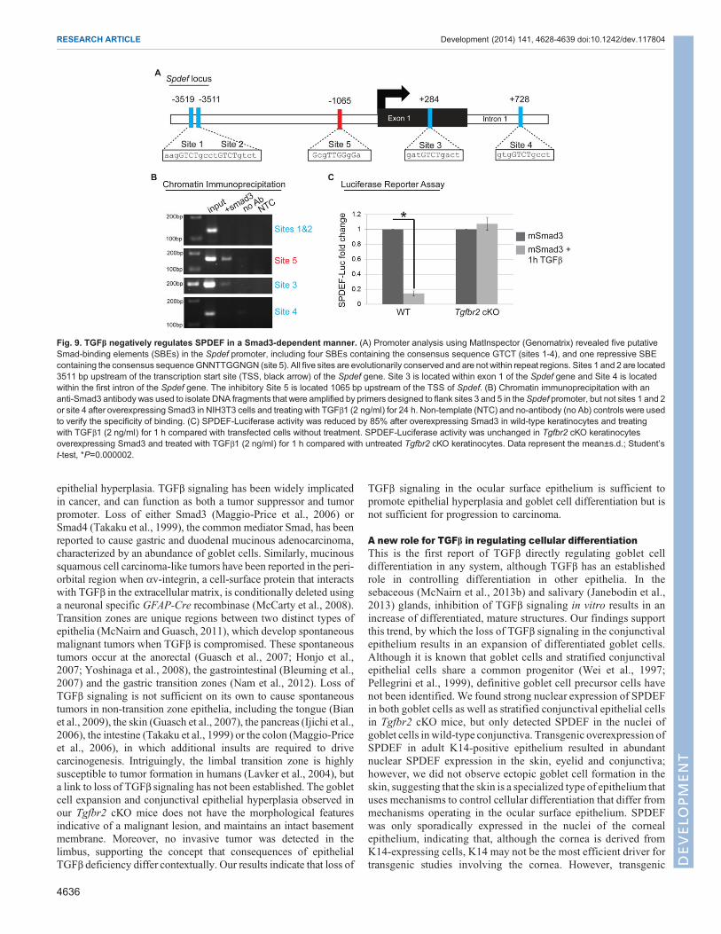
TGFβ negatively regulates SPDEF in a Smad3-dependentmannerBecause loss of function of Tgfbr2 and gain of function of Spdefboth resulted in expansion of the goblet cell compartment in themouse eye, we hypothesized that TGFβ directly repressed Spdefto control goblet cell differentiation. Inspection of the promoterregion of the mouse Spdef gene revealed five putative Smad-binding elements (SBE) that are evolutionarily conserved andnot found in repeat regions of the genome (Fig. 9A). Four ofthese sites contained the consensus SBE sequence GTCT (sites1-4) (Derynck et al., 1998; Massague and Wotton, 2000),whereas one site (site 5) contained the consensus repressive SBEsequence GNNTTGGNGN (Kerr et al., 1990; Frederick et al.,2004). We performed chromatin immunoprecipitation todetermine whether Smad3, a canonical effector of TGFβsignaling, bound the potential SBE in the Spdef promoter.Smad3 bound to the Spdef promoter at sites 3 and 5, but not sites1, 2 or 4 (Fig. 9B), indicating that Spdef may be a previouslyunidentified target of TGFβ signaling. To test directly whetherTGFβ regulated the transcriptional activity of Spdef, weperformed luciferase reporter assays. Overexpression of Smad3in K14-positive cells and treatment with TGFβ resulted in an85% reduction in luciferase activity driven by 7 kb of the mouseSpdef promoter (Fig. 9C). In parallel, we performed theseluciferase promoter assays using K14-positive cells from Tgfbr2
Fig. 7. The hyperplastic Tgfbr2-deficient conjunctivaexhibits increased SPDEF expression associated withthe goblet cell expansion. (A-C) Immunohistochemistryon the conjunctival epithelium in wild-type (A) and Tgfbr2cKO mice (B), and the invaginated conjunctival epitheliumwithin the subepithelial stroma of Tgfbr2 cKO eyes (C)showed similar expression of KLF5. (D-F) Alcian Blue-positive goblet cells within the conjunctiva of wild-typemice expressed SPDEF (D, black arrows). Alcian Blue-positive goblet cells (black arrows), as well as somestratified Alcian Blue-negative epithelial cells (whitearrows) within the conjunctiva (E) and the epithelialinvaginations (F) of Tgfbr2 cKO mice strongly expressedSPDEF. Nuclear Fast Red stains all nuclei in red. Scalebars: 20 µm. (G-H′) Conjunctival fornix (G,H) from wild-type (G,G′) and Tgfbr2 cKO (H,H′) mice were subjected tolaser-capture microdissection and RNA extraction.(I) Tgfbr2 cKO conjunctival epithelium expressed Spdefover 400,000 fold more than in wild type. Data representthe mean±s.d.; Student’s t-test, *P=0.00006. f, fornix.
4634
RESEARCH ARTICLE Development (2014) 141, 4628-4639 doi:10.1242/dev.117804
DEVELO
PM
ENT
cKO mice and found no change in the transcriptional activity ofSpdef after treatment with TGFβ (Fig. 9C), indicating that loss ofTGFβRII abolishes regulation of Spdef in response to TGFβsignaling. Taken together, these data indicate that TGFβRII isessential for modulating Spdef expression and goblet celldifferentiation in K14-positive cells.
DISCUSSIONLoss of TGFβ signaling in the ocular surface epithelium is notsufficient to drive carcinogenesisPresent findings elucidate a new role for TGFβ signaling in theocular surface epithelium. We found that loss of TGFβ signaling inthe conjunctiva results in goblet cell expansion and conjunctival
Fig. 8. Overexpression of SPDEF in K14-positive epithelium results in ectopic goblet cell formation in vivo and upregulation of goblet cell-associatedgenes in vitro. (A) K14-rtTA mice were crossed to TRE2-Spdef mice and fed doxycycline to induce SPDEF expression in K14-positive epithelium at threedifferent timepoints (P0, P30 and P120), and analyzed at P18, P42 and P148, respectively. (B,C) Immunofluorescence staining with an antibody against Muc5ACrevealed that adult K14-rtTA×TRE2-Spdef mice (C), which were induced with doxycycline from P120 to P148, exhibited increased goblet cell density in theconjunctival fornix, compared with K14-rtTA control littermates (B). Dotted lines indicate the basal layer. (D-K) Immunohistochemistry with an antibodyagainst SPDEF revealed that adult K14-rtTA control mice expressed SPDEF exclusively in the nuclei of conjunctival goblet cells (D,E). However, adultK14-rtTA×TRE2-Spdefmice, induced at P120, expressed SPDEF abundantly throughout eyelid epithelium and conjunctival epithelium (F,G), and formed ectopicAlcian Blue-positive goblet cells in the eyelid epithelium (arrows, F) and peripheral cornea (arrows, G), which also expressed PAS (H,I, arrows) and Muc5AC (J,K,arrows). (J,K) Co-immunofluorescence staining with an antibody against keratin 12 revealed goblet cells in the keratin 12-positive peripheral corneal epithelium.(L,M) Lentiviral infection of K14-positive cells with the full-length Spdef gene resulted in a 10,000-fold enrichment of Spdef mRNA compared with cellsinfected with empty vector control (*P=0.00004) (L), and significantly upregulated genes associated with goblet cell differentiation, including Muc5aC(*P=0.00005), Foxa3 (*P=0.006) and Gcnt3 (*P=0.002) (M). Data represent the mean±s.d.; Student’s t-test. Scale bars: 50 µm. f, fornix; el, eyelid; co, cornea;p, palpebral conjunctiva; lim, limbus.
4635
RESEARCH ARTICLE Development (2014) 141, 4628-4639 doi:10.1242/dev.117804
DEVELO
PM
ENT
epithelial hyperplasia. TGFβ signaling has been widely implicatedin cancer, and can function as both a tumor suppressor and tumorpromoter. Loss of either Smad3 (Maggio-Price et al., 2006) orSmad4 (Takaku et al., 1999), the common mediator Smad, has beenreported to cause gastric and duodenal mucinous adenocarcinoma,characterized by an abundance of goblet cells. Similarly, mucinoussquamous cell carcinoma-like tumors have been reported in the peri-orbital region when αv-integrin, a cell-surface protein that interactswith TGFβ in the extracellular matrix, is conditionally deleted usinga neuronal specific GFAP-Cre recombinase (McCarty et al., 2008).Transition zones are unique regions between two distinct types ofepithelia (McNairn and Guasch, 2011), which develop spontaneousmalignant tumors when TGFβ is compromised. These spontaneoustumors occur at the anorectal (Guasch et al., 2007; Honjo et al.,2007; Yoshinaga et al., 2008), the gastrointestinal (Bleuming et al.,2007) and the gastric transition zones (Nam et al., 2012). Loss ofTGFβ signaling is not sufficient on its own to cause spontaneoustumors in non-transition zone epithelia, including the tongue (Bianet al., 2009), the skin (Guasch et al., 2007), the pancreas (Ijichi et al.,2006), the intestine (Takaku et al., 1999) or the colon (Maggio-Priceet al., 2006), in which additional insults are required to drivecarcinogenesis. Intriguingly, the limbal transition zone is highlysusceptible to tumor formation in humans (Lavker et al., 2004), buta link to loss of TGFβ signaling has not been established. The gobletcell expansion and conjunctival epithelial hyperplasia observed inour Tgfbr2 cKO mice does not have the morphological featuresindicative of a malignant lesion, and maintains an intact basementmembrane. Moreover, no invasive tumor was detected in thelimbus, supporting the concept that consequences of epithelialTGFβ deficiency differ contextually. Our results indicate that loss of
TGFβ signaling in the ocular surface epithelium is sufficient topromote epithelial hyperplasia and goblet cell differentiation but isnot sufficient for progression to carcinoma.
A new role for TGFβ in regulating cellular differentiationThis is the first report of TGFβ directly regulating goblet celldifferentiation in any system, although TGFβ has an establishedrole in controlling differentiation in other epithelia. In thesebaceous (McNairn et al., 2013b) and salivary (Janebodin et al.,2013) glands, inhibition of TGFβ signaling in vitro results in anincrease of differentiated, mature structures. Our findings supportthis trend, by which the loss of TGFβ signaling in the conjunctivalepithelium results in an expansion of differentiated goblet cells.Although it is known that goblet cells and stratified conjunctivalepithelial cells share a common progenitor (Wei et al., 1997;Pellegrini et al., 1999), definitive goblet cell precursor cells havenot been identified. We found strong nuclear expression of SPDEFin both goblet cells as well as stratified conjunctival epithelial cellsin Tgfbr2 cKO mice, but only detected SPDEF in the nuclei ofgoblet cells in wild-type conjunctiva. Transgenic overexpression ofSPDEF in adult K14-positive epithelium resulted in abundantnuclear SPDEF expression in the skin, eyelid and conjunctiva;however, we did not observe ectopic goblet cell formation in theskin, suggesting that the skin is a specialized type of epithelium thatuses mechanisms to control cellular differentiation that differ frommechanisms operating in the ocular surface epithelium. SPDEFwas only sporadically expressed in the nuclei of the cornealepithelium, indicating that, although the cornea is derived fromK14-expressing cells, K14 may not be the most efficient driver fortransgenic studies involving the cornea. However, transgenic
Fig. 9. TGFβ negatively regulates SPDEF in a Smad3-dependent manner. (A) Promoter analysis using MatInspector (Genomatrix) revealed five putativeSmad-binding elements (SBEs) in the Spdef promoter, including four SBEs containing the consensus sequence GTCT (sites 1-4), and one repressive SBEcontaining the consensus sequenceGNNTTGGNGN (site 5). All five sites are evolutionarily conserved and are not within repeat regions. Sites 1 and 2 are located3511 bp upstream of the transcription start site (TSS, black arrow) of the Spdef gene. Site 3 is located within exon 1 of the Spdef gene and Site 4 is locatedwithin the first intron of the Spdef gene. The inhibitory Site 5 is located 1065 bp upstream of the TSS of Spdef. (B) Chromatin immunoprecipitation with ananti-Smad3 antibody was used to isolate DNA fragments that were amplified by primers designed to flank sites 3 and 5 in theSpdef promoter, but not sites 1 and 2or site 4 after overexpressing Smad3 in NIH3T3 cells and treating with TGFβ1 (2 ng/ml) for 24 h. Non-template (NTC) and no-antibody (no Ab) controls were usedto verify the specificity of binding. (C) SPDEF-Luciferase activity was reduced by 85% after overexpressing Smad3 in wild-type keratinocytes and treatingwith TGFβ1 (2 ng/ml) for 1 h compared with transfected cells without treatment. SPDEF-Luciferase activity was unchanged in Tgfbr2 cKO keratinocytesoverexpressing Smad3 and treated with TGFβ1 (2 ng/ml) for 1 h compared with untreated Tgfbr2 cKO keratinocytes. Data represent the mean±s.d.; Student’st-test, *P=0.000002.
4636
RESEARCH ARTICLE Development (2014) 141, 4628-4639 doi:10.1242/dev.117804
DEVELO
PM
ENT
overexpression of SPDEF was sufficient to drive goblet cellmetaplasia in the eyelid epithelium and the peripheral cornea,similar to the mechanism of goblet cell metaplasia described in thelung (Park et al., 2007; Chen et al., 2009) and the intestine (Noahet al., 2010), indicating that SPDEF is sufficient to drive gobletcell differentiation in some K14-positive ocular cell types. Onlya subset of SPDEF-expressing K14-positive epithelial cellsunderwent metaplasia into goblet cells in vivo, suggesting thatthese epithelia employ additional mechanisms to restrict goblet celldifferentiation. Our data demonstrate that TGFβ is one such factorrequired for restriction of goblet cell differentiation in adult mice,and that it acts upstream of Spdef.
TGFβ is a novel regulator of SPDEFWe found that Smad3 bound two distinct sites on the Spdefpromoter and that treatment of K14-positive cells with TGFβinhibited SPDEF activation, thereby identifying a novelmechanistic role for TGFβ in the regulation of goblet celldifferentiation. Although the role of Spdef in goblet celldifferentiation is consistent between the lung, the intestine andthe conjunctiva, transcriptional control of Spdef remains unclear,especially in the conjunctiva. Conditional deletion of Klf4(Swamynathan et al., 2007; Gupta et al., 2011) or Klf5(Kenchegowda et al., 2011), which are structurally related andexpressed in the ocular surface epithelium, results in a loss ofconjunctival goblet cells and reduced Spdef mRNA expression(Gupta et al., 2011; Marko et al., 2013). Gene expression analysisof Klf4-deficient mice yielded Spdef, but not elements of the TGFβsignaling pathway, as direct targets of KLF4 in the conjunctiva(Gupta et al., 2011). We detected no change in KLF5 expression inTgfbr2 cKO conjunctiva, suggesting that TGFβ may functionindependently of KLF4/5 in a novel mechanism of transcriptionalregulation of goblet cell differentiation. Because SPDEF isrequired for goblet cell differentiation in many organs, ourfindings support the concept that TGFβ may have a more globalrole in spatial or temporal restriction of goblet cell differentiation.
TGFβ as a therapeutic target for human goblet cellpathologiesSPDEF is known to play a role in goblet cell function in humans.SPDEF is expressed in the nuclei of healthy human conjunctivalgoblet cells, and samples obtained from individuals with Sjögren’ssyndrome, an autoimmune disorder that causes dry eye, displaydecreased Spdef mRNA expression (Marko et al., 2013).Individuals with Sjögren’s syndrome also display hyposalivationand exogenous TGFβ1 production in the salivary gland of miceresults in a dry mouth phenotype (Hall et al., 2010), supporting thepossibility of TGFβ as a therapeutic target for this type of disorder.Treatment for dry eye, including Sjögren’s syndrome, currentlyrelies largely on managing symptoms of disease with artificial tears.Our findings present exciting opportunities for development oftherapeutic strategies that target TGFβ signaling to treat disorders ofgoblet cell differentiation, including dry eye syndromes, Sjögren’ssyndrome and mucinous adenocarcinoma, in humans.
MATERIALS AND METHODSMice and genotypingThe conditional knockout Tgfbr2flox/flox×K14-Cre mouse model (Leveenet al., 2002; Guasch et al., 2007) is derived in a pure C57BL/6background and backcrossed (McCauley and Guasch, 2013) into a mousereporter containing an enhanced yellow fluorescent protein gene (EYFP)inserted into the Gt(ROSA)26Sor locus (Srinivas et al., 2001) and called
R26R-eYFPflox-STOP-flox (Jackson Laboratory). Control mice were eitherTgfbr2flox/flox×R26R-eYFPflox-STOP-flox or Tgfbr2+/+×R26R-eYFPflox-STOP-flox×K14-Cre, all in a C57BL/6 background.
Mice expressing SPDEF were in a mixed background and were generatedby crossing K14-rtTAmice (Nguyen et al., 2006) (Jackson Laboratory) withTRE2-Spdefmice (Park et al., 2007). Control mice were K14-rtTA alone. Toinduce transgene expression, adult compound transgenic mice were fed withDox-chow (doxycycline 1 g/kg chow, Bioserv) ad libitum. Neonates wereadministered doxycycline at birth (P0) by feeding the nursing mother withDox-chow ad libitum.
All experiments were approved by the Cincinnati Children’s HospitalResearch Foundation Institutional Animal Care and Use Committee andwere carried out using standard procedures. Genotyping was conducted byPCR of tail skin DNA using mouse Cre, Tgfbr2, EYFP, K14-rtTA andTRE2-Spdef primers, as described previously (Soriano, 1999; Nguyen et al.,2006; Guasch et al., 2007; Park et al., 2007).
Histological analysisAfter sacrificing mice by carbon dioxide inhalation, eyes, including the skin,intact eyelids and eyeball, were dissected and fixed in 4% paraformaldehydefor 48 h. The samples were then dehydrated and embedded in paraffin orcryopreserved in 30% sucrose and embedded in OCT compound (Tissue-Tek),and stored at –80°C as previously described (McCauley and Guasch, 2013).Deparaffinized sections (5 µm) were stained with Hematoxylin and Eosin(Ventana Medical Systems), periodic acid-Schiff (PAS; Sigma-Aldrich) orAlcian Blue (Poly Scientific) according to the manufacturer’s instructions.
Immunostainings and antibodiesDeparaffinized tissue sections (5 µm) were subjected to antigen retrieval andimmunostaining as previously described (Tompkins et al., 2009). Frozentissue sections (10 µm) were subjected to immunofluorescence labeling aspreviously described (Runck et al., 2010). Antibodies used and imageacquisition are described in the methods in the supplementary material.
Detection of cellular proliferationMice were injected intraperitoneally with 10 mg/ml of 5-bromo-2-deoxyuridine (BrdU, Sigma-Aldrich) 2 h before sacrifice, and eyes wereharvested, sectioned and stained as described in the methods in thesupplementary material, with additional treatment of slides with 1 N HClfor 40 min at 37°C prior to incubation with antibody against BrdU (Abcam,ab6326, 1/100). The percentages of BrdU-positive cells were determined bycounting the total number of nuclei in the basal layer and the number ofBrdU-positive basal epithelial cells using a 40× objective in z-stackcombined images. Data represent quantification of eyes from threewild-typeand six Tgfbr2 cKO mice.
Fluorescence-activated cell sorting (FACS)Eyes, including the surrounding skin, intact eyelids and eyeball, were removedfrom Tgfbr2flox/flox×R26R-eYFPflox-STOP-flox×K14-Cre mice and dissociatedinto a single cell suspension as previously described (McCauley and Guasch,2013). Dead cells were excluded using 7-amino-actinomycin D (7-AAD;eBioscience) incorporation and the remaining live YFP-positive and YFP-negative cells were collected directly into cell lysis buffer containing β-mercaptoethanol using a FACS Aria II (BD) and FACSDiva software (BD).Sorted cells were vortexed and stored at –80°C until RNA extraction.
Laser-capture microdissectionEyes were dissected from adult wild-type and Tgfbr2 cKO mice andprocessed for laser capture microdissection as previously described (Potterand Brunskill, 2012). Once samples were obtained, RNAwas isolated usinga ZR RNAMicroPrep kit (Zymo Research) and amplified using the OvationRNA-Seq System V2 (Nugen) according to the manufacturer’s directionsbefore being subjected to qPCR as described below.
Real-time PCRTotal RNA was isolated using a Qiagen RNeasy Mini Kit and used toproduce cDNA (Maxima first strand cDNA synthesis kit, Fermentas).
4637
RESEARCH ARTICLE Development (2014) 141, 4628-4639 doi:10.1242/dev.117804
DEVELO
PM
ENT
Real-time PCR was performed using the CFX96 real-time PCR System,CFX Manager Software and SsoFast EvaGreen Supermix reagents (Bio-Rad) or StepOnePlus real-time PCR system and TaqMan reagents (AppliedBiosystems). All reactions were run three times in triplicate and analyzedusing the ΔΔCT method with relative expression normalized to Gapdh or18S. Primers are described in the methods in the supplementary material.
Isolation of primary keratinocytes and cell cultureWild-type and Tgfbr2 cKO keratinocytes were isolated from newbornC57BL/6 mice at P1. Detailed protocol is described in the methods in thesupplementary material. Loss of Tgfbr2 has been verified by qPCR and lossof TGFβ responsiveness confirmed as previously described (Guasch et al.,2007).
Chromatin immunoprecipitationNIH3T3 cells were seeded in 10 cm plates at 80% confluence and transientlytransfected with a pCMV-driven mouse Smad3 (Sino Biological) using X-treme Gene transfection reagent (Roche Applied Science) for 24 h, thentreated with recombinant human TGFβ1 (R&D Systems, 2 ng/ml) for anadditional 24 h. Cells were crosslinked with 1% formaldehyde and subjectedto ChIP using an antibody against Smad3 (Abcam, ab28379) using a ChIPassay kit (Millipore) according to the manufacturer’s instructions. Afterpurification, DNA obtained from the ChIP assay was used as PCR templatesto verify the interaction between DNA and protein, using primers designed toamplify distinct sites in the mouse Spdef promoter. Primers are described inthemethods in the supplementarymaterial. PCR products were then subjectedto gel electrophoresis on a 3% agarose gel using a molecular weight marker toverify the size of migrating bands.
Cloning and luciferase reporter assayUsing SalI and EcoRI restriction enzymes, a 7634 bp fragment of the mouseSpdef promoter, containing∼3.7 kb upstream of the transcriptional start site,the first exon (399 bp) and the first intron (∼3.5 kb), was isolated from theWI1 - 1250E21 Fosmid containing the mouse Spdef gene (SpectraGenetics).The fragment was gel purified and ends were blunted with Klenow fragmentfollowed by alkaline phosphatase to prevent re-ligation. In parallel, thepGL3 Basic Luciferase Vector (Promega) was cut with the blunt-end cuttingSmaI restriction enzyme, followed by alkaline phosphatase to prevent re-ligation. The Quick Ligase kit (NEB) was used to combine the openedpGL3 Basic Vector with the 7634 bp Spdef fragment, and the resultingligation was transformed into DH5α competent cells and selected on LB-ampicillin plates overnight. Colonies were subjected to enzymatic digestionfollowed by sequencing to confirm the integration.
Primary wild-type and Tgfbr2 cKO keratinocytes were seeded in six-wellplates at 80% confluence and were transiently transfected with the pGL3basic luciferase vector containing 7634 bp of the mouse Spdef promoter andpCMV-mSmad3 (Sino Biological) using X-treme gene transfection reagent(Roche Applied Science). The pcDNA3.1 empty vector and pCMV-β-galactosidase vector (Addgene) were used to normalize total DNA andtransfection efficiency, respectively. A Foxa3 expression vector (Chen et al.,2009, 2014) was used as a positive control. Forty-eight hours aftertransfection, cells were treated with recombinant human TGFβ1 (R&DSystems, 2 ng/ml) for 1 h at 37°C before measuring luciferase activity usinga Luciferase Assay System kit (Promega). Experiments were performedthree times in triplicate and statistical significance was determined using apaired two-tailed Student’s t-test.
TheSPDEF-expressing lentivirus has been previously described (Chen et al.,2009). Seventy-two hours after infection, cells were trypsinized andGFP+ cellswere isolated by FACS and processed for RNA extraction and qPCR.
AcknowledgementsWe thank Dr Abbot Spaulding and Dr Keith Stringer for initial pathologicalinterpretations; Angie Keiser and Joe Kitzmiller for assisting with the KLF5 andSPDEF immunohistochemistry; Andrew Potter for assisting with laser-capturemicrodissection; and Dr Anil Jegga for expertise in analyzing the Spdef promoter.
Competing interestsThe authors declare no competing financial interests.
Author contributionsH.A.M., C.-Y.L. and G.G. conceived and designed the experiments; H.A.M., C.-Y.L.,A.C.A., Y.Z. and G.G. performed the experiments; H.A.M., C.-Y.L., K.A.W.-B., J.A.W.and G.G. analyzed the data; and H.A.M. and G.G. wrote the paper.
FundingAll flow cytometric data were acquired using equipment maintained by the ResearchFlow Cytometry Core in the Division of Rheumatology at Cincinnati Children’sHospital Medical Center, supported in part by the National Institutes of Health (NIH)[AR-47363, DK78392 and DK90971]. This work was supported by grants from theV Foundation and the Sidney Kimmel Foundation (G.G.), and the NIH [EY21501 toC.-Y.L. and Heart, Lung and Blood Institute HL095580 to J.A.W.]. Deposited in PMCfor release after 12 months.
Supplementary materialSupplementary material available online athttp://dev.biologists.org/lookup/suppl/doi:10.1242/dev.117804/-/DC1
ReferencesAronson, B. E., Stapleton, K. A., Vissers, L. A. T. M., Stokhuijzen, E., Bruijnzeel,
H. and Krasinski, S. D. (2014). Spdef deletion rescues the crypt cell proliferationdefect in conditional Gata6 null mouse small intestine. BMC Mol. Biol. 15, 3.
Barsky, S., Siegal, G., Jannotta, F. and Liotta, L. (1983). Loss of basementmembrane components by invasive tumors but not by their benign counterparts.Lab. Invest. 49, 140-147.
Bian, Y., Terse, A., Du, J., Hall, B., Molinolo, A., Zhang, P., Chen, W., Flanders,K. C., Gutkind, J. S., Wakefield, L. M. et al. (2009). Progressive tumor formationin mice with conditional deletion of TGF-β signaling in head and neck epithelia isassociated with activation of the PI3K/Akt pathway. Cancer Res. 69, 5918-5926.
Bleuming, S. A., He, X. C., Kodach, L. L., Hardwick, J. C., Koopman, F. A., tenKate, F. J., van Deventer, S. J. H., Hommes, D. W., Peppelenbosch, M. P.,Offerhaus, G. J. et al. (2007). Bone morphogenetic protein signaling suppressestumorigenesis at gastric epithelial transition zones in mice. Cancer Res. 67,8149-8155.
Chen, G., Korfhagen, T. R., Xu, Y., Kitzmiller, J., Wert, S. E., Maeda, Y.,Gregorieff, A., Clevers, H. and Whitsett, J. A. (2009). SPDEF is required formouse pulmonary goblet cell differentiation and regulates a network of genesassociated with mucus production. J. Clin. Invest. 119, 2914-2924.
Chen, G., Korfhagen, T. R., Karp, C. L., Impey, S., Xu, Y., Randell, S. H.,Kitzmiller, J., Maeda, Y., Haitchi, H. M., Sridharan, A. et al. (2014). Foxa3induces goblet cell metaplasia and inhibits innate antiviral immunity.Am. J. Respir. Crit. Care Med. 189, 301-313.
Cornec, D., Jamin, C. and Pers, J.-O. (2014). Sjogren’s syndrome: where do westand, and where shall we go? J. Autoimmun. 51, 109-114.
DePaiva, C. S., Volpe, E. A., Gandhi, N. B., Zhang, X., Zheng, X., Pitcher, J. D.,III, Farley, W. J., Stern, M. E., Niederkorn, J. Y., Li, D.-Q. et al. (2011). Disruptionof TGF-β signaling improves ocular surface epithelial disease in experimentalautoimmune keratoconjunctivitis sicca. PLoS ONE 6, 1-9.
Derynck, R., Zhang, Y. and Feng, X.-H. (1998). Transcriptional activators of TGF-βresponses: Smads. Cell 95, 737-740.
Feng, X.-H. and Derynck, R. (2005). Specificity and versatility in TGF-β signalingthrough smads. Annu. Rev. Cell Dev. Biol. 21, 659-693.
Frederick, J. P., Liberati, N. T., Waddell, D. S., Shi, Y. and Wang, X.-F. (2004).Transforming growth factor β-mediated transcriptional repression of c-myc isdependent on direct binding of smad3 to a novel repressive smad bindingelement. Mol. Cell. Biol. 24, 2546-2559.
Gregorieff, A., Stange, D. E., Kujala, P., Begthel, H., van den Born, M., Korving,J., Peters, P. J. and Clevers, H. (2009). The Ets-domain transcription factorSpdef promotes maturation of goblet and paneth cells in the intestinal epithelium.Gastroenterology 137, 1333-1345.e3.
Guasch, G., Schober, M., Pasolli, H. A., Conn, E. B., Polak, L. and Fuchs, E.(2007). Loss of TGFβ signaling destabilizes homeostasis and promotessquamous cell carcinomas in stratified epithelia. Cancer Cell 12, 313-327.
Gupta, D., Harvey, S. A. K., Kaminski, N. and Swamynathan, S. K. (2011). Mouseconjunctival forniceal gene expression during postnatal development and itsregulation by Kruppel-like factor 4. Invest. Opthalmol. Vis. Sci. 52, 4951-4962.
Hall, B. E., Zheng, C., Swaim, W. D., Cho, A., Nagineni, C. N., Eckhaus, M. A.,Flanders, K. C., Ambudkar, I. S., Baum, B. J. and Kulkarni, A. B. (2010).Conditional overexpression of TGF-β1 disrupts mouse salivary glanddevelopment and function. Lab. Invest. 90, 543-555.
Honjo, Y., Bian, Y., Kawakami, K., Molinolo, A., Longenecker, G., Boppana, R.,Larsson, J., Karlsson, S., Gutkind, J. S., Puri, R. K. et al. (2007). TGF-βreceptor I conditional knockout mice develop spontaneous squamous cellcarcinoma. Cell Cycle 6, 1360-1366.
Huang, J., Dattilo, L. K., Rajagopal, R., Liu, Y., Kaartinen, V., Mishina, Y., Deng,C.-X., Umans, L., Zwijsen, A., Roberts, A. B. et al. (2009). FGF-regulated BMPsignaling is required for eyelid closure and to specify conjunctival epithelial cellfate. Development 136, 1741-1750.
4638
RESEARCH ARTICLE Development (2014) 141, 4628-4639 doi:10.1242/dev.117804
DEVELO
PM
ENT

Ijichi, H., Chytil, A., Gorska, A. E., Aakre, M. E., Fujitani, Y., Fujitani, S., Wright,C. V. E. and Moses, H. L. (2006). Aggressive pancreatic ductal adenocarcinomain mice caused by pancreas-specific blockade of transforming growth factor-βsignaling in cooperation with active Kras expression.Genes Dev. 20, 3147-3160.
Janebodin, K., Buranaphatthana, W., Ieronimakis, N., Hays, A. L. and Reyes, M.(2013). An in vitro culture system for long-term expansion of epithelial andmesenchymal salivary gland cells: role of TGF-β1 in salivary gland epithelial andmesenchymal differentiation. BioMed Res. Int. 2013, 1-20.
Kenchegowda, D., Swamynathan, S., Gupta, D., Wan, H., Whitsett, J. andSwamynathan, S. K. (2011). Conditional disruption of mouse Klf5 results indefective eyelids with malformed meibomian glands, abnormal cornea and loss ofconjunctival goblet cells. Dev. Biol. 356, 5-18.
Kerr, L. D., Miller, D. B. and Matrisian, L. M. (1990). TGF-β1 inhibition of transin/stromelysin gene expression is mediated through a Fos binding sequence. Cell61, 267-278.
Lavker, R. M., Tseng, S. C. G. andSun, T.-T. (2004). Corneal epithelial stem cells atthe limbus: looking at some old problems from a new angle. Exp. Eye Res. 78,433-446.
Leveen, P., Larsson, J., Ehinger, M., Cilio, C. M., Sundler, M., Sjostrand, L. J.,Holmdahl, R. and Karlsson, S. (2002). Induced disruption of the transforminggrowth factor b type II receptor gene in mice causes a lethal inflammatory disorderthat is transplantable. Blood 100, 560-568.
Li, W., Chen, Y.-T., Hayashida, Y., Blanco, G., Kheirkah, A., He, H., Chen, S.-Y.,Liu, C.-Y. and Tseng, S. C. G. (2007). Down-regulation ofPax6 is associated withabnormal differentiation of corneal epithelial cells in severe ocular surfacediseases. J. Pathol. 214, 114-122.
Lu, S.-L., Herrington, H., Reh, D., Weber, S., Bornstein, S., Wang, D., Li, A. G.,Tang, C.-F., Siddiqui, Y., Nord, J. et al. (2006). Loss of transforming growthfactor-β type II receptor promotes metastatic head-and-neck squamous cellcarcinoma. Genes Dev. 20, 1331-1342.
Maggio-Price, L., Treuting, P., Zeng, W., Tsang, M., Bielefeldt-Ohmann, H. andIritani, B. M. (2006). Helicobacter infection is required for inflammation and coloncancer in Smad3-deficient mice. Cancer Res. 66, 828-838.
Mantelli, F. and Argueso, P. (2008). Functions of ocular surface mucins in healthand disease. Curr. Opin. Allergy Clin. Immunol. 8, 477-483.
Marko, C. K., Menon, B. B., Chen, G., Whitsett, J. A., Clevers, H. and Gipson,I. K. (2013). Spdef null mice lack conjunctival goblet cells and provide a model ofdry eye. Am. J. Pathol. 183, 35-48.
Massague, J. and Wotton, D. (2000). Transcriptional control by the TGF-β/Smadsignaling system. EMBO J. 19, 1745-1754.
McCarty, J. H., Barry, M., Crowley, D., Bronson, R. T., Lacy-Hulbery, A. andHynes, R. O. (2008). Genetic ablation of αv integrins in epithelial cells of the eyelidskin and conjunctiva leads to squamous cell carcinoma. Am. J. Pathol. 172,1740-1747.
McCauley, H. A. and Guasch, G. (2013). Serial orthotopic transplantation ofepithelial tumors in single-cell suspension. Methods Mol. Biol. 1035, 231-245.
McNairn, A. J. and Guasch, G. (2011). Epithelial transition zones: mergingmicroenvironments, niches, and cellular transformation. Eur. J. Dermatol. 21,21-28.
McNairn, A. J., Brusadelli, M. and Guasch, G. (2013a). Signaling moderation:TGF-β in exocrine gland development, maintenance, and regulation.Eur. J. Dermatol. 23, 31-38.
McNairn, A. J., Doucet, Y., Demaude, J., Brusadelli, M., Gordon, C. B., Uribe-Rivera, A., Lambert, P. F., Bouez, C., Breton, L. and Guasch, G. (2013b). TGFβsignaling regulates lipogenesis in human sebaceous glands. BMC Dermatol. 13,2.
Nam, K. T., O’Neal, R., Lee, Y. S., Lee, Y. C., Coffey, R. J. and Goldenring, J. R.(2012). Gastric tumor development in Smad3-deficient mice initiates fromforestomach/glandular transition zone along the lesser curvature. Lab. Invest.92, 883-895.
Nguyen, H., Rendl, M. and Fuchs, E. (2006). Tcf3 governs stem cell features andrepresses cell fate determination in skin. Cell 127, 171-183.
Noah, T. K., Kazanjian, A., Whitsett, J. and Shroyer, N. F. (2010). SAM pointeddomain ETS factor (SPDEF) regulates terminal differentiation and maturation ofintestinal goblet cells. Exp. Cell Res. 316, 452-465.
Ouyang, H., Xue, Y., Lin, Y., Zhang, X., Xi, L., Patel, S., Cai, H., Luo, J., Zhang,M.,Zhang, M. et al. (2014). WNT7A and PAX6 define corneal epitheliumhomeostasis and pathogenesis. Nature 511, 358-361.
Pajoohesh-Ganji, A., Pal-Ghosh, S., Tadvalkar, G. and Stepp, M. A. (2012).Corneal goblet cells and their niche: implications for corneal stem cell deficiency.Stem Cells 30, 2032-2043.
Park, K.-S., Korfhagen, T. R., Bruno, M. D., Kitzmiller, J. A., Wan, H., Wert, S. E.,Hershey, G. K. K., Chen, G. andWhitsett, J. A. (2007). SPDEF regulates gobletcell hyperplasia in the airway epithelium. J. Clin. Invest. 117, 978-988.
Pellegrini, G., Golisano, O., Paterna, P., Lambiase, A., Bonini, S., Rama, P. andDe Luca, M. (1999). Location and clonal analysis of stem cells and theirdifferentiated progeny in the human ocular surface. J. Cell Biol. 145, 769-782.
Potter, S. S. and Brunskill, E. W. (2012). Laser capture. Methods Mol. Biol. 886,211-221.
Radtke, F. andClevers, H. (2005). Self-renewal and cancer of the gut: two sides of acoin. Science 307, 1904-1909.
Ren, X., Shah, T. A., Ustiyan, V., Zhang, Y., Shinn, J., Chen, G., Whitsett, J. A.,Kalin, T. V. and Kalinichenko, V. V. (2013). FOXM1 promotes allergen-inducedgoblet cell metaplasia and pulmonary inflammation. Mol. Cell. Biol. 33, 371-386.
Runck, L. A., Kramer, M., Ciraolo, G., Lewis, A. G. and Guasch, G. (2010).Identification of epithelial label-retaining cells at the transition between the analcanal and rectum in mice. Cell Cycle 9, 3039-3045.
Soriano, P. (1999). Generalized lacZ expression with the ROSA26 Cre reporterstrain. Nat. Genet. 21, 70-71.
Srinivas, S., Watanabe, T., Lin, C.-S., William, C. M., Tanabe, Y., Jessell, T. M.and Constantini, F. (2001). Cre reporter strains produced by targeted insertion ofEYFP and ECFP into the ROSA26 locus. BMC Dev. Biol. 1, 4.
Swamynathan, S. K. (2013). Ocular surface development and gene expression.J. Ophthalmol. 2013, 1-22.
Swamynathan, S. K., Katz, J. P., Kaestner, K. H., Ashery-Padan, R., Crawford,M. A. and Piatigorsky, J. (2007). Conditional deletion of the mouse Klf4 generesults in corneal epithelial fragility, stromal edema, and loss of conjunctival gobletcells. Mol. Cell. Biol. 27, 182-194.
Takaku, K., Miyoshi, H., Matsunaga, A., Oshima, M., Sasaki, N. and Taketo,M. M. (1999). Gastric and duodenal polyps in Smad4 (Dpc4) knockout mice.Cancer Res. 59, 6113-6117.
Terai, K., Call, M. K., Liu, H., Saika, S., Liu, C.-Y., Hayashi, Y., Chikama, T.-I.,Zhang, J., Terai, N., Kao, C. W.-C. et al. (2011). Crosstalk between TGF-β andMAPK signaling during corneal wound healing. Invest. Ophthalmol. Vis. Sci. 52,8208-8215.
Tompkins, D. H., Besnard, V., Lange, A. W., Wert, S. E., Keiser, A. R., Smith,A. N., Lang, R. and Whitsett, J. A. (2009). Sox2 is required for maintenance anddifferentiation of bronchiolar Clara, ciliated, and goblet cells. PLoS ONE 4, e8248.
Wei, Z.-G., Wu, R.-L., Lavker, R. M. and Sun, T.-T. (1993). In vitro growth anddifferentiation of rabbit bulbar, fornix, and palpebral conjunctival epithelia. Invest.Ophthalmol. Vis. Sci. 34, 1814-1828.
Wei, Z.-G., Cotsarelis, G., Sun, T.-T. and Lavker, R. M. (1995). Label-retainingcells are preferentially located in fornical epithelium: implications on conjunctivalepithelial homeostasis. Invest. Ophthalmol. Vis. Sci. 36, 236-246.
Wei, Z.-G., Lin, T., Sun, T.-T. and Lavker, R. M. (1997). Clonal analysis of the in vivodifferentiation potential of keratinocytes. Invest. Ophthalmol. Vis. Sci. 38, 753-761.
Yoshinaga, K., Obata, H., Jurukovski, V., Mazzieri, R., Chen, Y., Zilberberg, L.,Huso, D., Melamed, J., Prijatelj, P., Todorovic, V. et al. (2008). Perturbation oftransforming growth factor (TGF)-β1 association with latent TGF-β bindisngprotein yields inflammation and tumors. Proc. Natl. Acad. Sci. USA 105,18758-18763.
Zhang, Y., Lam, O., Nguyen, M.-T. T., Ng, G., Pear, W. S., Ai, W., Wang, I.-J., Kao,W. W.-Y. and Liu, C.-Y. (2013). Mastermind-like transcriptional co-activator-mediated Notch signaling is indispensable for maintaining conjunctival epithelialidentity. Development 140, 594-605.
4639
RESEARCH ARTICLE Development (2014) 141, 4628-4639 doi:10.1242/dev.117804
DEVELO
PM
ENT