tension pneumo-orbit:a rare tension
TRANSCRIPT

1
TENSION PNEUMO-ORBIT:A RARE TENSIONPRESENTING AUTHOR:
DR.ANKITA MULCHANDANICO AUTHORS:
DR. MEHUL SHAHDR.SHREYA SHAH
DR.ANAND TIBDEWALDR. NARAYAN ALANE
TOKEN NO: 0347
The authors have no financial interests to disclose

2
INTRODUCTION• Orbital floor fractures are generally the result of blowout of
orbitand may be associated with orbital emphysema also called as pneumoorbit
• It is a benign condition which is caused due to trauma in the lamina papyracea or the orbital floor. Lyoid reported that 50% of the orbital fractures have radiological evidence of air, especially those involving the medial wall
• Emphysema is harmless unless there is a check valve which prevents the air from leaving the orbit.
• If large amount of air enters the orbit, the intraorbital pressure may increase, may constrain blood flow and cause the orbital compartment syndrome

3
• A 16-year-old boy sustained orbital fractures during road traffic accident. He presented to us 12 days later with painful proptosis in right eye
• A cursory ophthalmologic examination showed left subcutaneous and subconjunctival emphysema
• The visual acuilty was no perception of light, and a relative afferent pupillary defect in right eye

4
• There was limitation in right eye movements in all gazes,but able to close the eye and no paresthesia of the cheek or teeth.
• A slit-lamp examination showed normal anterior segment.The posterior segment showed signs suggestive of optic atrophy.
• The intraocular pressure measured by indentation tonometry was 20 mmHg.
• There was 4mm of proptosis in the right eye compared with the left eye, associated with crepitus• The left eye was normal .

5
• An orbital computed tomography scan showed a right linear, non displaced fracture of the medial wall,orbital floor and roof associated with peribulbar and retrobulbar emphysema without muscle entrapment or fat herniation.

6
• Under local anesthesia right lower eyelid,20-gauge needle attached to a 5-cm3 syringe filled with some normal saline was introduced transcutaneously into the inferior orbit at the junction of the middle and lateral thirds of the inferior orbital margin.
• The plunger was forced back spontaneously as the air pocket was entered with aid of an assistant by squeezing the lesion.
• Finally, a total of 6 mL of air was aspirated•The patient was treated with prophylactic broad -spectrum oral antibiotics, lubricating eye drops and systemic corticosteroid

7
Relief of proptosis, free movementin all gazes but no improvement in vision

8
Discussion• Pneumo orbit is the abnormal presence of air within the fascial
layers of the orbit.• About 63% of cases occur as a result of blunt orbital or facial trauma
involving nose and paranasal sinuses .Infrequent sources of intraorbital air include postoperative complications, infection, esophageal rupture, spontaneous pneumothorax ,etc .Predisposing factors include sneezing and nose blowing.
• Orbital emphysema is often a benign and self-limited but if orbital soft tissues lock the bony defect ,produce a one-way check valve,it may elevate pressure high enogh to occlude retinal vasculature by causing orbital compartment syndrome
• Diagnosis of orbital emphysema is made from the history with sudden periorbital swelling and closing of the eyelids on the affected side, and signs of crepitation, tenderness, pain and ecchymosis and confirmed by orbital CT

9
• Conservative non-surgical approach includes treatment with antibiotics, antiinflammatory drugs, and high-dose corticostroids
• Surgical treatment of tension emphysema includes lateral canthotomy or cantholysis, orbital decompression via needle aspiration, transconjunctival or lateral blepharoplasty approach and bone decompression
• Needle decompression is a simple, rapid technique, described by Lindberg,and provides a treatment option for patients with orbital emphysema associated with the presence of proptosis, elevated IOP, and progressive loss of vision due to vascular compromise or stretching of the optic nerve.
• Potential complications require careful consideration, such as retrobulbar hemorrhage, scleral perforation and optic nerve damage.
10
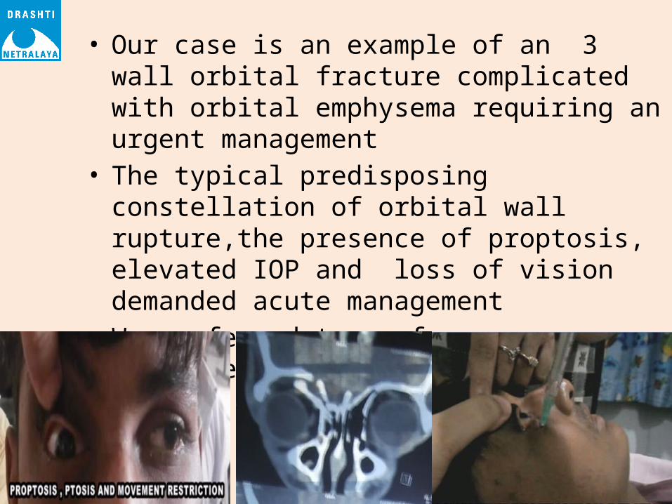
• Our case is an example of an 3 wall orbital fracture complicated with orbital emphysema requiring an urgent management
• The typical predisposing constellation of orbital wall rupture,the presence of proptosis, elevated IOP and loss of vision demanded acute management
• We preferred to perform decompression with a 20G needle .

11
Conclusion
• If tension emphysema occurs, the orbit must be decompressed to relieve the compressive effect of the air accumulation giving consideration to vision and therapeutic complications

12
References1. Singh, M., Phua, V.M., and Sundar, G. Sight-threatening
orbital emphysema treated with needle decompression. Clin Experiment Ophthalmol. 2007; 35: 386–387
2. Lee, S.L., Mills, D.M., Meyer, D.R., and Silver, S.M. Orbital emphysema. Ophthalmology. 2006; 113:2113.e1–2113
3. Bastion, M.L. and Wong, Y.C. A case of sneezing-related orbital emphysema treated by aspiration-decompression in the office. Ophthal Plast Reconstr Surg. 2006; 26: 500–501
4. Dobler, A.A., Nathenson, A.L., Cameron, J.D., Carpel, E.T., Janda, A.M., and Pederson, J.E. A case of orbital emphysema as an ocular emergency. Retina. 1993; 13: 166–168