temperature-sensitive cell division mutants ofescherichia
TRANSCRIPT
JOURNAL OF BACTERIOLOGY, July 1977, p. 293-305Copyright C) 1977 American Society for Microbiology
Vol. 131, No. 1Printed in U.S.A.
Temperature-Sensitive Cell Division Mutants of Escherichiacoli with Thermolabile Penicillin-Binding Proteins
BRIAN G. SPRATTDepartment of Genetics, University of Leicester, Leicester LEl 7RH, England
Received for publication 11 March 1977
The thermostability of the penicillin-binding proteins (PBPs) of 31 tempera-ture-sensitive cell division mutants ofEscherichia coli has been examined. Twoindependent cell division mutants have been found that have highly thermola-bile PBP3. Binding of ['4C]benzylpenicillin to PBP3 (measured in envelopesprepared from cells grown at the permissive temperature) was about 30% of thenormal level at 300C, and the ability to bind [14C]benzylpenicillin was rapidlylost on incubation at 420C. The other PBPs were normal in both mutants. At30°C both mutants were slightly longer than their parents and on shifting to420C they ceased dividing, but cell mass and deoxyribonucleic acid synthesiscontinued and long filaments were formed. At 420C division slowly recom-menced, but at 440C this did not occur. The inhibition of division at 42°C was
suppressed by 0.35 M sucrose, and in one of the mutants it was partiallysuppressed by 10 mM MgCl2. PBP3 was not stabilized in vitro at 42°C by theseconcentrations of sucrose or MgCl2. Revertants that grew as normal rods at 420Cregained both the normal level and the normal thermostability of PBP3. Theresults provide extremely strong evidence that the inactivation of PBP3 at 420Cin the mutants is the cause of the inhibition of cell division at this temperatureand identify PBP3 as an essential component of the process of cell division in E.coli. It is the inactivation of this protein by penicillins and cephalosporins thatresults in the inhibition of division characteristic of low concentrations of manyof these antibiotics.
The process of bacterial cell division is poorlyunderstood (17). Physiological studies withEscherichia coli have shown that over a widerange of growth rates division follows the ter-mination of a round of deoxyribonucleic acid(DNA) replication by a fixed interval (the Dperiod) of about 25 min at 3700 (7). Soon afterthe termination of a round ofDNA replication,cell division becomes independent of furtherribonucleic acid and protein synthesis (3, 5).Presumably, at this time all the enzymes andstructural or regulatory proteins required forthe assembly of the membrane and peptidogly-can layers of the septa have been synthesized.The first visible sign of division is the ingrowthof the inner membrane and a double layer ofpeptidoglycan, commencing about 15 min be-fore division and being completed only 1 to 2min before division, resulting in the physiologi-cal separation ofthe cell into two compartments(3, 13, 24). The final assembly of the outermembrane between the double layer of peptido-glycan and the physical separation of the cellsoccupies the last 1 or 2 min of the division cycle(3, 13, 24).
Biochemical and genetic approaches to theprocess of cell division are severely limited bythe inability to study division at the subcellularlevel. Genetic analysis has indentified at leasteight genes in E. coli that are specifically re-quired for cell division (8, 14, 15, 17); tempera-ture-sensitive mutants at these loci (ftsA-H)cease division abruptly and grow into long fila-ments when transferred from the permissive tothe restrictive temperature. The usefulness ofthese mutants is restricted, since the identifica-tion of the defective gene products has not beenachieved.One approach to the biochemistry of cell divi-
sion is to study specific inhibitors ofthe process.In theory, it should be possible to identify thetarget with which the inhibitor interacts andthereby to identify a specific component of thedivision process. Although many chemicals re-sult in the filamentation of bacterial cells, theonly ones known to act specifically on divisionare the penicillins and cephalosporins (2, 4, 16,18). Low concentrations of most of these /3-lac-tam antibiotics inhibit cell division in E. coliwithout influencing DNA synthesis or cell
293

294 SPRATTJ
mass increase and result in the formation oflong filaments. At higher concentrations theseantibiotics inhibit cell elongation, resulting incell lysis and the production of spheroplasts (16,18). 8-Lactam antibiotics inhibit several en-
zymes involved in the terminal stages of pepti-doglycan metabolism (2), and it has been pro-
posed that their differential effects on cell divi-sion and cell elongation are due to the involve-ment of separate enzyme systems in the twotypes of peptidoglycan growth (6, 12, 16, 18, 23).Inactivation of a penicillin-sensitive enzyme
(PSE) specifically required for cell division willresult in the growth of the cell as long fila-ments, and therefore mutants that produce ahighly thermolabile form of this enzyme shouldbe temperature-sensitive cell division mutants.The screening of cell division mutants for alter-ations in the thermostability of one of the sev-
eral PSEs is impracticable, but fortunatelythese enzymes can be conveniently detectedand studied as those proteins that bind[14C]benzylpenicillin (2, 19, 22). Six penicillin-binding proteins (PBPs) are consistently de-tected in E. coli envelopes, and two additionalPBPs are less consistently detected (19). Pre-vious studies on the properties of the E. coliPBPs have implicated PBP3 as the target atwhich penicillins and cephalosporins interact toinhibit cell division (18-20). We have examinedthe thermostability ofthe individual PBPs in 31temperature-sensitive cell division mutants ofE. coli and have identified two independentmutants that have highly thermolabile PBP3.One ofthese mutants has been briefly describedpreviously (18). The properties ofthese mutantsand their revertants provide strong evidencethat PBP3 is specifically required for the divi-
sion of E. coli and is the target at which ,B-lactam antibiotics interact to inhibit cell divi-sion and cause filamentation.
MATERIALS AND METHODSBacterial strains. Table 1 lists the E. coli strains
used in this work. LEB18 and its derivatives were
B/r strains, whereas all other strains were K-12 de-rivatives. Temperature-sensitive mutants of KN126and LEB18 were isolated after mutagenesis for 30min at 370C with 100 g.g of N-methyl-N'-nitro-N-nitrosoguanidine per ml in 50 mM tris(hydroxy-methyl)aminomethane-maleate buffer, pH 6.0 (1).Cells were washed free ofmutagen, grown overnightin broth at 300C to allow mutations to be expressed,and plated on nutrient agar at 300C. Colonies were
replica plated to the same agar at 42°C, and temper-ature-sensitive mutants were purified and grown inbroth at 30 and 42°C. Those mutants that were nor-
mal rods at 30°C, but which filamented when shiftedto 42°C, were kept for further study. In the mutantselection in which SP63 was obtained, the mutagen-ized cells were subjected to a primary selection at300C with cephalexin (5 ,ug/ml), cephalexin-resist-ant colonies were replica plated to 42°C, and temper-ature-sensitive mutants were screened as above(18).Media and conditions of growth. Cells were
grown in Penassay broth (Difco antibiotic mediumno. 3) in all experiments unless otherwise stated.The minimal medium used in some experiments wasM9 supplemented with 0.2% glucose and required L-amino acids (20 ,ug/ml). Cultures were initiated bytaking a loop of bacterial growth from a stock agarplate into about 20 ml of medium and growing thecells at the appropriate temperature overnight. Inthe morning, the cultures were diluted and grownwith vigorous aeration for at least a further five celldoublings (maintaining the absorbance below 0.4 byperiodic dilution) before experiments were started.Measurement of cell growth. Growth of the cul-
tures was followed by measuring the absorbance at
TABLE 1. Bacterial strainsStrain Relevant genotype or properties Source
KN126 tryE(Am)9829 tyr(Am) ilv sup-126 T. Nagata (11)SP63 Temperature-sensitive cell division mutant derived This paper and reference
from KN126 after nitrosoguanidine mutagenesis 18 (strain 6-30)SP631 and SP632 Spontaneous temperature-resistant revertants of This paper
SP63LEB18 lacZ rpsL P. MeacockSP25 Temperature-sensitive mutant derived from LEB18 This paper
after nitrosoguanidine mutagenesisSP258 Spontaneous partial revertant of SP25 This paperSP2581 and SP2582 Spontaneous temperature-resistant revertants of This paper
SP258T84 ftsA J. L. Strominger (15)MFT84 ftsB F. Jacob (15)MFT108 ftsC J. L. Strominger (15)MFT103 ftsD J. L. Strominger (15)MFT99 ftsE F. Jacob (15)MFT1141 ftsF F. Jacob (15)ASH501 ftsH I. B. Holland (8)
J. BACTZRIOL.

PENICILLIN-BINDING PROTEINS AND CELL DIVISION 295
550 nm (A550) (for broth cultures) or A450 (for mini-mal medium cultures) in a Gilford model 300 auto-matic sampling spectrophotometer using a 1-cm-path length cuvette. Total cell numbers were mea-
sured using a model B Coulter counter. Cells were
diluted into ice-cold membrane-filtered saline con-
taining 0.37% formaldehyde and were counted witha 30-,um probe. To measure cell size, exponentiallygrowing cultures were sampled in triplicate, and theabsorbances and total cell numbers were measured.Average cell size was expressed as A550 per cellnumber. DNA synthesis was measured by incor-poration of [3H]thymidine in the presence of 1.5 mMuridine, as described previously (21).
Preparation of envelopes and measurement ofPBPs. For all experiments, cell envelopes (consist-ing of inner membrane, outer membrane, and pepti-doglycan) were prepared as described previously (19)from cells that had been grown in Penassay broth at30°C. For the standard assay of PBPs, 200 ,lI ofenvelopes (5 to 10 mg/ml in 50 mM sodium phos-phate buffer, pH 7.0) were incubated for 10 min at30°C with 20 ,A of a 50-,uCi/ml amount of[14C]benzylpenicillin. The final concentration of[E4C]benzylpenicillin for all samples in all experi-ments was 31.3 ,.Lg/ml. The binding was terminated,and the inner membrane proteins were solubilizedby the addition of 10 Al of 20% (wt/vol) Sarkosyl NL-97 and 5 Al of a 120-mg/ml amount of nonradioac-tive benzylpenicillin (19). After 20 min at room tem-perature, the Sarkosyl-insoluble outer membraneand peptidoglycan were pelleted by centrifugationat 40,000 x g for 60 min at 10°C in the SM-24 rotor ofa Sorvall RC2B centrifuge. The Sarkosyl-soluble in-ner membrane proteins were fractionated, accordingto apparent molecular weight, on a 10% sodiumdodecyl sulfate-polyacrylamide slab gel, and thePBPs were detected by fluorography on prefogged(9) Kodak RP Royal X-ray film at -80°C as de-scribed previously (19).The in vitro thermostability of the PBPs in tem-
perature-sensitive mutants was examined by pre-
paring envelopes from cells that had been grown atthe permissive temperature (30°C) and measuringtheir ability to bind [14C]benzylpenicillin at 30°C (asabove) and at 42°C, after various periods of preincu-bation at this temperature.
Quantitation of the levels of [14C]benzylpenicillinbound to each PBP was measured by densitometry ofthe X-ray films, using a Joyce-Loebl MKlllc mi-crodensitometer.
Measurement of minimal inhibitory concentra-tions. The minimal inhibitory concentrations of f3-
lactam antibiotics were measured by serial dilution.Tubes containing 1 ml of Penassay broth and twofoldserial dilutions of the antibiotic were inoculatedwith 0.1 ml of a 10-4 dilution of an overnight cultureof bacteria. After incubation for 24 h at 30°C, theminimal inhibitory concentrations were read as thelowest concentration that inhibited turbid growth.
Chemicals. [14C]benzylpenicillin (specific activ-ity, 53 mCi/mmol) was obtained from the Radi-ochemical Centre, Amersham, England. Unlabeledbenzylpenicillin, cephalexin, and cephaloridine
were kindly provided by Glaxo Laboratories, Green-ford, England. Sarkosyl NL-97 was obtained fromGeigy Industrial Chemicals, New York.
RESULTS
Isolation of mutants. Twenty-four tempera-ture-sensitive cell division mutants were iso-lated after mutagenesis of strains KN126 andLEB18 as described above. The temperaturesensitivity of the binding of [14C]benzylpeni-cillin to the individual PBPs was measured inthese mutants and in seven previously de-scribed cell division mutants. There were nodefects in the PBPs of any of the fts mutantswe examined (listed in Table 1), but two ofthe newly isolated mutants had thermolabilePBP3. One of these mutants (SP25) was iso-lated from LEB18, and the other (SP63) wasisolated from KN126. Both mutants retainedthe nutritional markers of their respective par-ents and had not gained any additional require-ments during mutagenesis.SP63 formed colonies on nutrient agar at
42°C with a frequency of 2.1 x 10⁻⁵. This wasreduced to 1.3 x 10⁻⁷ on nutrient agar at 44°C.Many of these colonies were not true revert-ants, since they grew as mixtures of filamentsand normal cells at 42°C. Two true revertants,which grew as normal rods at 42°C, were, how-ever, isolated and designated SP631 and SP632.These revertants had the same nutritional re-quirements as their parent.SP25 formed short filaments at 42°C and then
lysed, with the production of spheroplasts. Norevertants of SP25 that form colonies on nutri-ent agar at 42°C (reversion rate, >1:109) havebeen obtained, and this strain is almost cer-tainly a double mutant, with one defect thatresults in filamentation and another that re-sults in lysis at the restrictive temperature. Insupport of this, we have been able to isolatederivatives of SP25 that form long filamentsbut do not lyse at 42°C by plating cells onnutrient agar at 37°C. Under these conditionsapproximately 1:107 cells formed colonies abovea background lawn of growth, and 8 of 13 ofthese partial revertants filamented withoutlysis at 42°C. One of these latter strains waschosen for further study and was designatedSP258. It grew at the same rate as SP25 and nolysis has been observed, at either 42 or 44°C.SP258 produced revertants on nutrient agar at42°C, and two of these, which grew as normalrods, were designated SP2581 and SP2582.
Properties of the PBPs of mutants SP25and SP63. Envelopes of mutant SP25 and itsparent (LEB18) were prepared from cells grown
VOL. 131, 1977
296 SPRAJT
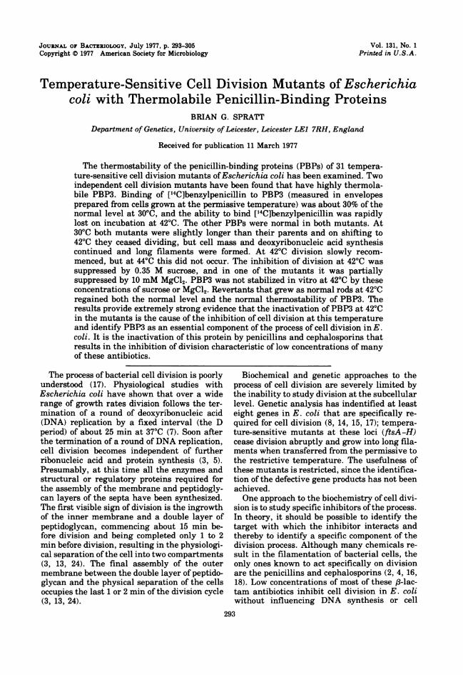
at the permissive temperature, and the level of['4C]benzylpenicillin bound to each of the PBPswas measured at 30 and 420C. The mobilities ofthe PBPs on the gels in these B/r strains wereidentical to those in E. coli K-12 (Fig. 1A andJ), although a PBP with an apparent molecularweight of 32,000 (PBP7) was consistently de-tected in the B/r strains, but less consistently,and in lesser amounts, in K-12 strains (19). Thelevel of [14C]benzylpenicillin binding to PBP3 ofmutant SP25 at 300C was about 30% of that inthe parent LEB18 (compare the ratio of PBP2/PBP3 in Fig. 1A and D). The low level of PBP3in mutant SP25 at 300C was not due to a failureto saturate the protein with ['4C]benzyl-penicillin, since raising the concentration ofthelatter from 31 to 116 ug/ml did not increase thelevel of binding. As PBP3 in this mutant washighly thermolabile (see below), it was possiblethat it was being partially inactivated duringenvelope preparation or during the assay at30°C. Attempts to increase the level of PBP3 bytaking additional precautions to prevent loss ofactivity during envelope preparation (e.g., re-placing the standard 10 mM sodium phosphatebuffer with 50 mM phosphate buffer containing10% glycerol and only washing the membranesonce) or during the PBP assay (e.g., by reduc-ing the assay temperature to 200C and includ-ing 10% glycerol in the assay mix) had no effect.Binding of [14C]benzylpenicillin to the PBPs
of LEB18 was not reduced by preincubation ofthe envelopes for 8 min at 42°C; i.e., the PBPswere thermostable for at least 8 min at 420C.Envelopes of the mutant SP25 rapidly lost theability to bind ['4C]benzylpenicillin to PBP3when incubated at 420C, although the otherPBPs were thermostable (Fig. 1E through G).The inactivation of PBP3 was irreversible un-der these conditions, since little recovery ofbinding activity was observed after the enve-lopes had been returned from 42 to 300C (Fig.1H and I).The second mutant with a thernolabile PBP3
has been described briefly previously (18,where it was called 6-30), and it had penicillin-binding properties very similar to those ofSP25. Envelopes prepared from cells of SP63grown at the permissive temperature bound alow level of [14C]benzylpenicillin to PBP3 at300C, and binding was thermolabile at 420C(18). The inactivation ofPBP3 at 420C was irre-versible, since after 10 min of preincubation at420C no binding of [14Clbenzylpenicillin to PBP3was detected either at 42°C (Fig. 2B) or for atleast 30 min after returning the envelopes to30°C (Fig. 2C through F). As with mutant SP25,the level of binding to PBP3 of SP63 at 300C
A B C D E F GH I
_ _ __ _- - - W
32j.4
7 _
FIG. 1. Binding of ['4C]benzylpenicillin to enve-lopes ofLEB18 and SP25. ['4C]benzylpenicillin wasbound to envelopes ofLEB18 for 10 min at 30°C (A).Envelopes of LEB18 were incubated at 42°C, andafter 4 min (B) or 8 min (C) [14C]benzylpenicillin wasadded for a further 2 min at this temperature.[14C]benzylpenicillin was added to envelopes ofSP25for 10 min at 30°C (D). Envelopes of SP25 wereincubated at 42°C, and after 0 min (E), 4 min (F), or8 min (G), [14C]benzylpenicillin was added for afurther 2 min at this temperature. A portion ofenve-lopes of SP25 was incubated at 42°C for 10 min andthen transferred back to 30°C, and after 1 min (H) or7 min (I) [14C]benzylpenicillin was added for a fur-ther 10 min at this temperature. The PBPs of an E.coli K-12 strain (KN126) are included for compari-son (J). The binding was terminated, the sampleswere loaded on a 10% sodium dodecyl sulfate-poly-acrylamide slab gel, and a fluorograph was preparedby exposure of the gel to X-ray film for 66 days asdescribed in the text.
could not be increased by raising the concentra-tion of [14C]benzylpenicillin or by taking addi-tional precautions to prevent inactivation dur-ing envelope preparation and PBP assay.
Properties of the PBPs of revertants ofSP25 and SP63. Figures 3 and 4 show thethermostability of the PBPs in revertants ofSP25. The partial revertant SP258, which fila-mented at 420C but did not lyse, was identicalto SP25 in the level and thermolability of PBP3(Fig. 3). The full revertants SP2581 and SP2582,which grew as nonnal rods at 4200, had re-gained both the nonnal level and the normalthermostability of PBP3 (Fig. 4). Revertants ofSP63 that grew as normal rods at 420C alsoregained the normal level and thermostabilityofPBP3 (Fig. 5). In this figure PBP1 is split intotwo bands. Recent experiments using a differ-
MP %NMNO %ME~ p
M--
J. BACTERIOL.

PENICILLIN-BINDING PROTEINS AND CELL DIVISION
A B CD E F G
3
FIG. 2. Binding of["4C]benzylpenicillin to envelopes ofSP63. ["C]benzylpenicillin was added to envelopesof SP63 for 10 min at 30°C (A, G). Envelopes of SP63 were incubated at 42°C for 10 min, and["4C]benzylpenicillin was added to one portion for a further 5 min at 42°C (B), while the remainder was
transferred back to 30°C and ["4C]benzylpenicillin was added after 0 min (C), 10 min (D), 20 min (E), or 30min (F) for a further 10 min at this temperature. Samples were fractionated, and a fluorograph was preparedby exposure of the slab gel to X-ray film for 49 days.
ent polyacrylamide gel system have shown thatPBP1 is at least two proteins (U. Schwarz andB. G. Spratt, unpublished experiments), as pre-viously suspected (19). On this gel PBP7 fails tobind ['4C]benzylpenicillin at 42°0. However,PBP7 of the parent and other derivatives wasalso thermolabile in many experiments and ispresumably a rather unstable protein.Growth and divison of SP258 and SP63. Mu-
tants SP258 and SP63 grew rapidly in broth at300C with mean generation times that wereindistinguishable from those of their parentstrains (32 to 34 min for SP258 and 34 to 36 minfor SP63). At 30°C the size (A550 per cell) ofSP258 was 22% greater than that of LEB18, andSP63 was 29% greater than KN126. The in-creased size of the mutants was due to an in-creased average cell length.
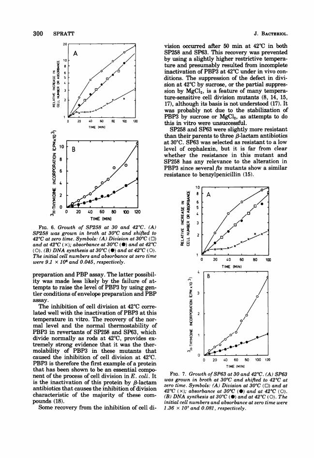
After a shift from 30 to 420C, both mutantsaccelerated their rate ofDNA synthesis and cellmass increase, but cell division ceased after aresidual increment of 20 to 25%. After about 50
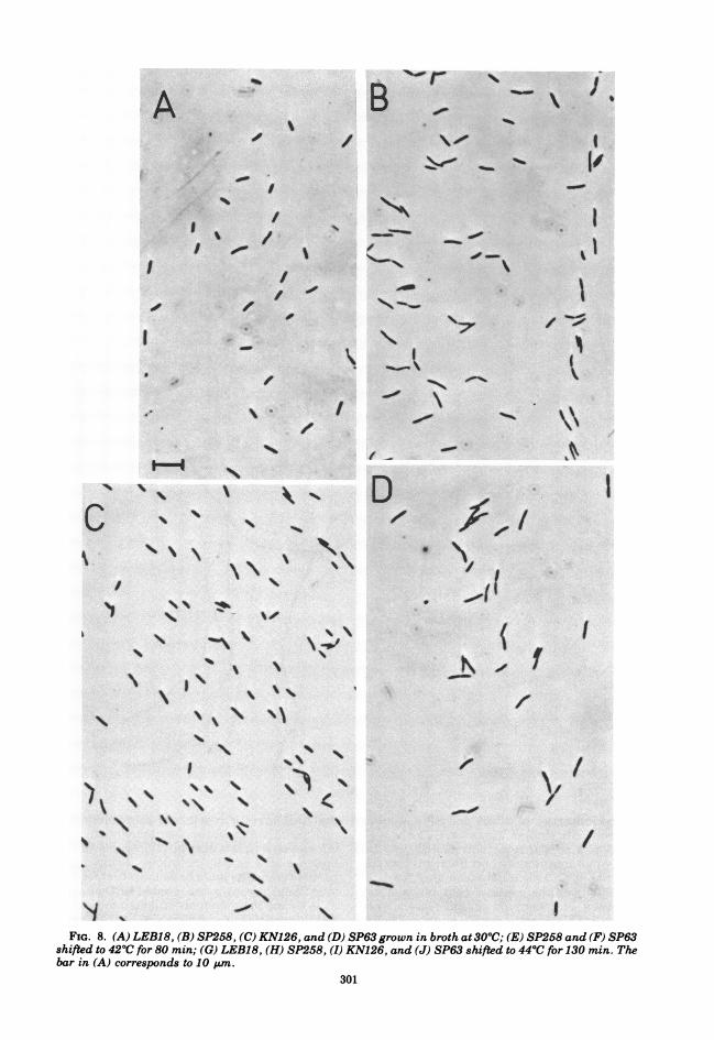
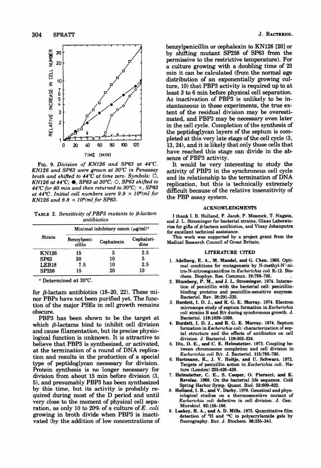
min at 4200, cell division slowly recommencedand a doubling in cell number, over that at thetime of the temperature shift, occurred by 80min (Fig. 6 and 7). At this time cell mass hadincreased 9-fold in SP63 and 11-fold in SP258,and long filaments were observed under thephase-contrast microscope (Fig. 8). The slowrecovery of cell division at 4200 was found bothin minimal medium and in broth; it occurredwhether the cell number at the time of thetemperature shift was low (e.g., 3 x 10⁶ cells/ml) or relatively high (e.g., 3 x 10⁷ cells/ml).When 4400 was used as the restrictive tempera-ture, the residual increment in cell number ofSP258 and SP63 was slightly smaller (10 to20%) than that at 42°C, and recommencementof division during continued incubation at 4400did not occur. Figure 9 shows the division ofSP63 and KN126 at 4400 in broth. After 130 minat 440, cell number had only increased by 20%whereas cell mass had increased by 20-fold,resulting in the production of extremely long
4
6
297VOL. 131, 1977

A B C D E F 6 H
1
__
FIG. 3. Thermostability of the PBPs in SP25 and SP258. ['4C]benzylpenicillin was bound to envelopes ofSP25 for 10 min at 30°C (A). Envelopes ofSP25 were incubated at 42°C, and after 0 min (B), 4 min (C), or 8min (D) ['4C]benzylpenicillin was added for a further 2 min at this temperature. ['4C]benzylpenicillin wasbound to envelopes ofSP258 for 10 min at 30°C (E). Envelopes ofSP258 were incubated at 42°C, and after 0min (F), 4 min (G), or 8 min (H) ['4C]benzylpenicillin was added for a further 2 min at this temperature.Samples were fractionated, and a fluorograph was prepared by exposure of the slab gel to X-ray film for 55days.
filaments (Fig. 8). Shifting SP63 back to 300C,after 40 min at 44°C, resulted in a burst ofdivision after a further 25 min. Filaments ofSP63 and SP258, as well as those formed bytheir parents in the presence of low concentra-tions of 18-lactam antibiotics, often showed asingle, centrally positioned incomplete septumwhen observed under phase-contrast or Nor-marski optics.The inhibition of cell division at 420C was
overcome by the addition of 0.35 M sucrose tobroth in strain SP63 and was partially over-come in strain SP258. MgCl2 (10 mM) also par-tially reversed the inhibition of division instrain SP63 but had no effect on SP258. DL-Diaminopimelic acid (40 ,ug/ml), D-alanine (20,Ag/ml), NaCl (0.34 M), and cyclic adenosine 5'-monophosphate (1 mM) failed to stimulate divi-sion at 420C in either mutant. The suppressionof the defect in cell division at 42°C by sucroseor MgCl2 could be the result of the stabilizationof PBP3 under these conditions. We compared
the thermolability of [14C]benzylpenicillin bind-ing to PBP3 in the presence or absence of 0.35Msucrose or 10 mM MgCl2. Neither compoundstabilized PBP3 in the mutants.
Additional properties of mutants SP258 andSP63. Table 2 shows the minimal inhibitoryconcentrations ofthe mutants and their parentsfor three 18-lactam antibiotics.SP63 was isolated in a strain that contains a
temperature-sensitive amber suppressor (sup-126; 11), and the lesion in the gene coding forPBP3 could have been an amber mutation. Ifthis had been the case, revertants of the tem-perature-sensitive suppressor should form colo-nies at 42°C and at the same time should losetheir requirements for tryptophan and tyrosine(which are also due to amber mutations). How-ever, although the amber suppressor revertedto thermostability with a frequency of 6 x 10⁻⁶(measured as the reversion rate of KN126 totryptophan and tyrosine independence at 420C),we did not obtain colonies of SP63 that grew at
2467 -------
.....1.
298 SPRATT J. BACTERIOL.

PENICILLIN-BINDING PROTEINS AND CELL DIVISION 299
A B C D E to be demonstrably altered in its enzymatic orphysicochemical properties, and, second, re-vertants that divide normally at the restrictivetemperature should regain the normal proper-ties of the protein.The knowledge that penicillins and cephalo-
sporins inhibit cell division by inactivating anenzyme involved in peptidoglycan metabolismand the availability of a convenient method for
1 _ ~ __wscreening mutants for an alteration in this pro-tein have resulted in the identification of twomutants, SP258 and SP63, in which the above
2 __ __ _ criteria have been satisfied.3 - Envelopes of SP258 and SP63 prepared from
cells grown at the permissive temperature pos-4t_ , _ sessed a PBP3 that was active when assayed at
30°C but was rapidly inactivated on incubation5 in vitro at 4200. The level of binding of6 [14C]benzylpenicillin (and the rate of binding
[unpublished experiments]) to PBP3 was lower7 _ _ - 1 -i ",, in the mutants at 300C than in their parents.
The lower activity of PBP3 at the permissivetemperature correlated well with the increasedlength of both mutants, suggesting that divi-sion even at this temperature was to some ex-tent impaired by the alteration of PBP3. Thereason for the lower level of PBP3 is unclear; itcould be due to a decreased synthesis, to adecreased ability of the altered protein to be
FIG. 4. Thermostability of the PBPs in revertants correctly inserted into the cytoplasmic mem-
of SP258. ['4C]benzylpenicillin was bound to enve- brane, or to a loss of activity during envelopelopes of SP25 for 10 min at 30°C (A). Envelopes ofSP25 (B), SP258 (C), SP2581 (D), and SP2582 (E)were incubated at 42°C for 8 min, and ['4C]benzyl-penicillin was added for a further 2 min at this tem-perature. Samples were fractionated, and a fluoro-graph was prepared by exposure of the slab gel toX-ray film for 48 days.
420C in the absence of tryptophan and tyrosine.The lesion in the gene coding for PBP3 of SP63was therefore not an amber mutation.
DISCUSSIONThe identification of the defective gene prod-
ucts in temperature-sensitive cell division mu-tants is difficult. Since most of these mutantshave been isolated after mutagenesis, particu-lar care is needed to show that any differencebetween the mutant and its parent is the causeof the defect in division. Loss of a protein orchanges in its rate of synthesis on shifting acell division mutant from the permissive to therestrictive temperature are not sufficient evi-dence for a role of that protein in the tempera-ture-sensitive phenotype. Two criteria shouldbe satisfied: first, the protein should be shown
A B C D E F
23--
FIG. 5. Thermostability of PBP3 in SP63 and itsrevertants. [14C]benzylpenicillin was bound to enve-lopes of (A) SP63, (B) SP631, and (C) SP632 for 10min at 30°C. Envelopes of (D) SP63, (E) SP631, and(F) SP632 were incubated at 42°C for 5 min, and[14C]benzylpenicillin was added for a further 5 minat this temperature. Samples were fractionated, anda fluorograph was prepared by exposure of the slabgel to X-ray film for 31 days.
qm.-
VOL. 131, 1977
300 SPRATT
Wz I9
tr
:t w
p
W 6tr o
0 20 40 60 80 100 120
TIME (MIN)
0
x
a. 10
R 6 -
0
z 4
0 20
z 0 20 40 60 80 100 120TIME (MIN)
FIG. 6. Growth of SP258 at 30 and 42°C. (A)SP258 was grown in broth at 30°C and shifted to420C at zero time. Symbols: (A) Division at 30°C (0)and at 42°C (x); absorbance at 300C (0) and at 42°C(0). (B) DNA synthesis at 30°C (0) and at 42°C (0).The initial cell numbers and absorbance at zero timewere 9.1 x 10⁶ and 0.045, respectively.
preparation and PBP assay. The latter possibil-ity was made less likely by the failure of at-tempts to raise the level of PBP3 by using gen-tler conditions ofenvelope preparation and PBPassay.The inhibition of cell division at 42°C corre-
lated well with the inactivation ofPBP3 at thistemperature in vitro. The recovery of the nor-
mal level and the normal thermostability ofPBP3 in revertants of SP258 and SP63, whichdivide normally as rods at 420C, provides ex-tremely strong evidence that it was the ther-molability of PBP3 in these mutants thatcaused the inhibition of cell division at 42°C.PBP3 is therefore the first example of a proteinthat has been shown to be an essential compo-
nent of the process of cell division in E. coli. Itis the inactivation of this protein by (3-lactamantibiotics that causes the inhibition of divisioncharacteristic of the majority of these com-
pounds (18).Some recovery from the inhibition of cell di-
vision occurred after 50 min at 420C in bothSP258 and SP63. This recovery was preventedby using a slightly higher restrictive tempera-ture and presumably resulted from incompleteinactivation of PBP3 at 420C under in vivo con-ditions. The suppression of the defect in divi-sion at 420C by sucrose, or the partial suppres-sion by MgCl2, is a feature of many tempera-ture-sensitive cell division mutants (8, 14, 15,17), although its basis is not understood (17). Itwas probably not due to the stabilization ofPBP3 by sucrose or MgCl2, as attempts to dothis in vitro were unsuccessful.SP258 and SP63 were slightly more resistant
than their parents to three 83-lactam antibioticsat 3000. SP63 was selected as resistant to a lowlevel of cephalexin, but it is far from clearwhether the resistance in this mutant andSP258 has any relevance to the alteration inPBP3 since several fts mutants show a similarresistance to benzylpenicillin (15).
WI
WW
10
8
6
5
4
3
2
4
x
z
r0
z
I
I
3
2
0
0 20 40 60
TIME (MIN)
80 100
0 20 40 60 a0 100 120TIME (MIN)
FIG. 7. Growth ofSP63 at 30 and 42°C. (A) SP63was grown in broth at 30°C and shifted to 420C atzero time. Symbols: (A) Division at 300C (0) and at420C (x); absorbance at 30°C (0) and at 420C (0).(B) DNA synthesis at 300C (-) and at 420C (0). Theinitial cell numbers and absorbance at zero time were
1.36 x 10⁷ and 0.081, respectively.
0-1'
0 0/0/0
0 / x --
0 X,
x -,
B
0
0
00*
"I
.- , --
" 0 0
J. BACTERIOL.
I
A Br %
r
/r\f. ~~~I'I,
I
II
I
_/ . _M/00 %0
*6
-C,
I
, _,-r00doi
/S
1%
-4
I
I
II'4
"-V ~ .01k
-p
m--I N
C '949 9
'4'''1 94% ,
'I \
N\'lb-
nLid-r4z~~~~-
AI
I
(
N_w-
''S
I
/9
1% \ '9""I,
N%
i94% 9% ')9%\ 9%
9% re94
C N/
/94I%
N.000YI
FIG. 8. (A) LEB18, (B) SP258, (C) KN126, and (D) SP63 grown in broth at3O0C; (E) SP258 and (F) SP63shifted to 42°C for 80 min; (G) LEB18, (H) SP258, (I) KN126, and (J) SP63 shifted to 44°C for 130 min. Thebar in (A) corresponds to 10 uim.
301
1% 1%\
qm
I \'k
I

302 SPRATT
E EF~~~
.-7r
Ga
/
4e4-
/ _r
The properties of SP258 and SP63 were ex-traordinarily similar. However, SP258 was iso-lated as a partial revertant of SP25, and thislatter mutant grew as short filaments and thenlysed at 4200. This could mean that the defectin PBP3 was the cause of both filamentationand lysis, but there are several reasons to be-lieve that SP25 was a double mutant. The factthat PBP3 was thermolabile in SP25 suggests
that this was the result of a point mutation,and the failure to obtain revertants that fonnedcolonies on nutrient agar at 420C argues for thepresence of a second mutation. Furthennore,partial revertants could be isolated that fonnedlong filaments but never lysed at 420C or evenat 4400, and these mutants still maintained thefull thermolability of PBP3. Finally, SP63 hada thermolabile PBP3 and yet never lysed at
J. BACTERIOL.
.w
09LIO d
I --ft
m
.P .0OP

PENICILLIN-BINDING PROTEINS AND CELL DIVISION 303
I Vo
/
do
'aI
I',
J0I
42°C. The cause of lysis in SP25 was presum-ably due to a defect in cell wall synthesis, but itwas not due to the defect in PBP3 and none ofthe other PBPs were thermolabile.SP258 and SP63 are considered to be muta-
tions in the structural gene for PBP3, and Ipropose to call this gene pbpB (penicillin-bind-ing protein). pbpA is the structural gene forPBP2 defined by temperature-sensitive cellshape mutants that have thermolabile PBP2(B. G. Spratt, manuscript in preparation). ThepbpB mutations of SP258 and SP63 have notbeen mapped, but they appear to be distinctfrom those of the fts mutants that we examined(Table 1), as these latter mutants had normalPBP3.PBP3 has an apparent molecular weight of
60,000, and a single rapidly growingE. coli cellhas been estimated to contain about 50 mole-cules of this protein (19). PBP3 is presumed tocorrespond to a minor PSE involved in peptido-glycan metabolism specific to cross-wall forma-tion at division. From its abundance, molecularweight, and penicillin-binding properties, it isclearly distinct from the two major PSEs thathave been purified from E. coli (6, 12, 23).Purely on the basis of a difference in the sensi-tivities of the two PSEs to f-lactam antibiotics,it has been suggested that one is involved indivision and one in cell elongation (6, 12, 23).More recent work has shown that these assign-ments were premature, and it is now believedthat it is minor PSEs, corresponding to minorPBPs (e.g., PBP3), that are the killing targets
VOL. 131, 1977
,e..
304 SPRATT
Er30-
20 2~~~~~~0
w/
0
z /100
w7,
FIG. 9. Division of KN126 and SP63 at 44°C.KN126 and SP63 were grown at 30°C in Penassay
broth and shifted to 44°C at time zero. Symbols: EO,KN126 at 44°C; *, SP63 at 30°C; 0, SP63 shifted to
44°C for 40 mm and then returned to 30°C; x, SP63
at 44°C. Initial cell numbers were 9.9 x 1051m1 for
KN126 and 8.8 x I1Of/ml for SP63.
TABLE 2. Sensitivity ofPBP3 mutants to /3-lactam
antibiotics
Minimal inhibitory concn (p.g/ml)aStrain Benzylpeni Cephalori-
cBllpn Cephalexin dineKN126 15 5 2.5SP63 30 10 5LEB18 7.5 10 2.5SP258 15 20 10
a Determined at 30C.
for f8-lactam antibiotics (18-20, 22). These mi-nor PBPs have not been purified yet. The func-tion of the major PSEs in cell growth remainsobscure.PBP3 has been shown to be the target at
which /8-lactams bind to inhibit cell divisionand cause filamentation, but its precise physio-logical function is unknown. It is attractive tobelieve that PBP3 is synthesized, or activated,at the termination of a round of DNA replica-tion and results in the production of a specialtype of peptidoglycan necessary for division.Protein synthesis is no longer necessary fordivision from about 15 min before division (3,5), and presumably PBP3 has been synthesizedby this time, but its activity is probably re-
quired during most of the D period and untilvery close to the moment of physical cell sepa-
ration, as only 10 to 20% of a culture ofE. coligrowing in broth divide when PBP3 is inacti-vated (by the addition of low concentrations of
benzylpenicillin or cephalexin to KN126 [20] orby shifting mutant SP258 of SP63 from thepermissive to the restrictive temperature). Fora culture growing with a doubling time of 23min it can be calculated (from the normal agedistribution of an exponentially growing cul-ture, 10) that PBP3 activity is required up to atleast 3 to 6 min before physical cell separation.As inactivation of PBP3 is unlikely to be in-stantaneous in these experiments, the true ex-tent of the residual division may be overesti-mated, and PBP3 may be necessary even laterin the cell cycle. Completion of the synthesis ofthe peptidoglycan layers of the septum is com-pleted at this very late stage of the cell cycle (3,13, 24), and it is likely that only those cells thathave reached this stage can divide in the ab-sence of PBP3 activity.
It would be very interesting to study theactivity of PBP3 in the synchronous cell cycleand its relationship to the termination ofDNAreplication, but this is technically extremelydifficult because of the relative insensitivity ofthe PBP assay system.
ACKNOWLEDGMENTSI thank I. B. Holland, F. Jacob, P. Meacock, T. Nagata,
and J. L. Strominger for bacterial strains, Glaxo Laborato-ries for gifts of (3-lactam antibiotics, and Vinay Jobanputrafor excellent technical assistance.
This work was supported by a project grant from theMedical Research Council of Great Britain.
LITERATURE CITED
1. Adelberg, E. A., M. Mandel, and G. Chen. 1965. Opti-mal conditions for mutagenesis by N-methyl-N'-ni-tro-N-nitrosoguanidine in Escherichia coli K-12. Bio-chem. Biophys. Res. Commun. 18:788-795.
2. Blumberg, P. M., and J. L. Strominger. 1974. Interac-tion of penicillin with the bacterial cell: penicillin-binding proteins and penicillin-sensitive enzymes.Bacteriol. Rev. 38:291-335.
3. Burdett, I. D. J., and R. G. E. Murray. 1974. Electronmicroscope study of septum formation in Escherichiacoli strains B and B/r during synchronous growth. J.Bacteriol. 119:1039-1056.
4. Burdett, I. D. J., and R. G. E. Murray. 1974. Septumformation in Escherichia coli: characterization of sep-tal structure and the effects of antibiotics on celldivision. J. Bacteriol. 119:303-324.
5. Dix, D. E., and C. E. Helmstetter. 1973. Coupling be-tween chromosome completion and cell division inEscherichia coli B/r. J. Bacteriol. 115:786-795.
6. Hartmann, R., J. V. Holtje, and U. Schwarz. 1972.Targets of penicillin action in Escherichia coli. Na-ture (London) 235:426-429.
7. Helmstetter, C. E., S. Cooper, 0. Pierucci, and E.Revelas. 1968. On the bacterial life sequence. ColdSpring Harbor Symp. Quant. Biol. 33:809-822.
8. Holland, I. B., and V. Darby. 1976. Genetical and phys-iological studies on a thermosensitive mutant ofEscherichia coli defective in cell division. J. Gen.Microbiol. 92:156-166.
9. Laskey, R. A., and A. D. Mills. 1975. Quantitative filmdetection of 3H and l4C in polyacrylamide gels byfluorography. Eur. J. Biochem. 56:335-341.
J. BACTERIOL.

PENICILLIN-BINDING PROTEINS AND CELL DIVISION 305
10. Mitchison, J. M. 1971. The biology of the cell cycle.Cambridge University Press, Cambridge.
11. Nagata, T., and T. Horiuchi. 1973. Isolation and char-acterisation of a temperature-sensitive amber sup-pressor mutant of Escherichia coli K12. Mol. Gen.Genet. 123:77-88.
12. Nguyen-Disteche, M., J. J. Pollock, J. M. Ghuysen, J.Puig, P. Reynolds, H. R. Perkins, J. Coyette, and M.R. J. Salton. 1974. Sensitivity to ampicillin and ceph-alothin ofenzymes involved in wall peptide crosslink-ing in Escherichia coli K12, strain 44. Eur. J. Bio-chem. 41:457-463.
13. Onken, A., and W. Messer. 1973. Cell division inEsche-richia coli. Septation during synchronous growth.Mol. Gen. Genet. 127:349-358.
14. Reeve, J. N., D. J. Groves, and D. J. Clark. 1970.Regulation of cell division in Escherichia coli: charac-terization of temperature-sensitive division mutants.J. Bacteriol. 104:1052-1064.
15. Ricard, M., and Y. Hirota. 1973. Process of cellulardivision in Escherichia coli: physiological study ofthermosensitive mutants defective in cell division. J.Bacteriol. 116:314-322.
16. Schwarz, U., A. Asmus, and H. Frank. 1969. Autolyticenzymes and cell division of Escherichia coli. J. Mol.Biol. 41:419-429.
17. Slater, M., and M. Schaechter. 1974. Control of celldivision in bacteria. Bacteriol. Rev. 38:199-221.
18. Spratt, B. G. 1975. Distinct penicillin-binding proteinsinvolved in the division, elongation and shape ofEscherichia coli K12. Proc. Natl. Acad. Sci. U.S.A.72:2999-3003.
19. Spratt, B. G. 1977. Properties of the penicillin-bindingproteins of Escherichia coli K12. Eur. J. Biochem.72:341-352.
20. Spratt, B. G. 1977. Comparison of the binding proper-
ties of two 6,8-amidinopenicillanic acid derivativesthat differ in their physiological effects on Esche-richia coli. Antimicrob. Agents Chemother. 11:161-166.
21. Spratt, B. G., and R. J. Rowbury. 1971. Physiologicaland genetical studies on a mutant of Salmonella ty-phimurium which is temperature-sensitive for DNAsynthesis. Mol. Gen. Genet. 114:35-49.
22. Spratt, B. G., and J. L. Strominger. 1976. Identificationof the major penicillin-binding proteins of Esche-richia coli as D-alanine carboxypeptidase 1A. J. Bac-teriol. 127:660-663.
23. Tamura, T., Y. Imae, and J. L. Strominger. 1976. Puri-fication to homogeneity and properties of two D-ala-nine carboxypeptidases 1 from Escherichia coli. J.Biol. Chem. 251:414-423.
24. Woldringh, C. L. 1976. Morphological analysis of nu-
clear separation and cell division during the life cycleofEscherichia coli. J. Bacteriol. 125:248-257.
VOL. 131, 1977