tearlab clinical utility guide clinical utility...regulate osmolarity of body fluids within very...
TRANSCRIPT

1 1 0 2 5 R o s e l l e S t . , S u i t e 1 0 0 , S a n D i e g o , C A 9 2 1 2 1 · P h o n e 8 5 8 . 4 5 5 . 6 0 0 6 · F a x 8 5 8 . 8 1 2 . 0 5 4 0 · w w w . T e a r L a b . c o m
930070Rev B · Page 1
TearLab™ Osmolarity System · Clinical Utility Guide
The TearLab Osmolarity System is intended to measure the osmolarity of human tears to aid in the diagnosis of dry eye disease in patients suspected of having dry eye disease,
in conjunction with other methods of clinical evaluation.
– TearLab FDA 510(k)

1 1 0 2 5 R o s e l l e S t . , S u i t e 1 0 0 , S a n D i e g o , C A 9 2 1 2 1 · P h o n e 8 5 8 . 4 5 5 . 6 0 0 6 · F a x 8 5 8 . 8 1 2 . 0 5 4 0 · w w w . T e a r L a b . c o m
930070Rev B · Page 2
Understanding the Osmolarity Test Result
Abnormal tear osmolarity is a failure of homeostatic osmolarity regulation, a key feature of dry eye disease (DED). When left unchecked, hyperosmolar tears in early stage DED will lead to damage of the cornea and conjunctiva evident in later stage disease. The higher the osmolarity, the more severe the dry eye. TearLab recently conducted a clinical trial that recruited over 300 patients in which over 600 eyes were evaluated. Using patients selected from the general patient population, osmolarity was found to have 88% specificity, 75% sensitivity in mild/moderate disease and 95% sensitivity in severe disease at a diagnostic cutoff of 308 mOsms/L. Osmolarity values above 308 mOsms/L are therefore indicative of dry eye disease1. These data was presented at the American Academy of Ophthalmology in November of 2009. While there are several suggested cut-off values in the literature, historical studies have been based either on trials with a small sample size or populations weighted toward more severe disease (Gilbard and Farris, Tomlinson et. al.). These studies identify a diagnostic cutoff of 316 mOsms/L for more moderate/severe patients. While this cutoff is still relevant using the TearLab, i.e., 316 mOsms/L is a good cutoff for more moderate/severe patients, TearLab found that 308 mOsms/L represented a superior value for delineating the earlier, more mild forms of the disease in patients selected from the general population.
1 1 0 2 5 R o s e l l e S t . , S u i t e 1 0 0 , S a n D i e g o , C A 9 2 1 2 1 · P h o n e 8 5 8 . 4 5 5 . 6 0 0 6 · F a x 8 5 8 . 8 1 2 . 0 5 4 0 · w w w . T e a r L a b . c o m
930070Rev B · Page 3
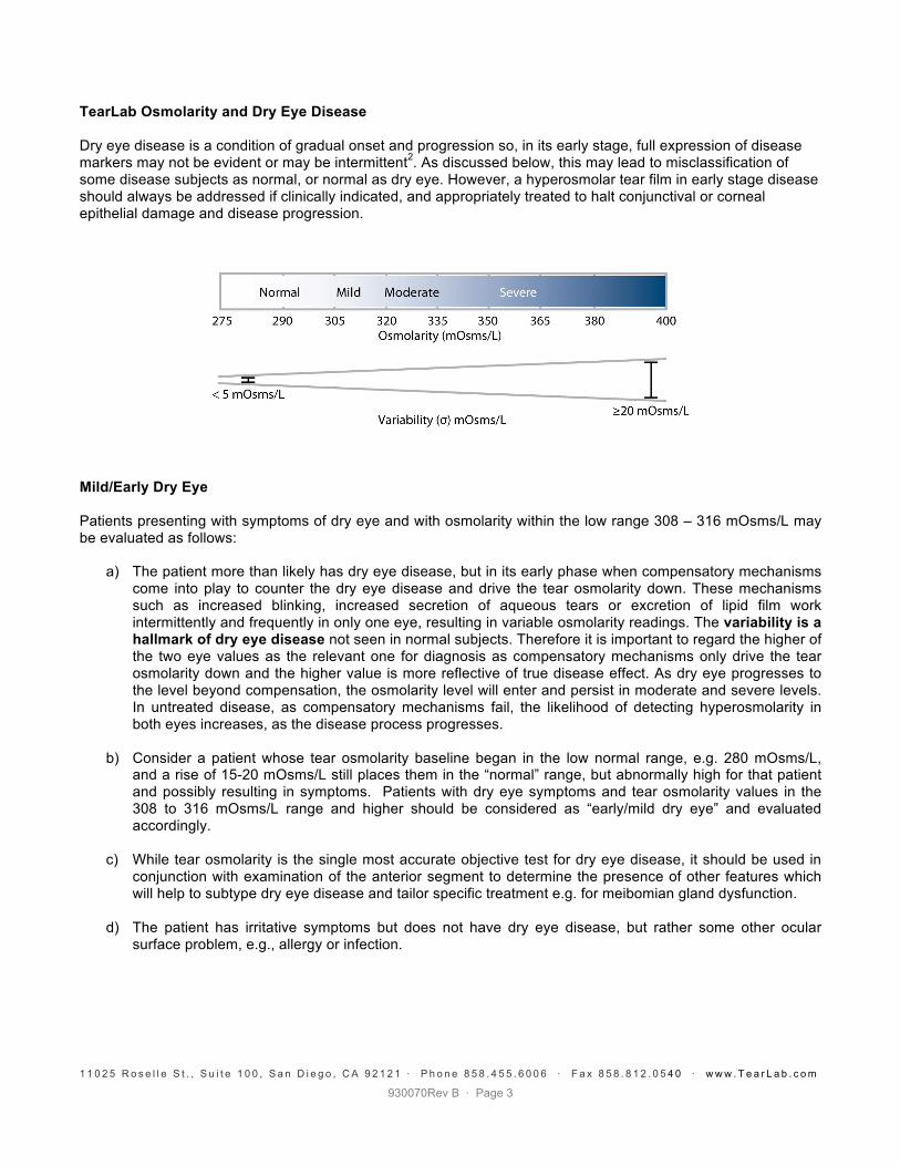
TearLab Osmolarity and Dry Eye Disease Dry eye disease is a condition of gradual onset and progression so, in its early stage, full expression of disease markers may not be evident or may be intermittent2. As discussed below, this may lead to misclassification of some disease subjects as normal, or normal as dry eye. However, a hyperosmolar tear film in early stage disease should always be addressed if clinically indicated, and appropriately treated to halt conjunctival or corneal epithelial damage and disease progression.
Mild/Early Dry Eye Patients presenting with symptoms of dry eye and with osmolarity within the low range 308 – 316 mOsms/L may be evaluated as follows:
a) The patient more than likely has dry eye disease, but in its early phase when compensatory mechanisms
come into play to counter the dry eye disease and drive the tear osmolarity down. These mechanisms such as increased blinking, increased secretion of aqueous tears or excretion of lipid film work intermittently and frequently in only one eye, resulting in variable osmolarity readings. The variability is a hallmark of dry eye disease not seen in normal subjects. Therefore it is important to regard the higher of the two eye values as the relevant one for diagnosis as compensatory mechanisms only drive the tear osmolarity down and the higher value is more reflective of true disease effect. As dry eye progresses to the level beyond compensation, the osmolarity level will enter and persist in moderate and severe levels. In untreated disease, as compensatory mechanisms fail, the likelihood of detecting hyperosmolarity in both eyes increases, as the disease process progresses.
b) Consider a patient whose tear osmolarity baseline began in the low normal range, e.g. 280 mOsms/L, and a rise of 15-20 mOsms/L still places them in the “normal” range, but abnormally high for that patient and possibly resulting in symptoms. Patients with dry eye symptoms and tear osmolarity values in the 308 to 316 mOsms/L range and higher should be considered as “early/mild dry eye” and evaluated accordingly.
c) While tear osmolarity is the single most accurate objective test for dry eye disease, it should be used in
conjunction with examination of the anterior segment to determine the presence of other features which will help to subtype dry eye disease and tailor specific treatment e.g. for meibomian gland dysfunction.
d) The patient has irritative symptoms but does not have dry eye disease, but rather some other ocular surface problem, e.g., allergy or infection.

1 1 0 2 5 R o s e l l e S t . , S u i t e 1 0 0 , S a n D i e g o , C A 9 2 1 2 1 · P h o n e 8 5 8 . 4 5 5 . 6 0 0 6 · F a x 8 5 8 . 8 1 2 . 0 5 4 0 · w w w . T e a r L a b . c o m
930070Rev B · Page 4
Analytical Variability of the TearLab Osmolarity System The TearLab Osmolarity System has demonstrated the ability to measure nanoliter volumes of tear fluid with the same analytical performance as laboratory osmometers (Table 1). Precision and accuracy are a reflection of repeatability. The TearLab Osmolarity System provides precision which exceeds that of other common in vitro diagnostic tests (e.g. glucose and cholesterol).
Table 1. Performance of the TearLab™ Osmolarity System
Precision and Accuracy on Nanoliter Volumes of Fluid4
Precision (CV) ≈ 1.5%
Standard Deviation (Stdev) 5.0 mOsms/L
Accuracy r2 = 0.95 Biological Variability of Dry Eye Patients Significant variability in osmolarity within eye, between eyes and/or from day-to-day is itself an indication of tear film instability and dysfunction. This variability is not seen in normal subjects and is considered a hallmark of dry eye disease (Table 2 – data from two patients from a TearLab clinical trial). Normal patients with inter-eye osmolarity readings typically fall within +/- 5 mOsms/L. Inter-eye or test-to-test variability greater than 10 mOsms/L is suggestive of dry eye disease.
Table 2. Variability of Normal and Dry Eye Patients5
Mild/Moderate Dry Eye
Patient OSDI = 22.92
Normal Patient OSDI = 4.17
Right Eye Left Eye Right Eye Left Eye Day 1 1 min 311 326 286 288 2 min 304 324 285 289 3 min 308 308 281 281 4 min 337 334 287 286 Day 2 1 min 315 321 296 284 2 min 305 313 296 291 3 min 315 323 285 291 4 min 297 343 291 287 Day 3 1 min 308 307 290 292 2 min 320 312 287 291 3 min 307 309 286 286 4 min 333 332 292 295
Mean 313 321 289 288 Stdev 11.8 11.5 4.6 3.9

1 1 0 2 5 R o s e l l e S t . , S u i t e 1 0 0 , S a n D i e g o , C A 9 2 1 2 1 · P h o n e 8 5 8 . 4 5 5 . 6 0 0 6 · F a x 8 5 8 . 8 1 2 . 0 5 4 0 · w w w . T e a r L a b . c o m
930070Rev B · Page 5
Dry Eye Severity “Regardless of which individual risk factor or group of factors initiates the disease process, the final common expression involves tear hyperosmolarity and tear instability leading to ocular surface damage. Since both aqueous tear deficiency and increased evaporative tear loss occur in most cases of dry eye disease and are linked by common pathogenetic mechanisms, expert clinicians are increasingly basing treatment decisions on an assessment of severity rather than discrete deficiencies. The group believed that a classification of disease based on severity would be of considerable value in clinical practice, particularly in terms of guiding therapeutic decisions … By establishing these definitions and classification of dry eye disease, we believe clinicians will be better able to determine the level of DED, as well as the best treatment course for their patients3.” Osmolarity Relative to Dry Eye Severity Results from a 300 subject, 11-site prospective clinical trial (n = 218 F, n = 81 M, average age = 46.3), presented at the 2009 American Academy of Ophthalmology meeting are reported below1. This study created a composite disease severity index from the following standard dry eye tests: TBUT, Schirmer I, corneal & conjunctival staining, meibomian grading, osmolarity and the Ocular Surface Disease Index (OSDI) for symptomology. Each patient was assigned a severity grade between 0 & 1, with 0 representing the absence of disease and 1 representing the most severe form of dry eye. Performance of each sign was plotted against this composite disease severity index, as shown in the graphs below. At a diagnostic cutoff of 308 mOsms/L, osmolarity was found to have the following performance, 88% specificity, and 75% sensitivity in mild/moderate disease and 95% sensitivity in severe disease. This performance was dramatically superior in the Mild to Moderate cohort as compared to the other common DED tests, as indicated in the graphs below. These data also suggest that symptoms alone are insufficient to grade dry eye severity.

1 1 0 2 5 R o s e l l e S t . , S u i t e 1 0 0 , S a n D i e g o , C A 9 2 1 2 1 · P h o n e 8 5 8 . 4 5 5 . 6 0 0 6 · F a x 8 5 8 . 8 1 2 . 0 5 4 0 · w w w . T e a r L a b . c o m
930070Rev B · Page 6
Dry Eye Disease Severity and Disease Management*
Recognizing that tear osmolarity correlates with disease severity, a dry eye severity index can provide the clinician with a valuable tool, allowing them to pause, flag those patients requiring additional work-up in order to properly classify the disease, and finally select the appropriate treatment which can then be monitored over time relative to both treatment efficacy and patient compliance. The following is an example of a clinical aid designed to facilitate this three-step process:
1. Determine disease severity
2. Classify
Aqueous Tear Deficiency Additional Signs: Evidence of low meniscus, low Schirmer test and/or decreased tear clearance, corneal/conjunctival staining Mild Dry Eye Moderate Dry Eye Severe Dry Eye Consider adding Consider adding: Consider adding: Tear Replacement Osmoprotectives Anti-inflammatories,
Stabilizing polymer tear replacement Punctal Plugs Omega-3 Supplements Viscoelastics
Anti-inflammatories, Immunomodulators Lid/Meibomian Gland Disease Additional Signs: Evidence of inflammation on lid, poor expressibility of lipids, corneal/conjunctival staining Mild Dry Eye Moderate Dry Eye Severe Dry Eye Consider adding Consider adding: Consider adding: Lubrication, Lipid Replacement Topical Antibiotics Viscoelastics Lid Hygiene Systemic Antibiotics Omega-3 Supplements Mixed Dry Eye Disease
Treat both forms of the disease as signs present 3. Reschedule patient for follow-up testing to monitor therapeutic efficacy & compliance
*For more information, see 2007 Report of the Dry Eye WorkShop. Ocul Surf 2007; 5[2]: 65-204.

1 1 0 2 5 R o s e l l e S t . , S u i t e 1 0 0 , S a n D i e g o , C A 9 2 1 2 1 · P h o n e 8 5 8 . 4 5 5 . 6 0 0 6 · F a x 8 5 8 . 8 1 2 . 0 5 4 0 · w w w . T e a r L a b . c o m
930070Rev B · Page 7
Dry Eye and Osmolarity: A Clinical Perspective Osmolarity is a basic and essential aspect of physiologic homeostasis in body fluids. Small deviations in osmolarity activate physiological mechanisms to return that variable to its set point. The body is normally able to regulate osmolarity of body fluids within very narrow limits through various mechanisms of osmo-regulation, such as the compensation and correction of fluid volume and the oils, lipids, proteins and mucins that make up the tear. Hyperosmolarity of any body fluid, including tear fluid, indicates a disorder in the ability to maintain homeostasis and is a basic indication of a physiological disorder. Tear hyperosmolarity is regarded as the central mechanism causing ocular surface inflammation, damage, and symptoms, and the initiation of compensatory events in dry eye. Tear hyperosmolarity arises as a result of either (a) abnormal water evaporation from the exposed ocular surface - Evaporative Dry Eye Disease usually associated with meibomian gland dysfunction, (b) low aqueous tear production - Aqueous Tear Deficient Dry Eye Disease or (c) in many cases a combination of both2. Tear osmolarity has been shown to have the highest accuracy and predictive value for diagnosing dry eye disease of any single test. Although there are other tests that may be helpful for identifying subsets of dry eye, osmolarity is a global test of dry eye, and is the key biomarker to identify both aqueous deficient and evaporative Dry Eye Disease. The following excerpts, from “The Definition and Classification of Dry Eye Disease: Report of the Definition and Classification Subcommittee of the International Dry Eye Workshop” The Ocular Surface, 2007;5(2):75-92, (DEWS Report) are meant to clarify the intended role of hyperosmolarity in the diagnosis of Dry Eye Disease.
Definition of Dry Eye Disease & Etiology “Dry eye is a multifactorial disease of the tears and ocular surface that results in symptoms of discomfort, visual disturbance, and tear film instability with potential damage to the ocular surface. It is accompanied by increased osmolarity of the tear film and inflammation of the ocular surface. The core mechanisms of dry eye are driven by tear hyperosmolarity and tear film instability. Tear hyperosmolarity causes damage to the surface epithelium by activating a cascade of inflammatory events at the ocular surface and a release of inflammatory mediators into the tears. Epithelial damage involves cell death by apoptosis, a loss of goblet cells, and disturbance of mucin expression, leading to tear film instability. This instability exacerbates ocular surface hyperosmolarity and completes the vicious circle. Tear film instability can be initiated, without the prior occurrence of tear hyperosmolarity, by several etiologies, including xerophthalmia, ocular allergy, topical preservative use, and contact lens wear. Tear hyperosmolarity is regarded as the central mechanism causing ocular surface inflammation, damage, and symptoms, and the initiation of compensatory events in dry eye. Tear hyperosmolarity arises as a result of water evaporation from the exposed ocular surface, in situations of a low aqueous tear flow, or as a result of excessive evaporation, or a combination of these events. The place of tear osmolarity measurement in dry eye diagnosis is well established, and its adoption has several attractions. There is considerable value in assessing a parameter that is directly involved in the mechanism of dry eye. Tear hyperosmolarity may reasonably be regarded as the signature feature that characterizes the condition of ‘ocular surface dryness.’ In the past, although the measurement of tear osmolarity has been offered as a ’gold standard‘ in dry eye diagnosis, its general utility as a test has been hindered by the need for expert technical support; thus, its use has been confined to a small number of specialized laboratories. The feasibility of this objective test is greatly enhanced by the imminent availability of a commercial device that will make the technology generally available.” (i.e., the TearLab Osmolarity System)
Therefore, although there are other tests that may be helpful for identifying subsets of dry eye disease, tear osmolarity is a global test of homeostatic osmo-regulation of the tears, and is the key biomarker to identify both aqueous deficient and evaporative Dry Eye Disease in early, middle and late stages.

1 1 0 2 5 R o s e l l e S t . , S u i t e 1 0 0 , S a n D i e g o , C A 9 2 1 2 1 · P h o n e 8 5 8 . 4 5 5 . 6 0 0 6 · F a x 8 5 8 . 8 1 2 . 0 5 4 0 · w w w . T e a r L a b . c o m
930070Rev B · Page 8
References
1. Foulks GN, Lemp MA, Berg M, Bhola R, Sullivan BD. TearLab™ Osmolarity as a biomarker for disease severity in mild to moderate dry eye disease. American Academy of Ophthalmology PO382, 2009.
2. “Definition and Classification of Dry Eye. Report of the Diagnosis and Classification Subcommittee of the Dry Eye Workshop (DEWS).” The Ocular Surface 5(2): 75-92, 2007.
3. Lemp MA. Foulks GN. “The Definition & Classification of Dry Eye Disease Guidelines from the 2007 International Dry Eye Workshop”. Ophthalmology Management April 2008.
4. TearLab Internal Data. 5. Keech A, Jones L, Senchyna M. TearLab Osmometer Validation Protocol. Centre for Contact Lens Research.
University of Waterloo School of Optometry. March 2009