task force report - spitjudms.ro · task force report guidelines on diagnosis and management of...
TRANSCRIPT

European Heart Journal (2000) 21, 1301–1336doi:10.1053/euhj.2000.2250, available online at http://www.idealibrary.com on
Task Force Report
Guidelines on diagnosis and management of acutepulmonary embolism1
Task Force on Pulmonary Embolism, European Society of Cardiology2:Core Writing Group: A. Torbicki (Chairman), E. J. R. van Beek (Editor), B. Charbonnier, G. Meyer,
M. Morpurgo, A. Palla and A. PerrierMembers: N. Galie, G. Gorge, C. Herold, S. Husted, V. Jezek, W. Kasper, M. Kneussl,
A. H. Morice, D. Musset, M. M. Samama, G. Simonneau, H. Sors, M. de Swiet and M. TurinaInternal reviewers: G. Kronik, J. Widimsky
Table of contents 1301Preamble 1301Introduction 1302
Epidemiology and predisposing factors 1302Pathophysiology 1304Natural history and prognosis 1305
DiagnosisClinical presentation and clinical evaluation
of pulmonary embolism 1306Lung scintigraphy 1307Pulmonary angiography 1309Spiral computed tomography 1311Echocardiography 1312Detection of deep vein thrombosis 1314D-dimer 1315Diagnostic strategies 1315
TreatmentHaemodynamic and respiratory support 1317Thrombolytic treatment 1318Surgical embolectomy 1320Anticoagulant therapy 1321Venous filters 1324
Specific problemsDiagnosis and treatment of PE in pregnancy 1325
References 1326
Preamble
Presented Guidelines were prepared by the ESC TaskForce on Pulmonary Embolism, as suggested by the
0195-668X/00/211301+36 $35.00/0
nucleus of the Working Group on Pulmonary Circu-lation and Right Ventricular Function and approved bythe ESC Board at its meeting on 17 June 1997 upon therecommendation of the Committee for Scientific andClinical Initiatives.
This Task Force consists of 21 Members, includingrepresentatives of the European Respiratory Society, theEuropean Association of Radiology, and an advisorybody consisting of two Internal Reviewers. The Mem-bers were appointed by the Board of the ESC uponsuggestions from the Working Group and from theBoards of Scientific Societies, invited to contribute to thedevelopment of the guidelines on pulmonary embolism.The Chairman and seven of the Members of the TaskForce formed a Core Writing Group (CWG), whichincluded an Editor, responsible for preparation ofthe final document. The Task Force Members metin September 1998 in Vienna and the Core WritingGroup in May 1999 in Warsaw and in January 2000 inParis. In addition, controversial issues were presentedand discussed with the Pulmonary Circulation Group ofthe European Respiratory Society during an openWorkshop organized at the ERS annual Congress inGeneva, in September 1998.
Review of the literature and position papers wereprepared by the Members according to their area ofexpertise. Their contributions were then posted on theTask Force WebBoard and submitted to discussion overthe internet. A second phase consisted of preparationand editing of the consecutive versions of the Guidelinesby the CWG, as discussed at the two consecutivemeetings as well as over the internet. At the requestof the Committee for Scientific and Clinical Initiatives,the Task Force Chairman reported to the Congress ofthe ESC in August 1999, indicating key points of theemerging guidelines.
Finally, the document was distributed for correctionand endorsement to all Members and independentlyreviewed for consistency by Internal Reviewers. Effortwas made to include all relevant evidence relating to the
Correspondence: A. Torbicki, Department of Chest Medicine,Institute of Tuberculosis and Lung Diseases, Warszawa, Poland.
1This document has been reviewed by members of the Committeefor Scientific and Clinical Initiatives and by members of the Boardof the European Society of Cardiology (see Appendix 1), whoapproved the document on 14 April 2000. The full text of thisdocument is available on the website of the European Society ofCardiology in the section ‘Scientific Information’, Guidelines.
2For affiliations of Task Force Members see Appendix 2.
� 2000 The European Society of Cardiology

1302 Task Force Report
diagnosis and treatment of pulmonary embolism. TheGuidelines were developed with the help of a budgetassigned to the Task Force by the European Society ofCardiology and without the involvement of any com-mercial organization. The list of all contributors is givenin the Appendix.
Introduction
Pulmonary embolism (PE) is a major internationalhealth problem with an annual estimated incidence ofover 100 000 cases in France, 65 000 cases among hos-pitalized patients in England and Wales, and at least60 000 new cases per year in Italy. The diagnosis is oftendifficult to obtain and is frequently missed. Mortality inuntreated PE is approximately 30%, but with adequate(anticoagulant) treatment, this can be reduced to 2–8%.Deep vein thrombosis (DVT) and PE are commoncauses of illness and death after surgery, injury, child-birth and in a variety of medical conditions[1,2]. Never-theless numerous cases go unrecognized and henceuntreated, with serious outcomes. Indeed, the prevalenceof PE at autopsy (approximately 12–15% in hospitalizedpatients) has not changed over three decades[3]. Asmodern medicine improves the longevity of patients withmalignancy and cardiac and respiratory disease, PE maybecome an even more common clinical problem.
In the immediate course, PE may be fatal: the recentICOPER study[4], which included 2454 consecutivepatients with acute PE observed in 52 hospitals,revealed a cumulative mortality at 3 months as high as17·5%. Sometimes PE represents the ‘coup de grace’that kills a patient already fated to die. However,‘preventable’ deaths range from 27% to 68% of variousautopsy series[5]. In the long-term, there is the risk ofdeveloping pulmonary hypertension from recurrentembolism or the absence of reperfusion of thepulmonary vasculature[6].
For clinical purposes this Task Force suggests that PEcan be classified into two main groups: massive andnon-massive. Thus, massive PE consists of shock and/orhypotension (defined as a systolic blood pressure<90 mmHg or a pressure drop of �40 mmHg for>15 min if not caused by new-onset arrhythmia, hypo-volemia or sepsis). Otherwise non-massive PE can bediagnosed. A subgroup of patients with non-massive PEmay be identified by echocardiographic signs of rightventricular hypokinesis. The Task Force proposes thatthis subgroup be called submassive, because there isgrowing evidence that the prognosis of this patientgroup may be different from those with non-massive PEand normal right ventricular function.
Epidemiology and predisposing factors
Estimated rates for DVT and PE in population-basedstudies had been reported in only a few countries, and
Eur Heart J, Vol. 21, issue 16, August 2000
available data must be analysed carefully, because dif-ferent diagnostic codes and criteria can be applied[7].The annual incidence for DVT and PE in the generalpopulation of the Western World may be estimated at1·0 and 0·5 per 1000 respectively[8]. The number ofclinically silent non-fatal cases cannot be determined.The use of death certificates with a diagnosis of PE isextremely inaccurate[9]. Furthermore, discrepanciesbetween clinical diagnosis and autopsy findings are wellknown.
Unsuspected PE in patients at post-mortem has notdiminished, even among individuals who die from acutemassive or submassive PE[2,10]. In autopsy studies,the prevalence of unsuspected PE, either fatal orcontributing to death, ranges from 3% to 8%[3,11–14].
A meta-analysis of 12 post-mortem studies carried outfrom 1971 through 1995 showed that more than 70% ofmajor PEs had been missed by the clinician[2,15]. How-ever, because necropsy is not systematically performed,autopsy studies do little to elucidate the prevalence ofvenous thromboembolic disease (VTE) or death by PE.In clinical studies, most cases of PE occur between ages60 and 70, compared to between 70 and 80 years inautopsy series[12–18].
The main primary and secondary risk factorsresponsible for VTE are summarized in Table 1[19,20].Various factors may obviously act together, but a recentFrench multicentre registry[18] revealed that almost onein two cases of PE and DVT occurred in the absence ofa classical predisposing factor.
Congenital predisposition to thrombosis is consideredto be a rare condition, but the true prevalence isunknown. It should be seriously considered in patientsdefined as having had a documented unexplained throm-botic episode below the age of 40, recurrent DVT or PEand a positive family history[6]. The most commongenetic defects that have been identified are: resistance toactivated protein C (which is caused by a point mutationof factor V in 90% of cases)21,22], factor II 20210Amutation[23], hyperhomocysteinemia[24,25] and deficienciesof antithrombin III, protein C and protein S[26,27].
The incidence rates of DVT and PE increase withage[28], but this trend may be due to an underlyingrelationship between age and other co-morbidities,which are the actual risk factors for VTE (e.g. cancer,myocardial infarction)[29,30].
Thromboembolic complications have been reportedin 30–60% of patients with stroke (paralysed leg),in 5–35% of patients with acute myocardial infarction,and in over 12% of patients with congestive heartfailure[10,15,31–33].
As to immobilization, even short-term (one week)immobilization may predispose to VTE. The frequencyof DVT in surgical patients is approximately 5% in thoseundergoing herniorrhaphy, 15%–30% in cases of majorabdominal surgery, 50%–75% in cases of operated hipfracture, and from 50% up to 100% in spinal cordinjuries[31,34]. PE is rare after isolated valve replacement;however, it is not uncommon (3%–9% of cases) aftercoronary bypass surgery[35,36]. About one fourth of all

Acute pulmonary embolism 1303
postoperative PEs occur after hospital discharge;this rate is even greater in the subgroup of patientsundergoing so-called low-risk surgery[37].
The risk of VTE is five times greater in a pregnantwoman than in a non-pregnant woman of similarage — 75% of DVT occurring ante-partum, 66% of PEoccurring post partum[38]. Oral contraceptives increasethe risk of DVT threefold, but the baseline incidence inyoung women is very low (approximately 0·3/10 000 peryear)[39]. Latest results provide reasonably strong evi-dence that in users of third-generation oral contracep-tives (containing either desogestrel or gestodene, as theprogestagen component), the risk of VTE is furtherincreased, to 1 to 2/10 000/year[40,41]. This risk may befurther increased in the presence of congenital throm-bophilia, such as resistance to activated protein C.Post-menopausal hormone replacement therapy (HRT),is also associated with a threefold increase in the riskof VTE, as demonstrated by large recent prospectivestudies[42,43]. However, the baseline risk is again low(approximately 15/10 000 women treated by HRT peryear), and most experts agree that a history of VTE isnot an absolute contraindication to HRT, particularly inwomen at high risk of coronary artery disease, unlessthe episode of VTE is recent (less than one year).Finally, smoking is an independent risk factor forpulmonary embolism, as shown recently in the Nurses’study[44].
An association between VTE and overt cancer is welldocumented, and recent studies suggest that patientswith so-called idiopathic PE develop subsequent malig-nant neoplasms in approximately 10% of cases[17]. How-ever, searching for a malignancy in patients with PErequires only a careful history and physical examination,and routine tests such as chest X-ray, complete bloodcount, and basic laboratory. More extensive work-up isuniformly disappointing[45–47].
With regard to the presumed origin of thromboemboliand the relationship between DVT and PE, in clinicaland autopsy studies the source of thromboemboli hasbeen detected in 50%–70% of cases, because thrombiwithin the calf veins are not easily diagnosed by non-invasive methods, and dissection of the veins under theknee is not routinely done post-mortem[2,16]. Further-more, thrombus detachment and migration may betotal, especially in surgical patients[48], so that the pointof origin can no longer be identified. Among those inwhom the source of thromboembolism can be identified,70%–90% have one or more thromboses in the area ofthe inferior vena cava, more frequently at the level of thefemoral and iliac veins. Recent post-mortem data[15]
show an increasing number of thromboemboli arisingfrom the pelvic veins, namely from peri-prostatic andperi-uterine plexuses.
In approximately 10%–20% of cases, emboli arisefrom thrombi located in the area of the superior venacava. Recently, upper extremity venous thrombosis hasbecome more frequent[15,16,49,50] as a result of invasivediagnostic and therapeutic procedures (e.g. indwellingvenous catheters, intravenous chemotherapeutic agents).Upper extremity venous thrombosis may be associatedwith PE in up to 40% of cases[52,53]. The cardiac origin ofPE plays only a minor role in the overall incidence of thedisease[15,53].
A correlation between thrombosis location and theincidence and severity of PE has been demonstratedby a prospective clinical study[54]. The incidence ofPE was 46% if DVT was confined to the calf,increased to 67% with involvement of the thigh, and upto 77% if the pelvic veins were involved. In severe PEs,most emboli arise from thrombi in the proximal veins.Many of these thrombi, however, originate in thecalf and progress into the proximal veins beforeembolization[55].
Table 1 Risk factors for venous thromboembolism (adapted from references[19,20])
(A) PrimaryAntithrombin deficiency Protein C deficiencyCongenital dysfibrinogenemia Factor V Leiden (APC-R)Thrombomodulin Plasminogen deficiencyHyperhomocysteinemia DysplasminogenemiaAnticardiolipin antibodies Protein S deficiencyExcessive plasminogen activator inhibitor Factor XII deficiencyProthrombin 20210A mutation
(B) SecondaryTrauma/fractures SurgeryStroke ImmobilisationAdvanced age Malignancy�chemotherapyCentral venous catheters ObesityChronic venous insufficiency Heart failureSmoking Long distance travelPregnancy/puerperium Oral contraceptivesCrohn’s disease Lupus anticoagulantNephrotic syndrome Prosthetic surfacesHyperviscosity (Polycythemia, Waldenstrom’s macroglobulinemia)Platelet abnormalities
Eur Heart J, Vol. 21, issue 16, August 2000
1304 Task Force Report
Summaryv The annual incidence of DVT and PE is esti-mated at 1·0 and 0·5 per 1000 in the Westernworld, respectively.v DVT and PE are both part of one entity: venousthromboembolism (VTE).v Both acquired and inherited risk factors havebeen identified
Pathophysiology
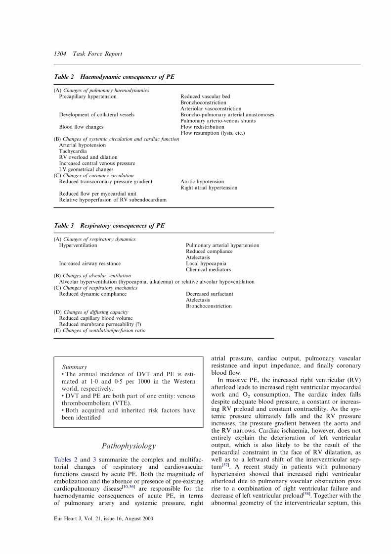
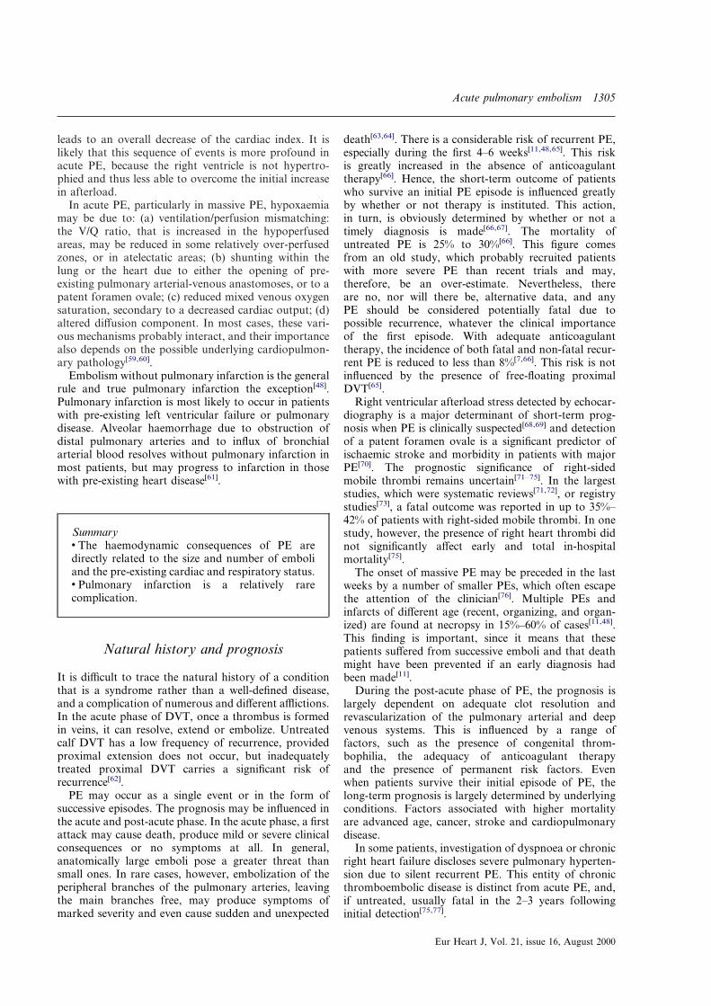
Tables 2 and 3 summarize the complex and multifac-torial changes of respiratory and cardiovascularfunctions caused by acute PE. Both the magnitude ofembolization and the absence or presence of pre-existingcardiopulmonary disease[10,56] are responsible for thehaemodynamic consequences of acute PE, in termsof pulmonary artery and systemic pressure, right
Eur Heart J, Vol. 21, issue 16, August 2000
atrial pressure, cardiac output, pulmonary vascularresistance and input impedance, and finally coronaryblood flow.
In massive PE, the increased right ventricular (RV)afterload leads to increased right ventricular myocardialwork and O2 consumption. The cardiac index fallsdespite adequate blood pressure, a constant or increas-ing RV preload and constant contractility. As the sys-temic pressure ultimately falls and the RV pressureincreases, the pressure gradient between the aorta andthe RV narrows. Cardiac ischaemia, however, does notentirely explain the deterioration of left ventricularoutput, which is also likely to be the result of thepericardial constraint in the face of RV dilatation, aswell as to a leftward shift of the interventricular sep-tum[57]. A recent study in patients with pulmonaryhypertension showed that increased right ventricularafterload due to pulmonary vascular obstruction givesrise to a combination of right ventricular failure anddecrease of left ventricular preload[58]. Together with theabnormal geometry of the interventricular septum, this
Table 2 Haemodynamic consequences of PE
(A) Changes of pulmonary haemodynamicsPrecapillary hypertension Reduced vascular bed
BronchoconstrictionArteriolar vasoconstriction
Development of collateral vessels Broncho-pulmonary arterial anastomosesPulmonary arterio-venous shunts
Blood flow changes Flow redistributionFlow resumption (lysis, etc.)
(B) Changes of systemic circulation and cardiac functionArterial hypotensionTachycardiaRV overload and dilationIncreased central venous pressureLV geometrical changes
(C) Changes of coronary circulationReduced transcoronary pressure gradient Aortic hypotension
Right atrial hypertensionReduced flow per myocardial unitRelative hypoperfusion of RV subendocardium
Table 3 Respiratory consequences of PE
(A) Changes of respiratory dynamicsHyperventilation Pulmonary arterial hypertension
Reduced complianceAtelectasis
Increased airway resistance Local hypocapniaChemical mediators
(B) Changes of alveolar ventilationAlveolar hyperventilation (hypocapnia, alkalemia) or relative alveolar hypoventilation
(C) Changes of respiratory mechanicsReduced dynamic compliance Decreased surfactant
AtelectasisBronchoconstriction
(D) Changes of diffusing capacityReduced capillary blood volumeReduced membrane permeability (?)
(E) Changes of ventilation/perfusion ratio
Acute pulmonary embolism 1305
leads to an overall decrease of the cardiac index. It islikely that this sequence of events is more profound inacute PE, because the right ventricle is not hypertro-phied and thus less able to overcome the initial increasein afterload.
In acute PE, particularly in massive PE, hypoxaemiamay be due to: (a) ventilation/perfusion mismatching:the V/Q ratio, that is increased in the hypoperfusedareas, may be reduced in some relatively over-perfusedzones, or in atelectatic areas; (b) shunting within thelung or the heart due to either the opening of pre-existing pulmonary arterial-venous anastomoses, or to apatent foramen ovale; (c) reduced mixed venous oxygensaturation, secondary to a decreased cardiac output; (d)altered diffusion component. In most cases, these vari-ous mechanisms probably interact, and their importancealso depends on the possible underlying cardiopulmon-ary pathology[59,60].
Embolism without pulmonary infarction is the generalrule and true pulmonary infarction the exception[48].Pulmonary infarction is most likely to occur in patientswith pre-existing left ventricular failure or pulmonarydisease. Alveolar haemorrhage due to obstruction ofdistal pulmonary arteries and to influx of bronchialarterial blood resolves without pulmonary infarction inmost patients, but may progress to infarction in thosewith pre-existing heart disease[61].
Summaryv The haemodynamic consequences of PE aredirectly related to the size and number of emboliand the pre-existing cardiac and respiratory status.v Pulmonary infarction is a relatively rarecomplication.
Natural history and prognosis
It is difficult to trace the natural history of a conditionthat is a syndrome rather than a well-defined disease,and a complication of numerous and different afflictions.In the acute phase of DVT, once a thrombus is formedin veins, it can resolve, extend or embolize. Untreatedcalf DVT has a low frequency of recurrence, providedproximal extension does not occur, but inadequatelytreated proximal DVT carries a significant risk ofrecurrence[62].
PE may occur as a single event or in the form ofsuccessive episodes. The prognosis may be influenced inthe acute and post-acute phase. In the acute phase, a firstattack may cause death, produce mild or severe clinicalconsequences or no symptoms at all. In general,anatomically large emboli pose a greater threat thansmall ones. In rare cases, however, embolization of theperipheral branches of the pulmonary arteries, leavingthe main branches free, may produce symptoms ofmarked severity and even cause sudden and unexpected
death[63,64]. There is a considerable risk of recurrent PE,especially during the first 4–6 weeks[11,48,65]. This riskis greatly increased in the absence of anticoagulanttherapy[66]. Hence, the short-term outcome of patientswho survive an initial PE episode is influenced greatlyby whether or not therapy is instituted. This action,in turn, is obviously determined by whether or not atimely diagnosis is made[66,67]. The mortality ofuntreated PE is 25% to 30%[66]. This figure comesfrom an old study, which probably recruited patientswith more severe PE than recent trials and may,therefore, be an over-estimate. Nevertheless, thereare no, nor will there be, alternative data, and anyPE should be considered potentially fatal due topossible recurrence, whatever the clinical importanceof the first episode. With adequate anticoagulanttherapy, the incidence of both fatal and non-fatal recur-rent PE is reduced to less than 8%[7,66]. This risk is notinfluenced by the presence of free-floating proximalDVT[65].
Right ventricular afterload stress detected by echocar-diography is a major determinant of short-term prog-nosis when PE is clinically suspected[68,69] and detectionof a patent foramen ovale is a significant predictor ofischaemic stroke and morbidity in patients with majorPE[70]. The prognostic significance of right-sidedmobile thrombi remains uncertain[71–75]. In the largeststudies, which were systematic reviews[71,72], or registrystudies[73], a fatal outcome was reported in up to 35%–42% of patients with right-sided mobile thrombi. In onestudy, however, the presence of right heart thrombi didnot significantly affect early and total in-hospitalmortality[75].
The onset of massive PE may be preceded in the lastweeks by a number of smaller PEs, which often escapethe attention of the clinician[76]. Multiple PEs andinfarcts of different age (recent, organizing, and organ-ized) are found at necropsy in 15%–60% of cases[11,48].This finding is important, since it means that thesepatients suffered from successive emboli and that deathmight have been prevented if an early diagnosis hadbeen made[11].
During the post-acute phase of PE, the prognosis islargely dependent on adequate clot resolution andrevascularization of the pulmonary arterial and deepvenous systems. This is influenced by a range offactors, such as the presence of congenital throm-bophilia, the adequacy of anticoagulant therapyand the presence of permanent risk factors. Evenwhen patients survive their initial episode of PE, thelong-term prognosis is largely determined by underlyingconditions. Factors associated with higher mortalityare advanced age, cancer, stroke and cardiopulmonarydisease.
In some patients, investigation of dyspnoea or chronicright heart failure discloses severe pulmonary hyperten-sion due to silent recurrent PE. This entity of chronicthromboembolic disease is distinct from acute PE, and,if untreated, usually fatal in the 2–3 years followinginitial detection[75,77].
Eur Heart J, Vol. 21, issue 16, August 2000

1306 Task Force Report
Summaryv Untreated VTE has a high risk of (fatal ornon-fatal) recurrence.v Anticoagulant therapy reduces the mortality inpatients with PE by 75%.v The prognosis of treated, non-massive VTE ismainly dependent on co-existing illnesses, such asmalignancy or cardiovascular diseases.
Diagnosis
Clinical presentations and clinical evaluationof pulmonary embolism
As previously discussed, PE is a potentially fatal dis-order with a range of clinical presentations (fromhaemodynamic instability to silent). Evaluating the like-lihood of PE in an individual patient according to theclinical presentation is of utmost importance in theinterpretation of diagnostic test results and selection ofan appropriate diagnostic strategy. In 90% of cases, sus-picion of PE is raised by clinical symptoms such asdyspnoea, chest pain or syncope, either singly or incombination. In a classic series, dyspnoea, tachypnoea,or chest pain were present in 97% of patients with PEwithout cardiac or pulmonary disease[78]. Similarly, in arecent series in which 25% had a previous cardiac orpulmonary disease, recent onset dyspnoea, chest pain orsyncope were present in as many as 97% of patients withPE[79,80]. In 10% of cases, PE is suspected because ofincidental radiological findings, either on chest X-ray orhelical CT scan, in high risk situations.
Pleuritic chest pain, whether or not combined withdyspnoea, is one of the most frequent presentations ofPE (Table 4)[78–80]. This pain is usually due to distalemboli causing pleural irritation, and a consolidationmay be present on chest X-ray. This syndrome is oftenimproperly named ‘pulmonary infarction’, although thehistopathological correlate is an alveolar haemorrhage,which is only exceptionally associated with haemoptysis.
Isolated dyspnoea of rapid onset is usually due tomore central PE, not affecting the pleura. It may beassociated with substernal angina-like chest pain, prob-ably representing right ventricular ischaemia. Thehaemodynamic consequences are more prominent thanin the ‘pulmonary infarction’ syndrome. Occasionally,the onset of dyspnoea may be very progressive, overseveral weeks, and the diagnosis of PE is evoked by theabsence of other classic causes of progressive dyspnoea.In patients with pre-existing heart failure or pulmonarydisease, worsening dyspnoea may be the only symptomindicative of PE.
Finally, syncope or shock is the hallmark of centralPE with severe haemodynamic repercussions, and isaccompanied by signs of haemodynamic compromiseand reduced heart flow, such as systemic arterial hypo-
Eur Heart J, Vol. 21, issue 16, August 2000
tension, oliguria, cold limb extremities and/or clinicalsigns of acute right heart failure.
The presence or absence of risk factors for VTE isessential in the evaluation of the likelihood of PE.Moreover, it should be recognized that the risk of PEincreases with the number of risk factors present. How-ever, PE does occur frequently in individuals withoutany risk factors[18]. Individual clinical signs and symp-toms are not very helpful, as they are neither sensitivenor specific (Table 4). Chest X-ray is usually abnormal,and the most frequently encountered findings are plate-like atelectasis, pleural effusion or elevation of a hae-midiaphragm. However, these signs are not very specificand the chest X-ray is mainly useful to exclude othercauses of dyspnoea and chest pain. In the PISAPEDstudy[80], amputation of a hilar artery, oligemia and apleural-based wedge-shaped infiltrate appeared to beclosely associated with PE, and were present in 15% to45% of patients. However, this contradicts the findingsfrom previous series[78], and the chest X-rays in thePISAPED study[80] were interpreted by six pulmonaryphysicians, all of them experts in the field of PE diag-nosis. Hence, the practical value of these signs in othersettings remains to be demonstrated. PE is generallyassociated with hypoxaemia, but up to 20% of patientswith PE have a normal arterial oxygen pressure (PaO2).Since most are also hypocapnic, it was hoped that theoxygen alveolo-arterial difference (D(A-a)O2) would bemore sensitive for PE than PaO2, but clinical trials were
Table 4 Signs, symptoms and findings in suspected PE(from references[78,80])
PE(n=219)
no PE(n=546)
SymptomsDyspnoea 80% 59%Chest pain (pleuritic) 52% 43%Chest pain (substernal) 12% 8%Cough 20% 25%Haemoptysis 11% 7%Syncope 19% 11%
SignsTachypnoea (�20/min)** 70% 68%Tachycardia (>100/min) 26% 23%Signs of DVT 15% 10%Fever (>38·5 �C) 7% 17%Cyanosis 11% 9%
Chest X-rayAtelectasis or infiltrate 49% 45%Pleural effusion 46% 33%Pleural-based opacity (infarction) 23% 10%Elevated diaphragm 36% 25%Decreased pulmonary vascularity 36% 6%Amputation of hilar artery* 36% 1%
Blood gasesHypoxaemia** 75% 81%
ElectrocardiogramRight ventricular overload* 50% 12%
*Only observed in series of reference[78].**Only observed in series of reference[80].

Acute pulmonary embolism 1307
disappointing[79] revealing that 15 to 20% of patientswith proven PE also have a normal D(A-a)O2. Finally,ECG signs of right ventricular overload (S1Q3 pattern,inversion of T waves, V1 to V3 leads, right bundlebranch block) may be helpful. Nevertheless, suchchanges are generally associated with the more severeforms of PE, and may be found in right ventricularstrain of any cause.
Since the diagnostic value of individual symptoms,signs and common test findings is poor, one couldconclude that clinical evaluation is useless in suspectedPE. However, a large body of data contradicts this.Indeed, the combination of these variables, either em-pirically or implicitly by the clinician[83–85], or by aprediction rule[80,86–88], allows a fairly accurate indica-tion of suspected PE patients in three broad so-calledclinical or pre-test probability categories. Table 5 showsthe predictive value of clinical assessment by variousmethods. To recognize patients with a high likelihood ofthe disease, prediction rules appear to be more accuratethan empirical evaluation. However, the clinical useful-ness of distinguishing between an intermediate and highclinical probability may be limited.
Clinical probability has been used in combinationwith lung scans to rule out clinically significant PE. Arecent analysis of a database of 1034 consecutivepatients suspected of PE in the emergency ward showedthat the 3-month thromboembolic risk was very low(1·7%, 95% CI 0·4 to 4·9) in 175 suspected PE patientsnot treated on the grounds of low empirical clinicalprobability and a non-diagnostic lung scan, providedlower limb venous compression ultrasonography (US)did not show a proximal deep vein thrombosis(DVT)[84]. This combination was found in 21% ofpatients, who, therefore, did not undergo an angiogram.Similarly, Canadian investigators used a low to moder-ate score of the clinical probability of PE to avoidangiography in 702 of 1239 (57%) patients with anon-diagnostic scan and normal serial US (see diagnos-tic strategies), and the 3-month thromboembolic riskwas only 0·5% (95% CI 0·1 to 1·3)[86].
As shown in Table 5, the clinician must choosebetween empiricism and two prediction rules to assessthe clinical probability of PE. The obvious advantage ofa prediction rule is to allow a standardized and explicitevaluation. The reader should, however, be aware thatsubjectivity carries a great weight in one of the predic-tion rules[86]. Indeed, an important element in the scoreis the decision whether another diagnosis is as, or morelikely, than PE in a given patient. Moreover, a predic-tion rule should fulfil rigorous methodological standardsin order to be valid and transferable to clinicalpractice[89,90]. These standards have been met by oneseries[86], including external validation and clinical use-fulness evaluation, but not by the PISAPED study[80].Besides, this score is only valid in combination withspecific lung scan criteria developed by the PISAPEDstudy[91], which still awaits external validation. Finally,practical experience shows that, when confronted bya discrepancy between empirical evaluation and the
probability given by a score, the clinician generallychooses to rely on his or her own experience.
Summaryv PE has a wide range of clinical presentation.v A reasonable clinical suspicion is required toavoid missing the diagnosis of PE.v First line diagnostic tests, such as ECG, chestX-ray and blood-gas analysis are indicated toassess clinical probability of PE and generalcondition of the patient.v Clinical evaluation is accurate to discriminate asubgroup of patients with a low likelihood of PE.v Clinical probability may be estimated empiricallyor explicitly by a prediction rule.v Patients with a low clinical probability of PE, nolower limb deep vein thrombosis and a non-diagnostic lung scan have a very low risk of PE.
Lung scintigraphy
Lung scintigraphy has a pivotal role to play in thediagnostic management of suspected pulmonary embol-ism. The reasons for this are twofold: it is a non-invasivediagnostic technique and it has been evaluated in exten-sive clinical trials. It has been proven extremely safe toapply, and few allergic reactions have been described.
Lung scintigraphy consists of two components: per-fusion and ventilation imaging. Imaging is performed inat least six projections; the most commonly used areanterior, posterior, left lateral, left anterior oblique,right lateral and right anterior oblique. For perfusionimaging, 99m-Technetium (Tc) labelled macro-aggregates of albumin (MAA) are injected intravenouslywith the patient supine and breathing deeply[92]. Theresult is that the particles are trapped uniformly in thepulmonary capillary bed, where a fraction of capillarieswill be temporarily obstructed[93]. In case of occlusionof pulmonary arterial branches, the capillary bed ofthe more peripheral vascular bed will not receiveparticles, rendering the area ‘cold’ on subsequentimages. Ventilation imaging may be performed with avariety of agents, including 81m-Krypton, 99m-Tc-diethylene triamine penta-acetic acid (DTPA) aerosols,133-Xenon and 99m-Tc labelled carbon particles(Technegas)[94].
Classification of lung scintigraphy has been a matterof debate over several years. The first attempts to arriveat a classification stem from McNeil[95] and Biello[96].More recently, a large trial was performed in NorthAmerica (PIOPED), which came up with an even moresophisticated classification[85]. Due to the fact that PEwere angiographically proven in 16% of patients whowere classified as low probability, this classification hasreceived criticism[97]. Subsequently, the PIOPED criteriawere revised to try and improve the predictive values oflung scintigraphy[98,99].
Eur Heart J, Vol. 21, issue 16, August 2000

1308 Task Force Report
The PISAPED trial used a more clinically orientatedclassification solely using perfusion lung scintigraphy totry and eliminate indeterminate lung scan results[91].Using this classification, one of the principle PIOPEDinvestigators was able to correctly identify 91% ofpatients with proven PE and exclude 80% of those inwhom PE was refuted by angiography. This approachcan be extended to the classification of perfusion–ventilation lung scintigraphy, which may be classifiedinto three categories: PE excluded (normal), PE proven(high probability, defined as at least one segmental orgreater perfusion defect with locally normal ventilationor chest X-ray findings) and PE neither excluded norproven (non-diagnostic)[100–103].
The results of lung scans should be integrated with theclinical suspicion of the referring physician, as demon-strated in several large studies[85,91,99]. Hence, if the lungscan findings contradict clinical suspicion (low clinicalprobability of PE and high probability lung scan, orhigh clinical suspicion and normal lung scan), furtherdiagnostic tests may be warranted[101]. However, suchcombinations of findings are rare.
The inter-observer and intra-observer disagreementfor lung scan reporting amounts to approximately10% to 20%, and is independent of the classificationused[104–107]. It was demonstrated that consistent appli-cation of an anatomical lung segment chart significantlyimproved the consistency of reporting[107,108].
Three studies have specifically assessed the validity ofa normal perfusion lung scan[109–111]. One of thesestudies was a retrospective analysis of 68 patients[109],whereas the remaining two studies were prospectivestudies in patients with clinically suspected PE and anormal perfusion scan in whom anticoagulants werewithheld[110,111]. Hence, data on 693 patients exist, whichshowed one patient with a fatal PE and one patient witha non-fatal thromboembolic event during at least 3months of follow-up for a total event rate of 0·2% (95%CI: 0·1%–0·4%). Thus, it is regarded as safe practice towithhold anticoagulant therapy in patients with a nor-
Eur Heart J, Vol. 21, issue 16, August 2000
mal perfusion scan. An exception to this rule may be apatient with a very high clinical probability for thepresence of thromboembolism[101,102].
Several studies have compared perfusion–ventilationlung scintigraphy with pulmonary angiogra-phy[85,96,100,112–117]. In a total of 350 patients with atleast one segmental perfusion defect and focally normalventilation, the positive predictive value (PPV) was 88%(95% CI: 84%–91%). This PPV constitutes sufficientproof for the presence of PE to warrant the institution oflong-term anticoagulant therapy in most patients. Itmay be appropriate to perform pulmonary angiography,if the clinical suspicion is low, and the risk of bleedingcomplications is high (e.g. in the postoperativeperiod)[101,102].
In a total of 12 studies, 1529 patients in whom neithera normal nor a high probability lung scan was obtained(no matter what criteria were used) underwent pulmon-ary angiography[85,96,100,112–120]. PE was proven in 385patients (25%; 95% CI: 24%–28%). Hence, these lungscan abnormalities are insufficient to base any type oftreatment decision on (i.e. non-diagnostic result) andfurther diagnostic tests are required.
The PISAPED study exclusively used perfusion lungscintigraphy and chest radiography and incorporatedthe clinical suspicion to reach a diagnosis of normal,quasi-normal, PE unlikely, and PE likely[91]. Pulmonaryangiography yielded a definitive result in 386 of 607patients with abnormal perfusion lung scans, while afurther four patients died prior to angiography andunderwent post-mortem examination. PE was shown in236 patients (lung scan positive in 217, sensitivity 92%,PPV 92%), while PE was excluded in 154 patients (lungscan normal in 134, specificity 87%, NPV 88%). Further-more, follow-up was performed up to one year in themajority of patients. When taking clinical suspicion,lung scan data and follow-up data into consideration,it is clear that this approach increased the numberof patients with a definitive diagnosis, and thatpulmonary angiography could be greatly reduced.
Table 5 Assessment of clinical probability of pulmonary embolism (PE): comparison of various methods
Study Wells et al.[86] Miniati et al.[80] Perrier et al.[83] PIOPED[85]
Patients included ER and inpatients ER and inpatients ER ER and inpatientsNumber of patients* 1239 250 1034 887Prevalence of PE 17% 41% 28% 28%Means of assessment of clinical probability prediction rule‡ prediction rule† empirical empiricalPE prevalence in clinical probability subgroups
Low probability 3% (2–5) 11% (6–16) 8% (6–11) 9% (6–14)Intermediate probability 28% (23–32) — 36% (32–40) 30% (26–34)High probability 78% (70–86) 91% (84–96) 67% (57–76) 68% (57–77)
% patients with a low clinical probability of PE 60% 62% 41% 26%Prospective validation of prediction rule yes yes — —Outcome of clinical use assessed yes no yes no
*In the validation sample in case of a prediction rule.†Rule development based on expert consensus and a multivariate analysis.‡Rule development based on expert consensus only.Numbers between parentheses represent 95% confidence intervals.ER=emergency room.

Acute pulmonary embolism 1309
However, this study requires confirmation in a manage-ment study.
A final topic of interest refers to patients with pre-existing chronic obstructive pulmonary diseases(COPD). In these patients, the lung perfusion may becompromised due to reactive vasoconstriction from air-way obstruction. Although lung scan findings appear tobe non-diagnostic more often in patients with COPD,there still remains a role for lung scintigraphy in thissub-group[121].
Summaryv Approximately 25% of patients with suspectedPE will have the diagnosis refuted by a normalperfusion lung scan and anticoagulants may besafely withheld.v Around 25% of patients with suspected PEwill have a high probability lung scan andanticoagulant therapy may be instituted.v The remaining patients will require further diag-nostic tests as part of a wider diagnostic strategy.
Pulmonary angiography
Forssmann was the first to describe right-sided heartcatheterization in 1929, which he performed first onhimself (sic!) and later in dogs[122]. Selective pulmonaryangiography was not performed until 25 years later.Since then, several advances have been made, such as thecatheter introduction technique by Seldinger, the devel-opment of rapid imaging equipment (film-changers),digital subtraction angiography (DSA), the introductionof the pigtail catheter, the application of safer contrastagents and catheter and guide-wire materials. Theseimprovements have led to pulmonary angiography beinga relatively safe procedure. This increased safety ishighlighted, when comparing studies, which were pub-lished during the period 1960–1980[123–126] with thosethat were published during this decade[127–132]. Thisshows a 50% reduction in fatal and a fourfold reductionin non-fatal complications[133]. Currently, the risk offatal or serious complications is approximately 0·1% and1·5%, respectively.
Indications to perform pulmonary angiography differwith the availability of non-invasive diagnostic tests, theclinical status of the patient and the necessity to get anabsolute diagnosis. It is generally accepted that pulmon-ary angiography is the method of choice in all patients inwhom non-invasive tests are either inconclusive or notavailable. Angiography may also be indicated in the raresituation of an extremely high bleeding risk (for instanceafter neurosurgery) and an abnormal, even highprobability lung scan. Finally, angiography may beindicated in patients with (relative) contraindications forthrombolytic or heparin therapy.
Contraindications for pulmonary angiography havedeclined over the years. No absolute contraindications
currently exist, although several relative contraindi-cations should be noted[133]. These include: allergy toiodine containing contrast agents, impaired renal func-tion, left bundle branch block, severe congestive heartfailure, and severe thrombocytopenia. Severe pulmonaryhypertension (mean PAP >40mmHg) increases the risksof complications, but by reducing amounts of contrastand increasing linear rise this is well within reasonablelimits. Several studies have reported on the safety ofpulmonary angiography in large patient groups withpre-existing pulmonary hypertension[129,134,135]. Albeitthat these contraindications are relative, they are usuallypart of the decision not to perform pulmonary angiog-raphy. However, the general condition of the patient ismostly the deciding factor, as demonstrated in tworecent studies where pulmonary angiography couldnot be performed in 10%–20% of patients who werescheduled for the procedure[127,130].
The technique of pulmonary angiography is well-known. Patients who are entering an angiographysuite to undergo pulmonary angiography need to bemonitored. Furthermore, oxygen should be freelyavailable. Monitoring can include pulse oxymeter,automated blood pressure and pulse measurementdevice and ECG. Rhythm disorders are common, butusually self-limited, during passage of the right heartchambers. Anyone performing pulmonary angiographyshould be capable of recognizing the main rhythmdisorders and know how to treat them. A pigtail cath-eter, varying in size from 5F to 7F, is generally used[136].Balloon catheters may be helpful to pass a largeright atrium/ventricle. Furthermore, they may be able toreduce contrast load in selected patients by usingocclusion[128]. Introducer sheaths are not required.Guide-wires are not essential, but atraumatic wires maybe used[130,137].
Low-osmolar contrast agents, with a minimum of300 mg iodine . ml�1, should be used for pulmonaryangiography. Although the safety of any of the availablelow-osmolar contrast agent has been shown, non-ionicagents are generally preferred due to better tolerance bythe patient, reduced cough reflex and less nausea, whichall contribute to better images on the basis of less patientmovement[138–140].
Digital subtraction angiography is increasingly replac-ing fast film exchange systems. The spatial resolution ofconventional film remains superior to that of DSA.However, recent studies suggest that the use of cinematicreview and work station manipulation are beneficial forthe interpretation of pulmonary angiography. Thesebenefits are noticeable in terms of inter-observer vari-ation, adequacy of opacification of smaller branches anddiagnostic performance[137,141,142]. Hence, it is warrantedthat DSA should replace cut film angiography as themethod of choice for arteriography of the pulmonaryarteries.
It was previously suggested that intravenous digitalsubtraction angiography could adequately depict orexclude pulmonary emboli[143]. This would havethe benefit of peripheral contrast injection. Initially, a
Eur Heart J, Vol. 21, issue 16, August 2000

1310 Task Force Report
sensitivity of 75%–100% and a specificity of 96%–100%were obtained, but these figures used lung scintigraphyas the reference method[143]. Later, it was shown thatintravenous contrast is diluted, resulting in insufficientlyopacified segmental and subsegmental branches, anda suboptimal sensitivity and specificity[144,145]. Hence,it should be stressed that intra-arterial injection ofcontrast is a prerequisite for adequate interpretationof pulmonary angiography.
Using the Seldinger technique, both a brachial,jugular and (right) femoral venous approach may beapplied. Once the catheter is in the pulmonary trunk, atrial injection of contrast should be administered byhand to check for large central emboli. If present,it is advised that a full X-ray series be obtainedwith contrast injection in the right ventricle. However,if the trial injection does not reveal central emboli,the catheter may be advanced into the right or leftpulmonary artery. A small bolus injection must be givenprior to obtaining a full radiographic series to ascertainthat the catheter tip is positioned adequately, and is notwedged into a small side branch or in a subintimalposition[133].
Using an injection into the main pulmonary artery,adequate opacification of all segmental and subsegmen-tal branches is usually obtained. However, in patientswith atelectasis or with pain-related splinting of thediaphragm, it may be necessary to perform more selec-tive catheterization[133]. Contrast injection should beperformed using an automated injector system, a rate of20 ml . s�1 at a pressure of 600 PSI (42 kg . cm�2). Inpatients with pulmonary hypertension or with moreselective injections, the total amount of contrast isreduced to 10–15 ml . s�1 for 2 s[133].
A minimum of two radiographic series is required.The standard projections used are anterior–posterior,and 20� to 40� left and right posterior oblique for the leftand right lung, respectively[146]. However, additionalseries, such as lateral or magnification views, may berequired.
Haemodynamic measurements are an integral part ofpulmonary angiography. Nevertheless, some people feelthat they may be omitted since echocardiography ispresently able to adequately measure pressures non-invasively. If pulmonary hypertension is diagnosed, lesscontrast at lower pressure may be injected. Alterna-tively, one could resort to super-selective catheterizationof lobar or segmental arteries. These measures willreduce the risks of acute right ventricular overload inpatients with pulmonary hypertension[133–135].
The diagnostic criteria for acute PE were defined over30 years ago[123,147]. Large studies have validated thesecriteria subsequently. There are direct angiographicsigns of PE, which are: complete obstruction of a vessel(preferably with concave border of the contrast column)or a filling defect[123,127,148]. These criteria have shownthe reliability of various studies which assessedintra- and inter-observer variation[127,137,149]. Morerecently, it was demonstrated that the same criteria maybe applied in DSA[137,141,142]. However, one should be
Eur Heart J, Vol. 21, issue 16, August 2000
aware of the fact that the reliability of pulmonaryangiography decreases with diminishing calibre of thevessels, i.e. the interpretation is more difficult afterthe subsegmental level[149]. Patient selection may alsoinfluence the diagnostic accuracy of pulmonaryangiography. In 140 patients with a non-diagnosticlung scan who underwent angiography, the kappavalues of cut-film angiography ranged between 0·28 and0·59, which increased to a range of 0·66–0·89 forDSA[137]. Nevertheless, these values were lower thanthose obtained in non-selected patient popu-lations[127,149], possibly because underlying pulmonaryand cardiac diseases had a negative influence on theinterpretation of images.
Indirect signs of PE may be slow flow of contrastmedia, regional hypoperfusion and delayed or dimin-ished pulmonary venous flow. One should be aware thatthese signs could direct one’s attention to a specificregion, but none of these signs have been validated.One should not diagnose PE in the absence of directangiographic signs.
PEs vary greatly in size, and distribution of embolimay be important for other, less invasive modalities.In one study in 76 patients with proven PE, emboliwere located exclusively in subsegmental arterialbranches in 23 (30%) patients[150]. In the PIOPEDstudy, 6% of all patients who underwent pulmonaryangiography had their emboli limited to subsegmentalvessels, but this percentage increased to 17% ofpatients with a low probability lung scan result[85].Similarly, in a selected group of 140 patients whounderwent angiography following a non-diagnostic lungscan, the largest emboli were in subsegmental vesselsin three out of 20 patients (15%) in whom PE wasproven[130].
Pulmonary angiography is generally regarded as thereference method for the diagnosis and (maybe moreimportantly) the exclusion of PE. This does notmean that pulmonary angiography is infallible. Sinceangiography is the reference method, the sensitivity andspecificity of this technique cannot be formally evalu-ated. The clinical validity of a normal pulmonaryangiogram was assessed in five well-designedstudies[85,112,130,147,151,152]. Anticoagulants were withheldin 840 patients with clinically suspected PE inwhom a normal pulmonary angiogram was obtained.All patients were followed-up for a minimum of3 months. Recurrent VTE was demonstrated in 16patients (1·9%; 95% CI: 1·4%–3·2%), three of them fatal(0·3%; 95% CI: 0·09%–1·08%). Hence, it is regardedsafe clinical practice to withhold anticoagulants inpatients with chest symptoms and a normal pulmonaryarteriogram.
From these data, it may be concluded that thesensitivity of pulmonary angiography is in the region of98%. Similar, the specificity is thought to be between95% and 98%. This figure is slightly lower than thesensitivity due to other illnesses, which may mimic thecriteria for PE, such as obstruction of an artery due totumour.

Acute pulmonary embolism 1311
Summaryv The safety of pulmonary angiography hasimproved over the past decade.v Pulmonary angiography is the referencemethod, but should be reserved for patients inwhom non-invasive diagnostic tests remainindeterminate.v It is safe to withhold anticoagulant therapyin patients with suspected PE and normalangiogram.v Indirect signs of PE on angiography have notbeen validated.
Spiral computed tomography (sCT)
In recent years, technical advances in CT have promptedenormous interest in the use of this technique for thediagnosis of PE. Two methods, namely electron beamtomography and helical or spiral CT (sCT) angiographyhave revolutionized the approach to the evaluation ofpatients with suspected PE. In sCT, imaging acquisitiontimes and total scan times are significantly reducedcompared to conventional CT and the pulmonary vas-cular tree can be scanned at peak contrast opacification.Therefore, unlike V/Q scanning, modern CT imagingtechniques enable the direct visualization of pulmonaryemboli within the pulmonary arteries[153,154]. Because ofits increasing availability, this presentation will focus onsCT angiography and discuss technique and imageinterpretation and its value in the diagnosis of PE.
The design of the optimal imaging technique to thediagnosis of PE varies from institution to institution.Despite this heterogeneity, the general guidelines forimaging of the pulmonary artery can be specified. Inmost institutions, sCT angiography is performed as asingle contrast series through the thorax. About 90% ofpatients investigated for suspected PE can hold theirbreath sufficiently long for single breath-hold dataacquisition, while shallow breathing is used in theremainder[155].
The lung volume scanned should be large enough toinclude subsegmental vessels to allow for a meticulousanalysis. To achieve this, CT scanning should comprisea lung volume between the top of the aortic arch and thedome of the diaphragm. In most institutions, caudo-cranial scanning direction is preferred. Breathing arti-facts are significantly less intensive in the uppercompared to the lower portions of the lung, when thepatient breathes during the final phase of data acquisi-tion. Alternatively collimation and table feed may beincreased. Finally, the administration of pure oxygenprior to the CT scan has been advocated to improvebreath-hold time.
Most commonly, imaging is performed with 120 kV,210–250 mAs, a slice thickness of 3 mm, a table speed of5 mm . s�1, (pitch 1·7) and a reconstruction index of2 mm. Narrowing the collimation to 2 mm improves the
analysis of subsegmental vessels[156]. The advantage ofincreased pitch is that it allows for scanning of largervolumes without loss of resolution[157]. In obese patients,a slice thickness of 5 mm, a table speed of 5 mm and areconstruction index of 3 mm should be used to improvesignal–noise ratio.
The scan delay, i.e. the time interval between contrastinjection and data acquisition depends on the patient’sclinical status. In most patients, a scan delay of 15 s issufficient to allow for optimal vessel opacification. Inpatients with a history, signs and symptoms of pulmon-ary arterial hypertension, right ventricular failure andoverall cardiac failure, scan delay may vary between 15and 30 s and should be determined individually. In thepresence of a central venous catheter, a delay of 5 s issuitable.
The administration of contrast material requires theuse of a power injector. In most institutions, non-ioniccontrast media are preferred. Two basic contrast admin-istration strategies can be specified, each with goodresults. The low concentration–high flow approachinjects 120–150 ml of contrast medium with 120–200 mgiodine . ml�1 with a flow of 4 to 5 ml . s�1[153,158,159].The high concentration–low flow technique uses100–120 ml of contrast medium with 270–320 mgiodine . ml�1 at a rate of 2–3 ml . s�1[155,160]. Streakartifacts, which result from the high concentration ofcontrast material in the superior vena cava, and whichpotentially limit the diagnostic accuracy in pulmonarytrunk and right pulmonary artery, can be significantlyreduced using a low concentration contrast material.Recently, some institutions have adopted a highconcentration–high flow approach, where 140–180 ml ofcontrast medium with 270–300 ml iodine . ml�1 areadministered at 4–5 ml . s�1.
Image interpretation is usually performed using bothsoft tissue (mediastinum) and pulmonary parenchymalwindows. The side-by-side analysis of images displayedwith the two different window settings may be helpful indifferentiating pulmonary arteries which accompany thebronchi from venous structures which, in the early phaseof scanning, may be unenhanced[153]. In addition, cine-mode viewing may provide a dynamic impression of thepulmonary arteries and is generally considered helpfulin the analysis of acute PE. Also, the use of two-dimensional multi-planar reformations may aid in thediagnosis of PE[161].
Spiral CT angiography enables the direct visualizationof PE within the pulmonary arteries as low attenuationfilling defects within the vessel, partly or completelysurrounded by opacified blood, or as a complete fillingdefect which leaves the distal vessel totally unopaci-fied[153]. The value of indirect signs of PE, such aspleural-based densities, linear densities or plate-likeatelectases, central or peripheral dilatation of pulmon-ary arteries, and pleural effusions of variable sizes, is lessclear[162].
Pitfalls in the interpretation of sCT arteriograms canbe attributed to breathing artifacts, which potentiallyresult in a pseudo-hypoattenuating area mimicking a
Eur Heart J, Vol. 21, issue 16, August 2000

1312 Task Force Report
clot or a non-opacified area in the vessel. On the otherhand, prominent perivascular tissue may in some in-stances be confused with intravascular thromboembolicmaterial and thus mimic PE. In such instances, the useof additional imaging rendering tools such as cine view-ing, and multi-planar and three-dimensional imageanalysis may be helpful[161,163]. Eccentrically located,potentially calcified masses within the pulmonary ar-teries, contiguous with the vessel wall, abrupt cut-off oflobar or segmental arteries, and irregularities of thevessel diameter are considered findings suggestive ofchronic PE[164].
The diagnostic accuracy of sCT for PE has been amatter of debate. Initial studies have reported sensitivi-ties and specificities of spiral CT in the evaluation of PEboth approaching 100% compared to pulmonary angi-ography as the gold standard[153,154]. However, morerecent studies have added more information andsomewhat broadened the sensitivity and specificity spec-trum of spiral CT angiography with sensitivity rangingfrom 53% to 89%, and specificity from 78% to100%[155,158,165–167]. The reasons for this apparent het-erogeneity seem to be manifold and include differencesin study design, investigator experience with sCT, andanatomic extent of the pulmonary vascular tree studied.
Spiral CT provides excellent results for the detectionof emboli located in the main, lobar and segmentalpulmonary arteries. In cases where emboli are limited tosubsegmental and more peripheral arteries, the sensitiv-ity of spiral CT seems to be limited[160,166]. In manypatients, however, this disadvantage of sCT is counter-balanced by the fact that multiple emboli shower thelung when a large embolus is fragmented in the heart. Inone study, an average of more than six emboli werefound within the pulmonary arterial system in patientswith proven PE[153]. The prevalence of isolated subseg-mental PE ranges from 6% in the PIOPED populationto 17% in patients with a non-diagnostic lung scanresult[85,150].
Another factor potentially influencing the accuracy ofsCT in the diagnosis of PE is the incidence of PE in thecohort of patients investigated. Indeed, in the firstpublications the incidence of PE was as high as57%[153,158]. However, a more recent series with anincidence of PE of 23%[168] and 33%[165] showedsimilarly good results for sCT.
Finally, should sCT only be compared with pulmon-ary angiography? Pulmonary angiography has an excel-lent sensitivity and specificity in the diagnosis of PE, butis not perfect. A recent animal study compared sCT andpulmonary angiography using an independent goldstandard (a cast of the porcine vascular tree)[169]. Nosignificant differences were found between sCT andangiography, although the numbers were small.
Spiral-CT seems to be a cost-effective method. Acost-effectiveness analysis based on current scientificliterature showed that the five strategies with the lowestcost per life saved (and the five strategies with the lowestmortality) all included sCT angiography[170]. When costper life saved was the primary outcome parameter, spiral
Eur Heart J, Vol. 21, issue 16, August 2000
CT angiography of the pulmonary arteries and D-dimertests provided the lowest cost for work-up of patientswith suspected PE. With mortality as the primary out-come parameter, a combination of sCT angiographyand an ultrasound study of the legs was the beststrategy.
Spiral-CT still requires prospective managementstudies, where the safety of withholding anticoagulanttherapy in patients with normal sCT findings needs to bedemonstrated. One study followed a cohort of 164patients with clinically suspected PE, intermediate prob-ability at V/Q scanning, and a negative result atspiral-CT angiography[171]. In this study, three outof 164 patients with negative sCT angiography andinitially negative results at Duplex ultrasound of the legveins were found to have clots in the calf veins atshort-term follow-up, and were categorized as initiallyfalse-negative sCT angiograms. Another three patientsexperienced recurrent PE during 3 months follow-up(one patient died). Therefore, six out of 112 (5·4%)patients with normal findings at sCT, who did notreceive anticoagulant treatment, suffered recurrentevents. A second, retrospective study in 260 patientswho were followed following normal sCT, in whomanticoagulant therapy was withheld, showed only onerecurrent PE[172].
It seems reasonable to assume that sCT deserves aplace in the diagnostic algorithm for suspected PE[173].Based on the availability of sCT, the role will increase.In some institutions, sCT has already been incorporatedinto clinical routine[174]. Spiral CT is used as a primaryscreening test for PE or in combination with lungscintigraphy and ultrasonography. This strategy findsbetter acceptance among clinicians than a strategyinvolving pulmonary arteriography.
Finally, spiral CT might be useful for monitoringpatients undergoing thrombolytic therapy[175,176]. Inthese patients, CT allows the visualization of embolicmaterial without the need for a central venous puncture,thus reducing the risk of bleeding.
Summaryv Spiral CT is more accurate in the demonstrationof central or lobar PE than segmental PE.v A normal sCT does not rule out isolated sub-segmental PE.v The safety of withholding anticoagulant therapyin patients with a normal sCT angiogram needsfurther confirmation.
Echocardiography
Recent large clinical registries including patients with PEshowed that echocardiographic data were available in asmany as 47%–74%[4,177]. The non-invasive character andhigh emergence availability of this test in many clinical

Acute pulmonary embolism 1313
centres underscores the need for its optimal use andinterpretation in patients with suspected or confirmedPE.
Echocardiography might be useful for the differentialdiagnosis of acute dyspnoea, chest pain, cardiovascularcollapse and many other clinical situations that requirepulmonary embolism to be considered as a potentialdiagnosis. This is due to the established diagnostic valueof this test in myocardial infarction, infective endocar-ditis, aortic dissection, pericardial tamponade andothers. Moreover, echocardiography may suggest orreinforce clinical suspicion of PE if right ventricular(RV) overload and dysfunction is found in the presenceof Doppler signs of increased pulmonary arterial press-ure. A typical echocardiographic picture of haemody-namically significant PE includes dilated, hypokineticRV, an increased RV/LV ratio caused by interventricu-lar septal bulging into the LV, dilated proximal pulmon-ary arteries, increased velocity of the jet of tricuspidregurgitation (usually in the range of 3–3·5 m . s�1), anddisturbed flow velocity pattern in the RV outflow tract.Furthermore, the inferior vena cava is usually dilatedand does not collapse on inspiration. In 132 patientswith suspected PE and without known previous severecardiorespiratory disease, a combination of right-over-left ventricular diameter ratio greater than 0·5 andDoppler-derived tricuspid regurgitant flow peak velocitygreater than 2·5 m . s�1 was found 93% sensitive butonly 81% specific for diagnosis of PE. Echocardiographydetermined an alternative diagnosis in 55 patients[178].
Recently, RV regional systolic wall motion abnormali-ties were suggested as a more specific diagnostic sign ofacute PE. In contrast to other causes of RV systolicoverload and for not totally clear reasons, hypokinesisdoes not affect the apical segment of RV free wall when itis caused by acute PE. This sign was tested prospectivelyin 85 patients and found to be 77% sensitive and 94%specific for the diagnosis of acute PE, resolving duringsuccessful treatment[179]. According to another report, aseverely disturbed RV ejection pattern (acceleration time<60 ms) in the setting of only moderate elevation ofpulmonary arterial systolic pressure, as assessed bytrans-tricuspid systolic gradient <60 mmHg, was 98%specific although only 48% sensitive for acute PE among86 patients with various causes of pulmonary hyperten-sion[180]. Further studies are needed to assess thediagnostic value of those new echocardiographic signs.
Presently, echocardiographic and Doppler analysis ofright heart dimensions and RV function permits neitherdefinitive confirmation nor exclusion of suspected PE.However, haemodynamically important PE is unlikelyin a patient with a normal echocardiogram. Hypokinesisof the RV free wall was described in 90% of patients withperfusion defects exceeding one third of the total lungfields at scintigraphy[181]. The decreased collapsibilityindex of the inferior vena cava, defined as an inspiratorychange in the diameter of less than 40% of its maximumexpiratory value, was reported in 82% of 60 patientswith clinically important PE. This was also the firstechocardiographic sign to improve with treatment,
suggesting that right atrial pressure decreased below8 mmHg[182].
Echocardiographic criteria, which may distinguishacute from subacute PE, have been suggested[183]. Theuse of predefined indices such as RV free wall thickness>5 mm; tricuspid regurgitant jet velocity >3·7 m . s�1;the occurrence of both a dilated RV cavity with normalinterventricular septal motion, or an inspiratory collapseof the inferior vena cava correctly identified 11 of 13patients (85%) with subacute massive PE[183]. Thesecriteria require further validation in larger trials.
While echocardiographic signs of RV pressure over-load only indirectly support the diagnosis of PE,echocardiography may also definitively confirm thisdiagnosis by visualization of proximal pulmonaryarterial thrombi. Due to the shielding effect of the leftmain bronchus, the continuity of the left pulmonaryartery is usually lost during transoesophageal echocar-diographic (TEE) examination. For this reason earlierstudies reported mostly on right pulmonary arterialthrombi: in a study of 60 patients with confirmed PE andsigns of RV overload, 32 thrombi were located in theright and only six in the left pulmonary artery[184]. In aprospective study of 49 patients with unexplained RVoverload the distal part of the left pulmonary artery wasalso evaluated[183]. A direct comparison of the diagnosticpower of TEE and sCT was performed. While thesensitivity of sCT was higher (97·5% vs 79%) TEE was atleast as specific (100% vs 90% for s-CT) and had theadvantage of rapid, bedside performance without requir-ing radiation or contrast injection[185]. The sensitivity ofTEE in patients with suspected PE but without signs ofRV overload is unknown and probably low. However,six out of 14 critically ill patients in whom pulmonaryarterial thrombi were accidentally found at TEE had noRV overload at transthoracic echocardiography[186].Bedside TEE may be the first-choice diagnostic test, andmay confirm PE, in patients with shock[187] or duringcardiopulmonary resuscitation[188]. Unfortunately, it isnot known to what extent the learning curve might affectthe sensitivity and — more importantly — the specificityof this test when introduced outside experienced centres.
Echocardiography identified a subgroup of patientswith suspected PE, who present with right heartthrombi, usually in-transit from systemic veins to pul-monary arteries. There are several controversies regard-ing the prevalence, prognostic significance and optimaltreatment of such floating thrombi. Recently, the ICO-PER registry found right heart thrombi in 4% of 1135consecutive patients with PE[4]. In contrast, in a study inwhich echocardiography was performed within 24 hfrom the onset of symptoms their prevalence was as highas 18%[74]. Interestingly, this study failed to find anyeffect of such thrombi on the outcome, providedmedical — usually thrombolytic — treatment waspromptly introduced. Other series suggested them asmarkers of high early mortality[74,189,190]. Some authorsfavour surgical removal of these thrombi with concomi-tant pulmonary embolectomy, due to the perceived riskof dislodgement, which can result in massive PE[74]. This
Eur Heart J, Vol. 21, issue 16, August 2000

1314 Task Force Report
approach seems reasonable when the thrombus is notonly blocked in the foramen ovale, but also extends tothe left atrium, with impending paradoxical systemicembolism, though successful thrombolysis has also beenreported in such cases. Despite those controversies thereis a consensus that echocardiographically detected rightheart thrombi require immediate treatment. Specifically,right heart catheterization and angiography arecontraindicated.
One report suggested that normotensive patients withconfirmed PE and subjectively diagnosed RV hypokine-sis have worse survival when treated with heparin alonethan when initially treated with thrombolysis[181]. Earlyand late mortality was significantly higher in the pres-ence of moderate to severe RV dysfunction assessed withechocardiography in patients with confirmed PE[4,69].Interestingly, short-term prognosis was good in patientswith suspected PE, who did not present with signs of RVafterload stress, irrespective of the final diagnosis[68].Because of documented differences in clinical outcome,the Task Force proposes that patients with non-massivePE but presenting right ventricular hypokinesis atechocardiography be classified as submassive PE, todistinguish them from those with normal right ventricu-lar function (who have a better prognosis). There is anurgent need for prospective studies, which assess the roleof echocardiography in the identification of patients withPE who may benefit from thrombolytic therapy ratherthan heparin therapy, despite the absence of systemichypotension or shock. Recently, pulmonary arterialsystolic pressure >50 mmHg as assessed by Dopplerechocardiography at the time of diagnosis of PE wasfound to predict persistence of pulmonary hypertensiondespite medical therapy[192]. The presence of a patentforamen ovale, as assessed by contrast echocardiogra-phy, was found to be related to a higher incidence ofparadoxical embolism and more marked hypoxaemiaamong 85 patients who presented with haemodynami-cally significant PE. Mortality was not significantlyhigher in these patients than in those without patentforamen ovale (27% vs 19%), however, resuscitation,intubation, or catecholamines were more frequentlynecessary in the former group (48% vs 23%)[191].
Finally, it is possible to use catheter mounted ultra-sound probes for visualization of pulmonaryemboli[193–195]. This may be helpful in the pre-operativeassessment of patients with chronic thromboembolicpulmonary hypertension[196]. However, the techniquehas limited availability and no established clinical role indiagnosis of PE.
Summaryv Echocardiography is useful in patients withsuspected massive PE.v Whether echocardiography may identify patientswho could benefit from thrombolytic therapy inthe absence of systemic hypotension or shockremains to be confirmed in prospective studies.
Eur Heart J, Vol. 21, issue 16, August 2000
Detection of deep vein thrombosis
PE and DVT are different clinical manifestations of acommon disease entity, namely VTE. Indeed, autopsystudies have established that PE arises from a lower limbDVT in 90% of patients[197]. Moreover, when venogra-phy is systematically performed in patients with angio-graphically confirmed PE, a residual DVT is found in70% of cases[148]. Therefore, the search for a residualDVT in suspected PE patients is rational, since thedemonstration of a clot in the lower limbs warrantsanticoagulant treatment, rendering further (invasive)diagnostic procedures unnecessary.
Impedance plethysmography (IPG) was very popularin North America, because of its simplicity and lowcosts[198]. Its principle rests on the detection of volumechanges of the lower extremity before and after inflationof a cuff applied to the thigh. When the cuff is deflated,the rapidity with which the lower limb volume returns tothe baseline is used as an index of venous permeability.IPG was deemed to have a high sensitivity and speci-ficity for symptomatic proximal DVT as compared tovenography. More recent data, however, have shown alower sensitivity (approximately 60%), possibly dueto an increase in the frequency of non-occlusivethrombi[199]. Moreover, a direct comparison of IPG andcompression ultrasonography (US) showed the latter tobe more sensitive[200].
Duplex lower limb real-time B-mode compressionultrasonography allows the direct visualization of thefemoral and popliteal veins and their compression by theultrasound probe. Doppler may be helpful to identifythe vein, but is not systematically necessary. B-mode USmay show the thrombus as a hyperechogenic signalinside the lumen. However, the demonstration of anon-compressible vein is highly specific for DVT andconstitutes the only diagnostic criterion[201,202]. Thesensitivity and specificity of compression US for diag-nosing proximal DVT are very high in symptomaticpatients, 95 and 98%, respectively[201]. However,less favourable results are found for calf vein andasymptomatic DVT.
The majority of patients with PE have no symptomsor signs of DVT[78,203]. Nevertheless, the specificity ofUS in patients with PE remains high (97%)[82,204], as inother categories of asymptomatic patients such as ortho-paedic patients screened for venous thromboembolismafter hip surgery[205]. Several studies have shown thatultrasonography shows a DVT in approximately 30% to50% of patients with confirmed PE[83,204,206–210]. Thediagnostic efficacy of US depends on whether it isperformed before lung scan, or only in cases of anon-diagnostic lung scan. Indeed, a significant pro-portion of DVTs in patients with confirmed PE is foundin patients with a high probability lung scan, alreadyestablishing PE. In the Geneva series, which includedemergency ward patients suspected of PE, the diagnosticyield of US was 15% of the entire patient cohort whenUS was performed before lung scan, vs only 5% if USwas done only in the case of a non-diagnostic scan[207].

Acute pulmonary embolism 1315
The corresponding figures in another series, which in-cluded both outpatients and inpatients were 13% and2%, respectively[204]. In the most recent series, an initialUS also showed a DVT in 5% of the 736 patients with anon-diagnostic scan[86]. Finally, combining US to lungscan and angiography is cost-effective and reducescosts by 5 to 15%, provided US is done before lungscan[211–213].
Due to its low sensitivity (30% to 50%) in suspectedPE patients, a normal US cannot rule out PE. However,serial US or IPG may allow foregoing angiography inpatients with a non-diagnostic scan[86,214]. The rationalefor serial testing is the following: in a suspected PEpatient with a non-diagnostic scan and no DVT in thelegs, the thromboembolic risk should be very low,therefore anticoagulant treatment might be withheld.However, US and IPG cannot completely rule out DVT,because these tests are not sensitive for distal (calf-vein)DVT. Nevertheless, the embolic risk associated withisolated distal DVT is low, unless the thrombus extendsproximally[215,216]. Therefore, serial testing may allowthe detection of a proximal thrombus extension, and,thus, identify the patients in whom anticoagulant treat-ment would be necessary. Two serial testing protocolshave been validated in large outcome studies in sus-pected PE patients[86,214]. One strategy used serial IPG inboth in- and outpatients, but its application was limitedto patients with an adequate cardiorespiratory reserve,and required six IPG examinations over a 14-day period,limiting its clinical usefulness[214]. A more recent strategydid not exclude patients with preexisting cardiac orrespiratory disease, and is somewhat less resourceintensive[86].
Summaryv Ultrasonography shows a proximal DVT in 50%of patients with proven PE.v A normal ultrasonography exam of the leg veinsdoes not rule out PE.v Serial leg testing may replace angiography inpatients with non-diagnostic lung scan findings.However, its practical use seems limited.
D-dimer
Plasma D-dimer[217], a degradation product of cross-linked fibrin, has been extensively investigated in recentyears. D-dimer, when assayed by a quantitative ELISAor ELISA-derived method has been shown highly sensi-tive (more than 99%) in acute PE or DVT at a cutoffvalue of 500 �g . l�1. Hence, a D-dimer level below thisvalue reasonably rules out PE. On the other hand,although D-dimer is very specific for fibrin, the speci-ficity of fibrin for venous thromboembolism is poor.Indeed, fibrin is produced in a wide variety of con-ditions, such as cancer, inflammation, infection,necrosis. Hence, a D-dimer level above 500 �g . l�1 has
a poor positive predictive value for PE, and cannotreliably rule in the disease. Moreover, specificity ofD-dimer is even lower in the very elderly (9% in sus-pected PE patients older than 80 years[206,218], andinpatients experiencing suspected PE during their hospi-tal stay[219]). Hence, D-dimer measurement is unlikely tobe useful in such populations.
Table 6 summarizes the performances of various typesof D-dimer tests. Traditional latex tests have a lowsensitivity and negative predictive value, and should beabandoned. The labour-intensive traditional ELISAtests have been replaced by rapid unitary ELISA-derivedassays. Whole blood agglutination tests such as theSimplired� have proved disappointing, with a sensitivityof only 85% (95% CI, 83 to 87) in the largest seriespublished to date[220]. Very few tests have been validatedin clinical large scale outcome studies. D-dimer allowedruling out PE in 159 of 444 (36%) consecutive patientssuspected of PE in the emergency ward, who did notundergo other tests and were not treated by antico-agulants[83]. None of these patients had a VTE during3-month follow-up (0%, 95% CI: 0–2·3%).
Summaryv A normal D-dimer level by an ELISA assay maysafely exclude PE, provided the assay has beenvalidated in an outcome study.v Traditional latex and whole agglutination testshave a low sensitivity for PE and should not beused to rule out PE.v D-dimer is most useful in emergency wardpatients. In elderly or inpatients, D-dimer retains ahigh negative predictive value, but it is normal inless than 10% of patients. and, hence, not veryuseful.
Diagnostic strategies
The prevalence of PE in patients in whom the diseaseis suspected is low (15% to 35% in recent largeseries)[83,85,86]. Pulmonary angiography, the definitivecriterion standard is invasive, costly, and sometimesdifficult to interpret[85,228]. Hence, non-invasive diagnos-tic approaches are warranted, and various combinationsof clinical evaluation, plasma D-dimer measurement,lower limb venous compression ultrasonography (US)and lung scan have been evaluated, to reduce therequirement for pulmonary angiography[83,86,220]. Thesestrategies were applied either to patients presenting withsuspected PE in the emergency ward[83], during a hospi-tal stay[229], or both[86]. Moreover, suspected massive PEis a specific clinical situation, in which a differentalgorithm should be applied. Finally, it should be recog-nized that the approach to suspected PE may legiti-mately vary according to the local availability of tests inspecific clinical settings.
Eur Heart J, Vol. 21, issue 16, August 2000

1316 Task Force Report
Suspected PE in the emergency wardIn patients admitted to the emergency ward for sus-pected PE, a rapid D-dimer assay by the ELISA methodis the logical initial test. In a recent series of 444 patientsin which the prevalence of PE was 24%, a D-dimer level<500 �g . l�1 allowed PE to be safely ruled out in 36%of the entire study population[83]. These patients did notreceive anticoagulant treatment, and none had a venousthromboembolic event during a 3-month follow-up. Asalready discussed in the corresponding section, the diag-nostic yield of venous US is higher when performedbefore lung scan. Moreover, US is cheaper and morewidely available than lung scintigraphy. Hence, per-forming US as the second test in the sequence is cost-effective[214]. In the same study[83], US showed a DVT in17% of the patients with a D-dimer level �500 �g . l�1,i.e. 11% of the entire cohort. Patients with clinicallysuspected PE and a DVT shown by venous US shouldreceive anticoagulant therapy. Whether such patientsshould undergo a lung scan that would serve as abaseline exam in the event of a suspected PE recurrenceis controversial. Indeed, that situation is infrequent (lessthan 10% of adequately treated patients), and repeatedlung scans are frequently difficult to interpret. Therefore,systematic baseline lung scans are unlikely to be cost-effective. The diagnostic work-up should be pursued inpatients with a normal venous US and an abnormalD-dimer level, since 50% of patients with proven PEhave a normal US result. Lung scintigraphy is diagnostic(either normal or high probability) in approximately30% to 50% of emergency ward patients with suspectedPE[84,86,100,215]. This figure is not significantly influencedby performing D-dimer and US before lung scintigra-phy: among the patients with an abnormal D-dimer leveland a normal venous US, 16% had a normal/near-normal scan result, and 18% a high probability scan[83].Hence, combining D-dimer, venous US and lung scanallows a definite diagnosis in approximately 65% ofoutpatients with suspected PE. The number of patientswho require angiography may be further reduced bytaking clinical probability into account at this stage ofthe diagnostic work-up. Indeed, patients with a non-diagnostic lung scan, a normal venous US and a lowclinical probability of PE have a very low prevalence ofPE. In one trial, PE could be ruled out by this constel-lation in an additional 24% of patients[83]. Moreover, in
Eur Heart J, Vol. 21, issue 16, August 2000
a recent analysis of a large database of consecutiveoutpatients suspected of PE, 21% of the patients withsuch a combination of findings were left untreated, andthe 3-month thromboembolic risk was only 1·7%[84].Therefore, combining D-dimer, venous US, lung scanand clinical evaluation allowed diagnosing or ruling outPE in 89% of the study patients, reducing the patientsrequiring angiography to only 11%[83]. Figure 1 summar-izes this diagnostic strategy. Any step in the algorithmmay be skipped, if the corresponding test is not locallyavailable.
Admittedly, in many centres, lung scintigraphy and/orangiography would be replaced by spiral CT angiogra-phy in the diagnostic work-up. However, no large-scaleoutcome studies assessing that approach are available atpresent. Most experts would agree that patients in whomsCT shows PE may be treated without further tests.However, the best course of action in the presence of anegative sCT is still controversial. Indeed, in the onlyseries in which patients with a low probability lung scan,a normal venous US, and a normal sCT scan remaineduntreated, the 3-month thromboembolic risk was unac-ceptably high (5·4%)[171]. Therefore, more data areneeded to assign a definite position for sCT in a rationaland evidence-based diagnostic strategy for suspected PE.
Suspected PE in hospitalized patientsAlthough the incidence of suspected PE in patientsstaying in the hospital for another medical or surgicalproblem is lower than in the emergency ward, it remainsan important clinical problem. Furthermore, the contri-bution of some of the non-invasive diagnostic instru-ments is lower in that patient population. Indeed, due toa high prevalence of conditions other than VTE yieldingan elevated D-dimer level (infection, cancer, inflamma-tion, etc.), this test will allow PE to be ruled out in lessthan 10%[221,222,229]. Similarly, clinical evaluation is lessoften useful, since the vast majority of patients who havebeen operated on have at least an intermediate clinicalprobability of PE. In a study evaluating a combinationof tests similar to that detailed in Fig. 1 in hospitalizedpatients, 36% of the patients required an angiogram,compared to only 11% in emergency ward patients[229].Clearly, hospitalized patients with suspected PE wouldgreatly benefit from the replacement of angiography bysCT. However, insufficient data are available to forego
Table 6 Performances of various D-dimer assays in suspected pulmonary embolism
Series Patients,n
PE,n (%)
Sensitivity,% (95% CI)
Specificity,% (95% CI)
Classical ELISA*[206,221,222] 1579 537 (34) 98 (96–99) 43 (40–46)Rapid ELISA*[83,223] 635 152 (24) 100 (98–100) 44 (39–48)Classical latex tests[221,222] 364 167 (46) 92 (88–96) 68 (61–74)Microlatex (Liatest)[224,225] 887 293 (33) 100 (98–100) 40 (36–44)Whole blood latex test (Simplired�)[220,226,227] 1317 232 (18) 87 (82–91) 65 (62–68)
95% CI: 95% confidence interval.*Validated in an outcome study.

Acute pulmonary embolism 1317
angiography in hospitalized patients with suspected PE,a normal venous US, a non-diagnostic lung scan and anormal sCT.
Another diagnostic algorithm which uses lung scintig-raphy, clinical evaluation and serial US has recentlybeen validated in both inpatients and outpatients[86]. Inthat series, patients with a low or moderate clinicalprobability of PE, as assessed by a prediction rule,underwent a venous US on day 1, which showed a DVTin 3%. The remaining patients were managed by a serialUS protocol (US on days 3, 7, and 14), which diagnoseda DVT in an additional 2% of patients. Patients in whomserial US did not show a DVT were left untreated, andthe 3-month thromboembolic risk was very low (0·5%).However, the prevalence of PE was very low in thisseries, the prediction rule for assessing clinical prob-ability was complex, and 95% of the patients with a low
or moderate clinical probability and a non-diagnosticscan required the resource intensive serial US protocol.Although, this clearly limits the clinical usefulness ofthat approach, it might be an alternative in hospitalizedpatients in whom clinicians are reluctant to perform anangiogram.
Suspected massive PEPatients suspected of massive PE, i.e. presenting withshock, or hypotension, represent a distinct clinicalproblem. The clinical suspicion is usually high, and thedifferential diagnosis includes cardiogenic shock, tam-ponade and aortic dissection. Hence, the most usefulinitial test in that situation is echocardiography, whichwill usually show indirect signs of acute pulmonaryhypertension and right ventricular overload if acute PEis the cause of the haemodynamic consequences. In ahighly unstable patient, thrombolytic treatment (or evensurgery) for PE may be undertaken based on compatibleechocardiographic findings alone. If the patient isstabilized by supportive treatment, a definite diagnosisshould be sought. Due to the high thrombus load in thepulmonary circulation, both lung scan, sCT and bedsideTEE are usually able to confirm the diagnosis. Casereports of patients with massive PE and a normal lungscan result have been published, but should be consid-ered anecdotal. Hence, a normal lung scan or sCTangiogram should prompt the search for another causeof shock.
Treatment
Haemodynamic and respiratory support
Acute circulatory failure is the leading cause of death inpatients with acute massive PE and is primarily causedby a reduced cross-sectional area of the pulmonaryvascular bed and by pre-existing cardiac or pulmonarydisease[230,231]. When present in patients with acute PE,acute circulatory failure is mainly due to RV ischaemiaand LV diastolic dysfunction, which ultimately leads toLV failure.
The increase in RV afterload leads to an increase inRV work and oxygen consumption. Systemic hypo-tension or elevated mean RV pressure may reduce RVcoronary perfusion pressure and myocardial bloodflow[232]. The resulting RV ischaemia has been confirmedin patients with acute massive PE by an increase increatinine kinase MB isoenzyme, or even right ventricu-lar infarction at autopsy despite normal coronaryarteries[233,234].
RV failure results in reduced LV preload whereas theincrease in RV-end-diastolic pressure reverses thediastolic trans-septal pressure gradient resulting indiastolic ventricular interdependence and decreased LVend-diastolic volume which is further constrained bythe increased pericardial pressure[235]. The resulting
Clinically suspected non massive PE
Clinical probability of PE assessment (100%)
Plasma D-dimer (100%)
≥ 500 µg/L
US (64%)
no DVT
Lung scan (53%)
Non diagnostic
Clinical probabilityof PE intermediateor high (as assessed
initially)
Angiography (11%)
< 500 µg/L,no treatment
(36%)
DVTtreat (11%)
High probability,treat (10%)
Normal/near-normal,no treatment (8%)
Low clinicalprobability of
PE (as assessedinitially) no
treatment (24%)
Negative,no treatment (8%)
Positive,treat (3%)
Figure 1 Proposed diagnostic algorithm for non-massivesuspected pulmonary embolism in emergency roompatients. Numbers between parentheses represent the pro-portion of patients at each stage of the algorithm, from atrial performed in emergency ward patients (adapted fromref.[83]). Note that plasma D-dimer measurement is oflimited usefulness in suspected PE occurring in hospital-ized patients. Moreover, any step can be omitted from thediagnostic work-up in cases of non-availability of thecorresponding diagnostic test, except pulmonary angiog-raphy. Helical CT scan may replace lung scan and/orangiography in some centres (see text for comments).PE=pulmonary embolism; US=lower limb venous ultra-sonography; DVT=deep vein thrombosis.
Eur Heart J, Vol. 21, issue 16, August 2000

1318 Task Force Report
impaired LV diastolic function contributes to the lowcardiac index and systemic hypotension, and therebyfurther worsens RV ischaemia.
A significant number of the deaths caused by massivePE occur within the hours after the onset of symptoms.Initial supportive treatment could therefore have amajor role in patients with PE and circulatory failure.However, most of our knowledge on the effectiveness ofthis treatment arises from experimental studies withquestionable significance as regards clinical practice.
Most experimental studies indicate that fluid loadingfurther deteriorates the haemodynamic status of animalswith PE and hypotension[236]. Only two clinical studieshave addressed this issue. Ozier et al. reported anincrease in cardiac index from 1·7 to 2·0 l . mn�1 m�2
after 600 ml fluid loading in 13 patients with PE, half ofthem being mechanically ventilated[237]. More recently,Mercat et al. observed an increase in cardiac index from1·6 to 2·0 l . mn�1 m�2 after a 500 ml dextran 40infusion in 13 normotensive patients with acute PE andlow cardiac index. In the latter study, there was aninverse correlation between the increase in cardiac indexand RV end-diastolic volume index[238]. All in all, theseresults suggest that a 500 ml fluid challenge may result ina significant albeit modest increase in cardiac index inpatients with PE, low cardiac index and normal bloodpressure. However, available data in humans suggestthat the benefit is even slighter in cases of frank RVdistension and animal experiments even suggest that thistreatment may be harmful when systemic hypotension ispresent.
Isoproterenol is an inotropic drug, which producesPA vasodilatation, but its use is limited by tachycardiaand systemic vasodilatation. In dogs with shock andhypotension produced by PE, isoproterenol did notreverse systemic hypotension[232]. One case report fur-ther suggests that isoproterenol may be deleterious inpatients with PE and shock[239]. Norepinephrine canreverse hypotension and shock and increase the cardiacoutput in hypotensive animals with PE[232], whereas thiswas not seen in animals with PE, normal blood pressureand low cardiac output[240]. No clinical data are avail-able on the effect of this drug in PE patients, and its useshould probably be limited to hypotensive patients.
Dobutamine and dopamine both increase cardiacoutput and decrease pulmonary vascular resistances(PVR) in dogs with experimental PE[241]. In 10 patientswith PE, low cardiac index and normal blood pressure,Jardin et al. observed a 35% increase in cardiac indexwithout a significant change in heart rate, systemicarterial pressure and mean PAP[242]. The same group ofinvestigators observed a 57% increase in cardiac output,a 12% increase in heart rate and a 53% increase in PAPin 13 other patients[242]. According to these results, bothdobutamine and dopamine may be used in patients withPE, low cardiac index and normal blood pressure.
Epinephrine has not been investigated in experimentalPE studies, but a case report suggest a beneficial effect ofthis drug in patients with PE and shock[243]. Experimen-tal studies suggest a role for vasoconstriction in PE[244]
Eur Heart J, Vol. 21, issue 16, August 2000
but this phenomenon is probably limited in patientswith PE.
Although vasodilators decrease PAP and PVR inanimals and to a lesser extent in PE patients, they alsodecrease systemic arterial pressure limiting their clinicaluse[244,245]. More recently, inhaled nitric oxide (NO) wasshown to selectively decrease PAP and PVR in pigletswith experimental pulmonary hypertension induced bymicrospheres infusion[246]. It has also been shown in afew patients that inhalation of NO may improve thehaemodynamic status and gas exchange in patients withPE[247].
Hypoxaemia and hypocapnia are commonly encoun-tered in patients with PE but hypoxaemia is moderate inmost patients. Only 30 of 160 patients of the UPETstudy received oxygen and PaO2 was below 80 mmHg in89% of the 130 patients without oxygen[248]. In the samestudy, only 16% of the patients who did not receiveoxygen had PaO2 above 50 mmHg. Hypoxaemia isusually reversed with nasal oxygen and mechanicalventilation is rarely necessary[249]. When mechanicalventilation is required, care should be given to limit itshaemodynamic adverse effects. Positive intrathoracicpressures induced by mechanical ventilation reduce thevenous return and worsen RV failure in patients withmassive PE. Accordingly, some authors use low tidalvolumes (7 ml . kg�1) and advocate fluid loading.
Summaryv Dobutamine and dopamine may be used inpatients with PE, low cardiac index and normalblood pressure.v Vasopressive drugs may be used in hypotensivepatients with PE.v Monitored oxygen therapy is beneficial inpatients with PE and hypoxaemia.v The usefulness of fluid challenge is controversialand should not exceed 500 ml.
Thrombolytic treatment
The increase in RV afterload observed in patientswith massive PE may induce RV failure, systemichypotension and shock, all associated with a pooroutcome[68,250–252]. Thrombolytic therapy has beneficialeffects on these parameters. In patients with pulmonaryhypertension and low cardiac output due to PE, throm-bolytic therapy induces a 30% reduction in mean PAPand a 15% increase in cardiac index 2 h after treatmentinstitution[253]; at 72 h, a 80% increase in cardiac indexand a 40% decrease in PAP have been observed[254]. Asignificant reduction in mean RV end-diastolic area hasalso been recorded 3 h after treatment[251]. In the samestudies, heparin alone did not produce any change incardiac index, pulmonary artery pressure or echographicfindings at 2 or at 72 h after the beginning of therapy.

Acute pulmonary embolism 1319
Thrombolytic treatment also produces a faster reduc-tion in pulmonary vascular obstruction as compared toheparin alone. In the PAIMS 2 study, 100 mg of recom-binant tissue plasminogen activator (rtPA) induced a12% decrease in vascular obstruction at the end of the2 h infusion time, whereas no change was observed inpatients who were receiving heparin. At 24 h, a 35%reduction has been observed after rtPA, whereas thecorresponding figure was only 5% with heparin[251].However, on the 7th day, the decrease in vascularobstruction was similar in both groups[248,253].
Thrombolytic therapy also carries hazards. Severebleeding occurs in 14% of the patients with PE who arereceiving thrombolytic therapy after pulmonary angiog-raphy[255,256], which is twice the rate observed in heparintreated patients[257]. Bleeding at the venous puncture sitefor angiography represents the first source of bleedingand accounts for 36 to 45% of major haemor-rhages[248,258]. Intracranial bleeding occurs in 1·9% inthese patients[259]. Data on bleeding complications inpatients receiving thrombolytic therapy for PE diag-nosed by non-invasive tests are limited, but two studiesreported no major bleeding[251,259].
The outcome was assessed in only 431 patients in eightrandomized studies, which compared heparin andthrombolysis in patients with PE[248,251,253,254,260–263].Most patients included in seven of these studies did nothave massive PE. In these studies, the mortality in theheparin group varied from 0 to 18% and no survivaladvantage of thrombolysis over heparin was found.However, none of these studies had sufficient power todetect potentially important differences between the twotreatment regimes. The last randomized study includedonly eight patients with massive PE and cardiogenicshock, four have been allocated to heparin and died as aresult of pulmonary embolism, whereas the four whoreceived thrombolysis survived; this difference inoutcome was highly significant and the study wasinterrupted for ethical reasons[263].
In patients with PE and evidence of RV afterload, theuse of thrombolytic treatment is independently associ-ated with survival (Odds Ratio : 0·46 ; 95% CI: 0·21–1·00) but this result arises from an observational
uncontrolled study[252]. There is some indirect evidencethat thrombolysis may reduce mortality in patients withRV hypokinesis but this was the result of a subgroupanalysis on a limited number of patients[251].
Several thrombolytic drugs have been applied in clini-cal studies[248,251,253,254,260–273]. Although no difference inmortality was found between the different regimen ofrtPA, streptokinase or urokinase evaluated in controlledstudies, several differences do exist as regards earlyhaemodynamic efficacy and safety. The currently avail-able data indicates that a 2-h 100 mg infusion of rtPAacts more rapidly than urokinase given as a4400 IU . kg�1 . h�1 dose during 12 to 24 h[264,265]. Thisregimen also produces a faster haemodynamic improve-ment as compared to a 0·6 mg . kg�1 15 min rtPAinfusion or a 1·5 million IU dose of streptokinasedelivered over 2 h, although the difference between thesethree regimens is limited to the first hour[266–268].
The rate of major bleeding cannot be comparedbetween studies using different bleeding assessmentscores[258]. Using the same definition, two studiesobserved major bleeding in 28% of patients receivingurokinase as a 4400 IU . kg�1 . h�1 dose over 12 h, in21% to 24% of those treated by the 2 h 100 mg rtPAregimen and in 11% of patients receiving 0·6 mg . kg�1
of rtPA given over 15 min[265,266]. However, none ofthese differences was statistically significant as a result ofthe limited number of patients included in those studies.
Several contraindications exist for thrombolytictherapy. In a patient with massive PE most of these areconsidered only relative contraindications (Table 7).
Based on the available data, there is a survival benefitfrom thrombolytic therapy in patients with massive PE,i.e. those with shock and/or hypotension (defined as asystolic blood pressure <90 mmHg or a pressure drop of�40 mmHg for >15 min if not caused by new-onsetarrhythmia, hypovolaemia or sepsis).
In patients with normal blood pressure and clinical,haemodynamic or echocardiographic evidence of RVfailure (submassive PE), the benefit is less clear andhas not been established or ruled out in adequaterandomized studies. When vascular obstruction is lessthan 50% and in patients with neither massive nor
Table 7 Contraindication to fibrinolytic therapy in patients with massive PE
Absolute contraindicationsActive internal bleedingRecent spontaneous intracranial bleeding
Relative contraindicationsMajor surgery, delivery, organ biopsy or puncture of non-compressible vessels within 10 daysIschaemic stroke within 2 monthsGastrointestinal bleeding within 10 daysSerious trauma within 15 daysNeurosurgery or ophthalmologic surgery within 1 monthUncontrolled severe hypertension (systolic pressure >180 mmHg; diastolic pressure >110 mmHgRecent cardiorespiratory resuscitationPlatelet count <100 000/mm3, prothrombin time less than 50%PregnancyBacterial endocarditisDiabetic haemorrhage retinopathy
Eur Heart J, Vol. 21, issue 16, August 2000

1320 Task Force Report
submassive PE, mortality is less than 5% in the mostrecent studies[269,270] and any benefit of thrombolysis inthese patients in very unlikely. Finally, the choice of thedrug is not a major issue, although the faster haemody-namic effect of the 2 h 100 mg infusion of rtPA may berelevant in the sickest patients, whereas the low inci-dence of bleeding observed with the 0·6 mg . kg�1 rtPAbolus injection may be of value in patients with relativecontraindications.
Unless absolutely contraindicated, thrombolysisshould be given to all patients with massive PE. Inpatients with normal blood pressure, normal tissueperfusion and clinical or echocardiographic evidence ofRV dysfunction (i.e. submassive PE), thrombolytictherapy may be given in the absence of contraindi-cations. Thrombolytic therapy should not be given inpatients with neither massive nor submassive PE unlessthey have haemodynamic impairment due to previouscardiac or pulmonary disease.
When to start thrombolytic treatment? Angiographicconfirmation is no longer warranted in all patientssubjected to thrombolysis. It is time consuming, notwithout hazards in these patients[265] and is associatedwith an increase in bleeding complications[258]. A diag-nostic perfusion lung scan coinciding with a high clinicalprobability[91], proximal obstruction on sCT[153] or evi-dence of acute cor pulmonale coinciding with a highclinical suspicion in patients without major previouscardiac or respiratory disease all allow a sufficient levelof proof to start thrombolytic therapy if required.
Summaryv Thrombolytic therapy is indicated in patientswith massive PE, as shown by shock and/orhypotension.v Most contraindications for thrombolytic therapyin massive PE are relative.v Thrombolytic therapy should be based onobjective diagnostic tests.v The use of thrombolytic therapy in patients withsub-massive PE (RV hypokinesia) is controversial.v Thrombolytic therapy is not indicated in patientswithout right ventricular overload.
Surgical embolectomy
Although there are no randomized trials demonstratingimproved survival with thrombolytic therapy in PE, thistreatment is generally believed to be an effective thera-peutic strategy, and nowadays surgery is rarely per-formed. Nevertheless, there remains a limited role forsurgical treatment of PE.
In the pre-echocardiography era, diagnosis of massivePE was usually made by angiography, and this exami-nation was considered mandatory before any surgicalprocedures[272]. The angiography provided the vitalinformation necessary for planning the procedure, such
Eur Heart J, Vol. 21, issue 16, August 2000
as confirmation of diagnosis, size and location of PE andRV function. Currently this information is availablewith TEE in most cases with massive PE[185,187,275].
Indications for surgery currently consist of threepatient categories[276–278]: patients with acute, massivePE; patients with contraindications to thrombolytictreatment; and patients who lack a response to intensivemedical treatment and thrombolysis.
The optimal surgical candidate is a patient withsubtotal obstruction of the main pulmonary artery or itsmajor branches, without fixed pulmonary hypertension.The latter usually indicates a chronic thromboembolicobstruction, and emergency surgical treatment cannotrelieve the obstructions in distal pulmonary arterybranches. Although there is limited evidence to suggestthat some patients in shock might survive with throm-bolytic therapy alone, the decision regarding operationmust be made on a case-by-case basis.
The surgical technique consists of median sternotomy,with rapid cannulation of the ascending aorta and rightatrium and institution of normothermic cardiopulmon-ary bypass[279]. Aortic cross clamping is avoided. Theemboli are removed via a longitudinal incision in themain pulmonary artery, using common bile duct forcepsor malleable non-occlusive grasping instruments[280]. Astandard sucker is used to extract the most distal clots.
Normothermic bypass offers several advantages. Itprovides excellent resuscitation in cardiogenic shock,restoring blood flow and oxygen delivery to the tissues.It also gives the surgeon ample time to perform acomplete embolectomy and to explore side branches ofthe pulmonary artery. Furthermore, cardiopulmonarybypass can function as a circulatory assistance, becausea dilated, acutely failing right ventricle will need somepharmacological and mechanical support after comple-tion of the embolectomy. In patients under ongoingresuscitation, cardiopulmonary bypass can be institutedby cannulation of femoral vessels or with percutaneouscardiopulmonary support, thus providing oxygenatedblood to vital organs[281]. This technique can also behelpful if the systemic artery pressure falls under thecritical value required during induction of anaesthesia.
Some experts feel that pulmonary embolectomyshould be combined with vena cava filter inser-tion[282,283]. With the newer retrievable filters, one mayconsider removal if venography after 10–14 days revealsno distal thrombi. However, if no DVT is present at thetime of surgery and there are no contraindications foranticoagulation, filter insertion is controversial.
The main goal of the surgical procedure is to preventdeath from major pulmonary artery obstruction, causingacute right heart failure and preventing blood flowthrough the lungs. Results of surgical pulmonary em-bolectomy are poor, because it is usually performed inmoribund patients, often following prolonged resusci-tation[279,284,285]. Operative mortality for embolectomyranges from 20–50%. Operative mortality is determinedby resuscitation prior to surgery; age; duration of symp-toms; and number of episodes of PE. Long-term survivalrate is acceptable, with 71% surviving after 8 years

Acute pulmonary embolism 1321
(operative mortality of 20%). There is also a majorimprovement in functional status of the operatedpatients, with 84% in NYHA I and II after 8 years
Percutaneous catheter embolectomy or catheter dis-ruption of thrombi offers alternatives for patients whodo not require cardiopulmonary resuscitation. Percu-taneous endoluminal methods are difficult to initiate andtheir clinical use has been limited[286–288].
Summaryv Acute pulmonary thrombectomy has a limitedrole in massive, life-threatening PE.v If confirmation of massive central PE prior tosurgery can be obtained by echocardiography(preferably TEE) or sCT, angiography is notsystematically required.
Anticoagulant therapy
Unfractionated heparin (UFH) treatment in PE is wellestablished since the classic clinical trial performed byBarritt and Jordan in 1960[66]. At the recent Vth NorthAmerican Consensus Conference on Thrombosis andAntithrombotic agents in April 1998[289], the recommen-dation was that patients with DVT or PE should betreated with a dose of heparin sufficient to prolong theactivated partial thromboplastin time (aPTT) to a rangecorresponding to a plasma heparin level of 0·30–0·60anti-Xa IU as determined with an amidolytic as-say[269,270,289]. Low molecular weight heparin (LMWH)can be substituted for UFH in the treatment of stablepatients with PE[[269,270]
, but cannot be recommended formassive PE, as such patients were excluded from thetrials on PE treatment by LMWH.
An accurate diagnosis of PE is essential before pre-scribing prolonged anticoagulant treatment. However,heparin should be administered while awaiting adefinitive diagnosis, at least in patients with anintermediate or high clinical probability of PE. Initialtherapy with oral anticoagulant alone may be lesseffective or even dangerous[290]. Thus, heparin (UFHand possibly LMWH) is the cornerstone of the initialmanagement of acute PE. Finally, contraindicationsto anticoagulant therapy, such as active bleeding,haemostatic disorders, severe uncontrolled hyper-tension and recent surgery should be considered in
selecting anticoagulant treatment. However, most ofthese are relative contraindications in patients withproven PE.
The initial therapy consists of intensive anticoagu-lation with heparin, i.e. an i.v. bolus of UFH (usually5000 to 10 000 IU) followed by a continuous i.v. in-fusion. The infusion rate can be adapted to body weightaccording to existing nomograms[291] (Table 8), butshould not be lower than 1250 IU . h�1. Higher startinginfusion rates have been shown to result in fasterachievement of a therapeutic anticoagulant level[292], butthe evidence for a higher rate of recurrence in patientswith a subtherapeutic anticoagulation level is incon-clusive, provided the infusion rate is higher than1250 IU . h�1. The infusion rate should be adapted tothe level of anticoagulation ascertained by the aPTT(target aPTT:aPTT ratio patient/control 1·5 to 2·5). Thefirst aPTT should be measured 4 to 6 h after initiation ofthe heparin infusion. Due to the variability of aPTTresults with different reagents, it is recommended thateach laboratory should determine the range of aPTTratio with the reagent used. This should correspond to ananti-Xa activity ranging from 0·3 to 0·6 IU with anamidolytic method (these values correspond to the range0·20–0·40 IU with the protamine sulfate test)[289]. Inspecial circumstances, such as resistance of the aPTTratio response to recommended dose of heparin, anti-Xameasurement may be required[293]. In this special groupof patients, it has been shown that this phenomenoncould be related to an increased binding of heparin todifferent plasma proteins, the level of which could beincreased in some patients[294]. Another possibility couldbe the substitution of a LMWH preparation, since theseagents have less propensity to bind to plasma proteins.
The existence of an association between prolongedaPTT and bleeding is generally accepted. However, allthe experts have recognized that bleeding during heparintherapy is infrequent unless some invasive procedure isperformed or the patient has a local lesion or haemo-static abnormalities. The same applies to LMWH.The studies on the relationship between excessive pro-longation of aPTT levels and bleeding compli-cations are less conclusive[295,296]. However, it is likelythat a relationship exists.
Monitoring platelet count is important since heparininduced thrombocytopenia (HIT) is a rare, but lifethreatening side effect. Two forms of HIT have beendescribed: an early benign form, which is reversibleduring treatment, due to a non-immune mechanism. The
Table 8 Weight-based nomogram from Raschke et al.[291]
Initial dose 80 U . kg�1 bolus, then 18 U . kg�1 h�1
aPTT <35 s (<1·2�mean normal) 80 U . kg�1 bolus, then increase infusion rate by 4 U . kg�1 h�1
aPTT, 35 to 45 s (1·2 to 1·5�mean normal) 40 U . kg�1 bolus, then increase infusion rate by 2 U . kg�1 h�1
aPTT, 46 to 70 s (1·5 to 2·3�mean normal) No changeaPTT, 71 to 90 s (2·3 to 3·0�mean normal) Decrease infusion rate by 2 U . kg�1 h�1
aPTT >90 s (3�mean normal) Stop infusion 1 h, then decrease infusion rate by 3 U . kg�1 h�1
aPTT=activated partial thromboplastin time.
Eur Heart J, Vol. 21, issue 16, August 2000

1322 Task Force Report
second one is paradoxically associated with venous andarterial thrombotic complications and usually occursbetween 5–15 days of treatment. A sudden unexplaineddecrease in platelet count below 100�109 . l�1 or apronounced decrease >30% are important warning sig-nals. Discontinuation of heparin results in a progressiveincrease in platelet count in less than 10 days. Plateletcount should be monitored every second day duringtreatment.
The frequency of HIT is greater with UFH than withLMWH[297]. In the prophylactic treatment of patientswith total hip replacement, the frequency of HIT wasabout 1%. The diagnosis of HIT is difficult because thereare no gold standard criteria or routine laboratory tests.The reference test is based on serotonin release. TheFratantoni test is a platelet aggregation test, whichrequires very well defined standardization. Morerecently, an ELISA test has been made available, whichdetects heparin antibodies directed against platelet fac-tor 4-heparin complexes. Although it does not replacethe other tests mentioned, it is becoming an importantcomplementary test. The concordance between plateletaggregation tests and the newly developed ELISA test isonly approximately 80% for various reasons.
The safety and efficacy of low molecular weightheparin (LMWH) for the treatment of non-massive PEwas compared with UFH in two recent large, controlled,randomized studies[269,270]. Tinzaparin (single daily s.c.injection) was proven as effective and safe as UFH in612 patients[269]. In the COLUMBUS study among 1021patients with proven VTE, 271 had symptomatic PE atpresentation[270]. These patients were treated either withreviparin (2 s.c. injections/day) or with conventionalUFH. There was no difference between both treatmentsregarding the VTE recurrence rate, bleeding andmortality.
The use of LMWH may shorten hospital stay andimprove quality of life for patients. It is essential to usethe dosage which was used in these trials. Thus, tinza-parin is given at a dose of 175 anti Xa . kg�1 once daily,while reviparin was used at a dose of 85 anti-XaIU . kg�1 twice a day by subcutaneous route. It is notrecommended to extrapolate the dose from one LMWHpreparation to another. However, since tinzaparin andreviparin have widely different anti-Xa/anti-IIa activityratios, it is probably safe to treat PE with otherLMWHs, provided their efficacy and safety has beendemonstrated in the treatment of DVT, and local regu-lations allow their use for such treatment. Laboratorymonitoring is not required except for a regular plateletcount before treatment initiation and on the 5th day,then every 2 to 3 days if heparin treatment is continued.Since renal clearance of LMWH is important, the moni-toring of anti-Xa activity in elderly patients and thosewith chronic renal failure has been advocated, but thisremains controversial. Although recent studies haveshown that subcutaneous therapy with LMWH in thehome environment was efficacious and safe in selectedpatients with DVT[298–301], ambulatory treatment of PEcannot yet be recommended, pending further clinical
Eur Heart J, Vol. 21, issue 16, August 2000
trials. Finally, due to lack of evidence, LMWHs cannotbe recommended for the treatment of massive PE.
If PE occurs postoperatively, heparin therapy shouldnot be started until 12–24 h after major surgery. Treat-ment could be delayed even longer if there is anyevidence of bleeding from the surgical site[302]. Heparinshould be started without a bolus and at a dose moder-ately lower that the conventional dose. The aPTT shouldbe checked 4 h after initiation of the treatment. Arigorous surveillance of the surgical site and clinicalsupervision of the patient is required. (Temporary) venacava filter placement should be considered if patientshave a very high risk of bleeding.
Heparin may be administered during pregnancy. Itdoes not cross the placenta and is not dangerous for thefetus. During pregnancy a resistance to heparin is rela-tively common and an increase in the dosage should beconsidered. Subcutaneous UFH is usually used after theinitial phase of treatment with i.v. infusion. The dose isadjusted according to aPTT or, in some cases, accordingto anti-Xa[38]. The heparin treatment in pregnant womenwith PE should be prolonged until at least 6 weeks afterdelivery. Subcutaneous UFH is given twice a day andmonitored by the aPTT, blood being collected 6 h afterthe subcutaneous injection. LMWHs have been used inpregnant women with DVT but limited informationabout their use in PE is available[303]. After delivery, aswitch to oral anticoagulant is considered even in breastfeeding mothers since the oral anticoagulant is notfound in the milk, especially when warfarin is used.Long-term heparin treatment is associated with a risk ofearly osteoporosis.
The rate of recurrent VTE is increased in cancerpatients, which may indicate long-term treatment.LMWH may be used at the same dose as in patientswith DVT. Although no clear recommendations exist,anti-Xa measurement should be considered. Someauthors consider that heparins are more effective thanoral anticoagulants in the secondary prevention of VTEin cancer patients. Long-term LMWH therapy has beensuggested for patients with metastatic cancer, becauseoral anticoagulation alone may fail to prevent recurrentVTE[304].
The management of thrombosis in patients with anantiphospholipid (APL) syndrome may be complicated,since the APL syndrome is often associated with circu-lating anticoagulant, which prolongs aPTT. Anti-Xameasurement seems preferable to aPTT for the labora-tory monitoring of these patients since the circulatinganticoagulant will not influence anti-Xa measurementwith an amidolytic method[305,306].
The use of UFH and LMWH is contraindicated inpatients with HIT and starting oral anticoagulants with-out another immediately active anticoagulant may behazardous[307,308]. Various treatments have been used inthese patients. They include danaparoid sodium (a hep-arinoid preparation, containing mainly heparan sulphateand dermatan sulphate), r-hirudin and argatroban, achemical anti-thrombotic agent[309,310]. The use ofr-hirudin is restricted to patients with HIT associated

Acute pulmonary embolism 1323
with a new thrombotic episode or an aggravation of PE.Its monitoring requires repeated aPTT measurement.The same applies to danaparoid sodium, which has along half-life and potential cumulative effect. The doserecommended is 750 anti-Xa IU twice a day, givensubcutaneously. An intravenous route can also be usedin some patients. Anti-Xa measurement is commonlyused for patients monitoring.
The oral anticoagulants in current use in Europe aresodium warfarin, acenocoumarol and fluindione (war-farin being the predominant derivative in clinical use).They are well absorbed in the gut and transported inplasma highly bound to albumin (97%–99%). The drugsare metabolized in the liver and excreted in a hydroxy-lated form in the urine. The plasma half-life is about42 h for warfarin, 9 h for acenocoumarol and 31 h forfluindione. This half-life is important, as longer half-lifeis correlated with less fluctuation in the level of antico-agulation. The oral anticoagulants inhibit the synthesisof four vitamin K dependent coagulant proteins (factorsII, VII, IX, X) in the liver and limit the carboxylationof proteins C and S (anticoagulant proteins) whichdecrease their activity[311].
Patients who require long-term anticoagulant therapyneed to be switched from heparin to oral management.It is recommended in most patients to initiate OAT onthe first or second day of heparin treatment. The startof coumarin therapy should be with the expectedmaintenance dose, which is 5 mg daily for warfarin,3 mg daily for acenocoumarol and 20 mg daily forfluindione. Adjustment of dose depends on INR moni-toring. A loading dose of oral anticoagulants is notuseful to obtain an INR in the therapeutic range(2·0–3·0) more rapidly than maintenance dose[312]. Thismay even be harmful by inducing a temporary hyper-coaguable state due to the shorter half-life of proteins Cand S compared to other coagulation factors (II, VII,IX, X). Therefore, concomitant heparin treatment mustbe continued for 4–5 days until a therapeutic rangeof INR has been obtained for at least 2 consecutivedays[290,313,314].
INR monitoring is usually performed daily until thetherapeutic range has been achieved, then twice or threetimes weekly during the first 2 weeks and then once aweek or less often depending of the stability of the INRresults[311]. During long-term treatment, frequency ofcontrols can be reduced to intervals as long as every4 weeks.
The intensity of anticoagulation may be adjustedaccording to the clinical setting. Evidence frommany studies indicates that effective therapy in VTEis obtained with an INR value between 2·0 to3·0[269,270,315]. When INR is between 3·0 and 4·5, theincidence of VTE recurrence was not decreased butbleeding complications occurred four times morefrequently[315].
The duration of anticoagulation is dependent on thetype of event and the coexistence of prolonged riskfactors. The risk of recurrence of VTE is less in patientswith a temporary or reversible risk (e.g. thrombosis
secondary to surgery or trauma) than it is in those withcontinuing risk factor (e.g. malignancy) or with idio-pathic VTE[316,317]. In a controlled trial that addressedduration of treatment, 12 weeks was better than 4 weeksparticularly in ‘medical patients’[318]. In another study,patients with a first episode of VTE received anticoagu-lation during 6 weeks or 6 months[319]. The incidence ofrecurrence over 2 years of follow-up was higher (18·1%)in the patient group treated over 6 weeks than in thegroup treated over 6 months (9·5%). Recent trials dem-onstrated the benefit of prolonged treatment (6 months)with a decrease of recurrence, particularly in patientswith idiopathic VTE[320]. For patients with recurrentVTE, a recent trial comparing 6 months of anticoagu-lation and indefinite duration (>2 years) also showeda reduced recurrent rate with prolonged treatment pre-dominantly in patients with idiopathic episodes[321].Therefore, the low incidence of recurrence of VTE inpatients with temporary risk factors suggest that 3–6months of treatment might be appropriate, whereaslong-term oral anticoagulant therapy for 6 monthsshould be considered for patients without predisposingrisk factors after a first episode. An indefinite durationof oral anticoagulant therapy should be considered inpatients with VTE associated with active malignantdisease or with recurrent episodes. A number of trialsare in progress to test these recommendations and yieldmore information on these situations.
The most common complication of oral anticoagulanttherapy is bleeding and the risk is related to the intensityof anticoagulation. There is sufficient evidence thatbleeding is more common when INR is above 3·0[315,322].Multivariate analysis of cohort studies suggest thatbleeding risk is influenced by underlying clinical dis-orders and age[295]. Bleeding complications tend to occurearly after induction of treatment and may unmask alesion such as renal tumour, gastro-intestinal tumour orulcer, or cerebral aneurysm[322].
If clinically indicated, oral anticoagulant effect can becorrected either by withholding therapy or by oral orparenteral administration of vitamin K1 (1–2 mg). If thepatient has serious bleeding, a rapid reversal of theanticoagulant effect could be obtained with i.v. vitaminK and fresh frozen plasma or prothrombin complexconcentrate[323].
The most important non-haemorrhagic side effectof oral anticoagulant therapy is skin necrosis,which may occur during the first week of treatment.This complication has been associated with proteinC deficiency[324], protein S deficiency[325] andmalignancy[326].
During pregnancy, oral anticoagulants cross the pla-centa and are responsible for abortion and embryo-pathies during the first trimester[327]. Therefore, oralanticoagulant therapy should be replaced by heparintreatment during the first trimester of pregnancy andalso during the last 6 weeks before delivery due to thebleeding risks. Subcutaneous adjusted UFH or LMWHare the long-term treatment of choice in a pregnantwoman[328].
Eur Heart J, Vol. 21, issue 16, August 2000

1324 Task Force Report
Summaryv Patients with PE should be treated with intra-venous, weight-adapted UFH, with an adjustedaPTT between 1·5 to 2·5 control (anti Xa activity0·3–0·6 IU).v LMWH may be used in patients with sympto-matic non-massive PE.v Oral anticoagulant treatment should be initiatedduring the first 3 days with an overlap withheparin treatments for at least 4 to 5 days.Heparins could be discontinued when the INRhas been therapeutic (range 2·0 to 3·0) for2 consecutive days.v Patients with a first episode of PE should betreated for at least 3 months if they have areversible risk factor and for at least 6 months isthey have idiopathic VTE.v Oral anticoagulants should be continued for alonger period, possibly indefinitely, in patientswith recurrent VTE, or continuing risk factorssuch as cancer.
Venous filters
Interruption of the inferior vena cava (IVC) to preventPE was performed percutaneously routinely in the early1980s[329]. The percutaneous approach allowed the pro-cedure to be performed easily and the use of filters hasextensively increased over the years[328]. A new conceptof temporary filters has recently appeared, which wasdesigned to prevent PE in high risk patients during ashort period of time[331,332].
Many devices were developed for the last 15 years,whose principle was to stop emboli and to maintaincaval patency. The five most used are: Titanium Green-field[333], LGM/Venatech[334], Simon Nitinol[335] andBird’s nest[336] filters. There is no comparative largestudy to determine which one of these is the mosteffective in preventing PE. The Greenfield filter hasproved to be safe in the supra-renal position and the lowocclusion rate could be an argument for using it inprophylaxis of PE in young patients[337,338]. If the venacava is greater than 30 mm diameter, the Bird’s nestfilter is indicated. Nevertheless, all these filters are proneto complications[339]: penetration of the wall of the venacava and caudal migration were noticed with Greenfieldfilters and LGM/Venatech once. Insertion site complica-tions, such as DVT and haematomas should be reducedwith the use of smaller diameter introducers and jugularvein access.
Prevention of PE: recurrent PE and death are notfrequent after IVC interruption but the effectiveness ofthe filters is difficult to determine because follow-up inmost series is incomplete and non-systematic and more-over did not always include objective tests for PE. Whenpooling multiple studies with Greenfield filters, PE wasreported in 2·4% (26/1094 patients) and 2·9% (42/1428)
Eur Heart J, Vol. 21, issue 16, August 2000
with the latest devices[340,341]. In the only randomizedstudy (PREPIC), 400 patients with DVT (with or with-out PE) were treated either with anticoagulant (standardheparin vs low molecular weight heparin plus oralanticoagulant) alone or with anticoagulant plus venacava filter[342]. During the first 12 days, the PE rate was1·1% with filter vs 4·8% with anticoagulant alone(P=0·03). However, during the 2-year follow-up, thedifference became non-significant — 3·4% vs 6·3%(P=0·16). Although there is no difference in the totalmortality at 12 days (2·5% in each group), four of fivedeaths in the non-filter group were due to PE vs none offive deaths in the filter group.
Inferior vena cava occlusion and DVT recurrences:filter occlusion may be due to its thrombogenic potentialor due to the efficacy against clot migration. Whenpatency was evaluated, obstruction was noticed in 5/81(6·2%) with Greenfield filters[340]. Higher incidence ofIVC thrombosis was noticed with new devices 30/272(11%)[340] and recently a long-term follow-up (6 years)showed that 30% of the LGM/Venatech filters wereoccluded[341]. In PREPIC, at 2 years, the recurrence ofDVT was significantly more frequent with filters (21%)as compared with conventional treatment without anyfilter (12%)[342]. Furthermore, after a 6 year follow-up,59% of patients had clinical evidence of venousinsufficiency.
Anticoagulant treatment with vena cava filters: ad-junctive anticoagulant treatment after filter insertion, ifnot contra-indicated, might be useful to prevent recur-rence of DVT, vena cava occlusion and insertion siteDVT. There is no randomized study, but in a Greenfieldseries with contra-indication to anticoagulant, theocclusion rate was 15%[343], whereas in two otherseries of new filters and anticoagulant treatment thisocclusion rate was only 8%[344,345]. Thus, long-termanticoagulant treatment, if not contra-indicated,should be recommended with an INR in the rangeof 2·0 to 3·0.
Indications for IVC interruption: the three majorindications for IVC filters were to prevent PE in patientswith DVT or PE who either could not be anticoagulatedor who suffered from a PE or recurrent VTE despiteadequate anticoagulant treatment and finally after sur-gical pulmonary embolectomy. Because of a relativesafety of the procedure, filters are also used for PEprevention in other situations. In case of free floatingthrombus, many filters were inserted. In a recent series,the PE recurrence rate with adequate anticoagulanttreatment is low, 3·3% as compared to 3·7% in cases ofocclusive thrombus, so this indication is no longerwarranted[65]. Other prophylactic indications have beensuggested: in high risk situations prior to orthopaedicsurgery in elderly patients with a history of VTE[346], inpatients with minimal cardiopulmonary reserve and/orpulmonary hypertension, prior to thrombolysis of proxi-mal DVT or massive PE[331]. Filter insertion was alsosuggested routinely in trauma patients with head orspinal injury[347,348]. But concerning these prophylacticindications, the risks and benefits still remain to be

Acute pulmonary embolism 1325
determined compared to LMWH prophylaxis. More-over, in such a situation with a short period of time atrisk (i.e. after pelvic fracture or hip surgery) the devel-opment of a retrievable vena cava filter should beparticularly appealing, but so far there is no ideal deviceand no adequate study[332,349].
Summaryv IVC filters are indicated to prevent PE in patientswith either absolute anticoagulation contra-indications or patients who suffer from recurrentVTE despite adequate anticoagulant treatment.v IVC filters are probably indicated after surgicalembolectomy.v Retrievable IVC filters require further study tovalidate their use.
Specific problems
Diagnosis and treatment of PE in pregnancy
PE is an uncommon but important cause of maternaldeath during pregnancy[350,351]. It poses specific prob-lems about safety of maternal investigations on thefetus, particularly when these investigations involve ion-izing radiation. There is also confusion about the effectsthat any maternal therapy may have on fetal well-being.No recent data are available on the incidence of DVTand PE during pregnancy. In classic venographicstudies, the incidence of DVT during pregnancy wasapproximately 0·5 per 1000[352,353]. The risk of DVT isapproximately four times higher in the post-partumperiod and 20 times higher after a caesarean section. Theincidence of PE in pregnancy varies between 1 per 1000and 1 per 3000 deliveries[354,355]. PE remains the leadingcause of pregnancy-related maternal death in developedcountries[350,351]. The increased risk of VTE during preg-nancy is due to a combination of hormonal, mechanicand blood composition modifications. A reduction offemoral venous blood flow has been documented inpregnancy[356,357], resulting from the mechanic compres-sion of the iliac veins by the enlarged uterus anddecreased venous tone in response to hormonal modifi-cations. The effect is more marked in the veins of the leftleg and DVT is more frequent on this side[358]. Finally,coagulation factors II, VII and X increase by the thirdtrimester, whereas the levels of coagulation inhibitorprotein S and plasma fibrinolytic activity both de-crease[38]. In summary, pregnancy is a hypercoaguablestate, and haemostasis returns to normal 2 weeks afterdelivery.
The clinical features of PE are no different in preg-nancy compared to the non-pregnant state. A recentreview suggests that 90% of those with pulmonaryembolus have dyspnoea and tachypnoea[359]. Hence, theabsence of these features excludes PE in most cases.However, pregnant women often present with breath-
lessness, and that symptom should be interpreted withcaution, especially when isolated, and neither severe norof acute onset. Basic diagnostic tests (chest X-ray, bloodgases and ECG) suffer from the same limitations as inthe non-pregnant state. Arterial oxygen pressure (PaO2)is normal during pregnancy. However, arterial bloodshould be drawn in the upright position, as the PaO2
may be up to 2 kPa lower in the supine position duringthe third trimester[360].
The amount of radiation absorbed by the fetus fordifferent diagnostic tests is shown in Table 9. A singlematernal chest radiograph represents less than 10 µGy, atrivial quantity compared to the 50 000 µGy consideredto be the upper limit with regard to danger of injury forthe fetus[361]. The radiation from one perfusion lungscintigram varies between 10 and 350 �Gy and theinjected dose may be reduced further in pregnancy[38].Nursing mothers should not breast feed for 15 h afterthe scan, as significant quantities of 99mTc are secretedin their milk[359]. It is likely that ventilation scans addlittle diagnostic precision in healthy young women.However even if a ventilation scan is used this onlyincreases the radiation by a further 40–190 µGy[361]. Theradiation from pulmonary angiography is significantlyhigher (2210 to 3740 µGy), although it is still muchbelow the 50 000 µGy limit, and can be further reducedby using the brachial route. Spiral CT also delivers asignificant amount of radiation, however, this is directedat the chest. Recent investigations show that, dependingon the fetal age, the fetal dose varies between 13 µGy (12weeks) and 300 µGy (full-term), and sCT is consideredsafe in this setting[362]. In spite of all these figures beinglower than the upper limit, it is better to use as littleradiation as possible and use other means.
As in the non-pregnant state, plasma D-dimermeasurement should be the initial test in suspected PE.However, as D-dimer plasma concentration rises physio-logically throughout pregnancy[363], the proportion ofpregnant patients in whom a normal D-dimer level canrule out PE will be lower than in a usual outpatientpopulation. In patients with an abnormal D-dimer level,leg vein imaging should be performed by compressionUS. The same diagnostic criteria apply to pregnant
Table 9 Estimated radiation absorbed by fetus in pro-cedures for diagnosing PE[361,362]
Test Estimated radiation(�Gy)
Chest radiography <10Perfusion lung scan with Technetium-99m
macroaggregated albumin (1–2mCi)60–120
Ventilation lung scanwith Technetium-99m sulfur colloid 10–50with Technetium-99m pentetate 70–350with Xenon-133 40–190
Pulmonary angiography by femoral route 2210–3740Pulmonary angiographyn by brachial route <500Helical CT (increases with gestational age) 13–300
Eur Heart J, Vol. 21, issue 16, August 2000

1326 Task Force Report
patients, and finding a DVT warrants the prescription ofanticoagulant treatment. Lung scintigraphy should onlybe performed in patients with an elevated D-dimer leveland a normal US, or if these tests are unavailable.Patients with a non-diagnostic lung scan and a lowclinical probability of PE may be discharged withoutanticoagulant treatment. Finally, most experts agreethat the inconvenience of prolonged anticoagulanttreatment in a pregnant patient in whom PE is notcertain is greater than that of performing pulmonaryangiography.
The treatment of PE in pregnancy rests mainly onheparin, either UFH or LWMH, because neither crossthe placenta and they are not found in breast milk in anysignificant amount[364]. UFH is still the standard for lackof data on the use of LMWH for treating VTE inpregnancy. After an initial course of i.v. treatment of5–10 days, at the dose necessary to prolong the aPTT to1·5–2·5 times the control value, UFH may be givensubcutaneously twice a day, with the same target aPTT.Blood should be sampled 6 h after the subcutaneousinjection. The heparin treatment should be giventhroughout the entire pregnancy. After delivery, antico-agulation by warfarin may replace heparin treatment.Anticoagulant treatment should be administered until 6weeks post-partum, or up to 3 months after acute PE ifthat exceeds the 6-week period. This can be administeredby oral anticoagulants even in breast feeding mothers.The advantages of LMWH in the treatment of VTEduring pregnancy are obvious: no monitoring required,and reduced risk of osteoporosis and heparin-inducedthrombocytopenia[365]. However, published experience isstill limited[328,366]. The dose of LMWH should be either200 IU . kg�1 once a day or 100 IU . kg�1 twice a day.
Vitamin K antagonists cross the placenta, and war-farin is associated with a characteristic embryopathyduring the first trimester[367,368]. Its administration in thethird trimester can also result in fetal and neonatalhaemorrhage and placental abruption[369]. Althoughwarfarin may be associated with central nervous systemanomalies in any trimester in pregnancy, that risk is verylow. Hence, for women having a PE during the firsttrimester, some experts recommend the cautious use ofwarfarin during the second trimester of pregnancy, byanalogy to a frequently used regimen in pregnantwomen with mechanical heart valves[38], providing thepatient be fully informed and consenting.
To manage labour and delivery, numerous schemesare advocated, of which none have been compared inclinical trials. Subcutaneous heparin injections should bediscontinued at the onset of regular uterine contrac-tions[38]. Some even advocate that labour be planned, sothat heparin may be discontinued 24 h before labour[370].Whether small doses of intravenous UFH (5000 IUevery 12 h) should be given during labour is unclear[371].Finally, for patients with PE within the last 3 months,peri-partum intravenous heparin treatment may be used,the infusion being discontinued 4 to 6 h prior to theexpected time of delivery[372]. The use of epidural anal-gesia in patients having received their last dose of
Eur Heart J, Vol. 21, issue 16, August 2000
heparin in the previous 12 to 24 h (either UF or LMW)is debated. Although the risk of an epidural haematomais probably very low, several authorities argue against it.
There is published information on 36 women treatedwith thrombolytic agents in pregnancy, massive PEbeing the indication in about one third[373]. Strepto-kinase was used most frequently. Streptokinase (andprobably other thrombolytic drugs) does not cross theplacenta. However in the mother, bleeding is the majorside effect, usually from the genital tract and oftensevere; the overall incidence of bleeding is about 8%.This risk does not seem unreasonable compared to thatof massive PE treated by heparin alone. At the time ofdelivery, thrombolytic treatment should not be usedunless it appears that the patient is likely to dieand surgical embolectomy is not immediately available.Indications for caval filters in pregnancy are similar tothose in the non-pregnant state.
Summaryv An accurate diagnosis is mandatory, as pulmon-ary embolism requires a prolonged course ofheparin in pregnant women.v All diagnostic modalities, including CT scan andangiography, may be used without a significantrisk to the fetus.v The indication for anticoagulant treatment is thesame as in the non-pregnant state.v Coumarins are formally contraindicated duringthe first trimester and last 6 weeks of pregnancy.v Long courses of treatment by unfractionatedheparin should be administered subcutaneously.v Low-molecular heparins are probably safeduring pregnancy.
References
[1] Karwinski B, Svendsen E. Comparison of clinical and post-mortem diagnosis of pulmonary embolism. J Clin Path 1989;42: 135–9.
[2] Mandelli V, Schmid C, Zogno C et al. ‘False negatives’ and‘false positives’ in acute pulmonary embolism: A clinical-postmortem comparison. Cardiologia 1997; 42: 205–10.
[3] Stein PD, Henry JW. Prevalence of acute pulmonary embo-lism among patients in a general hospital and at autopsy.Chest 1995; 108: 78–81.
[4] Goldhaber SZ, Visani L, De Rosa M. Acute pulmonaryembolism: clinical outcomes in the International CooperativePulmonary Embolism Registry (ICOPER). Lancet 1999; 353:1386–9.
[5] Tibbut DA, Chesterman CN. Pulmonary embolism: currenttherapeutic concepts. Drugs 1976; 11: 161–92.
[6] Nicolaides AN, Arcelus J, Belcaro G et al. Prevention ofvenous thromboembolism. European consensus conference.Int Angiology 1992; 11: 151–9.
[7] Goldhaber SZ, Morpurgo M. Diagnosis, treatment andprevention of pulmonary embolism. Report of the WHO/ISFC Task Force. JAMA 1992; 268: 1727–33.
[8] Van Beek EJR, ten Cate JW. The diagnosis of venousthromboembolism: an overview. In: Hull RD, Raskob GE,

Acute pulmonary embolism 1327
Pineo GF, eds. Venous Thromboembolism: an evidence-based atlas. Armonk: Futura Publishing Co, 1996: 93–9.
[9] Dismuke SE, VanderZwaag R. Accuracy and epidemiologi-cal implications of the death certificate diagnosis of pulmon-ary embolism. J Chron Dis 1984; 37: 67–73.
[10] Stein PD. Pulmonary Embolism. Baltimore: William &Wilkins, 1996: 41–53.
[11] Morpurgo M, Schmid C. Clinico-pathologic correlations inpulmonary embolism: a posteriori evaluation. Prog Resp Dis1980; 13: 8–15.
[12] Goldhaber SZ. Strategies for diagnosis. In: Goldhaber SZ,ed. Pulmonary Embolism and Deep Vein Thrombosis.Philadelphia: Saunders, 1985: 79–97.
[13] Rubinstein I, Murray D, Hoffstein V. Fatal pulmonaryemboli in hospitalized patients. An autopsy study. ArchIntern Med 1988; 148: 1425–6.
[14] Gross JS, Neufeld RR, Libow LS, Gerber I, Rodstein M.Autopsy study of the elderly institutionalized patient. Reviewof 234 autopsies. Arch Intern Med 1988; 148: 173–6.
[15] Morpurgo M, Schmid C, Mandelli V. Factors influencing theclinical diagnosis of pulmonary embolism: analysis of 229postmortem cases. Int J Cardiol 1998; 65 (Suppl I): S79–S82.
[16] Diebold J, Loehrs U. Venous thrombosis and pulmonaryembolism. A study of 5039 autopsies. Path Res Pract 1991;187: 260–6.
[17] Giuntini C, Di Ricco G, Marini C et al. Epidemiology. Chest1995; 107 (Suppl I): 3S–9S.
[18] Ferrari E, Baudouy M, Cerboni P et al. Clinical epidemiol-ogy of venous thrombo-embolic disease. Results of a Frenchmulticentre registry. Eur Heart J 1997; 18: 685–91.
[19] Lane DA, Mannucci PM, Bauer KA et al. Inherited throm-bophilia: Part 1. Thromb Haemost 1996; 76: 651–62.
[20] Lane DA, Mannucci PM, Bauer KA et al. Inherited throm-bophilia: Part 2. Thromb Haemost 1996; 76: 824–34.
[21] Dahlback B. Inherited thrombophilia: resistance to activatedprotein C as a pathogenetic factor of venous thromboembo-lism. Blood 1995; 85: 607–14.
[22] Bertina RM, Koeleman BPC, Koster T et al. Mutation inblood coagulation factor V associated with resistance toactivated protein C. Nature 1994; 369: 64–7.
[23] Poort SR, Rosendaal FR, Reitsma PH, Bertina RM. Acommon genetic variation in the 3�-untranslated region of theprothrombin gene is associated with elevated plasma pro-thrombin levels and an increase in venous thrombosis. Blood1996; 88: 3698–703.
[24] Den Heyer M, Koster T, Blom HJ et al. Hyperhomocysteine-mia as a risk factor for deep-vein thrombosis. N Engl J Med1996; 334: 759–62.
[25] Fermo I, Vigano D, Angelo S et al. Prevalence of moderatehyperhomocysteinemia in patients with early-onset venousand arterial occlusive disease. Ann Intern Med 1995; 123:747–53.
[26] Demers C, Ginsberg JS, Hirsh J, Henderson P, BlajchmanMA. Thrombosis in antithrombin III deficient persons. AnnIntern Med 1992; 116: 754–61.
[27] Heyboer H, Brandjes DPM, Buller HR, Sturk A, ten CateJW. Deficiencies of coagulation-inhibiting and fibrinolyticproteins in outpatients with deep-vein thrombosis. N Engl JMed 1990; 323: 1512–5.
[28] Piccioli A, Prandoni P, Goldhaber SZ. Epidemiologic char-acteristics, management, and outcome of deep vein throm-bosis in a tertiary care hospital: The Brigham and Women’sHospital DVT Registry. Am Heart J 1996; 132: 1010–4.
[29] Lilienfeld DE, Goldbold JH, Burke GL et al. Hospitalizationand case fatality for pulmonary embolism in the twin cities:1979–1984. Am Heart J 1990; 120: 392–5.
[30] Anderson FA, Brownell-Wheeler H, Goldberg RJ et al. Apopulation-based perspective of the hospital incidence andcase-fatality rates of deep vein thrombosis and pulmonaryembolism. Arch Intern Med 1991; 151: 933–8.
[31] Bergqvist D, Lindblad B. Incidence of venous thrombo-embolism in medical and surgical patients. In: Bergqvist D
et al., eds. Prevention of Venous Thromboembolism.London: Med-Orion, 1994: 3–15.
[32] Kniffin WD, Baron JA, Barrett J et al. The epidemiology ofdiagnose pulmonary embolism and deep vein thrombosis inthe elderly. Arch Intern Med 1994; 154: 861–6.
[33] Gallus AS. Medical patients. In: Hull RD, Raskob GE,Pineo GF, eds. Venous Thromboembolism: an evidence-based atlas. Armonk: Futura Publishing Co, 1996: 67–73.
[34] Turpie AGG, Leclerc JR. Prophylaxis of venous throm-boembolism. In: Leclerc JR, ed. Venous ThromboembolicDisorders. Philadelphia: Lea Febiger, 1991: 303–45.
[35] Josa M, Siouffi SY, Silverman AB et al. Pulmonary embo-lism after cardiac surgery. J Am Coll Cardiol 1993; 21:990–6.
[36] Malone KM. Coronary artery bypass grafting. In:Goldhaber SZ, ed. Prevention of Venous Thromboembolism.New York: Marcel Dekker, 1993: 439–44.
[37] Huber O, Bounameaux H, Borst F et al. Postoperativepulmonary embolism after hospital discharge. Arch Surg1992; 127: 310–3.
[38] Toglia MR, Weg JG. Venous Thromboembolism DuringPregnancy. N Engl J Med 1996; 335: 108–13.
[39] WHO. Venous thromboembolic disease and combined oralcontraceptives: results of international multicentre case-control study. Lancet 1995; 346: 1575–82.
[40] Jick H, Jick SS, Gurevich V et al. Risk of idiopathiccardiovascular death and non-fatal venous thromboem-bolism in women using oral contraceptives with differentprostagen compounds. Lancet 1995; 346: 1589–93.
[41] Spitzer WO, Lewis MA, Heinemann LAJ et al. Third gen-eration oral contraceptives and risk of venous thromboem-bolic disorders: an international case-control study. Br Med J1996; 312: 83–8.
[42] Grodstein F, Stampfer MJ, Goldhaber SZ et al. Prospectivestudy of exogenous hormones and risk of pulmonaryembolism in women. Lancet 1996; 348: 983–7.
[43] Hulley S, Grady D, Bush T et al. Randomized trial ofestrogen plus progestin for secondary prevention of coronaryheart disease in postmenopausal women. Heart andEstrogen/progestin Replacement Study (HERS) ResearchGroup. JAMA 1998; 280: 605–13.
[44] Goldhaber SZ, Grodstein F, Stampfer MJ et al. A prospec-tive study of risk factors for pulmonary embolism in women.JAMA 1997; 277: 642–5.
[45] Baron JA, Gridley G, Weiderpass E, Nyren O, Linet M.Venous thromboembolism and cancer. Lancet 1998; 351:1077–80.
[46] Cornuz J, Pearson SD, Creager MA, Cook EF, Goldman L.Importance of findings on the initial evaluation for cancer inpatients with symptomatic idiopathic deep venous thrombo-sis. Ann Intern Med 1996; 125: 785–93.
[47] Sorensen HT, Mellemkjaer L, Steffensen FH, Olsen JH,Nielsen GL. The risk of a diagnosis of cancer after primarydeep venous thrombosis or pulmonary embolism. N Engl JMed 1998; 338: 1169–73.
[48] Hume M, Sevitt S, Thomas DP. Venous Thrombosis andPulmonary Embolism. Cambridge: Harvard UniversityPress, 1970: 206–7.
[49] Monreal M, Lafoz E, Ruiz J et al. Upper-extremity deepvenous thrombosis and pulmonary embolism. Chest 1991;99: 280–3.
[50] Polak JF. Venous ultrasound and doppler sonography. In:Goldhaber SZ, ed. Prevention of Venous Thromboembolism.New York: Marcel Dekker, 1993: 87–126.
[51] Moser KM. Pulmonary Embolism. Am Rev Resp Dis 1977;115: 829–52.
[52] Prandoni P, Polistena P, Bernardi E et al. Upper-extremitydeep vein thrombosis. Risk factors, diagnosis, and compli-cations. Arch Intern Med 1997; 157: 57–62.
[53] Coon WW, Coller FA. Clinicopathologic correlation inthromboembolism. Surg Gyn Obst 1959; 109: 259–69.
Eur Heart J, Vol. 21, issue 16, August 2000

1328 Task Force Report
[54] Koehn H, Koenig B, Mostbeck A. Incidence and clinicalfeature of pulmonary embolism in patients with deep veinthrombosis: a prospective study. Eur J Nucl Med,1987; 13:S11–S15.
[55] Kakkar VV, Flanc C, Howe CT, Clarke MB. Natural historyof postoperative deep-vein thrombosis. Lancet 1969; 2:230–2.
[56] Sasahara AA, McIntyre KM, Cella G et al. The clinical andhemodynamic features of acute pulmonary embolism. CurrPulmonol 1988; 9: 305–46.
[57] Colman NC. Pathophysiology of pulmonary embolism.In: Leclerc JR, ed. Venous Thromboembolic Disorders.Philadelphia: Lea Febiger, 1991: 65–73.
[58] Menzel T, Wagner S, Kramm T et al. Pathophysiologyof impaired right and left ventricular function in chronicembolic pulmonary hypertension. Chest 2000 (in press).
[59] Dantzker DR. Pulmonary gas exchange. In: Dantzker DR,ed. Cardiopulmonary Critical Care. Orlando: GruneStratton, 1986: 25–46.
[60] Sergysels R. Pulmonary gas exchange abnormalities in pul-monary embolism. In: Morpurgo M, ed. Pulmonary Embo-lism. New York: Marcel Dekker, 1994: 89–96.
[61] Dalen JE, Haffajee CI, Alpert JS et al. Pulmonary embolism,pulmonary hemorrhage and pulmonary infarction. N Engl JMed 1977; 296: 1431–5.
[62] Leclerc JR. Natural history of venous thromboembolism.In: Leclerc JR, ed. Venous Thromboembolic Disorders.Philadelphia: Lea Febiger, 1991: 166–75.
[63] Neuhof H. Venous Thrombosis and Pulmonary Embolism.New York: Grune Stratton, 1948: 92–105.
[64] Morpurgo M, Rustici A. Lo spettro dell’embolia polmonare.Cardiologia 1988; 33: 1105–8.
[65] Pacouret G, Alison D, Pottier JM et al. Free-floating throm-bus and embolic risk in patients with angiographically con-firmed proximal deep venous thrombosis. a prospectivestudy. Arch Intern Med 1997; 157: 305–8.
[66] Barritt DW, Jordan SC. Clinical features of pulmonaryembolism. Lancet 1961; 1: 729–32.
[67] Goldhaber SZ, Hennekens CH, Evans D, Newton EC,Godleski JJ. Factors associated with correct antemortemdiagnosis of major pulmonary embolism. Am J Med 1982;73: 822–6.
[68] Kasper W, Konstantinides S, Tiede N et al. Prognosticsignificance of right ventricular afterload stress detectedby echocardiography in patients with clinically suspectedpulmonary embolism. Heart 1997; 77: 346–9.
[69] Ribeiro A, Lindmarker P, Juhlin-Dannfelt A et al. Echocar-diography doppler in pulmonary embolism: right ventriculardysfunction as a predictor of mortality rate. Am Heart J1997; 134: 479–87.
[70] Konstantinides S, Geibel A, Kasper W et al. Patent foramenovale is an important predictor of adverse outcome inpatients with major pulmonary embolism. Circulation 1988;97: 1946–51.
[71] Farfel Z, Shechter M, Vered Z et al. Review of echocardio-graphically diagnosed right heart entrapment of pulmonaryemboli-in-transit with emphasis on management. Am Heart J1987; 113: 171–8.
[72] Kinney REL, Wright RJ. Efficacy of treatment of patientswith echocardiographically detected right-sided heartthrombi: A meta-analysis. Am Heart J 1989; 118: 569–73.
[73] Kronik G. The European cooperative study on the clinicalsignificance of right heart thrombi. Eur Heart J 1989; 10:1046–59.
[74] Chartier L, Bera J, De Lomez M et al. Free floating thrombiin the right heart, diagnosis, management and prognosticindexes in 38 consecutive patients. Circulation 1999; 99:2779–83.
[75] Casazza F, Bongarzoni A, Centonze F et al. Prevalence andprognostic significance of right-sided cardiac mobile thrombiin acute massive pulmonary embolism. Am J Cardiol 1997;79: 1433–5.
Eur Heart J, Vol. 21, issue 16, August 2000
[76] Widimsky J. Mechanisms in embolic pulmonary hyperten-sion. In: Wagenvoort CA, Denolin H, eds. Pulmonary Cir-culation, Advances and Controversies. Amsterdam: Elsevier,1989: 75–86.
[77] Riedel M, Stanek V, Widimsky J, Prerovsky L. Long-termfollow-up of patients with pulmonary thromboembolism.Late prognosis and evaluation of hemodynamic and respir-atory data. Chest 1982; 81: 151–8.
[78] Stein PD, Terrin ML, Hales CA et al. Clinical, laboratory,roentgenographic, and electrocardiographic findings inpatients with acute pulmonary embolism and no pre-existingcardiac or pulmonary disease. Chest 1991; 100: 598–603.
[79] Palla A, Petruzzelli S, Donnamaria V, Giuntini C. The roleof suspicion in the diagnosis of pulmonary embolism. Chest1995; 107: 21S–24S.
[80] Miniati M, Prediletto R, Formichi B et al. Accuracy ofclinical assessment in the diagnosis of pulmonary embolism.Am J Respir Crit Care Med 1999; 159: 864–71.
[81] Stein PD, Goldhaber SZ, Henry JW, Miller AC. Arterialblood gas analysis in the assessment of suspected acutepulmonary embolism. Chest 1996; 109: 78–81.
[82] Perrier A, Bounameaux H, Morabia A et al. Diagnosis ofpulmonary embolism by a decision analysis-based strategyincluding clinical probability, D-dimer levels, and ultra-sonography: a management study. Arch Intern Med 1996;156: 531–6.
[83] Perrier A, Desmarais S, Miron MJ et al. Noninvasive diag-nosis of venous thromboembolism. Lancet 1999; 353: 190–5.
[84] Perrier A, Miron MJ, Desmarais S et al. Combining clinicalprobability and lung scan in suspected pulmonary embolism.Arch Intern Med 2000; 160: 512–6.
[85] The PIOPED Investigators. Value of the ventilation-perfusion scan in acute pulmonary embolism. JAMA 1990;263: 2753–9.
[86] Wells PS, Ginsberg JS, Anderson DR et al. Use of a clinicalmodel for safe management of patients with suspected pul-monary embolism. Ann Intern Med 1998; 129: 997–1005.
[87] Celi A, Palla A, Petruzzelli S et al. Prospective study of astandardized questionnaire to improve clinical estimate ofpulmonary embolism. Chest 1989; 95: 332–7.
[88] Hoellerich VL, Wigton RS. Diagnosing pulmonary embo-lism using clinical findings. Arch Intern Med 1986; 146:1699–704.
[89] Wasson JH, Sox HC. Clinical prediction rules. Have theycome of age? JAMA 1996; 275: 641–2.
[90] Wyatt JC, Atman DG. Prognostic models: clinicallly usefulor quickly forgotten? Br Med J 1995; 311: 1539–41.
[91] The PISA-PED Investigators. Value of perfusion lung scanin the diagnosis of pulmonary embolism: results of theprospective study of acute pulmonary embolism diagnosis(PISA-PED). Am J Respir Crit Care Med 1996; 154: 1387–93.
[92] Miller RF, O’Doherty MJ. Pulmonary nuclear medicine. EurJ Nucl Med 1992; 19: 355–68.
[93] Dalen JE, Haynes FW, Hoppin FG Jr, Evans GL, BhardwajP, Dexter L. Cardiovascular responses to experimental pul-monary embolism. Am J Cardiol 1967; 20: 3–9.
[94] White PG, Hayward MWJ, Cooper T. Ventilationagents — what agents are currently used? Nucl Med Comm1991; 12: 349–52.
[95] McNeil BJ, Holman BL, Adelstein SJ. The scintigraphicdefinition of pulmonary embolism. JAMA 1974; 227: 753–6.
[96] Biello DR, Mattar A, McKnight RC, Siegel BA. Ventilation-perfusion studies in suspected pulmonary embolism. Am JRoentgenol 1979; 133: 1033–7.
[97] Hull RD, Raskob GE. Low-probability lung scan findings: aneed for change. Ann Intern Med 1991; 114: 142–3.
[98] Gottschalk A, Sostman HD, Coleman RE et al. Ventilation-perfusion scintigraphy in the PIOPED study. Part II. Evalu-ation of the scintigraphic criteria and interpretations. J NuclMed 1993; 34: 1119–26.

Acute pulmonary embolism 1329
[99] Sostman HD, Coleman RE, DeLong DM, Newman GE,Paine S. Evaluation of revised PIOPED criteria for venti-lation perfusion scintigraphy in patients with suspectedpulmonary embolism. Radiology 1994; 193: 103–7.
[100] Hull RD, Hirsh J, Carter CJ et al. Diagnostic value ofventilation-perfusion lung scanning in patients with sus-pected pulmonary embolism. Chest 1985; 88: 819–28.
[101] Stein PD, Hull RD, Saltzman HA, Pineo G. Strategy fordiagnosis of patients with suspected acute pulmonary embo-lism. Chest 1993; 103: 1553–9.
[102] Van Beek EJR, Tiel-van Buul MMC, Buller HR, van RoyenEA, ten Cate JW. The value of lung scintigraphy in thediagnosis of pulmonary embolism. Eur J Nucl Med 1993; 20:173–81.
[103] Ginsberg JS. Management of venous thromboembolism.N Engl J Med 1996; 335: 1816–28.
[104] Bateman NT, Coakley AJ, Croft DN, Lyall JRW.Ventilation-perfusion lung scans for pulmonary embolism.Accuracy of reporting. Eur J Nucl Med 1977; 2: 201–3.
[105] Hoey JR, Farrer PA, Rosenthall LJ, Spengler RF. Inter-observer and intra-observer variability in lung scan readingin suspected pulmonary embolism. Clin Nucl Med 1980;5: 509–13.
[106] Sullivan DC, Coleman RE, Mills SR, Ravin CE, HedlundLW. Lung scan interpretation: effect of different observersand different criteria. Radiology 1983; 149: 803–7.
[107] Lensing AWA, van Beek EJR, Demers C et al. Ventilation-perfusion lung scanning and the diagnosis of pulmonaryembolism: improvement of observer agreement by the use ofa lung segment reference chart. Thromb Haemost 1992; 68:245–9.
[108] Van Beek EJR, Tiel-van Buul MMC, Hoefnagel CA, JagtHHT, van Royen EA. Reporting of perfusion-ventilationlung scintigraphy with the use of an anatomical lung segmentchart — a prospective study. Nucl Med Commun 1994; 15:746–51.
[109] Kipper MS, Moser KM, Kortman KE, Ashburn WL. Long-term follow-up of patients with suspected pulmonary embo-lism and a normal lung scan. Chest 1982; 82: 411–5.
[110] Hull RD, Raskob GE, Coates G, Panju AA. Clinical validityof a normal perfusion lung scan in patients with suspectedpulmonary embolism. Chest 1990; 97: 23–6.
[111] Van Beek EJR, Kuyer PMM, Schenk BE, Brandjes DPM,ten Cate JW, Buller HR. A normal perfusion lung scanin patients with clinically suspected pulmonary embolism:frequency and clinical validity. Chest 1995; 108: 170–3.
[112] Cheely R, McCartney WH, Perry JR et al. The role ofnoninvasive tests versus pulmonary angiography in the diag-nosis of pulmonary embolism. Am J Med 1981; 70: 17–22.
[113] McBride K, LaMorte WW, Menzoian JO. Can ventilation-perfusion scans accurately diagnose acute pulmonary embo-lism? Arch Surg 1986; 121: 754–7.
[114] Hull RD, Raskob GE, Carter CJ et al. Pulmonary embolismin outpatients with pleuritic chest pain. Arch Intern Med1988; 148: 838–44.
[115] Alderson PO, Biello DR, Sachariah KG, Siegel BA. Scinti-graphic detection of pulmonary embolism in patients withobstructive pulmonary disease. Radiology 1981; 138: 661–6.
[116] Spies WG, Burstein SP, Dillehay GL, Vogelzang RL, SpiesSM. Ventilation-perfusion scintigraphy in suspected pulmon-ary embolism: correlation with pulmonary angiography andrefinement of criteria for interpretation. Radiology 1986;159: 383–90.
[117] Gray HW, McKillop JH, Bessent RG, Fogelman I, SmithML, Moran F. Lung scanning for pulmonary embolism:clinical and pulmonary angiographic correlations. Q J Med1990; 77: 1135–50.
[118] McNeil BJ. A diagnostic strategy using ventilation-perfusionstudies in patients suspected for pulmonary embolism. J NuclMed 1976; 17: 613–6.
[119] Fischer KC, McNeil BJ. The indeterminate lung scan: itscharacteristics and its association with pulmonary embolism.Eur J Nucl Med 1979; 4: 49–53.
[120] Van Beek EJR, Reekers JA, Batchelor D, Brandjes DPM,Peeters FLM, Buller HR. Feasibility, safety and clinicalutility of angiography in patients with suspected pulmonaryembolism and non-diagnostic lung scan findings. Eur Radiol1996; 6: 415–9.
[121] Lesser BA, Leeper KV, Stein PD et al. The diagnosis of acutepulmonary embolism in patients with chronic obstructivepulmonary disease. Chest 1992; 102: 17–22.
[122] Ludwig JW. Heart and coronaries — the pioneering age.In: Rosenbusch G, Oudkerk M, Amman E. Radiologyin medical diagnostics — evolution of X-ray applications1895–1995. Oxford: Blackwell Science, 1995: 213–24.
[123] Dalen JE, Brooks HL, Johnson LW et al. Pulmonary angi-ography in acute pulmonary embolism: indications, tech-niques, and results in 367 patients. Am Heart J 1971; 81:175–85.
[124] Sasahara AA, Hyers TM, Cole CM et al. The urokinasepulmonary embolism trial. Circulation 1973; 47: 1–108.
[125] Bell WR, Simon TL. A comparative analysis of pulmonaryperfusion scans with pulmonary angiograms. Am Heart J1976; 92: 700–6.
[126] Mills SR, Jackson DC, Older RA et al. The incidence,etiologies, and avoidance of complications of pulmonaryangiography in a large series. Radiology 1980; 136: 295–9.
[127] Stein PD, Athanasoulis C, Alavi A et al. Complicationsand validity of pulmonary angiography in acute pulmonaryembolism. Circulation 1992; 85: 462–8.
[128] Van Rooy WJJ, den Heeten GJ, Sluzewski M. Pulmonaryembolism: diagnosis in 211 patients with use of selectivepulmonary digital subtraction angiography with a flow-directed catheter. Radiology 1995; 195: 793–7.
[129] Hudson ER, Smith TP, McDermott VG et al. Pulmonaryangiography performed with iopamidol: complications in1434 patients. Radiology 1996; 198: 61–5.
[130] Van Beek EJR, Reekers JA, Batchelor D, Brandjes DPM,Peeters FLM, Buller HR. Feasibility, safety and clinicalutility of angiography in patients with suspected pulmonaryembolism and non-diagnostic lung scan findings. Eur Radiol1996; 6: 415–9.
[131] Zuckerman DA, Sterling KM, Oser RF. Safety of pulmonaryangiography in the 1990s. J Vasc Intervent Radiology 1996;7: 199–205.
[132] Nilsson T, Carlsson A, Mare K. Pulmonary angiography: asafe procedure with modern contrast media and technique.Eur Radiol 1998; 8: 86–9.
[133] Oudkerk M, van Beek EJR, Reekers JA. Pulmonary angiog-raphy: technique, indications and interpretations. In:Oudkerk M, van Beek EJR, ten Cate JW eds. PulmonaryEmbolism. Berlin: Blackwell Science, 1999: 135–59.
[134] Perlmutt LM, Braun SD, Newman GE, Oke EJ, DunnickNR. Pulmonary arteriography in the high-risk patient.Radiology 1987; 162: 187–9.
[135] Nicod P, Peterson K, Levine M et al. Pulmonary angiogra-phy in severe chronic pulmonary hypertension. Ann InternMed 1987; 107: 565–8.
[136] Grollman JH, Gyepes MT, Helmer E. Transfemoral selectivebilateral pulmonary arteriography with a pulmonary arteryseeking catheter. Radiology 1970; 96: 102–4.
[137] Van Beek EJR, Bakker AJ, Reekers JA. Interobserver varia-bility of pulmonary angiography in patients with non-diagnostic lung scan results: conventional versus digitalsubtraction arteriography. Radiology 1996; 198: 721–4.
[138] Saeed M, Braun SD, Cohan RH et al. Pulmonary angiogra-phy with iopamidol: patient comfort, image quality andhemodynamics. Radiology 1987; 165: 345–9.
[139] Tajima H, Kumazaki T, Tajima N, Ebata K. Effect ofiohexol and diatrizoate on pulmonary arterial pressure fol-lowing pulmonary angiography. Acta Radiol Scand 1988;Suppl 29: 487–90.
[140] Smit EMT, van Beek EJR, Bakker AJ, Reekers JA. A blind,randomized trial evaluating the hemodynamic effects of
Eur Heart J, Vol. 21, issue 16, August 2000

1330 Task Force Report
contrast media during pulmonary angiography for suspectedpulmonary embolism — ioxaglate vs iohexol. Acad Radiol1995; 2: 609–13.
[141] Johnson MS, Stine SB, Shah H, Harris VJ, Ambrosius WT,Trerotola SO. Possible pulmonary embolus: evaluationwith digital subtraction versus cut-film angiography —prospective study in 80 patients. Radiology 1998; 207: 131–8.
[142] Hagspiel KD, Polak JF, Grassi CJ, Faitelson BB, KandarpaK, Meyerovitz MF. Pulmonary embolism: comparison ofcut-film and digital pulmonary angiography. Radiology1998; 207: 139–45.
[143] Piers DB, Verzijlbergen F, Westermann CJJ, Ludwig JW. Acomparative study of intravenous digital subtraction angiog-raphy and ventilation perfusion scans in suspected pulmon-ary embolism. Chest 1987; 91: 837–44.
[144] Musset D, Rosso J, Petiptretz P et al. Acute pulmonaryembolism: diagnostic value of digital subtraction angiogra-phy. Radiology 1986; 166: 455–9.
[145] Pond GD, Ovitt TW, Capp MP. Comparison of conven-tional pulmonary angiography with intravenous digital sub-traction angiography for pulmonary embolic disease.Radiology 1983; 147: 345–50.
[146] Johnson BA, James AE. Oblique and selective pulmonaryangiography in diagnosis of pulmonary embolism. Am JRoentgenol 1973; 118: 801–8.
[147] Bookstein JJ. Segmental arteriography in pulmonary embo-lism. Radiology 1969; 93: 1007–12.
[148] Hull RD, Hirsh J, Carter CJ et al. Pulmonary angiography,ventilation lung scanning, and venography for clinicallysuspected pulmonary embolism with abnormal perfusionlung scan. Ann Intern Med 1983; 98: 891–9.
[149] Quinn MF, Lundell CJ, Klotz TA et al. Reliability ofselective pulmonary arteriography in the diagnosis of pul-monary embolism. Am J Roentgenol 1987; 149: 469–71.
[150] Oser RF, Zuckerman DA, Gutierrez FR, Brink JA. Ana-tomic distribution of pulmonary emboli at pulmonary angi-ography: implications for cross-sectional imaging. Radiology1996; 199: 31–5.
[151] Novelline RA, Baltarowich OH, Athanasoulis CA et al. Theclinical course of patients with suspected pulmonary embo-lism and a negative pulmonary arterio-gram. Radiology1978; 126: 561–7.
[152] Henry JW, Relyea B, Stein PD. Continuing risk of throm-boemboli among patients with normal pulmonary angio-grams. Chest 1995; 107: 1375–8.
[153] Remy-Jardin M, Remy J, Wattinne L, Giraud F. Centralpulmonary thromboembolism: diagnosis with spiralvolumetric CT with single-breath-hold technique —comparison with pulmonary angiography. Radiology 1992;185: 381–7.
[154] Teigen CL, Maus TP, Sheedy II PF, Johnson CM, StansonAW, Welch TJ. Pulmonary embolism: diagnosis withelectron-beam CT. Radiology 1993; 188: 839–45.
[155] Van Rossum AB, Pattynama PMT, Tjin A et al. Pulmonaryembolism: Validation of spiral CT-angiography in 124patients. Radiology 1996; 201: 467–70.
[156] Remy-Jardin M, Remy J, Artaud D, Deschildre F, DuhamelA. Peripheral pulmonary arteries: Optimization of the spiralCT acquisition protocol. Radiology 1997; 204: 157–63.
[157] Kalender WA, Polacin A. Physical performance character-istics of spiral CT scanning. Med Phys 1991; 18: 910–15.
[158] Remy-Jardin M, Remy J, Deschildre F et al. Diagnosis ofpulmonary embolism with spiral CT: comparison with pul-monary angiography and scintigraphy. Radiology 1996; 200:699–706.
[159] Schnyder P, Meuli R, Wicky S, Mayor B. Injection tech-niques in helical CT of the chest. Eur Radiol 1995; 5: 26–33.
[160] Goodman LR, Curtin JJ, Mewissen MW et al. Detection ofpulmonary embolism in patients with unresolved clinical andscintigraphic diagnosis: Helical CT versus angiography. AmJ Roentgenol 1995; 164: 1369–74.
Eur Heart J, Vol. 21, issue 16, August 2000
[161] Remy-Jardin M, Remy J, Cauvain O, Petyt L, WannebroucqJ, Beregi JP. Diagnosis of central pulmonary embolism withhelical CT: role of two-dimensional multiplanar refor-mations. Am J Roentgenol 1992; 165: 1131–8.
[162] Coche EC, Mueller NL, Kim K, Wiggs B, Mayo J. Acutepulmonary embolism: ancillary findings at spiral CT. Radi-ology 1998; 207: 753–8.
[163] Herold CJ, Kontrus M, Ziesche G, Fleischmann D, WegerleT, Huebsch. Evaluation of pulmonary embolism: value of 3Dand multiplanar cine spiral CT angiography. Radiology1993; 189 (P): 264.
[164] Schwickert HC, Schweden FJ, Schild HH et al. Pulmonaryarteries and lung parenchyma in chronic pulmonary embo-lism: preoperative and postoperative CT findings. Radiology1994; 191: 351–7.
[165] Mayo JR, Remy-Jardin M, Muller NL et al. Pulmonaryembolism: prospective comparison of spiral CT withventilation/perfusion scintigraphy. Radiology 1997; 205:447–52.
[166] Drucker E, Rivitz M, Shepard J et al. Acute pulmonaryembolism: assessment of helical CT for diagnosis. Radiology1998; 209: 235–41.
[167] Garg K, Welsh C, Feyerabend A et al. Pulmonary embolism:diagnosis with spiral CT and ventilation-perfusionscanning — correlation with pulmonary angiographic resultsor clinical outcome. Radiology 1998; 208: 201–8.
[168] Kim K, Muller NL, Mayo JR. Clinically suspected pulmon-ary embolism: utility of spiral-CT. Radiology 1999; 210:693–7.
[169] Baile EM, Mayo JR, King GG, Muller NL, Coche EC, ParePD. Contrast-enhanced spiral-CT is comparable to pulmon-ary angiography for the diagnosis of pulmonary embolism.Radiology 1999; 213 (P): 127.
[170] Van Erkel AR, van Rossum AB, Bloeum JL et al. Spiral CTangiography for suspected pulmonary embolism: cost-effectiveness analysis. Radiology 1996; 201: 29–36.
[171] Ferretti GR, Bosson J-L, Buffaz P-D et al. Acute pulmonaryembolism: role of helical CT in 164 patients with intermedi-ate probability at V/Q scintigraphy and normal results atduplex US of the legs. Radiology 1998; 205: 453–8.
[172] Krestan CR, Klein N, Kreuzer S, Minar E, Janeta J, HeroldCJ. Value of a negative spiral-CT angiography in patientswith suspected acute PE: retrospective analysis of PE recur-rence and outcome. Eur Radiol 2000 (in press).
[173] Goodman LR, Lipchik RJ. Diagnosis of acute pulmonaryembolism: time for a new approach. Radiology 1996; 199:25–7.
[174] Janata-Schwatczek K, Weiss K, Riezinger I, Bankier A,Domanovits H, Seidler D. Pulmonary embolism II: diagnosisand treatment. Sem Thromb Hemostas 1996; 22: 33–52.
[175] Curtin JJ, Mewissen MW, Crain MR, Lipchik RJ. Post-contrast CT in the diagnosis and assessment of response tothrombolysis in massive pulmonary embolism. J ComputAssist Tomogr 1994; 18: 133–5.
[176] Remy-Jardin M, Louvegny S, Remy J et al. Acute centralthromboembolic disease: post-therapeutic follow-up withspiral CT angiography. Radiology 1997; 203: 173–80.
[177] Kasper W, Konstantinides S, Geibel A et al. Managementstrategies and determinants of outcome in acute majorpulmonary embolism: results of a multicenter registry. J AmColl Cardiol 1997; 30: 1165–71.
[178] Nazeyrollas P, Metz D, Jolly D et al. Use of transthoracicDoppler echocardiography combined with clinical and elec-trocardiographic data to predict acute pulmonary embolism.Eur Heart J 1996; 17: 779–86.
[179] McConnell MV, Solomon SD, Rayan ME, Come PC,Goldhaber SZ, Lee RT. Regional right ventricular dysfunc-tion detected by echocardiography in acute pulmonaryembolism. Am J Cardiol 1996; 78: 469–73.
[180] Torbicki A, Kurzyna M, Ciurzynski M et al. Proximalpulmonary emboli modify right ventricular ejection pattern.Eur Respir J 1999; 13: 616–21.

Acute pulmonary embolism 1331
[181] Wolfe MW, Lee RT, Feldstein ML, Parker JA, Come PC,Goldhaber SZ. Prognostic significance of right ventricularhypokinesis and perfusion lung scan defects in pulmonaryembolism. Am Heart J 1994; 127: 1371–5.
[182] Cheriex EC, Sreeram N, Eussen YF, Pieters FA, Wellens HJ.Cross sectional Doppler echocardiography as the initialtechnique for the diagnosis of acute pulmonary embolism. BrHeart J 1994; 72: 52–7.
[183] Kasper W, Geibel A, Tiede N et al. Distinguishing betweenacute and subacute massive pulmonary embolism by conven-tional and Doppler echocardiography. Br Heart J 1993; 70:352–6.
[184] Wittlich N, Erbel R, Eichler A et al. Detection of centralpulmonary artery thromboemboli by transesophagealechocardiography in patients with severe pulmonary embo-lism. J Am Soc Echocardiogr 1992; 5: 515–24.
[185] Pruszczyk P, Torbicki A, Pacho R et al. Noninvasive diag-nosis of suspected severe pulmonary embolism: transesopha-geal echocardiography vs spiral CT. Chest 1997; 112: 722–8.
[186] Patel JJ, Chandrasekaran K, Maniet AR et al. Impact of theincidental diagnosis of clinically unsuspected central pulmon-ary artery thromboembolism in treatment of critically illpatients. Chest 1994; 105: 986–90.
[187] Krivec B, Voga G, Zuran I et al. Diagnosis and treatment ofshock due to massive pulmonary embolism: approach withtransesophageal echocardiography and intrapulmonarythrombolysis. Chest 1997; 112: 1310–6.
[188] Van der Wouw PA, Koster RW, Delemarre BJ et al.Diagnostic accuracy of transesophageal echocardiographyduring cardiopulmonary resuscitation. J Am Coll Cardiol1997; 30: 780–3.
[189] Torbicki A, Pasierski T, Uchman B, Miskiewicz Z. Rightatrial mobile thrombi: two-dimensional echocardiographicdiagnosis and clinical outcome. Cor Vasa 1987; 29: 293–303.
[190] Chapoutot L, Nazeyrollas P, Metz D et al. Floating rightheart thrombi and pulmonary embolism: diagnosis, outcomeand therapeutic management. Cardiology 1996; 87: 169–74.
[191] Kasper W, Geibel A, Tiede N, Just H. Patent foramen ovalein patients with haemodynamically significant pulmonaryembolism. Lancet 1992; 340: 561–4.
[192] Ribeiro A, Lindmarker P, Johnsson H, Juhlin-Dannfelt A,Jorfeldt L. Pulmonary embolism: one-year follow-up withechocardiography doppler and five-year survival analysis.Circulation 1999; 99: 1325–30.
[193] Porter TR, Taylor DO, Fields J et al. Direct in vivoevaluation of pulmonary arterial pathology in chroniccongestive heart failure with catheter-based intravascularultrasound imaging. Am J Cardiol 1993; 71: 754–7.
[194] Gorge G, Erbel R, Schuster S et al. Intravascular ultrasoundin diagnosis of acute pulmonary embolism. Lancet 1991; 337:623–4.
[195] Gorge G, Schuster S, Ge J et al. Intravscular ultrasound inpatients with acute pulmonary embolism after treatment withintravenous urokinase and high-dose heparin. Heart 1997;77: 73–7.
[196] Ricou F, Nicod PH, Moser KM et al. Catheter-basedintravascular ultrasound imaging of chronic thromboembolicpulmonary disease. Am J Cardiol 1991; 67: 749–52.
[197] Sevitt S, Gallagher NG. Venous thrombosis and pulmonaryembolism: a clinicopathologic study in injured and burnedpatients. Br J Surg 1961; 48: 475–82.
[198] Hull RD, Hirsh J, Carter C et al. Diagnostic efficacy ofimpedance plethysmography for clinically suspected deep-vein thrombosis. Ann Intern Med 1985; 102: 21–8.
[199] Ginsberg JS, Wells PS, Hirsh J et al. Reevaluation of thesensitivity of impedance plethysmography for the detectionof proximal deep vein thrombosis. Arch Intern Med 1994;154: 1930–3.
[200] Heijboer H, Buller HR, Lensing AWA, Turpie AGG, CollyLP, ten Cate JW. A comparison of real-time compressionultrasonography with impedance plethysmography for thediagnosis of deep-vein thrombosis in symptomatic out-patients. N Engl J Med 1993; 329: 1365–9.
[201] Becker DM, Philbrick JT, Abbitt PL. Real-time ultrasonog-raphy for the diagnosis of lower extremity deep venousthrombosis. The wave of the future? Arch Intern Med 1989;149: 1731–4.
[202] Lensing AWA, Prandoni P, Brandjes D et al. Detection ofdeep-vein thrombosis by real-time B-mode ultrasonography.N Engl J Med 1989; 320: 342–5.
[203] Stein PD, Henry JW, Gopalakrishnan D, Relyea B. Asym-metry of the calves in the assessment of patients withsuspected acute pulmonary embolism. Chest 1995; 107:936–9.
[204] Turkstra F, Kuijer PMM, van Beek EJR, Brandjes DPM, tenCate JW, Buller HR. Diagnostic utility of ultrasonographyof leg veins in patients suspected of having pulmonaryembolism. Ann Intern Med 1997; 126: 775–81.
[205] Wells PS, Lensing AWA, Davidson BL, Prins MH, Hirsh J.Accuracy of ultrasound for the diagnosis of deep venousthrombosis in asymptomatic patients after orthopedicsurgery. Ann Intern Med 1995; 122: 47–53.
[206] Killewich LA, Bedford GR, Beach KW, Strandness DE.Diagnosis of deep venous thrombosis. A prospective studycomparing duplex scaning to contrast venography. Circu-lation 1989; 79: 810–14.
[207] Perrier A, Desmarais S, Goehring C et al. D-dimer testing forsuspected pulmonary embolism in outpatients. Am J RespirCrit Care Med 1997; 156: 492–6.
[208] Smith LL, Conrad I, Sirr S. Pulmonary embolism: con-firmation with venous duplex US as adjunct to lungscanning. Radiology 1994; 191: 143–7.
[209] Quinn RJ, Nour R, Butler SP et al. Pulmonary embolismin patients with intermediate probability lung scans: diag-nosis with Doppler venous US and D-Dimer measurement.Radiology 1994; 190: 509–11.
[210] Beecham RP, Dorfman GS, Cronan JJ, Spearman MP,Murphy TP, Scola FH. Is bilateral lower extremity compres-sion sonography useful and cost-effective in the evaluation ofsuspected pulmonary embolism? Am J Roentgenol 1993; 161:1289–92.
[211] Oudkerk M, van Beek JR, van Putten WLJ, Buller HR.Cost-effectiveness analysis of various strategies in the diag-nostic management of pulmonary embolism. Arch InternMed 1993; 153: 947–54.
[212] Hull RD, Feldstein W, Stein PD, Pineo GF. Cost-effectiveness of pulmonary embolism diagnosis. Arch InternMed 1996; 156: 68–72.
[213] Perrier A, Buswell L, Bounameaux H et al. Cost-effectivenessof noninvasive diagnostic aids in suspected pulmonary em-bolism. Arch Intern Med 1997; 157: 2309–16.
[214] Hull RD, Raskob GE, Ginsberg JS et al. A noninvasivestrategy for the treatment of patients with suspected pulmon-ary embolism. Arch Intern Med 1994; 154: 289–97.
[215] Lagerstedt CI, Fagher BO, Olsson CG, O} qvist BW. Need forlong-term anticoagulant treatment in symptomatic calf-veinthrombosis. Lancet 1985; 2: 515–8.
[216] Kakkar VV, Flanc C, Howe CT, Clarke MB. Natural historyof postoperative deep-vein thrombosis. Lancet 1969; 2:230–2.
[217] Bounameaux H, de Moerloose P, Perrier A, Miron MJ.D-dimer testing in suspected venous thromboembolism: anupdate. Q J Med 1997; 90: 437–42.
[218] Righini M, Goehring C, Bounameaux H, Perrier A. Influ-ence of age on performances of common diagnostic tests insuspected pulmonary embolism. Am J Med 2000 (in press).
[219] Miron MJ, Perrier A, Bounameaux H et al. Contribution ofnoninvasive evaluation to the diagnosis of pulmonary embo-lism in hospitalized patients. Eur Respir J 1999; 13: 1365–70.
[220] Ginsberg JS, Wells PS, Kearon C et al. Sensitivity andspecificity of a rapid whole-blood assay for D-dimer in thediagnosis of pulmonary embolism. Ann Intern Med 1998;129: 1006–11.
[221] Bounameaux H, de Moerloose P, Perrier A, Reber G.Plasma measurement of D-dimer as diagnostic aid in
Eur Heart J, Vol. 21, issue 16, August 2000

1332 Task Force Report
suspected venous thromboembolism: an overview. ThrombHaemost 1994; 71: 1–6.
[222] Van Beek EJR, van den Ende B, Berckmans RJ et al. Acomparative analysis of D-dimer assays in patients withclinically suspected pulmonary embolism. Thromb Haemost1993; 70: 408–13.
[223] De Moerloose P, Desmarais S, Bounameaux H et al. Con-tribution of a new, rapid, individual and quantitative auto-mated D-dimer ELISA to exclude pulmonary embolism.Thromb Haemost 1996; 75: 11–3.
[224] Oger E, Leroyer C, Bressollette L et al. Evaluation of a new,rapid, and quantitative D-Dimer test in patients with sus-pected pulmonary embolism. Am J Respir Crit Care Med1998; 158: 65–70.
[225] Reber G, Bounameaux H, Perrier A, de Moerloose P.Performances of a new, rapid amnd automated microlatexD-dimer assay for the exclusion of pulmonary embolism insymptomatic outpatients. Thromb Haemost 1998; 80: 719–20.
[226] Ginsberg JS, Wells PS, Brill-Edwards P et al. Application ofa novel and rapid whole blood assay for D-Dimer in patientswith clinically suspected pulmonary embolism. ThrombHaemost 1995; 73: 379–86.
[227] Turkstra F, van Beek JR, ten Cate JW, Buller HR. Reliablerapid blood test for the exclusion of venous thromboembo-lism in symptomatic outpatients. Thromb Haemost 1996; 76:9–11.
[228] Carson JL, Kelley MA, Duff A, Palevitch M. The clinicalcourse of pulmonary embolism. N Engl J Med 1992; 326:1240–5.
[229] Miron MJ, Perrier A, Bounameaux H et al. Contribution ofnoninvasive evaluation to the diagnosis of pulmonary embo-lism in hospitalized patients. J Gen Intern Med 1998; 13: 20.
[230] McIntyre KM, Sasahara AA. Determinants of rightventricular function and hemodynamics after pulmonaryembolism. Chest 1974; 65: 534–43.
[231] Azarian R, Wartski M, Collignon MA et al. Lung perfusionscans and hemodynamics in acute and chronic pulmonaryembolism. J Nucl Med 1997; 38: 980–3.
[232] Molloy WD, Lee KY, Girling L, Schick GU, Prewitt RM.Treatment of shock in a canine model of pulmonary embo-lism. Am Rev Respir Dis 1984; 130: 870–4.
[233] Adams JE, Siegel BA, Goldstein ML et al. Elevation ofCK-MB following pulmonary embolism. Chest 1992; 101:1203–6.
[234] Ramirez-Rivera A, Gutierrez-Fajardo P, Jerjes-Sanchez Cet al. Acute right myocardial infarction without significantobstructive coronary lesions secondary to massive pulmon-ary embolism (Abstr). Chest 1993; 104: 80S.
[235] Belenkie I, Dani R, Smith ER, Tyberg JV. Ventricularinteraction during experimental acute pulmonary embolism.Circulation 1988; 78: 761–8.
[236] Belenkie I, Dani R, Smith ER, Tyberg JV. Effects of volumeloading during experimental acute pulmonary embolism.Circulation 1989; 80: 178–88.
[237] Ozier Y, Dubourg O, Farcot JC, Bazin M, Jardin F,Margairaz A. Circulatory failure in acute pulmonary embo-lism. Intensive Care Med 1984; 10: 91–7.
[238] Mercat A, Diehl JL, Meyer G, Teboul J-L, Sors H. Hemo-dynamic effects of fluid loading in acute massive pulmonaryembolism. Crit Care Med 1999; 27: 540–4.
[239] Jardin F, Gurdjian F, Margairaz A. Effet deletere del’isoprenaline au cours d’une embolie pulmonaire massive.Nouv Presse Med 1977; 6: 1878.
[240] Molloy DW, Lee KY, Jones D, Penner B, Prewitt RM.Effects of noradrenaline and isoproterenol on cardio-pulmonary function in a canine model of acute pulmonaryhypertension. Chest 1985; 88: 432–5.
[241] Ducas J, Stitz M, Gu S, Schick U, Prewitt RM. Pulmonaryvascular pressure-flow characteristics: effects of dopaminebefore and after pulmonary embolism. Am Rev Respir Dis1992; 148: 307–12.
Eur Heart J, Vol. 21, issue 16, August 2000
[242] Jardin F, Genevray B, Brun-Ney D, Margairaz A. Dob-utamine. A hemodynamic evaluation in pulmonary embo-lism shock. Crit Care Med 1985; 13: 1009–12.
[243] Boulain T, Lanotte R, Legras A et al. Efficacy of epinephrinetherapy in shock complicating pulmonary embolism. Chest1993; 104: 300–2.
[244] Delcroix M, Melot C, Lejeune P, Leeman M, Naeije R.Effects of vasodilators on gas exchange in acute canineembolic pulmonary hypertension. Anesthesiology 1990; 72:77–84.
[245] Huet Y, Brun-Buisson C, Lemaire F, Teisseire B, Lhoste F,Rapin M. Cardiopulmonary effects of ketanserin infusion inhuman pulmonary embolism. Am Rev Respir Dis 1987; 135:114–7.
[246] Bottiger BW, Motsch J, Dorsam J et al. Inhaled nitric oxideselectively decreases pulmonary artery pressure and pulmon-ary vascular resistance following acute massive pulmonarymicroembolism in piglets. Chest 1996; 110: 1041–7.
[247] Capellier G, Jacques T, Balay P et al. Inhaled nitric oxide inpatients with pulmonary embolism. Intensive Care Med1997; 1089–92.
[248] Urokinase Pulmonary Embolism Trial (UPET). A nationalcooperative study. Circulation 1973; 47 (Suppl II): 1–130.
[249] Jardin F, Gurdjian F, Desfonds P, Fouilladieu JL,Margairaz A. Hemodynamic factors influencing arterialhypoxemia in massive pulmonary embolism with circulatoryfailure. Circulation 1979; 59: 909–12.
[250] Alpert JS, Smith R, Carlson J et al. Mortality in patientstreated for pulmonary embolism. JAMA 1976; 236: 1477–80.
[251] Goldhaber SZ, Haire WD, Feldstein ML et al. Alteplaseversus heparin in acute pulmonary embolism: randomisedtrial assessing right-ventricular function and pulmonaryperfusion. Lancet 1993; 341: 507–11.
[252] Konstantinides S, Geibel A, Olschewski M et al. Associationbetween thrombolytic treatment and the prognosis of hemo-dynamically stable patients with major pulmonary embolism.Circulation 1997; 96: 882–8.
[253] Dalla-Volta S, Palla A, Santolicandro A et al. PAIMS 2:alteplase combined with heparin versus heparin in the treat-ment of acute pulmonary embolism. Plasminogen activatoritalian multicenter study 2. J Am Coll Cardiol 1992; 20:520–6.
[254] Tibbutt DA, Davies JA, Anderson JA et al. Comparison bycontrolled clinical trial of streptokinase and heparin intreatment of life-threatening pulmonary embolism. Br Med J1974; 1: 343–7.
[255] Stein PD, Hull RD, Raskob G. Risks for major bleedingfrom thrombolytic therapy in patients with acute pulmonaryembolism. Ann Intern Med 1994; 121: 313–7.
[256] Mikkola KM, Patel SR, Parker JA, Grodstein F, GoldhaberSZ. Increasing age is a major risk factor for hemorrhagiccomplications after pulmonary embolism thrombolysis. AmHeart J 1997; 134: 69–72.
[257] Meyer G, Charbonnier B, Stern M, Brochier M, Sors H.Thrombolysis in acute pulmonary embolism. In: Julian DG,Kubler W, Norris RM, Swan HJ, Collen D, Verstraete M,eds. Thrombolysis in cardiovascular disease. New-York,Marcel Dekker, 1989: 337–60.
[258] Meyer G, Gisselbrecht M, Diehl JL, Journois D, Sors H.Incidence and predictors of major hemorrhagic compli-cations from thrombolytic therapy in patients with massivepulmonary embolism. Am J Med 1998; 105: 472–7.
[259] Kanter DS, Mikkola KM, Patel SR, Parker JA, GoldhaberSZ. Thrombolytic therapy for pulmonary embolism. Fre-quency of intracranial hemorrhage and associated riskfactors. Chest 1997; 111: 1241–5.
[260] Levine M, Hirsh J, Weitz J et al. A randomized trial of asingle bolus dosage regimen of recombinant tissue plasmino-gen activator in patients with acute pulmonary embolism.Chest 1990; 98: 1473–9.
[261] The PIOPED Investigators. Tissue plasminogen activator forthe treatment of acute pulmonary embolism. Chest 1990; 97:528–33.

Acute pulmonary embolism 1333
[262] Ly B, Arnesen H, Eie H, Hol R. A controlled clinical trial ofstreptokinase and heparin in the treatment of major pulmon-ary embolism. Acta Med Scand 1978; 203: 465–70.
[263] Jerjes-Sanchez C, Ramirez-Rivera A, De Lourdes Garcia Met al. Streptokinase and heparin versus heparin alone inmassive pulmonary embolism: A randomized controlledtrial. J Thromb Thrombolys 1995; 2: 227–9.
[264] Goldhaber SZ, Heit J, Sharma GVRK et al. Randomisedcontrolled trial of recombinant tissue plasminogen activatorversus urokinase in the treatment of acute pulmonary embo-lism. Lancet 1988; 2: 293–8.
[265] Meyer G, Sors H, Charbonnier B et al. Effects of intravenousurokinase versus alteplase on total pulmonary resistancein acute massive pulmonary embolism: A European multi-center double-blind trial. J Am Coll Cardiol 1992; 19:239–45.
[266] Sors H, Pacouret G, Azarian R, Meyer G, Charbonnier B,Bassand JP, Simonneau G. Hemodynamic effects of bolusversus two hour infusion of alteplase in acute massivepulmonary embolism. A randomized controlled multicentertrial. Chest 1994; 106: 712–7.
[267] Goldhaber SZ, Agnelli G, Levine MN. Reduced dose bolusalteplase vs conventional alteplase infusion for pulmon-ary embolism thrombolysis. An international multicenterrandomized trial. Chest 1994; 106: 718–24.
[268] Meneveau N, Schiele F, Metz D et al. Comparative efficacyof a two hour regimen of streptokinase versus alteplase inacute massive pulmonary embolism: immediate clinical andhemodynamic outcome and one-year follow-up. J Am CollCardiol 1998; 32: 1057–63.
[269] The Columbus investigators. Low-molecular-weight heparinin the treatment of patients with venous thromboembolism.N Engl J Med 1997; 337: 657–62.
[270] Simonneau G, Sors H, Charbonnier B et al. A comparison oflow-molecular-weight heparin with unfractionated heparinfor acute pulmonary embolism. N Engl J Med 1997; 337:663–9.
[271] Urokinase-Streptokinase Embolism Trial. A cooperativestudy. Phase 2 results. JAMA 1974; 229: 1606–13.
[272] The UKEP study. Multicentre clinical trial on two localregimens of urokinase in massive pulmonary embolism. EurHeart J 1987; 8: 2–10.
[273] Goldhaber SZ, Kessler CM, Heit JA et al. Recombinanttissue-type plasminogen activator versus a novel dosingregimen of urokinase in acute pulmonary embolism: a ran-domized controlled multicenter trial. J Am Coll Cardiol1992; 20: 24–30.
[274] Berger RL, Ashraf MM. Letters to the editor: Pulmonaryembolectomy. J Thorac Cardiovasc Surg 1991; 94: 154–6.
[275] Vieillard-Baron A, Qanadli SD, Antakly Y et al. Trans-esophageal echocardiography for the diagnosis of pulmonaryembolism with acute cor pulmonale: a comparison withradiological procedures. Intensive Care Med 1998; 24: 429–33.
[276] Meyer G, Tamisier D, Sors H et al. Pulmonary embolec-tomy: a 20 year experience at one center. Ann Thorac Surg1991; 51: 232–6.
[277] Robinson RJ, Fehrenbacher J, Brown JW, Madura JA, KingH. Emergent pulmonary embolectomy: the treatment formassive pulmonary embolus. Ann Thorac Surg 1986; 42:52–5.
[278] Beall AC. Pulmonary embolectomy. Ann Thorac Surg 1991;51: 179.
[279] Bauer EP, Laske A, von Segesser L, Carrel T, Turina M.Early and late results after surgery for massive pulmonaryembolism. Thorac Cardiovasc Surg 1991; 39: 353–6.
[280] Von Segesser L, Bauer E, Jenni R, Turina M. Improvedforceps for pulmonary embolectomy. Ann Thorac Surg 1991;52: 1185–6.
[281] Ohteki H, Norita H, Sakai M, Narita Y. Emergency pul-monary embolectomy with percutaneous cardiopulmonarybypass. Ann Thorac Surg 1997; 63: 1584–6.
[282] Mohan CR, Hoballah JJ, Sharp WJ, Kresowik TF, Lu CT,Corson JD. Comparative efficacy and complications of venacaval filters. J Vasc Surg 1995; 21: 235–46.
[283] Haire WD. Vena caval filters for the prevention of pulmon-ary embolism. N Engl J Med 1998; 338: 463–4.
[284] Doerge HC, Schoendube FA, Loeser H, Walter M, MessmerBJ. Pulmonary embolectomy: review of a 15 year experienceand role in the age of thrombolytic therapy: early and lateresults. Thorac Cardiovasc Surg 1999; 47: 9–13.
[285] Stulz P, Schlapfer R, Feer R, Habicht J, Gradel E. Decisionmaking in the surgical treatment of massive pulmonaryembolism. Eur J Cardio-thorac Surg 1994; 8: 188–93.
[286] Meyer G, Diehl JL, Philippe B, Reynaud P, Sors H. Pulmon-ary embolectomy in pulmonary embolism: surgery and en-doluminal techniques. Arch Mal Couer Vaiss 1995; 88:1777–80.
[287] Greenfield LJ, Proctor MC, Williams DM et al. Long-termexperience with transvenous catheter pulmonary embolec-tomy. J Vasc Surg 1993; 18: 450–8.
[288] Timsit JF, Reynaud P, Meyer G, Sors H. Pulmonaryembolectomy by catheter device in massive pulmonary em-bolism. Chest 1991; 100: 655–8.
[289] Hyers TM, Agnelli G, Hull RD, Weg JG, Morris TA,Samama M, Tapson V. Anti-thrombotic therapy for venousthromboembolic disease. Chest 1998; 114/5 (Suppl): 561S–78S.
[290] Brandjes DPM, Heijboer H, Buller HR, de Rijk M, Jagt H,ten Cate JW. Acenocoumarol and heparin compared withacenocoumarol alone in the initial treatment of proximalvein thrombosis. N Engl J Med 1992; 327: 1485–9.
[291] Raschke RA, Reilly BM, Guidry JR et al. The weight-basedheparin dosing nomogram compared with a standard carenomogram a randomized controlled trial. Ann Intern Med1993; 119: 874–81.
[292] Anand SS, Bates S, Ginsberg JS et al. Recurrent venousthrombosis and heparin therapy: an evaluation of the impor-tance of early activated partial thromboplastin times. ArchIntern Med 1999; 159: 2029–32.
[293] Levine MN, Hirsh J, Gent M et al. A randomized trialcomparing activated thromboplastin time with haparin assayin patients with acute venous thromboembolism requirunglarge daily doses of heparin. Arch Intern Med 1994; 154:49–56.
[294] Young E, Prins M, Levine MN, Hirsh J. Heparin binding toplasma proteins: an important mechanism for heparin resist-ance. Thromb Haemost 1992; 67: 639–43.
[295] Levine MN, Raskob G, Landerfeld S, Hirsh J. Hemorrhagiccomplications of anticoagulant treatments. Chest 1995; 108(Suppl): 276S–90S.
[296] Hirsh J, Raschke R, Warkentin TE, Dalen JE, Deykin D,Poller J. Heparin: mechanism of action, pharmacokineties,dosing considerations, monitoring efficacy, and safety. Chest1995; 108 (Suppl): 258S–75S.
[297] Warkentin TF, Levine MN, Hirsh J et al. Heparin inducedthrombocytopenia in patients treated with low molecularweight heparin or unfractionated heparin. N Engl J Med1995; 332: 1330–5.
[298] Koopman MM, Prandoni P, Piovella F et al. Treatmentof venous thrombosis with intravenous unfrationatedheparin administered in the hospital as compared withsubcutaneous low molecular weight heparin administered athome. The TASMMAN Study group. N Engl J Med 1996;334: 682–7.
[299] Levine MN, Gent M, Hirsh J et al. A comparison of lowmolecular weight heparin administered primarily at homewith unfractionated heparin administered in the hospital forproximal deep vein thrombosis. N Engl J Med 1996; 334:677–81.
[300] Lindmarker P, Holmstrom M. Use of low molecular weightheparine (dalteparin) once daily, for the treatment of deepvein thrombosis. A feasibility and health economic study inan outpatient setting. J Intern Med 1996; 240: 395–401.
Eur Heart J, Vol. 21, issue 16, August 2000
1334 Task Force Report
[301] Koopman MMW, Buller HR. Low molecular weightheparins in the treatment of venous thromboembolism. AnnIntern Med 128; 12: 1037–9.
[302] Kearon C, Hirsh J. Management of anticoagulation beforeand after elective surgery. N Engl J Med 1997; 336: 1506–11.
[303] Thomson AJ, Walker ID, Greer IA. Low molecular weightheparins for immediate management of thromboembolicdisease in pregnancy. Lancet 1998; 352: 1904.
[304] Prandoni P, Lensing AWA, Cogo A et al. The long termclinical course of acute deep venous thrombosis. Ann InternMed 1996; 125: 1–7.
[305] Khamashta MA, Cuadrado MJ, Mujie F, Taub NA, HuntBJ, Hughes GRV. The management of thrombosis in theantiphospholipid antibody syndrome. N Engl J Med 1995;332: 993–7.
[306] Moll S, Ortel TL. Monitoring warfarin therapy in patientswith lupus anticoagulants, Ann Intern Med 1997; 127: 177–85.
[307] Warkentin TE, Kelton JG. A 14-year study of heparin-induced thrombocytopenia. Am J Med 1996; 101: 502–7.
[308] Warkentin TE, Elavathil L, Hayward CPM, Johnston MA,Russett H, Kelton JG. The pathogenesis of venous limbgangrene associated with heparin-induced thrombocyto-penia. Ann Intern Med 1997; 127: 804–12.
[309] Magnani HN. Heparin-induced thrombocytopenia (HIT): anoverview of 230 patients treated with Orgaran. ThrombHaemost 1993; 70: 554–61.
[310] Schwatz RP Jr, Becker JCP, Brooks RL et al. The preclinicaland clinical pharmacology of novastan (Argatroban): a smallmolecule, direct thrombin inhibitor. Clin Appl ThrombHemost 1997; 3: 1–15.
[311] Hirsch J, Dalen JE, Anderson DR et al. Oral anticoagulants.Mechanism of action, clinical effectiveness and optimaltherapeutic range. Chest 1998; 114/5 (Suppl): 445S–69S.
[312] Harrison L, Johnston M, Massicotte MP et al. Comparisonof 5 mg and 10 mg loading doses in initiation of warfarintherapy. Ann Intern Med 1997 ; 126: 133–6.
[313] Gallus A, Jackaman J, Tillett J et al. Safety and efficacy ofwarfarin started early after submassive venous thrombosis orpulmonary embolism. Lancet 1986; 2: 1293–6.
[314] Hull RD, Raskob GE, Rosenblum D et al. Heparin for5 days as compared with 10 days in the initial treatmentof proximal venous thrombosis. N Engl J Med 1990; 322:1260–4.
[315] Hull RD, Hirsh J, Jay R et al. Different intensities of oralanticoagulant therapy in the treatment of proximal veinthrombosis. N Engl J Med 1982; 307: 1676–81.
[316] Levine MN, Hirsh J, Gent M et al. Optimal duration of oralanticoagulant therapy. A randomised trial comparing fourweeks with three months of warfarin in patients with proxi-mal deep vein thrombosis. Thromb Haemost 1995; 74:606–11.
[317] Prandoni P, Lensing AWA, Buller HR et al. Deep veinthrombosis and the incidence of subsequent symptomaticcancer. N Engl J Med 1992; 327: 1128–33.
[318] Research Committee of the British Thoracic Society. Opti-mum duration of anticoagulation for deep vein thrombosisand pulmonary embolism. Lancet 1992; 340: 873–6.
[319] Schulman S, Rhedin AS, Lindmarker P et al. A comparisonof six weeks with six months of oral anticoagulant therapyafter a first episode of venous thromboembolism. N Engl JMed 1995; 332: 1661–5.
[320] Kearon C, Gent M, Hirsh J et al. A comparison of threemonths of anticoagulation with extended anticoagulationfor a first episode of idiopathic venous thromboembolism.N Engl J Med 1999; 340: 901–7.
[321] Schulman S, Granquist S, Holmstrom M et al. The durationof oral anticoagulant therapy after a second episode ofvenous thromboembolism. N Engl J Med 1997; 336: 393–8.
[322] Landefeld CS, Rosenblatt MW, Goldman L. Bleeding inoutpatients treated with warfarin: relation to the pro-thrombin time and important remediable lesions. Am J Med1989; 87: 153–9.
Eur Heart J, Vol. 21, issue 16, August 2000
[323] Crowther MA, Donovan D, Harrison L et al. Low doseoral vitamin K reliably reverses over-anticoagulation due towarfarin. Thromb Haemost 1998; 79: 1116–8.
[324] Brochmans AW, Bertina RM, Loeliger EA et al. Protein Cand the development of skin necrosis during anticoagulanttherapy. Thromb Haemost 1983; 49: 244–51.
[325] Grimaudo V, Gueissaz F, Hauert J et al. Necrosis of skininduced by coumarin in a patient deficient in protein S. BrMed J 1989; 298: 233–4.
[326] Everett RN, Jones FL. Warfarin induced skin necrosis: acutaneous sign of malignancy? Post Grad Med 1983; 79:97–103.
[327] Ginsberg JS, Hirsh J, Turner DC, Levine MN, Burrows R.Risks to the foetus of anticoagulant therapy during preg-nancy. Thromb Haemost 1989; 61: 197–203.
[328] Sanson BJ, Lensing AWA, Prinz MH et al. Safety of lowmolecular weight heparin in pregnancy: a systematic review.Thromb Haemost 1999; 81: 668–72.
[329] Tadavarthy SM, Castaneda-Zuniga W, Salomonwitz E et al.Kimray Greenfield vena cava filters: percutaneous introduc-tion. Radiology 1984; 151: 525–6.
[330] Greenfield LJ, Proctor MC. Current indications for cavalinterruption should they be liberalized in view of improvingtechnology? Semin Vasc Surg 1996; 9: 50–8.
[331] Thery C, Asseman P, Amrouni N et al. Use of a newremovable vena cava filter in order to prevent pulmonaryembolism in patients submitted to thrombolysis. Eur Heart J1990; 11: 334–41.
[332] Bovyn G, Gory P, Reynaud P, Ricco JB. The tempofilter�: amulticenter study of a new temporary caval filter implantablefor up to six weeks. Ann Vasc Surg 1997; 11: 520–8.
[333] Greenfield LJ, Cho KJ, Proctor M et al. Results of amulticenter study of the modified hook-titanium Greenfieldfilter. J Vasc Surg 1991; 14: 253–7.
[334] Ricco JB, Crochet D, Sebicotte P et al. Percutaneous trans-venous caval interruption with the ‘LGM’ filter: early resultsof a multicenter trial. Ann Vasc Surg 1988; 2: 242–7.
[335] Simon M, Athanasoulis CA, Kim D et al. Simon Nitinolinferior vena cava filter: initial clinical experience. Radiology1989; 172: 99–103.
[336] Roehm JOF Jr. The Bird’s nest filter: a new percutaneoustranscatheter inferior vena cava filter. J Vasc Surg 1984; 1:498–501.
[337] Greenfield LJ, Cho KJ, Proctor M, Sobel M, Sham S, WingoJ. Late results of supra-renal Greenfield filter placement.Arch Surg 1992; 127: 969–73.
[338] Stewart JR, Peyton JWR, Crute LL, Greenfield LJ. Clinicalresults of suprarenal placement of the Greenfield vena cavafilter. Surgery 1982; 92: 1–4.
[339] Ballew KA, Philbrick JT, Becker DM. Vena cava filterdevices. Clin Chest Med 1995; 16: 295–305.
[340] Becker DM, Philbrick JT, Selby JB. Inferior vena cava filters,indications, safety, effectiveness. Arch Intern Med 1992; 152:1985–94.
[341] Crochet DP, Stora O, Ferry D et al. Vena Tech-LGM filter:long term results of a prospective study. Radiology 1993;188: 857–60.
[342] Decousus H, Leizorovicz A, Parent F et al. A clinical trial ofvena cava filters with prevention of pulmonary embolism inpatients with proximal deep vein thrombosis. N Engl J Med1998; 338: 409–15.
[343] Kolachalam RB, Julian TB. Clinical presentation of throm-bosed Greenfield filters. Vasc Surg 1990; 9: 666–70.
[344] Castellani L, Nicaise M, Pietri J et al. Transvenous interrup-tion of the inferior vena cava. Int Angiol 1987; 6: 299–306.
[345] Roehm JOF Jr, Johnsrude IS, Barth MH, Gianturco C.The Bird’s nest inferior vena cava filter: progress report.Radiology 1988; 168: 745–9.
[346] Golveke PJ, Garrett WV, Thompson JE et al. Interruption ofthe vena cava by means of the Greenfield filter: expandingthe indications. Surgery 1988; 103: 111–7.

Acute pulmonary embolism 1335
[347] Patton JH, Fabian TC, Cruce MA, Minard G, Pritchard EF,Kudsk KA. Prophylactic Greenfield filters: acute compli-cations and long term follow-up. J Trauma 1996; 41: 231–7.
[348] Rogers FB, Strindberg G, Shackford SR et al. Five yearfollow-up of prophylactic vena cava filters in high-risktrauma patients. Arch Surg 1998; 133: 406–11.
[349] Vos LD, Tielbeek AV, Bom EP, Gooszen HC, VroegindeweijD. The Gunther temporary inferior vena cava filter for shortterm protection against pulmonary embolism. CardiovascIntervent Radiol 1997; 20: 91–7.
[350] Rochat RW, Koonin LM, Atrash HK, Jewett JF. Maternalmortality in the United States: report from the MaternalMortality Collaborative. Obstet Gynecol 1988; 72: 91–7.
[351] Sachs BP, Brown DA, Driscoll SG et al. Maternal mortalityin Massachusetts. Trends and prevention. N Engl J Med1987; 316: 667–72.
[352] Bergqvist A, Bergqvist D, Hallbook T. Deep vein thrombosisduring pregnancy. A prospective study. Acta Obstet GynecolScand 1983; 62: 443–8.
[353] Kierkegaard A. Incidence and diagnosis of deep vein throm-bosis associated with pregnancy. Acta Obstet Gynecol Scand1983; 62: 239–43.
[354] Rutherford S, Montoro M, McGehee W, Strong T. Throm-boembolic disease associated with with pregnancy: a 11-yearreview. Am J Obstet Gynecol 1991; 164: 286.
[355] Macklon NS, Greer IA. Venous thromboembolic disease inobstetrics and gynaecology: the Scottish experience. ScottMed J 1996; 41: 83–6.
[356] Ikard RW, Ueland K, Folse R. Lower limb venous dynamicsin pregnant women. Surg Gynecol Obstet 1971; 132: 483–8.
[357] Macklon NS, Greer IA, Bowman AW. An ultrasound studyof gestational and postural changes in the deep venoussystem of the leg in pregnancy. Br J Obstet Gynaecol 1997;104: 191–7.
[358] Ginsberg JS, Brill-Edwards P, Burrows RF et al. Venousthrombosis during pregnancy: leg and trimester of presen-tation. Thromb Haemost 1992; 67: 519–20.
[359] British Thoracic Society Consensus Committee. Suspectedacute pulmonary embolism: a practical approach. Thorax1997; 52: S2–S21.
[360] Ang CK, Tan TH, Walters WA, Wood C. Postural influenceon maternal capillary oxygen and carbon dioxide tension. BrMed J 1969; 4: 201–3.
[361] Ginsberg S, Hirsh J, Rainbow AJ, Coates G. Risks to thefetus of radiologic procedures used in the diagnosis ofmaternal venous thromboembolic disease. Thromb Haemost1989; 61: 189–96.
[362] Winer-Muram HT, Boone JM, Tankiwale A, Lombardo GL,Russi TJ, Muram D. Helical CT for pregnant patients withsuspected pulmonary embolism — is it safe? Radiology 1999;213 (P): 128.
[363] Francalanci I, Comeglio P, Liotta AA et al. D-dimer con-centrations during normal pregnancy, as measured byELISA. Thromb Res 1995; 78: 399–405.
[364] Omri A, Delaloye JF, Andersen H, Bachmann F. Lowmolecular weight heparin Novo (LHN-1) does not cross theplacenta during the second trimester of pregnancy. ThrombHaemost 1989; 61: 55–6.
[365] Lensing AW, Prins MH, Davidson BL, Hirsh J. Treatmentof deep venous thrombosis with low-molecular-weightheparins. A meta-analysis. Arch Intern Med 1995; 155:601–7.
[366] Melissari E, Parker CJ, Wilson NV et al. Use of lowmolecular weight heparin in pregnancy. Thromb Haemost1992; 68: 652–6.
[367] Shaul WL, Emery H, Hall JG. Chondrodysplasia punctataand maternal warfarin use during pregnancy. Am J Dis Child1975; 129: 360–2.
[368] Shaul WL, Hall JG. Multiple congenital anomalies associ-ated with oral anticoagulants. Am J Obstet Gynecol 1977;127: 191–8.
[369] Hall JG, Pauli RM, Wilson KM. Maternal and fetal sequelaeof anticoagulation during pregnancy. Am J Med 1980; 68:122–40.
[370] Ginsberg JS, Hirsh J. Use of antithrombotic agents duringpregnancy. Chest 1998; 114: 524S–30S.
[371] Dahlman TC, Hellgren MS, Blomback M. Thrombosisprophylaxis in pregnancy with use of subcutaneous heparinadjusted by monitoring heparin concentration in plasma. AmJ Obstet Gynecol 1989; 161: 420–5.
[372] Chan WS, Ginsberg JS. Management of venous thrombo-embolism in pregnancy. In: Oudkerk M, Van Beek EJR, TenCate JW, eds. Pulmonary Embolism. Berlin: BlackwellScience, 1999: 430–41.
[373] Turrentine MA, Braems G, Ramirez MM. Use of thrombo-lytics for the treatment of thromboembolic disease duringpregnancy. Obstet Gynecol Surv 1995; 50: 534–41.
Appendix 1: ESC Board and SCICommittee Reviewers
Lars Ryden MD, FESC, Department of Cardiology,Karolinska Hospital, Stockholm, SwedenMaarten Simoons MD, FESC, Thoraxcenter, UniversityHospital Rotterdam, NetherlandsAli Oto, MD, FESC, Medical Office, Cankaya, Ankara,TurkeyJean-Pierre Bassand MD, FESC, Service de Cardiologie,Pole Coeur-Poumons, C.H.U. Jean Minjoz, Besancon,FranceRaimund Erbel MD, FESC, Abteilung fur Kardiologie,Universitat GSH Essen, GermanyBarbara Mulder, Academic Medical Centre,Amsterdam, NL
Appendix 2: ESC Task Force onPulmonary Embolism
Core Writing Group:
Edwin J.R. van Beek MD PhD FRCR, Section ofAcademic Radiology, Royal Hallamshire Hospital,Sheffield, United Kingdom (Editor)Bernard Charbonnier MD, FESC, Department ofCardiology, University Hospital Trousseau, Tours,France Guy Meyer, MD, Respiratory and IntensiveCare, Hopital Laennec, Paris, FranceMario Morpurgo MD, FESC, Ospedale San CarloBorromeo, Milano, ItalyAntonio Palla, MD, Dipartimento Cardio ToracicoUniversita di Pisa, ItalyArnaud Perrier, MD, PD, Medical Clinics 1 and 2,Department of Internal Medicine, Geneva UniversityHospital, Geneva, SwitzerlandAdam Torbicki MD, FESC, Department of ChestMedicine, Institute of Tuberculosis and Lung Diseases,Warszawa, Poland (Chair
Members:
Nazzareno Galie, MD, FESC, Institute of Cardiology,University of Bologna, Italy (I)
Eur Heart J, Vol. 21, issue 16, August 2000

1336 Task Force Report
Gunter Gorge MD, FESC, Department of Cardiology,Lung Disease, Angiology, and Intensive Care KlinikumSaarbrucken, University of Saarland, Saarbrucken,GermanyChristian J. Herold, MD Department of DiagnosticRadiology, I University of Vienna General Hospital,Vienna, Austria (representative European Association ofRadiology)Steen E. Husted, MD, DMSc Department of Cardiologyand Medicine AAarhus Amtssygehus University Hospital of Aarhus,Aarhus C, DenmarkVlastimil Jezek, MD, FESC, Czech Republic (passedaway 1997)Meinhardt Kneussl MD, FESC, Department of Peu-monology I University of Vienna General Hospital,Vienna, Austria (A)Wolfgang Kasper MD, Medizinische Klinik 1, Kardi-ologie, Angiologie und Pneumologie, St. Josefs-Hospital, Wiesbaden, GermanyAlyn H. Morice, Academic Department of Medicine,Castle Hill HospitalCottingham, Hull, United Kingdom, (representativeEuropean Respiratory Society)
Eur Heart J, Vol. 21, issue 16, August 2000
Dominique Muset, MD, Department of Radiology,Hopital Antoine-Beclere, Paris XI University, Clamart,France (representative European Association of Radi-ology)Michel M. Samama, Laboratoire d’Hematologie, HotelDieu, Paris, FranceGerard Simonneau MD, Service de Pneumonologie,Hopital Antoine-Beclere, Paris XI University, Clamart,FranceHerve Sors (F), service de pneumologie-reanimation,Hopital Laennec, Paris, FranceMichael de Swiet, Queen Charlottes Hospital forWomen, London, United KingdomMarko Turina, MD, FESC, Herz- und Gefasschirurgie,Universitatsspital Zurich, Switzerland
Internal reviewers:
Gerhard Kronik, MD, FESC, Department of InternalMedicine, Krankenhaus Krems, Krems, AustriaJiri Widimsky MD, FESC, Department of Cardiology,Klinika Kardiologie IKEM, Praha, Czech Republic.