tandem mass spectrometry of kahalalides: identification of two new
TRANSCRIPT
1
Tandem mass spectrometry of kahalalides: Identification of two new cyclic depsipeptides, kahalalide P and Q from Elysia grandifolia.
Supriya Tilvi* & C G Naik National Institute of Oceanography, Dona Paula Goa, India
ABSTRACT
Spectra obtained using electrospray ionization mass spectrometry (ESI-MS) of mollusk
Elysia grandifolia showed a cluster of molecular ion peaks centered at a molecular mass of
1478 Da (kahalalide F, an anticancer agent). Two new molecules, kahalalide P (m/z 1464)
and Q (m/z 1492) were characterized using tandem mass spectrometry. The mass differences
of 14 Da suggest that they are homologous molecules. In addition, previously identified
kahalalide D and kahalalide G were also reported. While, ESI-MS of its algal diet Bryopsis
plumosa showed presence of only kahalalide F. The amino acid sequences of kahalalide P
and Q were proposed using collision induced dissociation (CID) experiments of singly and
doubly charged molecular ions and by comparison with the amino acid sequence of
kahalalide F. The pathway is presented for the loss of amino acid residues in kahalalide F. It
is observed that there is sequential loss of amino acids in the linear peptide chain but in the
cyclic part, the ring opens at the amide bond rather than at the lactone linkage and the loss of
amino acid residues is not sequential. The CID experiment of the alkali metal cationized
molecular ions shows that the sodium & potassium ions coordinate to the amide
nitrogen/oxygen in the linear peptide chain of the molecule and not lactone oxygen of the
lactone. In the case of kahalalide D, CID of the protonated peptide opens the depsipeptide
ring to form a linear peptide with acylium ion and fragment ions signals indicate losses of
amino acids in sequential order. In this study, tandem mass spectrometry provides detailed
information required to fully characterize the new peptides.
KEY WORDS: Bryopsis plumosa, Collision induced dissociation, Cyclic depsipeptide, Elysia grandifolia, ESI-MS/MS, Kahalalide and Tandem mass spectrometry.
*Corresponding author: Tel.: +91 (0) 832-2450394, 2450392 Fax: +91-(0) 832-2450603, 2450604 E-mail: [email protected]

2
INTRODUCTION
Among the sacoglossans, Elysia rufescens and more recently Elysia ornata are known to
accumulate toxic cyclic depsipeptides from their algal diet Bryopsis. These compounds,
called kahalalides, show very promising biologically activity, including antiviral, antimalarial
and, primarily antitumor properties.1 Eight cyclic depsipeptides, kahalalide A-F,2-7 K,8 O9,
and three linear peptides, kahalalide G,3 H and J10, ranging from a C31 tripeptide to a C75
tridecapeptide have been isolated from this mollusk. Among these, kahalalide A exhibited
antituberculosis activity11 and was antimalarial against Plasmodium falciparum. Kahalalide E
was selective in action against Herpes simplex II virus (HSV II).3 Kahalalide F (KF) showed
anticancer activity in vitro and in vivo against breast cancer, colon cancer, non-small cell lung
cancer (NSCLC), ovarian cancer and in particular against androgen-independent prostate
cancer.12 New formulation of kahalalide F for the early clinical studies is under
development.13 It is also active against some pathogenic microorganisms that cause the
opportunistic infection of HIV/AIDS. Its mode of action has been studied14 and it is currently
in phase II clinical trials in Europe.15-17
Multiple promising activities of kahalalides have attracted attention of researchers and the
search for new representatives from this genus is in progress. Although the cyclic
depsipeptides from genus Elysia are diverse they possess similar physiochemical properties,
making their separation and analysis difficult. Therefore, a need was felt to develop a reliable
analytical technique for rapid detection of peptides from genus Elysia. Because of the
sensitivity and selectivity, tandem mass spectrometry was found to be the most suitable
method for characterization of trace amount of peptides, present in the biological system,
using peptide sequencing.
In recent years, tandem mass spectrometry (MS/MS) has become a powerful technique for
sequencing peptides and small proteins. Tandem mass spectrometry of alkali metal cationized
peptides has been extensively studied by Grese,18,19 Teesch,20,21 Mallis,22 Russell,23 Tang24
and Lin.25 Russell23 suggested that the sodium ion interacted with the amino terminus of the
amide nitrogen in a small tyrosine containing peptide. Grese18,19 and Tang24 proposed that
metal cation binding occurs at the carbonyl oxygen of the C-terminus and the adjoining
amino acid residue; upon collisional activation, rearrangement of the alkali metal cationized
peptide and subsequent expulsion of the C-terminal residue ensues. Ngoka26,27 also presented
a strategy for the determination of amino acid sequences of cyclic depsipeptide antibiotics by

3
a highly specific sodium ion interaction with the backbone lactone opening the depsipeptide
ring to form a linear acylium ion or isomeric equivalent and fragmentation is charge-driven
deleting sequentially amino acid residues from the C-terminus of the acylium ion. According
to Lin,25 the sodium ion, which is bound to the carbonyl oxygen of the lactam bridge, acts as
a fixed charge site and directs a charge-remote, sequence-specific fragmentation of the ring-
opened peptide. Govaerts28 studied fragmentation of complex linear-ring peptides by
applying multistage tandem MS. The study showed that only the linear side chain could be
sequenced stepwise while the cyclic ring could not be sequenced stepwise. Similar studies
were applied for investigating impurities in gramicidin, polymyxin B, polymyxin E and
bacotracin using hyphenated technique i.e liquid chromatography coupled with ion trap mass
spectrometry.29 Das and co-workers also proposed that protonation of lactone oxygen opens
the ring to form a linear peptide with a C-terminal carboxylic acid that fragmented to furnish
the amino acid sequence.30
Several other studies have examined the dissociation pattern of metal-cationized cyclic
peptides. Williams31 used MSn experiments involving low energy collisionally activated
dissociation (CAD) in a quadrupole ion trap to characterize the fragmentation of alkali,
alkaline earth and transition metal complexes of five cyclic peptides (cyclosporin A,
valinomycin, enniantin A, iturin A and surfactin). It showed that the diagnostic utility of the
fragmentation information of the metal complexes is metal specific. Of all the metal ions
tested, those which provide the most useful complementary structural information to that of
the protonated peptides are Na+ and Pb2+ ions for the depsipeptides, and Ni2+, Sr2+, Ag+ and
Li+ for the other cyclic peptides. A detailed description of the fragmentation behaviour of
beauverolides using post source decay (PSD), MSn in the quadrupole ion trap and in-source
CID in APCI on a sector instrument has been reported. Contrary to laborious PSD and tricky
on-line LC/MS experiments, the in-source CID in APCI represents a powerful and robust tool
for providing unambiguous sequence results.32 The peptide amino acid sequence for
aspergillicins A-E was unambiguously assigned by MSn ion trap ESI mass spectrometry.33
Jegorov34 identified natural roseotoxins A-C and of minor destruxins A-B using an ion trap
ESI mass spectrometry. He proposed the dissociation pathway which involved opening of the
cyclopeptide ring via formation of oxazolone derivatives and fragmentation of the resulting
linear species have a free N-terminus and an oxazolone ring at the C-terminus.

4
In the present communication, the use of the ESI-MS/MS technique to characterize two new
cyclic depsipeptides, kahalalide P (4) and Q (5) along with the known kahalalides D (1), F
(2), and G (3) present in Elysia grandifolia, has been discussed. Its algal diet, Bryopsis
plumosa, showed only the presence of kahalalide F (2). Structures for all the kahalalides are
represented in Fig 1. The complete fragmentation of all the cyclic depsipeptides is reported
here for the first time. Kahalalides P (4) and Q (5) contain the same cyclic core structure as
that of kahalalide F (2) and differ only in the side chains attached to the backbone through the
amino group of threonine. The strategy is presented for ring opening of the cyclic part of the
molecule. The fragmentation pattern of the alkali cationized molecular ion of kahalalide F
and P is also discussed. The tandem mass spectrometry technique used in this study is much
faster as compared to NMR, highly sensitive and does not require a highly purified material
and thus, detection of minor peptides was possible.
EXPERIMENTAL
Biological material: The mollusk Elysia grandifolia and green seaweed Bryopsis plumosa
were collected from Malvan (16° 50’N, 17° 35’E), west coast of India.
Extraction and fractionation of mollusk Elysia grandifolia:
The mollusk Elysia grandifolia was finely ground and extracted with MeOH (1L). The
extract was decanted off and concentrated under vacuum to obtain crude extract, which was
partitioned with ethyl acetate. The ethyl acetate fraction was subjected to gel chromatography
(Sephadex LH-20) using methanol as mobile phase. The fractions obtained were monitored
by thin layer chromatography (TLC) on alumina backed sheets (Si gel 60 F254, 0.25mm
thick) using butanol:acetic acid:water (5:1:4) mobile phase and visualization with ninhydrin
reagent. The fraction rich in peptides was used for the MS analysis.
Extraction and fractionation of green algae Bryopsis plumosa:
Immediately after collection, seaweed was freed from extraneous matter and extracted with
methanol. The fractionation and purification procedure was carried out in the same manner as
given for Elysia.
ESI-QTOF MS/MS spectrometry:
The mass spectrometer used was a QTOF-XL MS/MS Applied Biosystem instrument
equipped with MDS Sciex Analyst software (Concord, Ontario, Canada). The sample
dissolved in 1:1 MeOH: 0.1% TFA and was directly infused at a constant flow rate of
5
10μL/min into the ion spray source using an integrated syringe pump. The instrument was
operated in positive ionization mode with the following settings: Nebulizer gas (N2) 28
(arbitrary units); curtain gas (N2) 18 (arbitrary units); Ion spray voltage-5700 V; Declustering
potential (DP)-120V; Focusing potential (FP)-365V; Declustering potential (DP2)-14V and
Collision gas (CAD) 3 (arbitrary units). Full-scan data acquisition was performed, scanning
from m/z 100 to m/z 2000 in profile mode and using a cycle time of 1s. The collision energy
(CE) 25 V was selected in case of kahalalide D to obtain the most intense precursors and 40
V for kahalalides F, G, P and Q and mass analyzed using the TOF analyzer of the instrument.
RESULTS AND DISCUSSION
Parent ion scan (ESI-MS) for the detection of peptides:
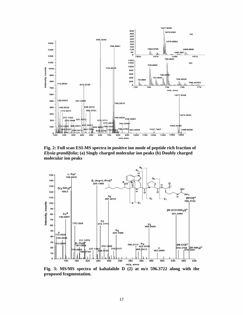
The ESI-MS of the peptide rich fraction of the E. grandifolia showed clusters of molecular
ion peaks (Fig 2). Closer examination of the spectra revealed the presence of both singly (+1)
and doubly (+2) charged molecular ion peaks in the mass range of 1400-1550 amu (Fig 2a)
and 700-800 amu (Fig 2b), respectively. The ethyl acetate fraction of B. plumosa was also
subjected to ESI-MS, which showed a peak for only one peptide, kahalalide F (m/z
1477.9350).
The theoretical monoisotopic m/z values of the peptides in various forms of +1 and +2
charges at m/z 596.3554, 1463.9706, 1477.9356, 1491.9870 and 748.0010 were calculated.
Based on the theoretical calculations singly and doubly charged ion peaks including sodium
and potassium adduct for five peptides could be detected in the full scan spectra, which are
shown in bold in Table 1. The literature reveals that [M+H]+ at m/z 596.3554, 1477.9346 and
doubly charged ion at m/z 748.0010 correspond to earlier reported cyclic depsipeptides,
kahalalide D (1), F (2) and linear peptide kahalalide G (3), respectively from E. rufescens and
its algal diet Bryopsis sp.3 The compounds with [M+H]+ at m/z 1463.9706 and 1491.9870 are
new to the literature. The relative abundance reveals kahalalide D as the major (100%) cyclic
depsipeptide followed by kahalalide F (2) with 40.33%. The other two new peptides,
designated as kahalalide P (4) (13.56%) and Q (5) (0.76%), are present as minor constituents.
The fragment ion peaks are labeled according to the nomenclature proposed by Roepstorff
and Fohlman,35 subsequently modified by Johnson et al.36 To retrieve a sequence from the
mass spectrometric data of the new peptides, we applied the criteria that the mass difference
between two adjacent peaks should precisely fit the mass of an amino acid residue.

6
Tandem mass spectrometry (MS/MS) analysis:
1) MS/MS at m/z 596.3722 of kahalalide D (1):
Fig 3 represents the MS/MS spectrum of a protonated peptide at m/z 596.3722. It is evident
from the spectrum that protonated lactone oxygen opens the ring to form the linear acylium
ion and the amino acid residues are sequentially deleted. The side fatty chain with the loss of
water gives a peak at m/z 140.0807 designated as b1º. The corresponding y3 fragment ion is
observed at m/z 454.2920. The fragment ions b2 and y2 were significantly observed at m/z
325.1985 and 272.1771 for the cleavage between tryptophan and N-terminal of the proline.
The fragment ions b3 and y1 were observed at m/z 422.2538 and 175.1205, respectively. The
peak at m/z 579.3461 was due to loss of ammonia while m/z 568.3784 was due to loss of CO.
The combined loss of CO and NH3 gives a peak at m/z 551.3499. Peaks at m/z 187.0908 and
159.0918 were observed for the tryptophan and its immonium ion, respectively. It is observed
that in case of kahalalide D (1) the protonation of lactone oxygen opens the ring to form a
linear peptide and the amino acids are deleted sequentially.
2) MS/MS at m/z 1477.9669 [M+H]+ and 739.4820 [M+2H]+2 of kahalalide F (2):
The MS/MS spectrum of kahalalide F (2) at m/z 1477.9666 (Fig 4a) showed intense series of
‘b’ and ‘y’ type fragment ions in the linear region. The spectrum shows the fragment at m/z
212.1449 which is consistent with D-val-(5-methylhexanoic acid), designated as b1 ion, and
results from the cleavage of the amide bond between the first valine and threonine residue in
the linear region of the molecule. The corresponding y12 ion was observed at m/z 1266.8085.
Similarly, the fragments at m/z 313.2144, 412.2842, 511.3546, 609.4066, 723.4777 and
835.5762 were assigned as b2, b3, b4, b5, b6 and b7, respectively. The corresponding y ions
were observed at m/z 1165.7494 (y11), 1066.6857 (y10), 967.6124 (y9), 868.5424 (y8),
756.4777 (y7), and 643.3934 (y6). The y9 ion due to the cleavage of the amide between the
valine at 4th position and proline was most prominent. This is because proline residue
contains a tertiary nitrogen atom and is thus more basic than other residues, and hence more
likely to be protonated, thereby leading to enhanced peptide-bond cleavage.37 The above
fragment ion observed confirms the peptide sequence 5-Me-Hex-V-T-V-V-P-Orn-I-,
containing seven amino acids with short fatty chain. Earlier studies by E. Stokis et al.38 report
the quantitative analysis of KF in human plasma done during a Phase I clinical trial, in
patients with androgen refractory prostate cancer using high performance liquid
chromatography (HPLC) coupled to API 2000 triple-quadrupole mass spectrometer equipped

7
with a electrospray ionization source (ESI-MS/MS). The study reported the fragmentation
leading to the formation of only b1 to b4 and y9 (969) and y10 (1068) fragment ions. This
reveals that the sensitivity is much better on QTOF instruments than on triple-quadrupole
mass spectrometers. The recently introduced Q2-pulsing function enhances the transmission
of fragment ions of a selected m/z window from the collision cell into the TOF part which
improves the sensitivity of precursor ion scans on a quadrupole TOF instrument.39-40
In the present study, it is noted that there is sequential loss of amino acid residues up to
isoleucine (b7 & y6) in the linear region while fragment ions y1 to y5 and its corresponding b8
to b12 were not detected, which suggests that there is no further sequential loss of amino acids
in the cyclic region in KF (Fig 4a).
In addition to ‘b’ and ‘y’ fragment ions ‘a’ and ‘c’ type of fragment ions were also observed.
Fragment ions at m/z 184.1705, 693.4750 and 850.5354 were assigned to a1, a6, c5 and c7,
respectively. Peaks at m/z 1469.9568 and 1460.9655 were observed due to the loss of H2O
and NH3. The fragmentation pathway for the loss of amino acid residues in the cyclic region
of KF is discussed below.
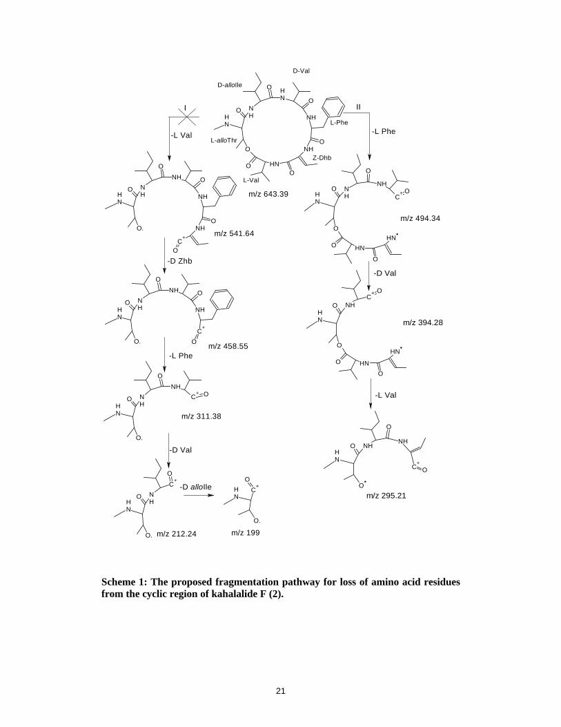
Fragmentation pathway in the cyclic region of kahalalide F (2):
For any cyclic depsipeptides the first dissociation step involves the opening of the cyclic ring
by cleavage of either the lactone group or one of the amide bonds. In most cases the most
basic site gets protonated first resulting in the weakening of the C-N or C-O bond.
In the case of KF, two pathways for the ring opening are possible as depicted in Scheme 1,
designated as ‘I’ and ‘II’. Pathway ‘I’ is for the ring opening at the lactone oxygen with the
first cleavage of L-Val, which would result in the formation of fragment ion at m/z 541,
which is not observed in the spectra. Similarly the sequential loss of other amino acid
residues Dhb, F, V, T would result in the fragment ions at m/z 558, 311, 212 and 199,
respectively which are also not observed in the spectra. This indicates that ring opening at the
lactone oxygen i.e. pathway ‘I’ is ruled out. Pathway ‘II’ is favored with the initial loss of
phenylalanine residue leading to the formation of the peak at m/z 494.3450 [y6-F]+ designated
as ‘A’. This loss can be attributed to N-C ring opening. The second loss of D-Val gave a peak
at m/z 394.2727 [A-V]+ designated as ‘B’. Species ‘B’ undergoes internal cyclization and the
loss L-Val which gives a very intense peak at m/z 295.2055 [B-V]+ designated as ‘C’. The
pathway proposed for the loss of the amino acids in the macrolidic region is outlined in
Scheme 1. The electron donating aromatic ring makes the phenylalanine group a more basic

8
residue, and hence it gets protonated in preference to lactone oxygen. Thus, the ring opens at
the amide bond of the phenylalanine group. It is also evident from Scheme 1 that loss of
amino acid residues from the cyclic region is not sequential.
The tandem mass spectra of doubly charged ion at m/z 739.4803 are remarkably similar to
Fig 4a differing only in the additional peaks being at m/z 1331.8930 and 1378.7940, which
are accounted for by elimination of F and V residues from the parent ion. Fragment ion peaks
b5, b7, y7 and y8 could not be detected or rather they may be less intense.
MS/MS of [M+Na]+ at m/z 1499.9488, [M+H+Na]+2 at m/z 750.4661 and [M+H+K]+2 at
m/z 758.4938 of kahalalide F (2):
In addition to the protonated molecular ion [M+H]+, CID was also obtained for the sodium
and potassium adducts of KF. Kahalalide F (2) displayed sodium adducts [M+Na]+ at m/z
1499.9488 (Fig 4b) and [M+H+Na]+2 at m/z 750.4661. The potassium adduct [M+H+K]+2
was observed at m/z 758.4938 (Fig 4c). The MS fragmentation pattern for the alkali metal
(Na/K) catalyzed molecular ion peaks differ significantly from the protonated molecular ion.
The sodium or potassium ion affects the fragmentation pattern depending upon the location
of the metal ion in the molecule.
In the MS/MS at m/z 1499.9488 of KF (2), an intense series of fragment ions with Na+
attached to the y and b ions was observed. The fragment ion peak with metal ion is denoted
by an asterisk (*) (Fig 4b). The fragment ion peaks from b1 to b5 were exactly matching with
CID of the protonated molecular ion. Its corresponding y ions, y12* (1288.8334), y11*
(1187.7263), y10* (1088.6648), y9* (989.5904) and y8* (890.5554) were ionized by sodium
ion attachment. It also shows peak at 1271.8 for [y12*-NH3]+. Peaks at m/z 505.3588,
647.4149, 700.4432, 744.4685 and 829.5602 were attributed to the sodiated fragment ions
a4*, c5*, [a6*-NH3]+, [b6*-NH3]+ and a7*, respectively. No change in the fragmentation of the
cyclic region was observed. It was identical to the fragmentation observed in MS/MS of
[M+H]+ ion. This indicated that Sodium is attached either to amide nitrogen or amide
carbonyl oxygen of the linear chain V-P-Orn-I. The Na+ does not bind to the lactone oxygen
of the cyclic ring, which opens the ring to give a linear peptide as described by Ngoka.26 This
suggests that the lactone linkage is stronger than any other amide bond in the linear as well as
cyclic part of the molecule and hence the new fragmentation pattern was observed. Fig 4b
also summarizes the fragmentation pattern for the sodium adduct. Combined loss of CO and
NH3 from sodiated molecular ion gives a peak at m/z 1455.9123 [M*-(CO+NH3)]+. Peaks

9
were also observed at m/z 1382.8642 and 1356.8642 for [M*-(H2O+V)]+ and [M*-
(CO+NH3+V)]+, respectively. Doubly charged ion [M+H+Na]+2 at m/z 750.4661 also
displayed similar peaks.
A similar fragmentation pattern was observed during CID of the potassium adduct
[M+H+K]+2 at m/z 758.4938 (Fig 4c). Fragment ion peaks from b1 to b4 were identical to that
of protonated molecular ion but the y fragment ions were ionized by potassium. The y*
fragment ion peaks were observed at m/z 1304.8310 (y12*), 1202.7209 (y11*), 1104.6974
(y10*), 1005.6188 (y9*), 906.5515 (y8*) and 794.4641 (y7*) but the y6 fragment is observed at
m/z 643.3944 without addition of potassium ion. No change was observed in the
fragmentation pattern of the cyclic ring. This indicated that potassium ion is not attached to
the lactone oxygen of the cyclic region but may be attached to the amide nitrogen or carbonyl
oxygen in the linear chain of the molecule.
3) Mass fragmentation of kahalalide G (3):
The doubly charged molecular ion at m/z 748.0010 for [M+2H]+2 corresponds to the
molecular mass 1496, which is 18 Da higher than kahalalide F. This suggest it is a ring
opened form i.e kahalalide G (3).12 It could also due to the formation of water adduct of
kahalalide F. But based on the literature we presumed it to be kahalalide G, which is reported
from the same source. The MS/MS experiment for the doubly charged ion m/z 748.0010 was
carried out at 25V, which showed identical b and y fragment ions as that of kahalalide F (2).
This suggests that KF is derived from kahalalide G (3) with the initial loss of water resulting
in the cyclisation via a lactone bond between the threonine (sixth residue from the C-
terminal) and carboxylic group. Thus, the fragmentation pattern is identical to that of (2).
4) Elucidation of the peptide sequence of new cyclic depsipeptides, kahalalide P (4) and
Q (5):
The CID spectra of [M+H]+ at m/z 1463.9541 (Fig 4a), [M+2H]+2 at m/z 732.4854 and
sodium adduct [M+H+Na]+2 at m/z 743.4582 (Fig 4b) of kahalalide P (4) were studied.
Molecular ion [M+H]+ showed peaks at m/z 212.1447 (b1) 313.2171 (b2), 412.2843 (b3),
511.3578 (b4), 609.3909 (b5) and 723.4675 (b6) identical to those observed in the MS/MS
spectrum of KF. The corresponding y ions were observed at 1252.7944 (y12), 1151.7452
(y11), 1052.6714 (y10), 953.6054 (y9), 839.5439 (y8-NH3) and 742.4637 (y7). The fragment
ion y6 at m/z 643.3909 (y6) reveals the presence of a cyclic part of the structure identical to
that observed in KF. Further it is also evident that the sequence of amino acids (5-MeHex-V-

10
T-V-V-P-Orn-) in the linear portion from b1 to b6 is identical to that of KF. The difference
was observed in the amino acid linking the cyclic moiety. The mass difference between the
821.5634 (b6) and 723.4675 (b7) is 99, which could be accounted for the amino acid valine
instead of isoleucine in KF. Also, the difference between y7 and y6 was exactly 99 amu
confirming the presence of valine residue (Fig 5a).
Thus the CID mass spectrum reveals a valine substitution for isoleucine at number 7. The one
line representation of the proposed structure of kahalalide P is given as Cyclo-8,13-(5MeHex-
V-T-V-V-P-Orn-V-T-I-V-F-Dhb-V) [Dhb-dehydroaminobutyric acid). In analogy with the
reported KF, similar stereochemistry may be assigned to this molecule. The CID of doubly
charged ion at m/z 732.4723 also showed similar peaks that were less intense. The peak at
1316.8573 was assigned to the loss of phenylalanine from the parent ion.
In the CID of doubly charged sodiated molecular ion [M+H+Na]+2 at m/z 743.4682, (Fig 5b),
sodium ion was found attached to y fragment ions from y12* to y7* in the linear peptide chain.
The y6 fragment ion is devoid of sodium ion. This confirms that sodium ion is bound either to
nitrogen or oxygen of the valine residue attached to the cyclic ring. The immonium ion of
phenylalanine is observed at m/z 120.0816. The cyclic ring shows fragment ions identical to
those found in [M+H]+.
In a similar fashion the MS/MS of the [M+H]+ at m/z 1491.9876 (Fig 6) showed identical
fragment ions b1 to b5, indicating linear peptide chain 5-MeHex-V-T-V-V-P- and the y6 peak
at m/z 643.3909 showed there is no change in the cyclic ring. This indicated that 14 units
extra should be present in the linear region of the molecule. The corresponding b fragment
ion for y6 (m/z 643.3909) was observed at m/z 849.5965 (b7). The mass difference between
643.3909 (y6) and newly observed peak at m/z 756.4822 was 113, which can be accounted by
the presence of isoleucine. Thus, the peak at m/z 756.4822 was assigned as the y7 fragment
ion. The corresponding b6 fragment ion could not be detected in the spectrum. The
corresponding y8 was observed at m/z 882.5586 for 609.3977 (b5). The peak m/z 864.5491
was attributed for loss of ammonia [y8-NH3]+. The mass difference between y8 (882.5586)
and y7 (756.4822) gives 128, which can be fitted by placing amino acid lysine at position six.
The peak at m/z 226.1566 was observed for [P+K]+ which was not observed in peptides
previously discussed.

11
This completes the peptide sequence of kahalalide Q (5) and the one line representation is
given as Cyclo-8,13-(5MeHex-V-T-V-V-P-K-I-T-I-V-F-Dhb-V) with the cyclic residue.
Peak at m/z 1374.9 was assigned to combined loss of water and valine [M-(H2O+V)]+.
CONCLUSION
The unreported peptides, kahalalide P (4) & Q (5) are close analogues of kahalalide F. In
kahalalide P, number 7th amino acid isoleucine is replaced by valine while in kahalalide Q,
number 6th amino acid ornithine is replaced by lysine. The presence of lysine was previously
reported in kahalalide J.10 ESI-MS/MS experiments provided data that was difficult to obtain
by the NMR technique, because of their low concentration. This technique requires about ten
thousand times less raw material than that of the NMR based procedure and all experiments,
including extraction, take only a few days. On the other hand, the basic limitation of the mass
spectrometric procedure is to determine the configuration of amino acid residues. The present
study also indicates that the loss of amino acid residues from depsipeptide is not always
sequential.
KF (2) exhibits significant bioactivity and is currently in Phase II clinical trials in Europe.
Hence it elicits maximum interest chemically. The two new cyclic depsipeptides, kahalalide P
and Q that were identified as being chemically similar to kahalalide F, may exhibit similar
bioactivity. Since the synthetic methods are well known for KF, the new cyclic depsipeptides
can be prepared to check for biological activity. As these compounds are typically
administered at very low doses, considerable demands are placed upon the sensitivity of
analytical methods to characterize their pharmacokinetic behaviour in humans. Thus, ESI-
MS/MS is the detection method of choice for the analysis of cyclic depsipeptides.
Acknowledgements
Due thanks are due to the Director, Satish R. Shetye, National Institute of Oceanography. The
author, Supriya Tilvi wishes to thank the Department of Ocean Development, for the
fellowship under the Project, Development of the Potential Drugs from the Indian Ocean.
We would also like to thank Dr. Solimabi Wahidullah for her valuable comments on the
manuscript.

12
REFERENCES
1. Fontana A, Ciavatta ML, D’souza L, Mollo E, Naik CG, Parameswaran PS, Wahidulla S, Cimino G. Selected chemo-ecological studies of marine opisthobranchs from Indian coasts. J. Indian Inst. Sci. 2001; 81: 403.
2. Hamann MT, Scheuer PJ. Kahalalide F: A bioactive depsipeptide from the sacoglossan mollusk Elysia rufescens and the alga Bryopsis sp. J. Am. Chem. Soc. 1993; 115: 5825.
3. Hamann MT, Otto CS, Scheuer PJ, Dunbar DC. Kahalalides: Bioactive peptides from a marine mollusk Elysia rufescens and its algal diet Bryopsis sp. J. Org. Chem. 1996; 61: 6594.
4. Goetz G, Nakao Y, Scheuer PJ. Two acyclic kahalalides from the sacoglossan mollusk Elysia rufescens. J. Nat. Prod. 1997; 60: 562.
5. Bonnard I, Manzanares I, Rinehart KL. Stereochemistry of kahalalide F. J. Nat. Prod. 2003; 66: 1466.
6. Goetz G, Yoshida WY, Scheuer PJ. The absolute stereochemistry of kahalalide F. Tetrahedron 1999; 55: 7739.
7. Lopez-Macia A, Jimenez JC, Royo M, Giralt E, Albericio F. Kahalalide B: Synthesis of a natural cyclodepsipeptide. Tetrahedron Lett. 2000; 41: 9765.
8. Kan Y, Fujita T, Sakiamoto B, Hokama Y, Nagai H. A new cyclic depsipeptide from the hawaiian green alga Bryopsis species. J. Nat. Prod. 1999; 62: 1169.
9. Horgen FD, Delossantos DB, Goetz G, Sakamoto B, Kan Y, Nagai H, Scheuer P J. A new depsipeptide from the sacoglossan mollusk Elysia ornata and the green alga Bryopsis species. J. Nat. Prod. 2000; 63: 152.
10. Goetz G, Nakao Y, Scheuer PJ. Two acyclic kahalalides from the sacoglossan mollusk Elysia rufescens. J. Nat. Prod. 1997; 60: 562.
11. El Sayed KA, Bartyzel P, Shen X, Perry TL, Zjawiony JK, Hamann MT. Marine natural products as antituberculosis agents. Tetrahedron 2000; 56: 949.
12. Lopez LL, Trigo JM, Jimeno J, Pazares L, Ruiz CA, Izquierdo MA, Schellens J. Kahalalide compounds for use in cancer theraphy. Patent Number EP 1435990, 2004.
13. Nuyen B, Weitman S, Faircloth, GT. Kahalalide F formulation. Patent Number US 2004067895, 2004.
14. Garcia-Rocha M, Bonay P, Avila J. The antitumor compound kahalalide F acts on cell lysosomes. Cancer Lett. 1996; 99: 43.
15. Ciruelos E, Trigo JM, Pardo B, Paz-Ares L, Estaun N, Cuadra C, Domínguez MJ, Marin A, Ruiz A, Jimeno J, Izquierdo MA. A phase I clinical and pharmacokinetic (PK) study with Kahalalide F (KF) in patients (pts) with advanced solid tumours
13
(AST) with a continuous weekly (W) 1-hour iv infusion schedule. Eur. J. Cancer, 2002; 38: S33.
16. Gomez SG, Bueren JA, Faireloth GT, Jimeno J, Albella B. In vitro toxicity of three new antitumoral drugs (Trabectedin, Aplidin and Kahalalide F) on hemotopoietic progenitors and stem cells. Experimental Hematology 2003; 31: 1104.
17. Jimenez JC, Rodrigues I, Giralt E, Lopes A, Royo M, Albericio F, Manzanares I. Kahalalide compounds, Patent Number PL 356800, 2004.
18. Grese RP, Gross ML. Interactions of lithium ions and dipeptides. J. Am. Chem. Soc. 1990; 112: 5098.
19. Grese RP, Cerny RL, Gross ML. Metal ion-peptide interactions in the gas phase: A tandem mass spectrometry study of alkali metal cationized peptides. J. Am. Chem. Soc. 1989; 111: 2835.
20. Teesch LM, Orlando RC, Adams J. Location of the alkali metal atom in gas phase peptide complexes. J. Am. Chem. Soc. 1991; 113: 3668.
21. Teesch LM, Adams J. Fragmentations of gas-phase complexes between alkali metal atoms and peptides: metal ion binding to carbonyl oxygens and other neutral functional groups. J. Am. Chem. Soc. 1991; 113: 812.
22. Mallis LM, Russell DH. Fast atom bombardment-tandem mass spectrometry studies of alkali-metal ions of small peptides. Anal. Chem. 1986; 58: 1076.
23. Russell DH, McGlohon ES, Mallis LM. Fast atom bombardment-tandem mass spectrometry studies of organo-alkali-metal ions of small peptides. Competitive interaction of sodium with basic amino acid substituents. Anal. Chem. 1988; 60: 1818.
24. Tang X, Ens W, Standing KG, Westmore JB. Daughter ion mass spectra from cationized molecules of small oligopeptides in a reflecting time-of-flight mass spectrometer. Anal. Chem. 1988; 60: 1791.
25. Lin S, Liehr S, Cooperman BS, Colter RJ. Sequencing cyclic peptide inhibitors of mammalian ribonucleotide reductase by electospray ionization mass spectrometry. J Mass Spectrom. 2001; 36: 658.
26. Ngoka LCM, Gross ML, Peter LT. Sodium-directed selective cleavage of lactones: a method for structure determination of cyclodepsipeptides. Int. J. Mass Spectrom. 1999; 182/183: 289.
27. Ngoka LCM, Gross ML, Peter LT. Multiple collisionally activated decomposition in an ion trap for the determination of the amino acid sequence and gas phase ion chemistry of lithium-coordinated valinomycin. Int. J. Mass Spectrom. 2000; 194: 247.
28. Govaerts C, Rozenski J, Orwa J, Roets E, Schepdael AV, Hoogmartens J. Mass spectrometric fragmentation of cyclic peptides belonging to the polymyxin and colistin antibiotics studied by ion trap and quadrupole/orthogonal-acceleration time-of-flight technology. Rapid Commun. Mass Spectrom. 2002; 16: 823.

14
29. Govaerts C, Adams E, Schepdael AV. Hypenation of liquid chromatography to ion trap mass spectrometry to identify minor components in polypeptide antibiotics. Anal. Bioanal. Chem. 2003; 377: 909.
30. Das BC, Varenne P, Taylor A. The identification of depsipeptides by chemical ionisation mass spectroscopy. J. Antibiot. 1979; 32: 569.
31. Williams SM, Brodbelt JS. MSn characterization of protonated cyclic peptides and metal complexes. J. Am. Soc. Mass Spectrom. 2004; 15: 1039.
32. Kuzma M, Jegorov A, kacer P, Havlicek V. Sequencing of new beauverolides by high performance liquid chromatography and mass spectrometry. J. Mass Spectrom. 2001; 36: 1108.
33. Capon RJ, Skene C, Steward M, Ford J, O’hair RAJ, Williams L, Lacey E, Gill JH, Heiland K, Friedel T. Aspergillicins A-E: five novel depsipeptides from the marine-derived fungus Aspergillus carneus. Org. Biomol. Chem. 2003; 1(11): 1856.
34. Jegorov A, Paizs B, Zabka M, Kuzma M, Havlicek V, Giannakopulos, Anastassios E, Derrick PJ. Profiling of cyclic hexadepsipeptides roseotoxins synthesized in vitro and in vivo: a combined tandem mass spectrometry and quantum chemical study. Eur. J. Mass Spectrom. 2003; 9(2): 105.
35. Roepstorff P, Fohlman J. Proposal for a common nomenclature for sequence ions in mass spectra of peptides. Biomed. Mass Spectrom. 1984; 11: 601.
36. Johnson, RS, Martin, SA, Biemann, K, Stults, JT, Watson, JT. Novel fragmentation process of peptides by collision-induced decomposition in a tandem mass spectrometer: differentiation of leucine and isoleucine. Anal. Chem. 1987; 59(21): 2621.
37. Nair H, Somogyi A, Wysocki VH. Effect of alkyl substitution at the amide nitrogen on amide bond cleavage: ESI/SID fragmentation of substance P and two alkylated analogs. J. Mass. Spectrom. 1996; 31: 1141.
38. Stokis E, Rosing H, Lopez-Lazaro LL, Rodriguez I, Jimeno JM, Supko JG, Schellens JHM, Beijnen JH. Quantitative analysis of the novel depsipeptide anticancer drug kahalalide F in human plasma by high-performance liquid chromatography under basic conditions coupled to electrospray ionization tandem mass spectrometry. J. Mass Spectrom. 2002; 37: 992.
39. Hanno S, Bernhard K, Matthias M. Quadrupole time-of-flight versus triple-quadrupole mass spectrometry for the determination of phosphopeptides by precursor ion scanning. J. Mass Spectrom. 2001; 36: 782.
40. Marchese S, Gentill A, Perret D, D’Ascenzo G, Pastori F. Quadrupole time-of-flight versus triple-quadrupole mass spectrometry for the determination of non-steroidal anti-inflammatory drugs in surface water by liquid chromatography/tandem mass spectrometry. Rapid Commun. Mass Spectrom. 2003; 17: 879.

15
Tab
le 1
: The
oret
ical
mon
oiso
topi
c m
/z v
alue
s of
pep
tides
in v
ario
us fo
rms
of +
1 an
d +2
cha
rges
. Mol
ecul
ar io
n pe
aks
of th
e pe
ptid
es
dete
cted
in E
SI-M
S ex
peri
men
t of E
lysi
a gr
andi
folia
are
show
n in
bol
d.
Pept
ides
[
M+H
]+ [
M+N
a]+
[M
+K]+
[M
+Na+
K]+
[M
+2H
]+2
[M
+H+N
a]+2
[M
+H+K
]+2
[M
+2N
a]+2
Kah
alal
ide
D
59
6.45
618
.3
6
34.3
65
7.3
2
98.2
2
309.
22
3
17.7
2
3
20.7
2 K
ahal
alid
e P
1
463.
97
148
5.9
1
501.
9
15
24.9
7
32.4
5
743
.45
751
.45
7
54.4
5 K
ahal
alid
e F
1
477.
93
149
9.9
15
15.9
153
8.9
73
9.46
7
50.4
6
7
58.4
6
761
.46
Kah
alal
ide
Q
149
2.00
1515
.0
1530
.0
1
553.
0
746.
00
757
.50
765
.50
7
68.5
0 K
ahal
alid
e G
1
497.
00
15
19.0
15
58.0
153
6.0
74
8.00
760
.00
768
.00
7
71.0
0

16
NO
NH
O
NH
NH2
NH
O
O
NH
O
NH
7-Me-3-Octol
L-Arg L-Pro
D-Trp
Kahalalide D (1)
NH
ONH
O
ONH O
NH
O
O NH
NH
O
ONH
R2
O
NH
R1
O
NO
NHO
NHO
NHOH
ONH
O
5-Me-HexD-Val
L-Thr
L-Val
D-Val
D-ProD-alloIle
L-Phe
Z-Dhb
L-Val
L-alloThrD-Val
Kahalalide F (2): R1= NH2; R2=Me Kahalaide P (4): R1= NH2; R2=H Kahalaide Q (5): R1= -CH2NH2; R2=Me
NH
ONH
ONH O
NH
O
O NH
NH
O
ONH
O
NH
NH2
O
NO
NHO
NHO
NHOH
ONH
O
OHOH
5-Me-HexD-Val
L-Thr
L-Val
D-Val
D-ProL-Orn
D-alloIleD-alloIle
L-Phe
Z-Dhb
L-Val
L-alloThrD-Val
Kahalalide G (3)
Fig 1: Structures of kahalalides.
17
Fig. 2: Full scan ESI-MS spectra in positive ion mode of peptide rich fraction of Elysia grandifolia; (a) Singly charged molecular ion peaks (b) Doubly charged molecular ion peaks
Fig. 3: MS/MS spectra of kahalalide D (2) at m/z 596.3722 along with the proposed fragmentation.

18
Fig. 4: MS/MS spectra of kahalalide F (2) at (a) m/z 1477.9669 [M+H]+, (b) 1499.9488 [M+Na]+ and (c) 758.4938 [M+H+K]+2 along with the proposed fragmentation pattern.

19
Fig. 5: MS/MS spectra of kahalalide P (4) at (a) m/z 1463.541 [M+H]+ and (b) 743.4682 [M+H+Na]+2 along with the proposed fragmentation pattern.

20
Fig. 6: MS/MS spectra of kahalalide Q (5) at m/z 1491.9876 [M+H]+ along with
the fragmentation pattern.
21
NH
O
O.
C+
O
NHO
O
NH
NHO
NH
NH
O
O.
NHO
O
NH
C+
O
NH
NH
O
O.
NHO
C+ O
NH
NH
O
C+
O.
O
NH
C+
O
O.
NH
NH
O
O
O NHO
NHO
O
NH
NHO
NH
L-Val
Z-Dhb
L-Phe
D-Val
D-alloIle
L-alloThr
NH
O
O
O NH
O
NH
O
C+ O
NH
NH
NHOC
+
O
O NHO
O
NH
NH
NHO
O
O
NH
NH
C+
O
-L Val -L Phe
m/z 643.39
m/z 494.34
-D Val
m/z 394.28
m/z 295.21
-L Val
m/z 541.64
-D Zhb
m/z 458.55-L Phe
m/z 311.38
-D Val
m/z 212.24
-D alloIle
m/z 199
I II
Scheme 1: The proposed fragmentation pathway for loss of amino acid residues from the cyclic region of kahalalide F (2).

22
LIST OF FIGURE LEGENDS
Fig. 1: Structures of kahalalide D, F, G, P & Q.
Fig. 2: Full scan ESI-MS spectra in positive ion mode of peptide rich fraction of
Elysia grandifolia; (a) Singly charged molecular ion peaks (b) Doubly charged
molecular ion peaks.
Fig. 3: MS/MS spectra of kahalalide D (2) at m/z 596.3722 along with the proposed
fragmentation.
Fig. 4: MS/MS spectra of kahalalide F (2) at (a) m/z 1477.9669 [M+H]+, (b)
1499.9488 [M+Na]+ and (c) 758.4938 [M+H+K]+2 along with the proposed
fragmentation pattern.
Fig. 5: MS/MS spectra of kahalalide P (4) at (a) m/z 1463.541 [M+H]+ and (b)
743.4682 [M+H+Na]+2 along with the proposed fragmentation pattern.
Fig. 6: MS/MS spectra of kahalalide Q (5) at m/z 1491.9876 [M+H]+ along with the
fragmentation pattern.