taeniasis learning outcomes by the end of the lecture, you should be able to: mention causal agent...
TRANSCRIPT


Learning outcomes• By the end of the lecture, you should be able to:• Mention causal agent of Taeniasis.• Mention systematic position of Taenia spp.• Mention geographic distribution of Taeniasis.• Describe morphology of adults, eggs & larvae.• Enumerate Intermediate, and Definitive hosts of Taenia spp.• Explain life cycle of Taenia spp.• Mention habitat of Taenia spp.• Mention mode of infection by Taenia spp.• Mention pathology and clinical features of Taeniasis.• Diagnose Taeniasis.• Mention prevention and conrol methodes of Taeniasis

Suggested Reading
• http://www.dpd.cdc.gov/dpdx/HTML/Taeniasis.htm

Causal Agents
• Taeniasis is a parasitic disease caused by tapeworms Taenia saginata (beef tapeworm) and Taenia solium (pork tapeworm).

Systematic position
• Phylum: Platyhelminthes• Class: Cestodae.g. Taenia saginataTaenia solium

Geographic Distribution
• Both species are worldwide in distribution.• Taenia solium is more prevalent in poor
communities where humans live in close contact with pigs and eat undercooked pork and is very rare in Muslim countries.
Morphology of Adult worm
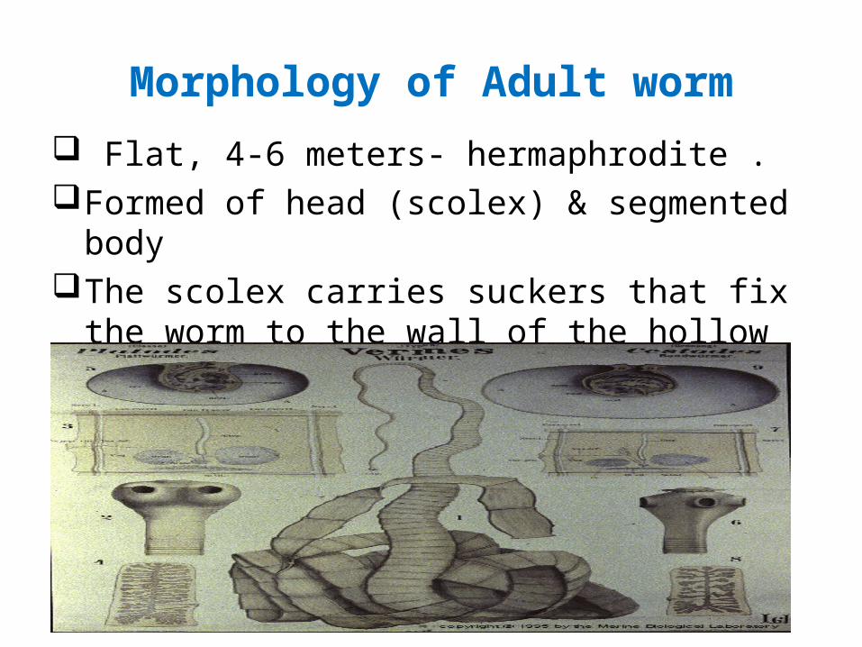
Flat, 4-6 meters- hermaphrodite .Formed of head (scolex) & segmented bodyThe scolex carries suckers that fix the worm to the wall
of the hollow small intestine

Adult worm (fresh /unstained)

Morphology of Adult ..cont
Segments (proglottids) are immature, mature and gravid
Mature segment contain mature male & female sexual organs
Gravid segments contain the uterus full of eggs

Scolex (head)
Taenia saginataScolex with four
suckers Taenia solium Scolex with four
suckers & Hooked rostellum

Mature & gravid segment (stained specimens)
Mature segmentwith mature male Gravid segment and female contains branchedreproductive uterus, full of eggs organs

Morphology of Eggs
OvoidYellowish brownRadically striated embryophoreContent: hexocanth embryo (oncosphere with 6 hooklets)

Morphology of Larva
In T. saginata larava termed Cysticercus bovis which is Ovoid, cystic, size =
a bean contains non hooked scolex inside + immature segments
In T. solium larava termed Cysticercus cellulosae which is Ovoid, cystic, size = a bean Contains scolex with rostellum & hooks inside + immature segments

Life cycle
1- The adult lives in the small intestine of man ( the definitive/ final host)
2- Mature eggs & gravid segments pass with stool (the diagnostic stages)
3- When Mature eggs & gravid segments are ingested by the cattle (the intermediate host of T. saginata), or the pig (the intermediate host of T. solium), the egg hatch, the embryo penetrates the intestinal wall, goes to the circulation.

Life cycle …cont
4- In the circulation, it passes from the write side of the heart to the left side of the heart to the brain, bones, muscles......etc; where it develops into cysticercus bovis in the case of T. saginata (the infective stage) or cysticercus cellulosae in the case of T. solium (the infective stage) after a bout 12 weeks.

Life cycle …cont5- When another man (the definitive host) eats under
cooked (raw or uncooked) beef meat containing viable cysticercus (the infective stage), meat is digested, cysticercus is liberated and grow to the adult worm which attaches itself to the intestinal mucosa and start new life cycle.
The scolex invaginates
in the bladder The scolex evaginates
Under stimulation of bile

Life Cycle

Mode of infection
• Humans become infected by ingesting raw or undercooked infected meat containing cysticercus.

Clinical Features
• Taenia saginata taeniasis produces only mild abdominal symptoms.
• Occasionally, appendicitis or cholangitis can result from migrating proglottids.
• The most important feature of Taenia solium taeniasis is the risk of development of cysticercosis.

It the infection of human tissue by cysticerci cellulosae, the larval stage of Taenia solium
How ??? Man is infected either by : Internal autoinfection : Taenia solium eggs hatch inside
the human body and the oncoshere (embryo) penetrate the intestinal wall, goes to the systemic circulation and from it to various body organs
External autoinfection: Man ingest Taenia solium eggs from intrinsic source (hand to mouth/ feco- oral) or from extrinsic source (contaminated food with human stool remnants)
Cysticercosis

Pathogenicity of Cysticercosis
• The cyst produce a foreign body inflammatory reaction which usually end in fibrosis & calcification in the affected tissue/organ.

The subcutaneous nodules are usually found in head, limbs, neck, abdomen and back. They are movable and painless.
Subcutaneous Cysticercosis

• •
Note this cysticercus in the tongue

The cysticercus is usually found in the vitreous body or subretina. Visual disturbance often occurs. The died body of worm may provokes local inflammation causing blindness.
Ocular Cysticercosis

Brain Cysticercosis:( neurocysticercosis)
Epilepsy is the most frequent symptoms of brain cysticercosis.
In frank words:
Neurocysticercosis is the most common cause of adult onset epilepsy.

Laboratory Diagnosis
• Microscopic identification of eggs and gravid segments (proglottids) in feces of infected human is the most common diagnostic method for taeniasis.

Laboratory Diagnosis….cont.
Taenisis prevention and control
• Prevention of contamination of areas, where cattle graze, with human feces
• Inspection of beef for cysticerci at slaughter houses and then infected meat are condemned
• Proper cooking of meat (at least 65 C° for 5 minutes ) or freezing at – 10 C° for 5-10 days
• Avoid suspected undercooked meat (grilled or pickled).
• Prompt treatment of infected persons.
• Infected persons must not work as a food handlers

1- One of the following parasites belongs to Cestoda:Taenia speceis2- Cysticercus bovis is the infective stage of:Taenia saginata3- Cysticercus cellulosae is the infective stage of:Taenia solium-

4- Egg containing onchosphere inTaenia speciesHymenolepis nana5- Egg and gravid segments are the infective stages ofTaenia species6- Cysticercosis occur inTaenia solium

Thank you