synthesis, characterisation and biological activities …
TRANSCRIPT
UNIVERSITY OF KWAZULU-NATAL
SYNTHESIS, CHARACTERISATION AND
BIOLOGICAL ACTIVITIES OF
HOMOISOFLAVONOIDS
2012
KAALIN GOPAUL
ii
SYNTHESIS, CHARACTERISATION AND
BIOLOGICAL ACTIVITIES OF
HOMOISOFLAVONOIDS
KAALIN GOPAUL
2012
A thesis submitted to the School of Chemistry and Physics, University of KwaZulu-Natal,
Westville, for the degree of Masters of Science.
As the candidate’s supervisor, I have approved this thesis for submission.
Supervisor:
Signed: --------------------------------Name: ------------------------- Date: --------------

iii
To my family…

iv
�ान शि�त है. “Knowledge is power”
v
DECLARATIONS
DECLARATION 1- PLAGIARISM
I, KAALIN GOPAUL, declare that the experimental work described in this dissertation was
carried out at the School of Chemistry and Physics, University of KwaZulu-Natal, Westville
campus under the supervision of Dr. N. A. Koorbanally, and that:
1. The research reported in this thesis is my original research, except where otherwise
indicated.
2. This thesis has not been submitted for any degree or examination at any other
university.
3. This thesis does not contain other persons’ data, pictures, graphs or other information,
unless specifically acknowledged as being sourced from other persons.
4. This thesis does not contain other persons' writing, unless specifically acknowledged
as being sourced from other researchers. Where other written sources have been
quoted, then:
a. Their words have been re-written but the general information attributed to them
have been referenced
b. Where their exact words have been used, then their writing has been placed in
italics and inside quotation marks, and referenced.
5. This thesis does not contain text, graphics or tables copied and pasted from the
Internet, unless specifically acknowledged, and the source being detailed in the thesis
and in the References sections.
Signed …………………………………………………………
vi
DECLARATION 2- PUBLICATIONS
DETAILS OF CONTRIBUTION TO PUBLICATIONS that form part and/or include
research presented in this thesis.
Publication 1
Kaalin Gopaul, Mahidansha Shaikh, Deresh Ramjugernath, Neil A. Koorbanally and Bernard
Omondia, 3-(3-Methoxybenzylidene)chroman-4-one, Acta Crystallographica Section E,
Accepted for publication: 4 March 2012
Publication 2
Kaalin Gopaul, Mahidansha M. Shaikh, Neil A. Koorbanally, Deresh Ramjugernath and
Bernard Omondia, (E)-3-(4-Cyclohexyl-3-fluorobenzylidene)chroman-4-one, Acta
Crystallographica Section E,
Accepted for publication: 28 May 2012
Publication 3
Kaalin Gopaul, Neil Anthony Koorbanally, Mahidansha M. Shaikh, Hong Su and Deresh
Ramjugernath, 3-(3,4-Dichlorobenzylidene)chroman-4-one, Acta Crystallographica Section
E,
Accepted for publication: 25 September 2012
From all the above publications, my role included carrying out the experimental work and
contributing to the writing of the publications along with my supervisor. The other co-
authors contribution was that of an editorial nature and checking on the scientific content and
my correct interpretation. Based on their expertise, they have added minor parts to the
manuscripts.
Signed: ………………………………….
vii
ACKNOWLEDGEMENTS
• To my parents, Prem and Preetha Gopaul, and my brother, Breneil Gopaul, I am
unconditionally grateful for your motivation and support. I would not be where I am
without you.
• To my supervisor, Dr. N.A. Koorbanally, Thank you for the opportunity to work with
you and for your guidance throughout this study and the long hours endured in writing
and editing this dissertation. Your time and dedication are sincerely appreciated. I
look forward to continuing my academic career under your supervision.
• To Dr M. Shaikh for imparting his practical knowledge to me on a daily basis, you
made my labwork enjoyable.
• Dr. H.Y. Chenia, at the School of Biochemistry, Genetics and Microbiology, UKZN
Westville for assisting me when carrying out the antibacterial testing.
• Dr B.O. Owaga and Dr. Hong Su for the collection and refinement of crystallographic
data.
• To my labmates in the Natural Products Research Group for always being there to
discuss any problem that I might have encountered through my studies.
• Mr. D. Jagjivan for assisitance with NMR experiments.
• To my friends, Vashen Moodley, Shirveen Sewpersad, Michael Nivendran Pillay
Rasmika Prithipal and Arisha Prithipal for sharing the fun times with me.
• To the technical and support staff of the School of Chemistry and Physics for being
ever willing to assist me.
• The National Research Foundation (NRF) for financial support through a Scarce
Skills Scholarship.
viii
LIST OF ABBREVIATIONS
1H NMR proton nuclear magnetic resonance spectroscopy 13C NMR carbon-13 nuclear magnetic resonance spectroscopy 19F NMR fluorine-19 nuclear magnetic resonance spectroscopy
DEPT distortionless enhancement by polarization transfer
COSY correlated nuclear magnetic resonance spectroscopy
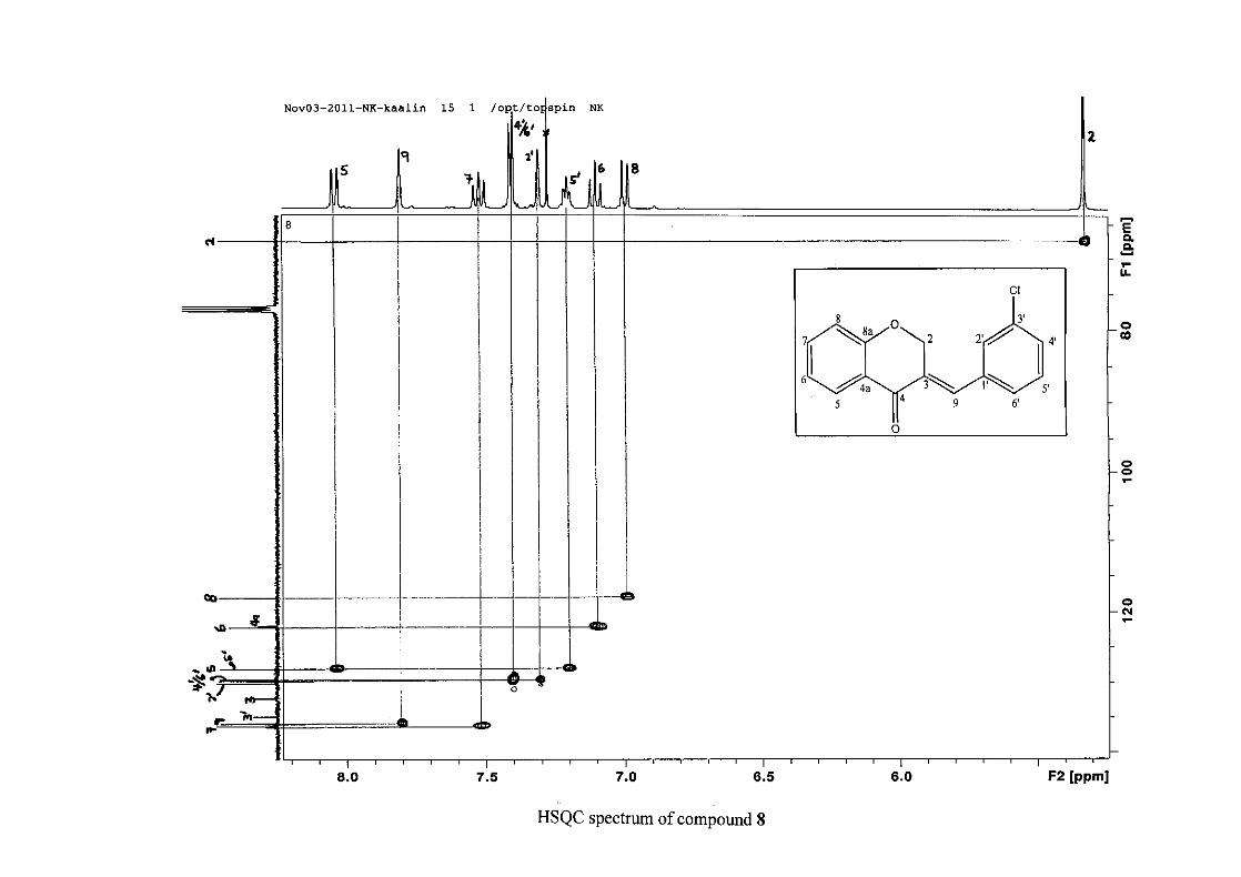
HSQC heteronuclear single quantum coherence
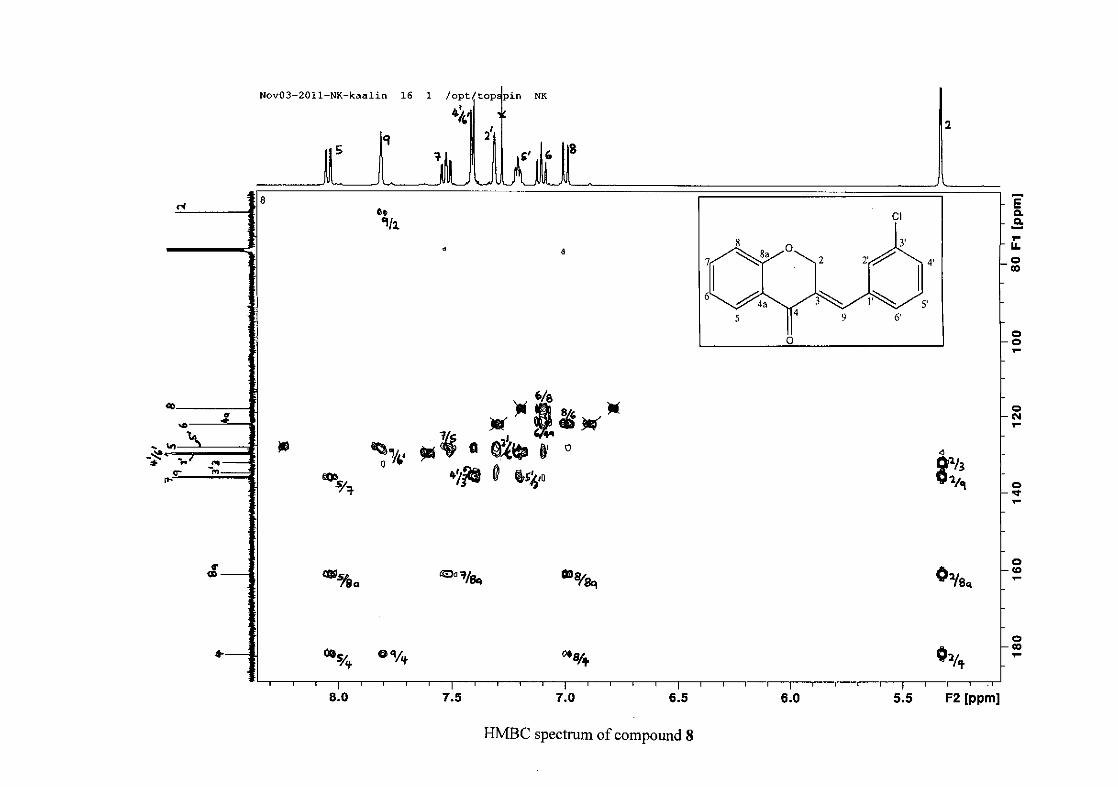
HMBC heteronuclear multiple bond coherence
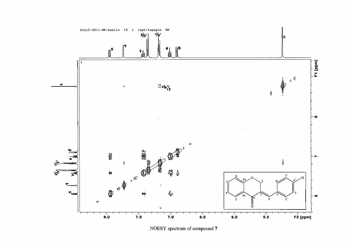
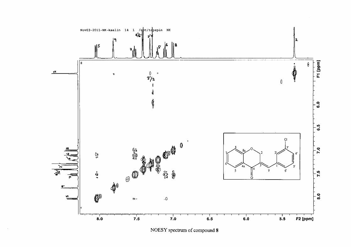
NOESY nuclear overhauser effect spectroscopy
CDCl3 deuterated chloroform
DMSO deuterated dimethyl sulfoxide
TMS tetramethylsilane
EtOH ethanol
PPA polyphosphoric acid
TCA trichloroacetic acid
EI-MS electron impact mass spectrometry
min minutes
hrs hours
Hz hertz
IR infrared
UV ultraviolet
m multiplet
d doublet
dd double doublet
ddd double double doublet
s singlet
t triplet
td triplet of doublets
m.p. melting point
°C degrees Celsius
TLC thin layer chromatography
MIC minimum inhibitory concentration
TET tetracycline
ix
AMP ampicillin
TSA trypticase soy agar
MH Mueller-Hinton
AI activity index
DPPH 2,2-diphenyl-1-picrylhydrazyl
FRAP ferric reducing antioxidant power
ABTS 2,2'-azino-bis(3-ethylbenzothiazoline-6-sulfonic acid)
ORAC oxygen radical absorbance capacity
INT p-iodonitrotetrazolium
ORTEP Oak Ridge Thermal Ellipsoid Plot
x
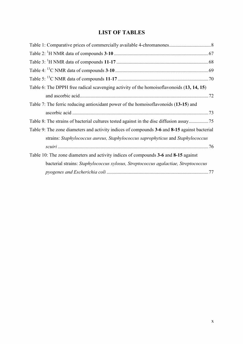
LIST OF TABLES
Table 1: Comparative prices of commercially available 4-chromanones .................................. 8
Table 2: 1H NMR data of compounds 3-10 ............................................................................. 67
Table 3: 1H NMR data of compounds 11-17 ........................................................................... 68
Table 4: 13C NMR data of compounds 3-10 ............................................................................ 69
Table 5: 13C NMR data of compounds 11-17 .......................................................................... 70
Table 6: The DPPH free radical scavenging activity of the homoisoflavonoids (13, 14, 15)
and ascorbic acid......................................................................................................... 72
Table 7: The ferric reducing antioxidant power of the homoisoflavonoids (13-15) and
ascorbic acid ............................................................................................................... 73
Table 8: The strains of bacterial cultures tested against in the disc diffusion assay ................ 75
Table 9: The zone diameters and activity indices of compounds 3-6 and 8-15 against bacterial
strains: Staphylococcus aureus, Staphylococcus saprophyticus and Staphylococcus
scuiri ........................................................................................................................... 76
Table 10: The zone diameters and activity indices of compounds 3-6 and 8-15 against
bacterial strains: Staphylococcus xylosus, Streptococcus agalactiae, Streptococcus
pyogenes and Escherichia coli ................................................................................... 77
xi
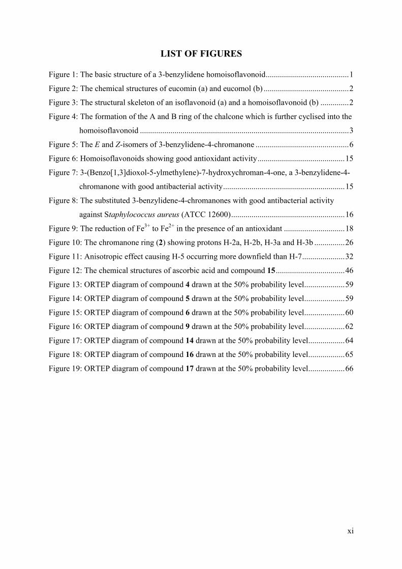
LIST OF FIGURES
Figure 1: The basic structure of a 3-benzylidene homoisoflavonoid ......................................... 1
Figure 2: The chemical structures of eucomin (a) and eucomol (b) .......................................... 2
Figure 3: The structural skeleton of an isoflavonoid (a) and a homoisoflavonoid (b) .............. 2
Figure 4: The formation of the A and B ring of the chalcone which is further cyclised into the
homoisoflavonoid ....................................................................................................... 3
Figure 5: The E and Z-isomers of 3-benzylidene-4-chromanone .............................................. 6
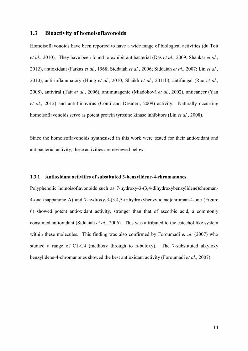
Figure 6: Homoisoflavonoids showing good antioxidant activity ........................................... 15
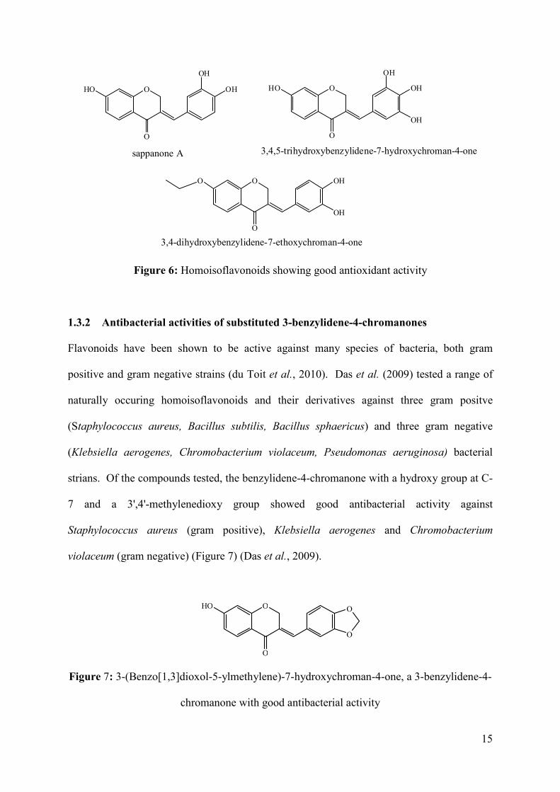
Figure 7: 3-(Benzo[1,3]dioxol-5-ylmethylene)-7-hydroxychroman-4-one, a 3-benzylidene-4-
chromanone with good antibacterial activity ............................................................ 15
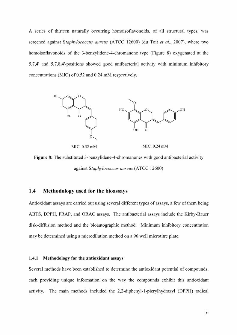
Figure 8: The substituted 3-benzylidene-4-chromanones with good antibacterial activity
against Staphylococcus aureus (ATCC 12600) ........................................................ 16
Figure 9: The reduction of Fe3+ to Fe2+ in the presence of an antioxidant .............................. 18
Figure 10: The chromanone ring (2) showing protons H-2a, H-2b, H-3a and H-3b ............... 26
Figure 11: Anisotropic effect causing H-5 occurring more downfield than H-7 ..................... 32
Figure 12: The chemical structures of ascorbic acid and compound 15 .................................. 46
Figure 13: ORTEP diagram of compound 4 drawn at the 50% probability level.................... 59
Figure 14: ORTEP diagram of compound 5 drawn at the 50% probability level.................... 59
Figure 15: ORTEP diagram of compound 6 drawn at the 50% probability level.................... 60
Figure 16: ORTEP diagram of compound 9 drawn at the 50% probability level.................... 62
Figure 17: ORTEP diagram of compound 14 drawn at the 50% probability level .................. 64
Figure 18: ORTEP diagram of compound 16 drawn at the 50% probability level .................. 65
Figure 19: ORTEP diagram of compound 17 drawn at the 50% probability level .................. 66
xii
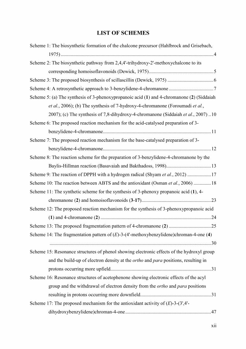
LIST OF SCHEMES
Scheme 1: The biosynthetic formation of the chalcone precursor (Hahlbrock and Grisebach,
1975) ........................................................................................................................... 4
Scheme 2: The biosynthetic pathway from 2,4,4'-trihydroxy-2'-methoxychalcone to its
corresponding homoisoflavonoids (Dewick, 1975) .................................................... 5
Scheme 3: The proposed biosynthesis of scillascillin (Dewick, 1975) ..................................... 6
Scheme 4: A retrosynthetic approach to 3-benzylidene-4-chromanone .................................... 7
Scheme 5: (a) The synthesis of 3-phenoxypropanoic acid (1) and 4-chromanone (2) (Siddaiah
et al., 2006); (b) The synthesis of 7-hydroxy-4-chromanone (Foroumadi et al.,
2007); (c) The synthesis of 7,8-dihydroxy-4-chromanone (Siddaiah et al., 2007) .. 10
Scheme 6: The proposed reaction mechanism for the acid-catalysed preparation of 3-
benzylidene-4-chromanone ....................................................................................... 11
Scheme 7: The proposed reaction mechanism for the base-catalysed preparation of 3-
benzylidene-4-chromanone ....................................................................................... 12
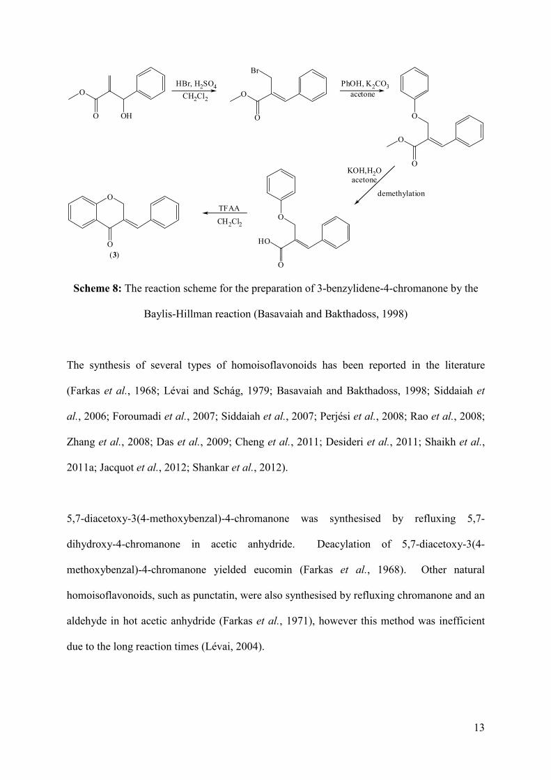
Scheme 8: The reaction scheme for the preparation of 3-benzylidene-4-chromanone by the
Baylis-Hillman reaction (Basavaiah and Bakthadoss, 1998).................................... 13
Scheme 9: The reaction of DPPH with a hydrogen radical (Shyam et al., 2012) ................... 17
Scheme 10: The reaction between ABTS and the antioxidant (Osman et al., 2006) .............. 18
Scheme 11: The synthetic scheme for the synthesis of 3-phenoxy propanoic acid (1), 4-
chromanone (2) and homoisoflavonoids (3-17)........................................................ 23
Scheme 12: The proposed reaction mechanism for the synthesis of 3-phenoxypropanoic acid
(1) and 4-chromanone (2) ......................................................................................... 24
Scheme 13: The proposed fragmentation pattern of 4-chromanone (2) .................................. 25
Scheme 14: The fragmentation pattern of (E)-3-(4'-methoxybenzylidene)chroman-4-one (4)
.................................................................................................................................. 30
Scheme 15: Resonance structures of phenol showing electronic effects of the hydroxyl group
and the build-up of electron density at the ortho and para positions, resulting in
protons occurring more upfield................................................................................. 31
Scheme 16: Resonance structures of acetophenone showing electronic effects of the acyl
group and the withdrawal of electron density from the ortho and para positions
resulting in protons occurring more downfield......................................................... 31
Scheme 17: The proposed mechanism for the antioxidant activity of (E)-3-(3',4'-
dihydroxybenzylidene)chroman-4-one ..................................................................... 47
xiii
Scheme 18: The preparation of 3-phenoxypropanoic acid (1) ................................................. 54
Scheme 19: The preparation of 4-chromanone (2) .................................................................. 55
Scheme 20: The preparation of homoisoflavonoids (3-17) ..................................................... 56
xiv
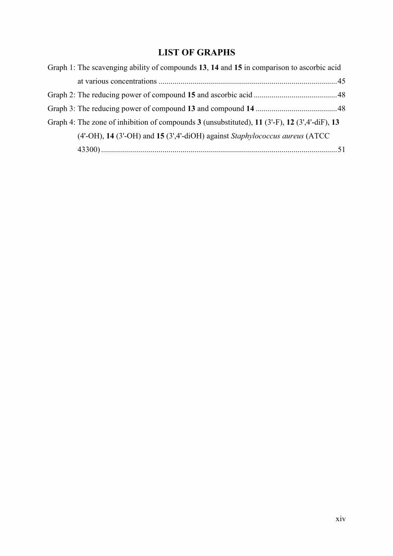
LIST OF GRAPHS
Graph 1: The scavenging ability of compounds 13, 14 and 15 in comparison to ascorbic acid
at various concentrations .......................................................................................... 45
Graph 2: The reducing power of compound 15 and ascorbic acid .......................................... 48
Graph 3: The reducing power of compound 13 and compound 14 ......................................... 48
Graph 4: The zone of inhibition of compounds 3 (unsubstituted), 11 (3'-F), 12 (3',4'-diF), 13
(4'-OH), 14 (3'-OH) and 15 (3',4'-diOH) against Staphylococcus aureus (ATCC
43300) ....................................................................................................................... 51
xv
ABSTRACT
Fifteen homoisoflavonoids (3-17) were synthesised using the base-catalysed aldol
condensation, thirteen of which were of the 3-benzylidene-4-chromanone type and the
remaining two of the 3-benzyl-4-chromanone type. The substitution patterns of the
homoisoflavonoids were varied by keeping the A-ring unsubstituted whilst changing the
substituent’s on the 3' and 4' positions of the B-ring. Methoxy, hydroxy, chloro, fluoro and
nitro groups were inserted on the B-ring of the homoisoflavonoids. All homoisoflavonoids
were characterised by NMR (1D and 2D), IR, UV spectroscopy and GC-MS. The crystal
structures were obtained for seven of the homoisoflavonoids. The homoisoflavonoids (3-17)
were tested for their antibacterial activity against ten gram-positive and six gram-negative
bacterial strains using the method of disc diffusion. Five compounds showed moderate
antibacterial activity whilst compound 14 showed good antibacterial activity against the gram
positive bacteria. The hydroxylated compounds were tested for their antioxidant activity
using the DPPH (2,2-diphenyl-1-picrylhydrazyl) radical scavenging method as well as the
FRAP (ferric reducing antioxidant power) method. Compound 15 showed good antioxidant
activity, comparable to that of ascorbic acid, due to the presence of a catechol system within
the molecule.
xvi
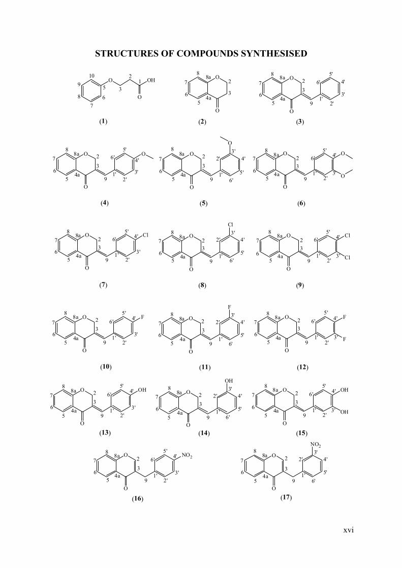
STRUCTURES OF COMPOUNDS SYNTHESISED
O OH
O
O
O
12
3
4a
5
6
7
8
8a9
102
3
5
6
7
8O
O
4a
8a2
3
5
6
7
8
91'
2'
3'
4'
5'
6'
O
O
4a
8a 2
3
5
6
7
8
91'
2'3'
4'
5'
6'O O
O
4a
8a 2
3
5
6
7
8
91'
2'3'
4'
5'
6'
O
O
O
4a
8a2
3
5
6
7
8
91'
2'3'
4'5'
6'O
O
O
O
4a
8a2
3
5
6
7
8
91'
2'
3'
4'5'
6'Cl O
O
4a
8a2
3
56
7
8
91'
2'3'
4'
5'
6'
Cl
O
O
4a
8a2
3
56
7
8
91'
2'3'
4'5'
6'Cl
Cl
O
O
4a
8a 2
3
5
6
7
8
91'
2'
3'
4'5'
6'F O
O
4a
8a 2
3
5
6
7
8
91'
2'3'
4'
5'
6'
F
O
O
4a
8a 2
3
5
6
7
8
91'
2'3'
4'5'
6'F
F
O
O
4a
8a 2
3
5
6
7
8
91'
2'
3'
4'5'
6'OH O
O
4a
8a 2
3
5
6
7
8
91'
2'3'
4'
5'
6'
OH
O
O
4a
8a2
3
5
6
7
8
91'
2'3'
4'5'
6'OH
OH
O
O
4a
8a 2
3
5
6
7
8
91'
2'
3'
4'5'
6'NO2 O
O
4a
8a 2
3
5
6
7
8
91'
2'3'
4'
5'
6'
NO2
(1) (3)(2)
(4) (6)(5)
(7) (9)(8)
(10) (12)(11)
(13) (15)(14)
(16) (17)

xvii
TABLE OF CONTENTS
Declaration 1- Plagiarism .......................................................................................................... v
Declaration 2- Publications ....................................................................................................... vi
Acknowledgements .................................................................................................................. vii
List of Abbreviations .............................................................................................................. viii
List of Tables ............................................................................................................................. x
List of Figures ........................................................................................................................... xi
List of Schemes ........................................................................................................................ xii
List of Graphs ......................................................................................................................... xiv
Abstract .................................................................................................................................... xv
Structures of compounds synthesised ..................................................................................... xvi
CHAPTER 1 INTRODUCTION ........................................................................................... 1
1.1 Classification, structure and biosynthesis of homoisoflavonoids .............................. 2
1.2 A review of the methods used to synthesise the 3-benzylidene-4 chromanones ....... 7
1.2.1 Synthesis of the 4-chromanone (2) intermediate ..................................................... 9
1.2.2 Synthesis of the 3-benzylidene-4-chromanones from the 4-chromanone intermediate...................................................................................................................... 10
1.2.3 Other methods used for the synthesis of 3-benzylidene-4-chromanones ............. 12
1.3 Bioactivity of homoisoflavonoids ............................................................................ 14
1.3.1 Antioxidant activities of substituted 3-benzylidene-4-chromanones .................... 14
1.3.2 Antibacterial activities of substituted 3-benzylidene-4-chromanones ................... 15
1.4 Methodology used for the bioassays ........................................................................ 16
1.4.1 Methodology for the antioxidant assays ................................................................ 16
1.4.1.1 DPPH radical scavenging assay ................................................................ 17
1.4.1.2 Ferric reducing antioxidant power assay ................................................... 17
1.4.1.3 ABTS assay ................................................................................................ 18
1.4.2 Methodology for the antibacterial assays .............................................................. 19
1.4.2.1 Kirby-Bauer disk-diffusion method............................................................ 19
1.4.2.2 Bioautographic methods ............................................................................. 19
1.4.2.3 Method of dilution ...................................................................................... 20
1.5 Hypothesis, aims and objectives .............................................................................. 20
CHAPTER 2 RESULTS AND DISCUSSION .................................................................... 22
xviii
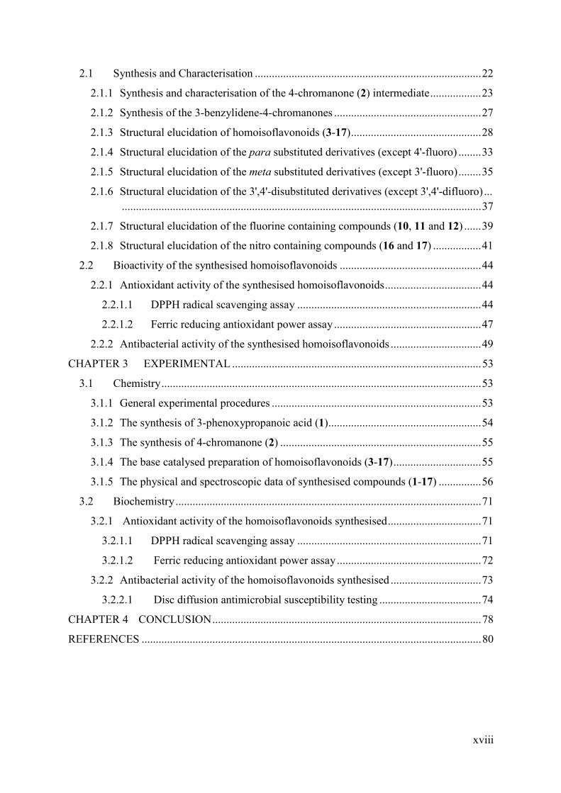
2.1 Synthesis and Characterisation ................................................................................ 22
2.1.1 Synthesis and characterisation of the 4-chromanone (2) intermediate .................. 23
2.1.2 Synthesis of the 3-benzylidene-4-chromanones .................................................... 27
2.1.3 Structural elucidation of homoisoflavonoids (3-17) .............................................. 28
2.1.4 Structural elucidation of the para substituted derivatives (except 4'-fluoro) ........ 33
2.1.5 Structural elucidation of the meta substituted derivatives (except 3'-fluoro) ........ 35
2.1.6 Structural elucidation of the 3',4'-disubstituted derivatives (except 3',4'-difluoro) ... ............................................................................................................................... 37
2.1.7 Structural elucidation of the fluorine containing compounds (10, 11 and 12) ...... 39
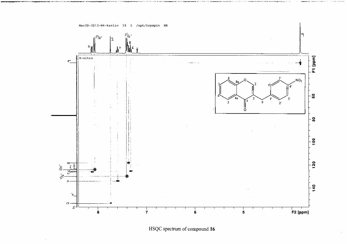
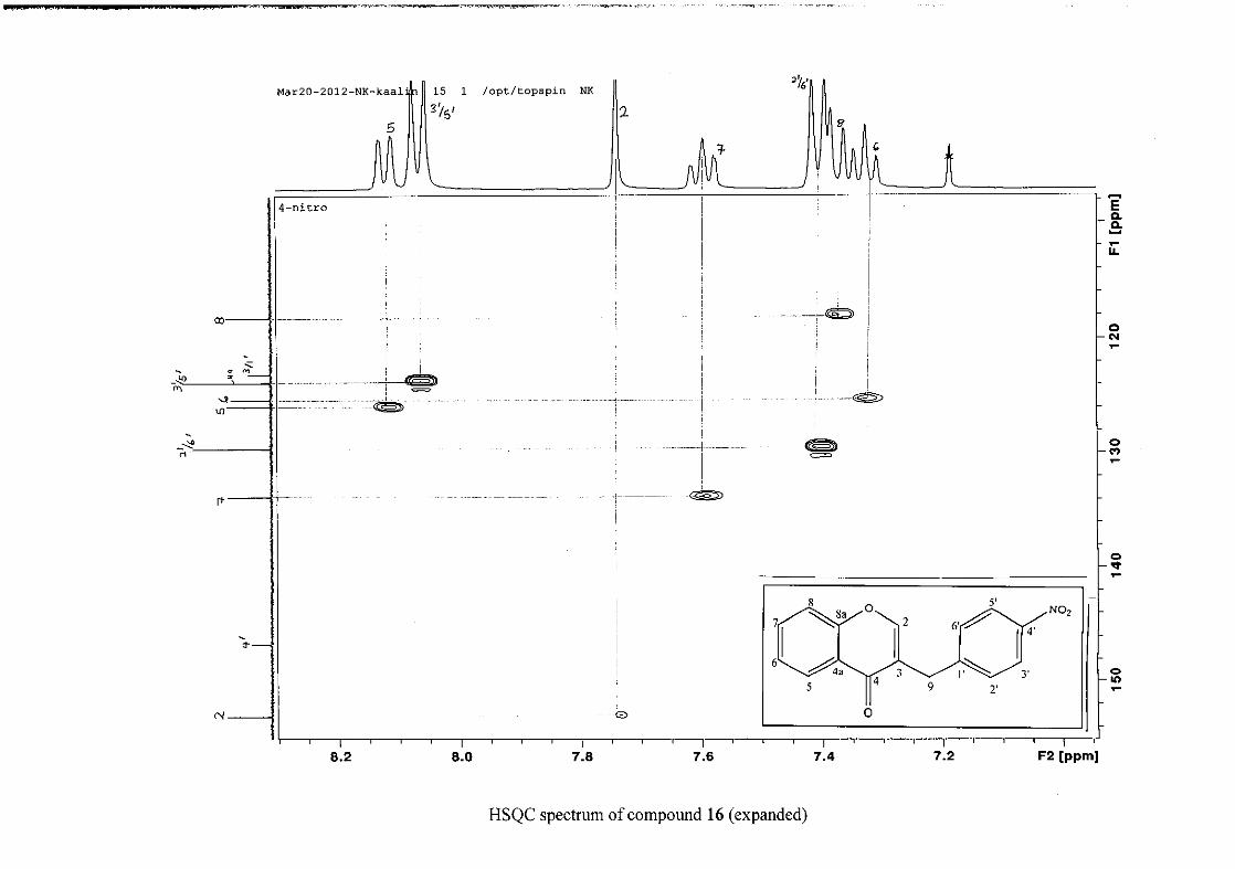
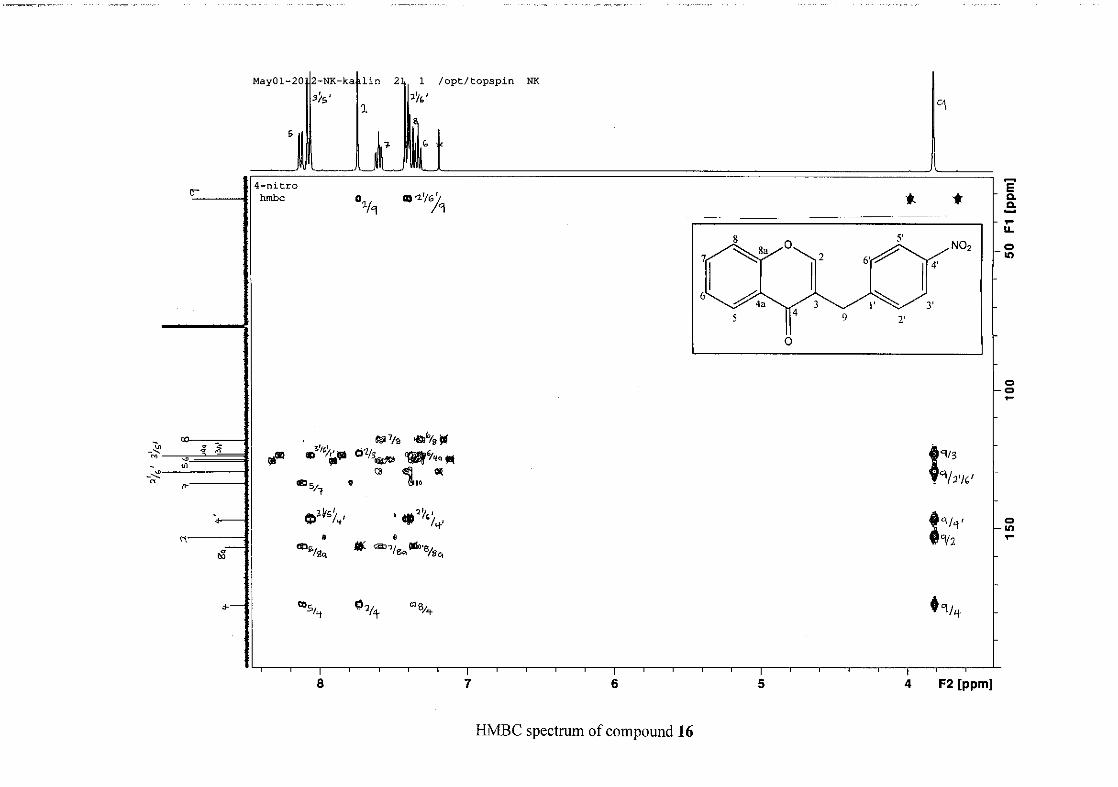
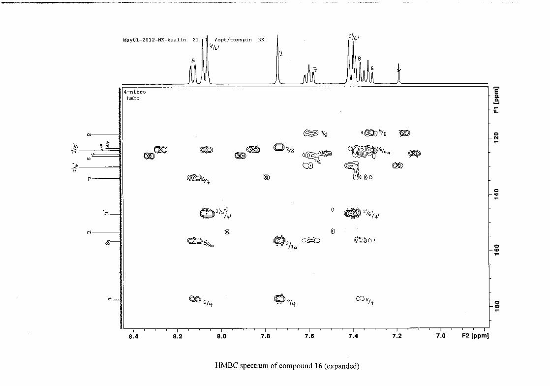
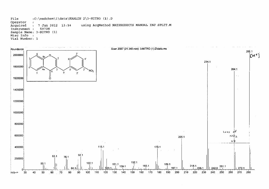
2.1.8 Structural elucidation of the nitro containing compounds (16 and 17) ................. 41
2.2 Bioactivity of the synthesised homoisoflavonoids .................................................. 44
2.2.1 Antioxidant activity of the synthesised homoisoflavonoids .................................. 44
2.2.1.1 DPPH radical scavenging assay ................................................................. 44
2.2.1.2 Ferric reducing antioxidant power assay .................................................... 47
2.2.2 Antibacterial activity of the synthesised homoisoflavonoids ................................ 49
CHAPTER 3 EXPERIMENTAL ........................................................................................ 53
3.1 Chemistry ................................................................................................................. 53
3.1.1 General experimental procedures .......................................................................... 53
3.1.2 The synthesis of 3-phenoxypropanoic acid (1) ...................................................... 54
3.1.3 The synthesis of 4-chromanone (2) ....................................................................... 55
3.1.4 The base catalysed preparation of homoisoflavonoids (3-17) ............................... 55
3.1.5 The physical and spectroscopic data of synthesised compounds (1-17) ............... 56
3.2 Biochemistry ............................................................................................................ 71
3.2.1 Antioxidant activity of the homoisoflavonoids synthesised ................................. 71
3.2.1.1 DPPH radical scavenging assay ................................................................. 71
3.2.1.2 Ferric reducing antioxidant power assay ................................................... 72
3.2.2 Antibacterial activity of the homoisoflavonoids synthesised ................................ 73
3.2.2.1 Disc diffusion antimicrobial susceptibility testing .................................... 74
CHAPTER 4 CONCLUSION ............................................................................................... 78
REFERENCES ........................................................................................................................ 80
1
CHAPTER 1 INTRODUCTION
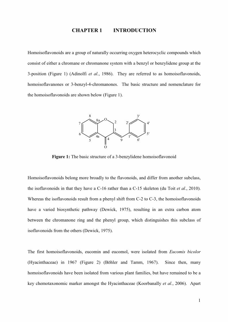
Homoisoflavonoids are a group of naturally occurring oxygen heterocyclic compounds which
consist of either a chromane or chromanone system with a benzyl or benzylidene group at the
3-position (Figure 1) (Adinolfi et al., 1986). They are referred to as homoisoflavonoids,
homoisoflavanones or 3-benzyl-4-chromanones. The basic structure and nomenclature for
the homoisoflavonoids are shown below (Figure 1).
O
O
2
3
44a
5
6
7
88a
91'
2'
3'
4'
5'
6'
Figure 1: The basic structure of a 3-benzylidene homoisoflavonoid
Homoisoflavonoids belong more broadly to the flavonoids, and differ from another subclass,
the isoflavonoids in that they have a C-16 rather than a C-15 skeleton (du Toit et al., 2010).
Whereas the isoflavonoids result from a phenyl shift from C-2 to C-3, the homoisoflavonoids
have a varied biosynthetic pathway (Dewick, 1975), resulting in an extra carbon atom
between the chromanone ring and the phenyl group, which distinguishes this subclass of
isoflavonoids from the others (Dewick, 1975).
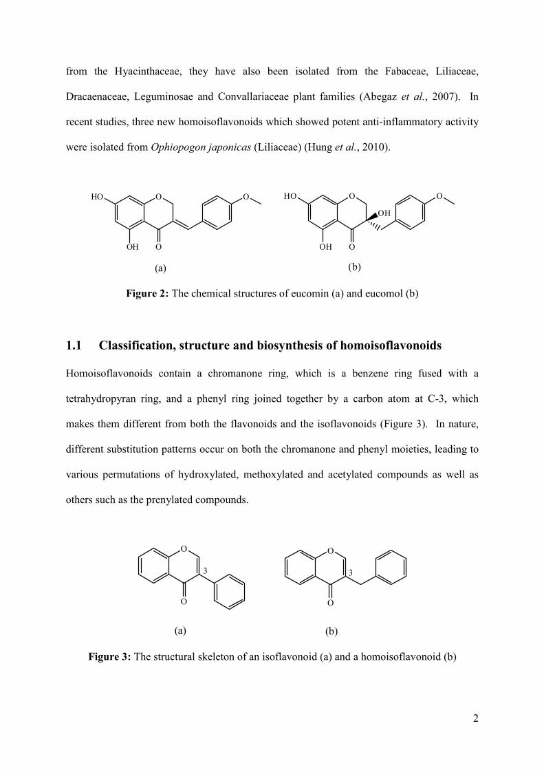
The first homoisoflavonoids, eucomin and eucomol, were isolated from Eucomis bicolor
(Hyacinthaceae) in 1967 (Figure 2) (Böhler and Tamm, 1967). Since then, many
homoisoflavonoids have been isolated from various plant families, but have remained to be a
key chemotaxonomic marker amongst the Hyacinthaceae (Koorbanally et al., 2006). Apart
2
from the Hyacinthaceae, they have also been isolated from the Fabaceae, Liliaceae,
Dracaenaceae, Leguminosae and Convallariaceae plant families (Abegaz et al., 2007). In
recent studies, three new homoisoflavonoids which showed potent anti-inflammatory activity
were isolated from Ophiopogon japonicas (Liliaceae) (Hung et al., 2010).
O
O
O
O
HO
OH
O
OH
HO
OH
O
(a) (b)
Figure 2: The chemical structures of eucomin (a) and eucomol (b)
1.1 Classification, structure and biosynthesis of homoisoflavonoids
Homoisoflavonoids contain a chromanone ring, which is a benzene ring fused with a
tetrahydropyran ring, and a phenyl ring joined together by a carbon atom at C-3, which
makes them different from both the flavonoids and the isoflavonoids (Figure 3). In nature,
different substitution patterns occur on both the chromanone and phenyl moieties, leading to
various permutations of hydroxylated, methoxylated and acetylated compounds as well as
others such as the prenylated compounds.
OO
O O
(a) (b)
33
Figure 3: The structural skeleton of an isoflavonoid (a) and a homoisoflavonoid (b)
3
Homoisoflavonoids are biosynthesised from chalcone precursors. The mechanism and
biosynthetic pathways by which homoisoflavonoids are formed were determined by labeling
studies with phenylalanine, sodium acetate and methionine in Eucomis comosum, where
labeled precursors were incorporated into the chalcone intermediates and further into the
homoisoflavonoid (Dewick, 1975).
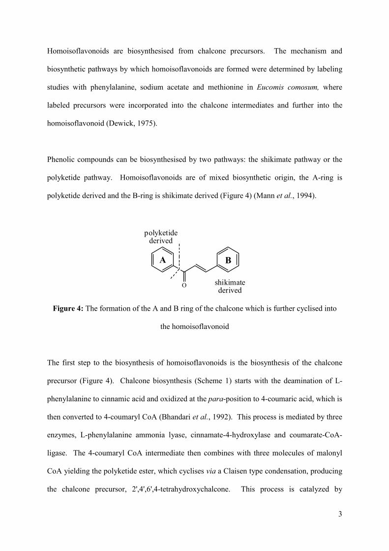
Phenolic compounds can be biosynthesised by two pathways: the shikimate pathway or the
polyketide pathway. Homoisoflavonoids are of mixed biosynthetic origin, the A-ring is
polyketide derived and the B-ring is shikimate derived (Figure 4) (Mann et al., 1994).
O
A B
polyketidederived
shikimatederived
Figure 4: The formation of the A and B ring of the chalcone which is further cyclised into
the homoisoflavonoid
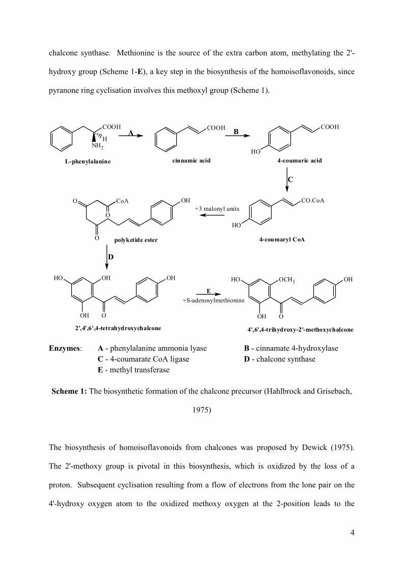
The first step to the biosynthesis of homoisoflavonoids is the biosynthesis of the chalcone
precursor (Figure 4). Chalcone biosynthesis (Scheme 1) starts with the deamination of L-
phenylalanine to cinnamic acid and oxidized at the para-position to 4-coumaric acid, which is
then converted to 4-coumaryl CoA (Bhandari et al., 1992). This process is mediated by three
enzymes, L-phenylalanine ammonia lyase, cinnamate-4-hydroxylase and coumarate-CoA-
ligase. The 4-coumaryl CoA intermediate then combines with three molecules of malonyl
CoA yielding the polyketide ester, which cyclises via a Claisen type condensation, producing
the chalcone precursor, 2',4',6',4-tetrahydroxychalcone. This process is catalyzed by
4
chalcone synthase. Methionine is the source of the extra carbon atom, methylating the 2'-
hydroxy group (Scheme 1-E), a key step in the biosynthesis of the homoisoflavonoids, since
pyranone ring cyclisation involves this methoxyl group (Scheme 1).
COOH
NH2
H
COOH COOH
HO
CO.CoA
HO
OH
O
CoAO
OH
OH
OHHO
O
OH
OH
OCH3HO
O
L-phenylalanine cinnamic acid 4-coumaric acid
A B
O
4-coumaryl CoApolyketide ester
C
D
E
2',4',6',4-tetrahydroxychalcone 4',6',4-trihydroxy-2'-methoxychalcone
+3 malonyl units
+S-adenosylmethionine
Enzymes: A - phenylalanine ammonia lyase B - cinnamate 4-hydroxylase C - 4-coumarate CoA ligase D - chalcone synthase E - methyl transferase
Scheme 1: The biosynthetic formation of the chalcone precursor (Hahlbrock and Grisebach,
1975)
The biosynthesis of homoisoflavonoids from chalcones was proposed by Dewick (1975).
The 2'-methoxy group is pivotal in this biosynthesis, which is oxidized by the loss of a
proton. Subsequent cyclisation resulting from a flow of electrons from the lone pair on the
4'-hydroxy oxygen atom to the oxidized methoxy oxygen at the 2-position leads to the
5
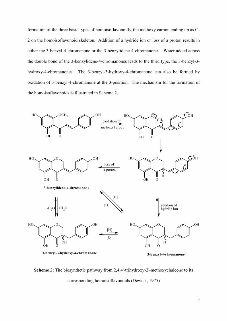
formation of the three basic types of homoisoflavonoids, the methoxy carbon ending up as C-
2 on the homoisoflavonoid skeleton. Addition of a hydride ion or loss of a proton results in
either the 3-benzyl-4-chromanone or the 3-benzylidene-4-chromanones. Water added across
the double bond of the 3-benzylidene-4-chromanones leads to the third type, the 3-benzyl-3-
hydroxy-4-chromanones. The 3-benzyl-3-hydroxy-4-chromanone can also be formed by
oxidation of 3-benzyl-4-chromanone at the 3-position. The mechanism for the formation of
the homoisoflavonoids is illustrated in Scheme 2.
OCH3HO
OH
OH
O
OHO
OH
OH
O
CH2
OHO
OH
OH
OH
OHO
OH
OH
O
OHO
OH
OH
OH
OHO
OH
OH
OOH
oxidation of
methoxyl group
loss of
a proton
-H2O +H2O
[H]
[O]
addition ofhydride ion
[H]
[O]
3-benzylidene-4-chromanone
3-benzyl-4-chromanone3-benzyl-3-hydroxy-4-chromanone
Scheme 2: The biosynthetic pathway from 2,4,4'-trihydroxy-2'-methoxychalcone to its
corresponding homoisoflavonoids (Dewick, 1975)
6
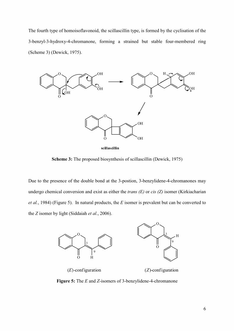
The fourth type of homoisoflavonoid, the scillascillin type, is formed by the cyclisation of the
3-benzyl-3-hydroxy-4-chromanone, forming a strained but stable four-membered ring
(Scheme 3) (Dewick, 1975).
O
O
OH
OHOH
O
O
OH
OH
H
O
O
OH
OH
scillascillin
Scheme 3: The proposed biosynthesis of scillascillin (Dewick, 1975)
Due to the presence of the double bond at the 3-postion, 3-benzylidene-4-chromanones may
undergo chemical conversion and exist as either the trans (E) or cis (Z) isomer (Kirkiacharian
et al., 1984) (Figure 5). In natural products, the E isomer is prevalent but can be converted to
the Z isomer by light (Siddaiah et al., 2006).
O
O
O
O
3
93
9
H
(E)-conf iguration (Z)-conf iguration
H
Figure 5: The E and Z-isomers of 3-benzylidene-4-chromanone
7
1.2 A review of the methods used to synthesise the 3-benzylidene-4
chromanones
Homoisoflavonoids have been synthesised since the mid twentieth century (Farkas et al.,
1968). The first synthesis of homoisoflavonoids was completed just a year after these
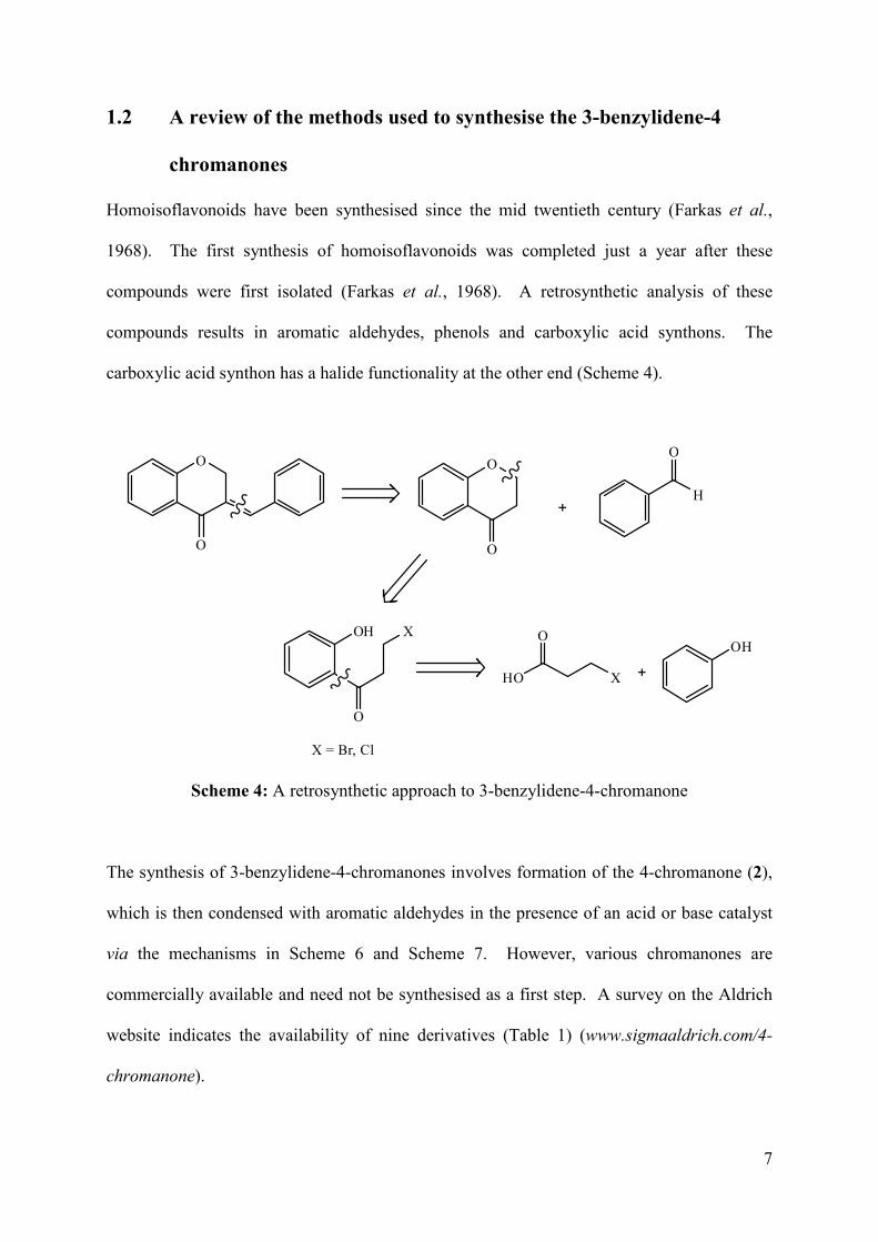
compounds were first isolated (Farkas et al., 1968). A retrosynthetic analysis of these
compounds results in aromatic aldehydes, phenols and carboxylic acid synthons. The
carboxylic acid synthon has a halide functionality at the other end (Scheme 4).
HO X
O
O
O
O
O
H
O
OH
O
X
X = Br, Cl
OH
Scheme 4: A retrosynthetic approach to 3-benzylidene-4-chromanone
The synthesis of 3-benzylidene-4-chromanones involves formation of the 4-chromanone (2),
which is then condensed with aromatic aldehydes in the presence of an acid or base catalyst
via the mechanisms in Scheme 6 and Scheme 7. However, various chromanones are
commercially available and need not be synthesised as a first step. A survey on the Aldrich
website indicates the availability of nine derivatives (Table 1) (www.sigmaaldrich.com/4-
chromanone).
8
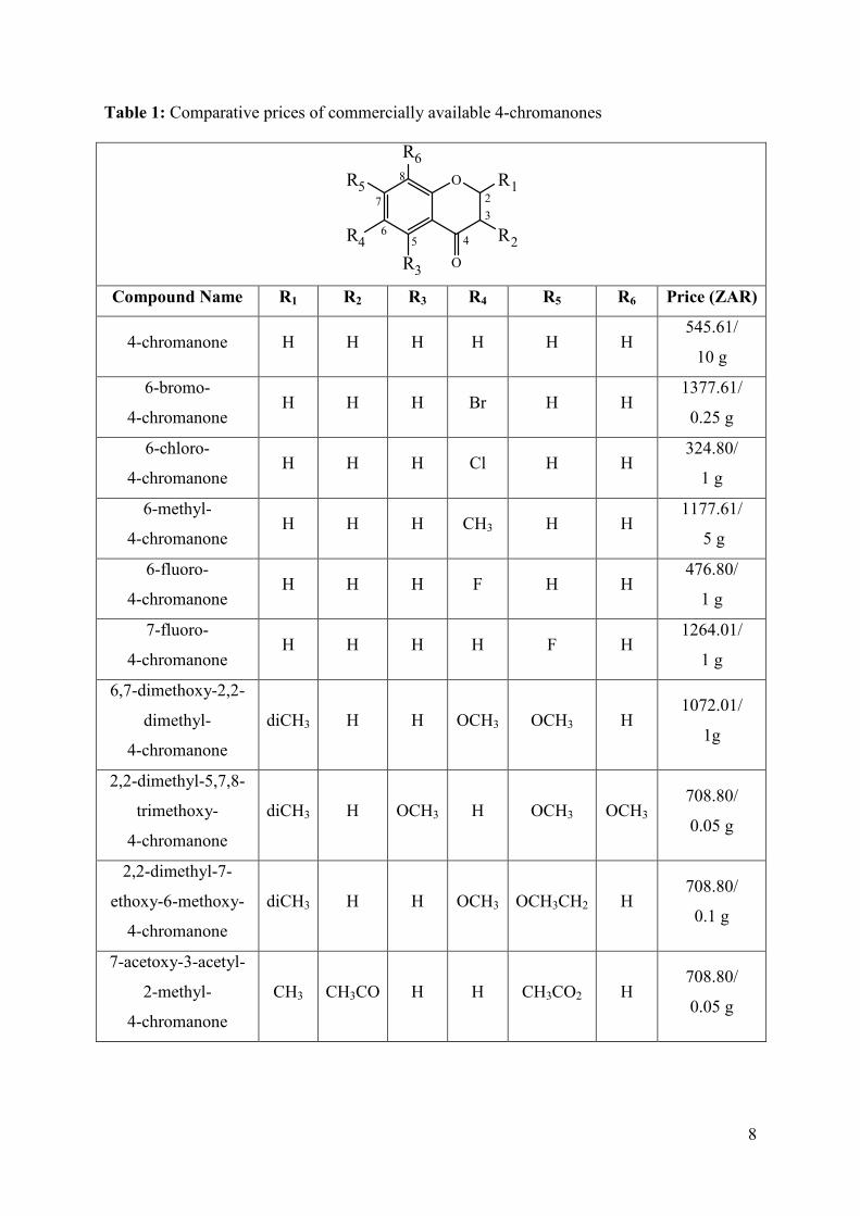
Table 1: Comparative prices of commercially available 4-chromanones
O
O
R1
R2
R3
R4
R5
R6
2
3
56
7
8
4
Compound Name R1 R2 R3 R4 R5 R6 Price (ZAR)
4-chromanone H H H H H H 545.61/
10 g
6-bromo-
4-chromanone H H H Br H H
1377.61/
0.25 g
6-chloro-
4-chromanone H H H Cl H H
324.80/
1 g
6-methyl-
4-chromanone H H H CH3 H H
1177.61/
5 g
6-fluoro-
4-chromanone H H H F H H
476.80/
1 g
7-fluoro-
4-chromanone H H H H F H
1264.01/
1 g
6,7-dimethoxy-2,2-
dimethyl-
4-chromanone
diCH3 H H OCH3 OCH3 H 1072.01/
1g
2,2-dimethyl-5,7,8-
trimethoxy-
4-chromanone
diCH3 H OCH3 H OCH3 OCH3 708.80/
0.05 g
2,2-dimethyl-7-
ethoxy-6-methoxy-
4-chromanone
diCH3 H H OCH3 OCH3CH2 H 708.80/
0.1 g
7-acetoxy-3-acetyl-
2-methyl-
4-chromanone
CH3 CH3CO H H CH3CO2 H 708.80/
0.05 g
9
Several of the chromanone derivatives that are commercially available are relatively
expensive, compared to the unsubstituted chromanone therefore derivatisation of the
chromanone may be a better alternative to purchasing the derivatives.
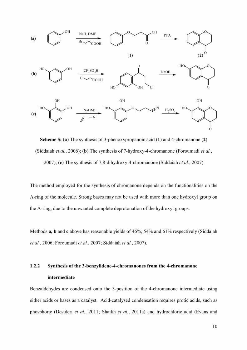
1.2.1 Synthesis of the 4-chromanone (2) intermediate
Even though the 4-chromanone intermediates are available, researchers in the field still
synthesise the intermediate en route to the homoisoflavonoids. Different methods were
employed for their synthesis (Scheme 5) (Siddaiah et al., 2006; Foroumadi et al., 2007;
Siddaiah et al., 2007; Shaikh et al., 2011a).
They can be formed from the reaction of:
(a) 3-bromo- or 3-chloropropanoic acids and phenols under basic conditions, producing a
phenoxypropanoic acid which can be cyclised with polyphosphoric acid (Siddaiah et
al., 2006; Shaikh et al., 2011a);
(b) 3-bromo- or 3-chloropropanoic acids and phenols under acidic conditions, producing
a benzophenone alkyl chloride which can be cyclised with sodium hydroxide
(Foroumadi et al., 2007);
(c) acrylonitrile and phenols under basic conditions forming a phenoxynitrile which is
followed by the cyclisation with sulfuric acid (Siddaiah et al., 2007).
In all cases, an activated carbon is produced in the intermediate. In the case of the acid and
nitrile intermediates, an acid is used as a catalyst for the cyclisation by activitating the
carboxyl or nitrile groups toward nucleophilic substitution and for the alkyl chloride, a base is
needed for the abstraction of the proton of the hydroxyl group, which is then followed by
nucleophilic substitution.
10
OH
OH
HO O
OH
HO N O
OH
HO
O
OH O OH
O
NaH, DMF
BrCOOH
PPAO
O
O
O
OHHO
HO OH
O
Cl
HO
NaOMe
N
H2SO4
Cl COOH
CF3SO3H NaOH
(a)
(b)
(c)
(1) (2)
Scheme 5: (a) The synthesis of 3-phenoxypropanoic acid (1) and 4-chromanone (2)
(Siddaiah et al., 2006); (b) The synthesis of 7-hydroxy-4-chromanone (Foroumadi et al.,
2007); (c) The synthesis of 7,8-dihydroxy-4-chromanone (Siddaiah et al., 2007)
The method employed for the synthesis of chromanone depends on the functionalities on the
A-ring of the molecule. Strong bases may not be used with more than one hydroxyl group on
the A-ring, due to the unwanted complete deprotonation of the hydroxyl groups.
Methods a, b and c above has reasonable yields of 46%, 54% and 61% respectively (Siddaiah
et al., 2006; Foroumadi et al., 2007; Siddaiah et al., 2007).
1.2.2 Synthesis of the 3-benzylidene-4-chromanones from the 4-chromanone
intermediate
Benzaldehydes are condensed onto the 3-position of the 4-chromanone intermediate using
either acids or bases as a catalyst. Acid-catalysed condensation requires protic acids, such as
phosphoric (Desideri et al., 2011; Shaikh et al., 2011a) and hydrochloric acid (Evans and
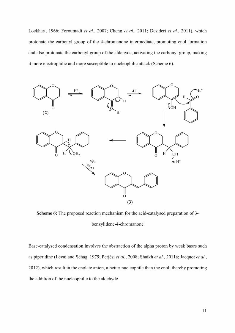
11
Lockhart, 1966; Foroumadi et al., 2007; Cheng et al., 2011; Desideri et al., 2011), which
protonate the carbonyl group of the 4-chromanone intermediate, promoting enol formation
and also protonate the carbonyl group of the aldehyde, activating the carbonyl group, making
it more electrophilic and more susceptible to nucleophilic attack (Scheme 6).
O
O
O
O
O
OH
H
H
O
O H OH
H+
O
O H OH2
H
O
O
(2)
(3)
-H+
H O
-H +-H
2 O
H+H+
Scheme 6: The proposed reaction mechanism for the acid-catalysed preparation of 3-
benzylidene-4-chromanone
Base-catalysed condensation involves the abstraction of the alpha proton by weak bases such
as piperidine (Lévai and Schág, 1979; Perjési et al., 2008; Shaikh et al., 2011a; Jacquot et al.,
2012), which result in the enolate anion, a better nucleophile than the enol, thereby promoting
the addition of the nucleophille to the aldehyde.
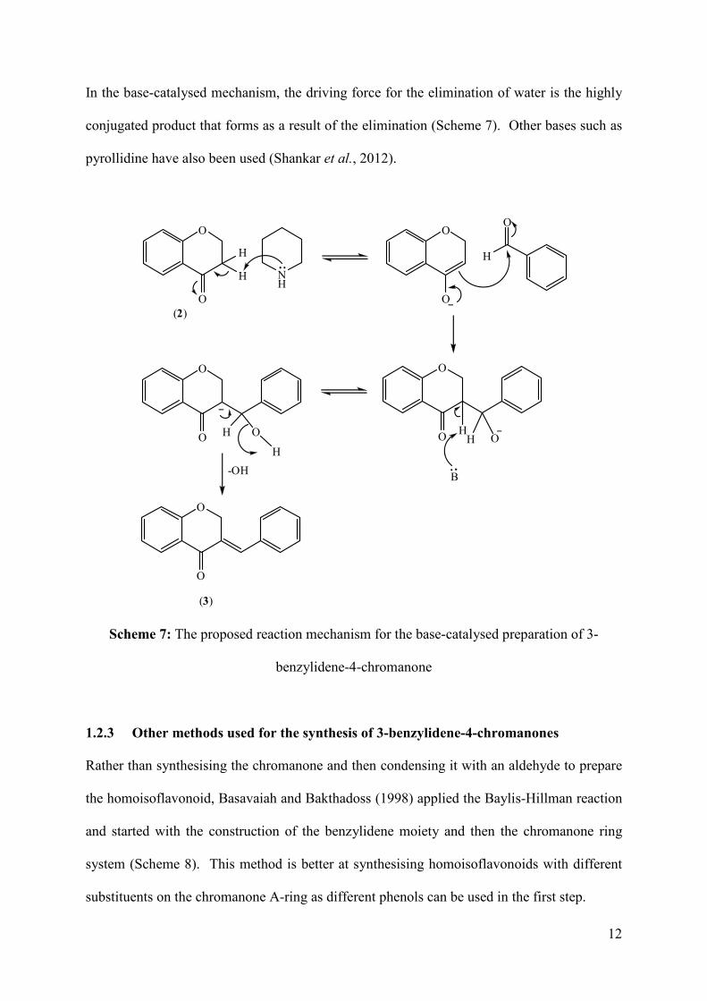
12
In the base-catalysed mechanism, the driving force for the elimination of water is the highly
conjugated product that forms as a result of the elimination (Scheme 7). Other bases such as
pyrollidine have also been used (Shankar et al., 2012).
O
O
O
O
H
O
O
O H O
O
O H O
O
O
(3)
NH
H
H
(2)
-OH
H
H
B
Scheme 7: The proposed reaction mechanism for the base-catalysed preparation of 3-
benzylidene-4-chromanone
1.2.3 Other methods used for the synthesis of 3-benzylidene-4-chromanones
Rather than synthesising the chromanone and then condensing it with an aldehyde to prepare
the homoisoflavonoid, Basavaiah and Bakthadoss (1998) applied the Baylis-Hillman reaction
and started with the construction of the benzylidene moiety and then the chromanone ring
system (Scheme 8). This method is better at synthesising homoisoflavonoids with different
substituents on the chromanone A-ring as different phenols can be used in the first step.
13
HBr, H2SO4
CH2Cl2
PhOH, K2CO3acetoneO
O OH
O
O
Br
O
O
O
demethylation
KOH,H2Oacetone
O
HO
O
O
O
TFAA
CH2Cl2
(3)
Scheme 8: The reaction scheme for the preparation of 3-benzylidene-4-chromanone by the
Baylis-Hillman reaction (Basavaiah and Bakthadoss, 1998)
The synthesis of several types of homoisoflavonoids has been reported in the literature
(Farkas et al., 1968; Lévai and Schág, 1979; Basavaiah and Bakthadoss, 1998; Siddaiah et
al., 2006; Foroumadi et al., 2007; Siddaiah et al., 2007; Perjési et al., 2008; Rao et al., 2008;
Zhang et al., 2008; Das et al., 2009; Cheng et al., 2011; Desideri et al., 2011; Shaikh et al.,
2011a; Jacquot et al., 2012; Shankar et al., 2012).
5,7-diacetoxy-3(4-methoxybenzal)-4-chromanone was synthesised by refluxing 5,7-
dihydroxy-4-chromanone in acetic anhydride. Deacylation of 5,7-diacetoxy-3(4-
methoxybenzal)-4-chromanone yielded eucomin (Farkas et al., 1968). Other natural
homoisoflavonoids, such as punctatin, were also synthesised by refluxing chromanone and an
aldehyde in hot acetic anhydride (Farkas et al., 1971), however this method was inefficient
due to the long reaction times (Lévai, 2004).
14
1.3 Bioactivity of homoisoflavonoids
Homoisoflavonoids have been reported to have a wide range of biological activities (du Toit
et al., 2010). They have been found to exhibit antibacterial (Das et al., 2009; Shankar et al.,
2012), antioxidant (Farkas et al., 1968; Siddaiah et al., 2006; Siddaiah et al., 2007; Lin et al.,
2010), anti-inflammatory (Hung et al., 2010; Shaikh et al., 2011b), antifungal (Rao et al.,
2008), antiviral (Tait et al., 2006), antimutagenic (Miadoková et al., 2002), anticancer (Yan
et al., 2012) and antirhinovirus (Conti and Desideri, 2009) activity. Naturally occurring
homoisoflavonoids serve as potent protein tyrosine kinase inhibitors (Lin et al., 2008).
Since the homoisoflavonoids synthesised in this work were tested for their antioxidant and
antibacterial activity, these activities are reviewed below.
1.3.1 Antioxidant activities of substituted 3-benzylidene-4-chromanones
Polyphenolic homoisoflavonoids such as 7-hydroxy-3-(3,4-dihydroxybenzylidene)chroman-
4-one (sappanone A) and 7-hydroxy-3-(3,4,5-trihydroxybenzylidene)chroman-4-one (Figure
6) showed potent antioxidant activity, stronger than that of ascorbic acid, a commonly
consumed antioxidant (Siddaiah et al., 2006). This was attributed to the catechol like system
within these molecules. This finding was also confirmed by Foroumadi et al. (2007) who
studied a range of C1-C4 (methoxy through to n-butoxy). The 7-substituted alkyloxy
benzylidene-4-chromanones showed the best antioxidant activity (Foroumadi et al., 2007).
15
O
O
HO
OH
OH
OH
O
O
HO OH
OH
sappanone A 3,4,5-trihydroxybenzylidene-7-hydroxychroman-4-one
O
O
O
OH
OH
3,4-dihydroxybenzylidene-7-ethoxychroman-4-one
Figure 6: Homoisoflavonoids showing good antioxidant activity
1.3.2 Antibacterial activities of substituted 3-benzylidene-4-chromanones
Flavonoids have been shown to be active against many species of bacteria, both gram
positive and gram negative strains (du Toit et al., 2010). Das et al. (2009) tested a range of
naturally occuring homoisoflavonoids and their derivatives against three gram positve
(Staphylococcus aureus, Bacillus subtilis, Bacillus sphaericus) and three gram negative
(Klebsiella aerogenes, Chromobacterium violaceum, Pseudomonas aeruginosa) bacterial
strians. Of the compounds tested, the benzylidene-4-chromanone with a hydroxy group at C-
7 and a 3',4'-methylenedioxy group showed good antibacterial activity against
Staphylococcus aureus (gram positive), Klebsiella aerogenes and Chromobacterium
violaceum (gram negative) (Figure 7) (Das et al., 2009).
O
O
HO
O
O
Figure 7: 3-(Benzo[1,3]dioxol-5-ylmethylene)-7-hydroxychroman-4-one, a 3-benzylidene-4-
chromanone with good antibacterial activity
16
A series of thirteen naturally occurring homoisoflavonoids, of all structural types, was
screened against Staphylococcus aureus (ATCC 12600) (du Toit et al., 2007), where two
homoisoflavonoids of the 3-benzylidene-4-chromanone type (Figure 8) oxygenated at the
5,7,4' and 5,7,8,4'-positions showed good antibacterial activity with minimum inhibitory
concentrations (MIC) of 0.52 and 0.24 mM respectively.
OHO
OH O
O
O
O
HO
OH O
OH
MIC: 0.52 mM MIC: 0.24 mM
Figure 8: The substituted 3-benzylidene-4-chromanones with good antibacterial activity
against Staphylococcus aureus (ATCC 12600)
1.4 Methodology used for the bioassays
Antioxidant assays are carried out using several different types of assays, a few of them being
ABTS, DPPH, FRAP, and ORAC assays. The antibacterial assays include the Kirby-Bauer
disk-diffusion method and the bioautographic method. Minimum inhibitory concentration
may be determined using a microdilution method on a 96 well microtitre plate.
1.4.1 Methodology for the antioxidant assays
Several methods have been established to determine the antioxidant potential of compounds,
each providing unique information on the way the compounds exhibit this antioxidant
activity. The main methods included the 2,2-diphenyl-1-picrylhydrazyl (DPPH) radical
17
scavenging, ferric reducing antioxidant power (FRAP) and the 2,2'-azino-bis(3-
ethylbenzothiazoline-6-sulfonic acid) ABTS method (Thaipong et al., 2006).
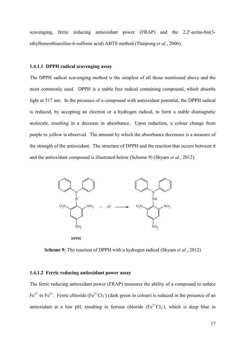
1.4.1.1 DPPH radical scavenging assay
The DPPH radical scavenging method is the simplest of all those mentioned above and the
most commonly used. DPPH is a stable free radical containing compound, which absorbs
light at 517 nm. In the presence of a compound with antioxidant potential, the DPPH radical
is reduced, by accepting an electron or a hydrogen radical, to form a stable diamagnetic
molecule, resulting in a decrease in absorbance. Upon reduction, a colour change from
purple to yellow is observed. The amount by which the absorbance decreases is a measure of
the strength of the antioxidant. The structure of DPPH and the reaction that occurs between it
and the antioxidant compound is illustrated below (Scheme 9) (Shyam et al., 2012).
N
N
O2N NO2
NO2
+ H
N
NH
O2N NO2
NO2
DPPH
Scheme 9: The reaction of DPPH with a hydrogen radical (Shyam et al., 2012)
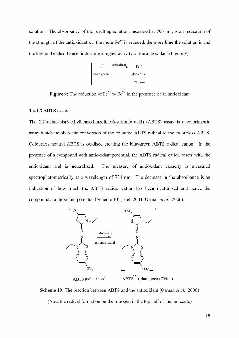
1.4.1.2 Ferric reducing antioxidant power assay
The ferric reducing antioxidant power (FRAP) measures the ability of a compound to reduce
Fe3+ to Fe2+. Ferric chloride (Fe3+Cl3-) (dark green in colour) is reduced in the presence of an
antioxidant at a low pH, resulting in ferrous chloride (Fe2+Cl2-), which is deep blue in
18
solution. The absorbance of the resulting solution, measured at 700 nm, is an indication of
the strength of the antioxidant i.e. the more Fe3+ is reduced, the more blue the solution is and
the higher the absorbance, indicating a higher activity of the antioxidant (Figure 9).
Fe3+ Fe2+
dark green deep blue
700 nm
antioxidant
Figure 9: The reduction of Fe3+ to Fe2+ in the presence of an antioxidant
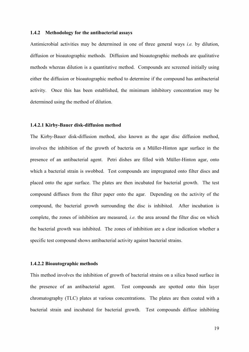
1.4.1.3 ABTS assay
The 2,2'-azino-bis(3-ethylbenzothiazoline-6-sulfonic acid) (ABTS) assay is a colorimetric
assay which involves the conversion of the coloured ABTS radical to the colourless ABTS.
Colourless neutral ABTS is oxidised creating the blue-green ABTS radical cation. In the
presence of a compound with antioxidant potential, the ABTS radical cation reacts with the
antioxidant and is neutralized. The measure of antioxidant capacity is measured
spectrophotometrically at a wavelength of 734 nm. The decrease in the absorbance is an
indication of how much the ABTS radical cation has been neutralised and hence the
compounds’ antioxidant potential (Scheme 10) (Erel, 2004; Osman et al., 2006).
SN
N
SO3-
N
NS
-O3S
SN
N
SO3-
N
NS
-O3S
ABTS (colourless) ABTS (blue-green) 734nm
oxidant
antioxidant
Scheme 10: The reaction between ABTS and the antioxidant (Osman et al., 2006)
(Note the radical formation on the nitrogen in the top half of the molecule)
19
1.4.2 Methodology for the antibacterial assays
Antimicrobial activities may be determined in one of three general ways i.e. by dilution,
diffusion or bioautographic methods. Diffusion and bioautographic methods are qualitative
methods whereas dilution is a quantitative method. Compounds are screened initially using
either the diffusion or bioautographic method to determine if the compound has antibacterial
activity. Once this has been established, the minimum inhibitory concentration may be
determined using the method of dilution.
1.4.2.1 Kirby-Bauer disk-diffusion method
The Kirby-Bauer disk-diffusion method, also known as the agar disc diffusion method,
involves the inhibition of the growth of bacteria on a Müller-Hinton agar surface in the
presence of an antibacterial agent. Petri dishes are filled with Müller-Hinton agar, onto
which a bacterial strain is swobbed. Test compounds are impregnated onto filter discs and
placed onto the agar surface. The plates are then incubated for bacterial growth. The test
compound diffuses from the filter paper onto the agar. Depending on the activity of the
compound, the bacterial growth surrounding the disc is inhibited. After incubation is
complete, the zones of inhibition are measured, i.e. the area around the filter disc on which
the bacterial growth was inhibited. The zones of inhibition are a clear indication whether a
specific test compound shows antibacterial activity against bacterial strains.
1.4.2.2 Bioautographic methods
This method involves the inhibition of growth of bacterial strains on a silica based surface in
the presence of an antibacterial agent. Test compounds are spotted onto thin layer
chromatography (TLC) plates at various concentrations. The plates are then coated with a
bacterial strain and incubated for bacterial growth. Test compounds diffuse inhibiting
20
bacterial growth. After incubation the plates are sprayed with an indicator solution of p-
iodonitrotetrazolium (INT) violet. This indicator solution colours the plate where the bacteria
is present, thereby clearly showing areas in which the bacterial growth was inhibited. Zones
of inhibition are measured as an indication of the compounds antibacterial activity (Valgas et
al., 2007).
1.4.2.3 Method of dilution
The method of dilution is employed to determine the minimum inhibitory concentration of
test compounds. A range of concentrations of the test compound are prepared. A 96-
microwell plate is prepared by adding a standard amount of test compound, Müller-Hinton
broth and bacterial standard into each well. The plate is then incubated for bacterial growth.
A small volume of INT violet is then added to each well. In the wells which have a negative
result, i.e. where the bacterial growth was not inhibited, the INT changes from yellow to
purple. The results may be read spectrophotometically or visually. The lowest concentration
at which the INT remained yellow is the minimum inhibitory concentration (Valgas et al.,
2007).
1.5 Hypothesis, aims and objectives
Since naturally occurring homoisoflavonoids of the 3-benzylidene type have shown
antibacterial and antioxidant activities (Siddaiah et al., 2007; Das et al., 2009), it was
hypothesised that various derivatives with chemical modifications to the phenyl ring could
produce enhanced activity in antibacterial and antioxidant assays. Investigations into various
constituents on the phenyl ring of the homoisoflavonoids were explored to see which of the
groups are essential for good biological activity.
21
The aim of the study was to synthesise and characterise a series of homoisoflavonoids with
modified phenyl rings and to test them for their antibacterial and antioxidant activity. To this
end, substitution on the B-ring of the homoisoflavonoids were varied with fluoro, chloro,
nitro, hydroxy and methoxy groups in order to determine which substituents as well as their
position on the phenyl ring will be the most biologically active. Mono- and di-substituted
derivatives were prepared to see whether or not substitution at more than one position could
also lead to enhanced activity.
The objective of the study is to find new target molecules for the development of more potent
antibacterial and antioxidant drugs.
22
CHAPTER 2 RESULTS AND DISCUSSION
This chapter includes a discussion of the synthesis and characterisation of 3-benzylidene-4-
chromanones as well as the antioxidant and antibacterial activities of the synthesised
compounds. The methods used to synthesise the compounds are discussed together with
mechanisms for the reactions. The characterisations of the compounds include a discussion
of the NMR data along with other data such as mass spectrometry, IR and UV to validate the
structures assigned to the synthesised products. The data for the antioxidant and antibacterial
assays as well as the interpretation of it are also included in this chapter.
2.1 Synthesis and Characterisation
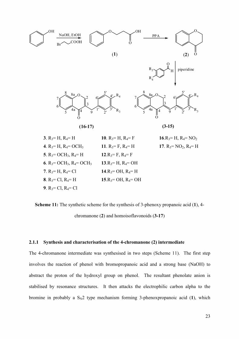
Thirteen 3-benzylidene-4-chromanones and two 3-benzyl-4-chromanones with different
substitution patterns on the phenyl ring (B-ring) (Scheme 11) were synthesised in good yields
of between 50 and 90% according to the modified procedure by Shaikh et al. (2011a). The
target molecules were chosen to examine the effect that the fluorine, chlorine, methoxy,
hydroxy and nitro groups have on different positions of the phenyl ring with regard to
reactivity and biological activity. This would enable us to study the structure-activity
relationship of the substituted benzylidene-4-chromanones with regard to antioxidant and
antibacterial activity.
The synthesis of the homoisoflavonoids from phenol is a three step reaction process; the
synthesis of 3-phenoxypropanoic acid (1) from phenol, the cyclisation of 3-
phenoxypropanoic acid to 4-chromanone (2), and the condensation of 4-chromanone with the
aromatic aldehyde to the homoisoflavonoid (3-17) (Scheme 11).
23
O
O
2
3
44a5
6
7
88a
91'
2'3'
4'5'
6'O
O
2
3
44a5
6
7
8 8a
91'
2'3'
4'5'
6'
(3-15)(16-17)
R4
R3
R4
R3
OHNaOH, EtOH
BrCOOH
O OH
O
PPAO
O
piperidineH
OR3
R4
(1) (2)
3. R3= H, R4= H 10. R3= H, R4= F 16.R3= H, R4= NO2
4. R3= H, R4= OCH3 11. R3= F, R4= H 17. R3= NO2, R4= H
5. R3= OCH3, R4= H 12.R3= F, R4= F
6. R3= OCH3, R4= OCH3 13.R3= H, R4= OH
7. R3= H, R4= Cl 14.R3= OH, R4= H
8. R3= Cl, R4= H 15.R3= OH, R4= OH
9. R3= Cl, R4= Cl
Scheme 11: The synthetic scheme for the synthesis of 3-phenoxy propanoic acid (1), 4-
chromanone (2) and homoisoflavonoids (3-17)
2.1.1 Synthesis and characterisation of the 4-chromanone (2) intermediate
The 4-chromanone intermediate was synthesised in two steps (Scheme 11). The first step
involves the reaction of phenol with bromopropanoic acid and a strong base (NaOH) to
abstract the proton of the hydroxyl group on phenol. The resultant phenolate anion is
stabilised by resonance structures. It then attacks the electrophilic carbon alpha to the
bromine in probably a SN2 type mechanism forming 3-phenoxpropanoic acid (1), which
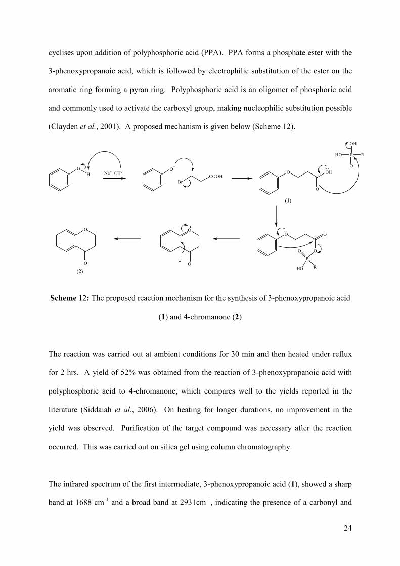
24
cyclises upon addition of polyphosphoric acid (PPA). PPA forms a phosphate ester with the
3-phenoxypropanoic acid, which is followed by electrophilic substitution of the ester on the
aromatic ring forming a pyran ring. Polyphosphoric acid is an oligomer of phosphoric acid
and commonly used to activate the carboxyl group, making nucleophilic substitution possible
(Clayden et al., 2001). A proposed mechanism is given below (Scheme 12).
OH
O
BrCOOH
Na+ OH-
(1)
O OH
O
HO P R
O
OH
O O
O
P
O
HO R
O
O
(2)
O
OH
Scheme 12: The proposed reaction mechanism for the synthesis of 3-phenoxypropanoic acid
(1) and 4-chromanone (2)
The reaction was carried out at ambient conditions for 30 min and then heated under reflux
for 2 hrs. A yield of 52% was obtained from the reaction of 3-phenoxypropanoic acid with
polyphosphoric acid to 4-chromanone, which compares well to the yields reported in the
literature (Siddaiah et al., 2006). On heating for longer durations, no improvement in the
yield was observed. Purification of the target compound was necessary after the reaction
occurred. This was carried out on silica gel using column chromatography.
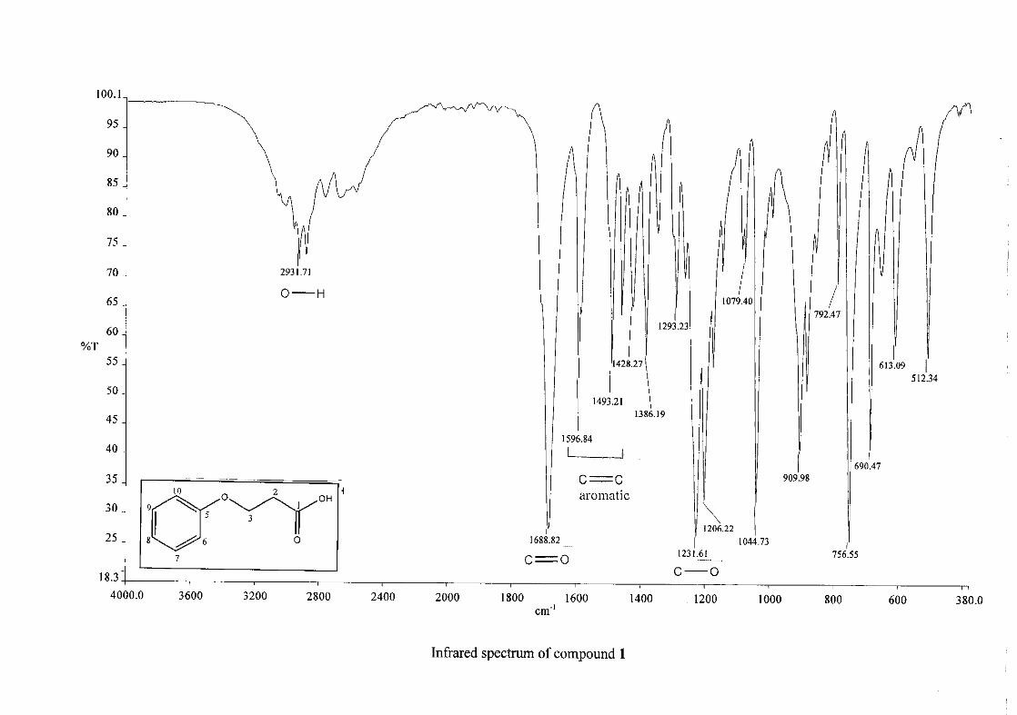
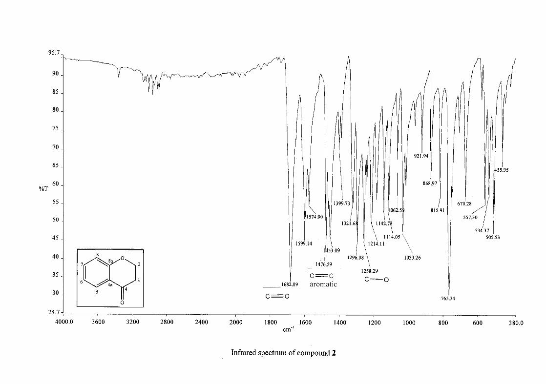
The infrared spectrum of the first intermediate, 3-phenoxypropanoic acid (1), showed a sharp
band at 1688 cm-1 and a broad band at 2931cm-1, indicating the presence of a carbonyl and
25
hydroxy group respectively. This serves as an indication for the formation of the acid. After
the cyclisation of 3-phenoxypropanoic acid to 4-chromanone (2), the carbonyl band shifted
from 1688 cm-1 to 1682 cm-1. Other characteristic absorption bands observed in 2 were that
of the aromatic ring, C=C stretching vibrations (1599, 1476, 1453 cm-1) and the C-O
stretching vibration at 1258 cm-1.
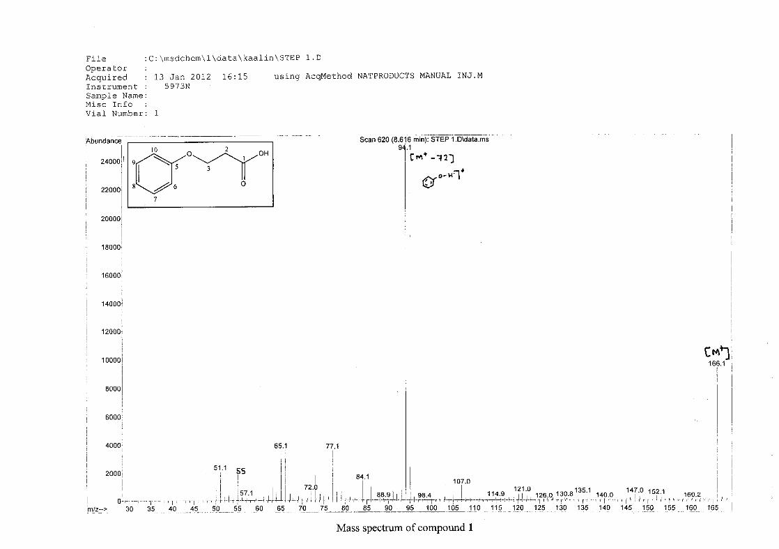
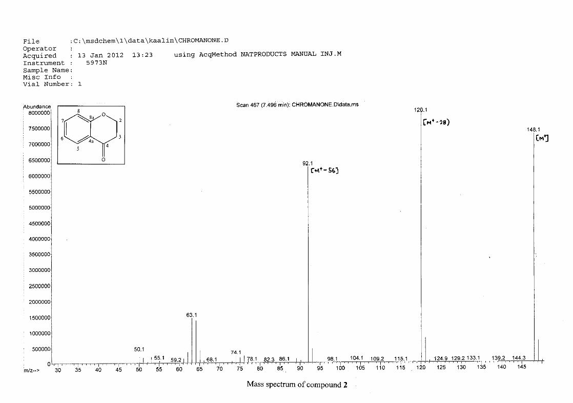
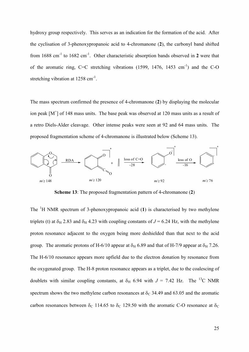
The mass spectrum confirmed the presence of 4-chromanone (2) by displaying the molecular
ion peak [M+] of 148 mass units. The base peak was observed at 120 mass units as a result of
a retro Diels-Alder cleavage. Other intense peaks were seen at 92 and 64 mass units. The
proposed fragmentation scheme of 4-chromanone is illustrated below (Scheme 13).
O
O
C
O
m/z 148 m/z 120
loss of C=O
-28
O
m/z 92 m/z 76
loss of O
-16RDA
O
Scheme 13: The proposed fragmentation pattern of 4-chromanone (2)
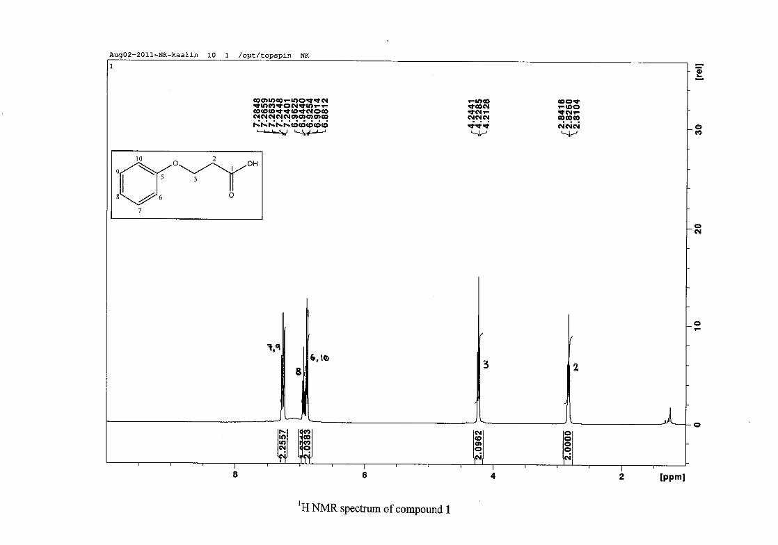
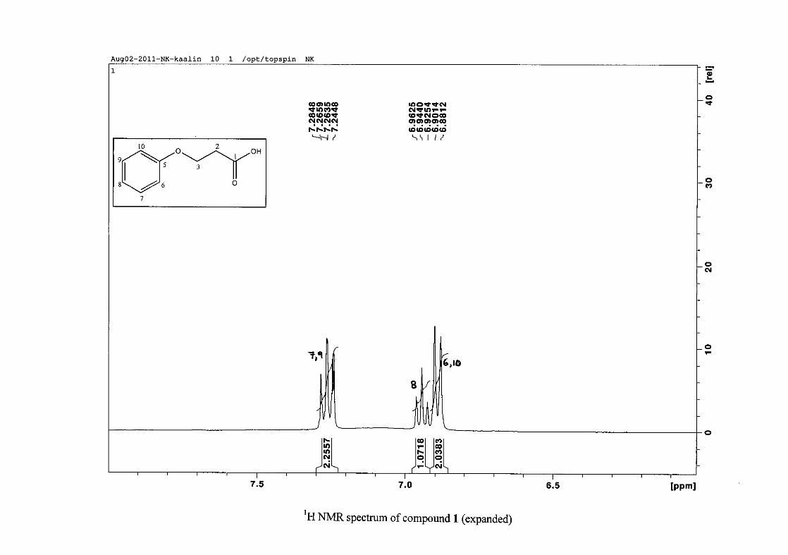
The 1H NMR spectrum of 3-phenoxypropanoic acid (1) is characterised by two methylene
triplets (t) at δH 2.83 and δH 4.23 with coupling constants of J = 6.24 Hz, with the methylene
proton resonance adjacent to the oxygen being more deshielded than that next to the acid
group. The aromatic protons of H-6/10 appear at δH 6.89 and that of H-7/9 appear at δH 7.26.
The H-6/10 resonance appears more upfield due to the electron donation by resonance from
the oxygenated group. The H-8 proton resonance appears as a triplet, due to the coalescing of
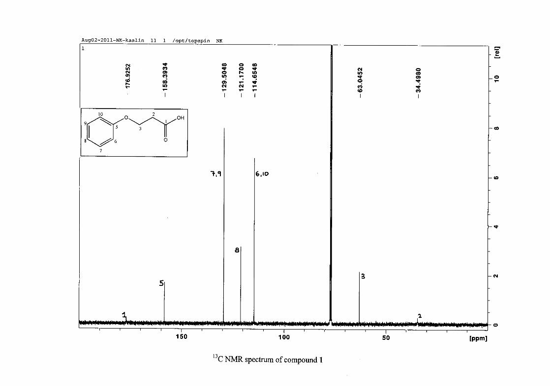
doublets with similar coupling constants, at δH 6.94 with J = 7.42 Hz. The 13C NMR
spectrum shows the two methylene carbon resonances at δC 34.49 and 63.05 and the aromatic
carbon resonances between δC 114.65 to δC 129.50 with the aromatic C-O resonance at δC
26
158.39 and an additional carbonyl resonance at δC 176.93 for the carboxylic acid carbonyl
group.
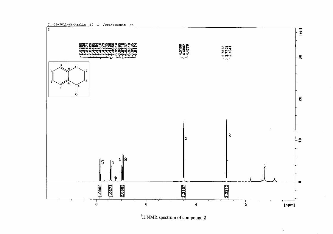
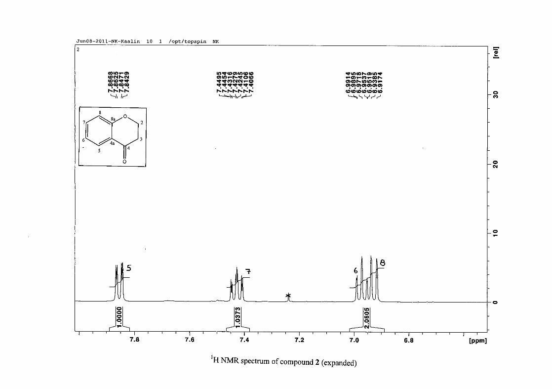
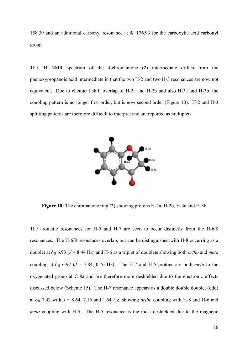
The 1H NMR spectrum of the 4-chromamone (2) intermediate differs from the
phenoxypropanoic acid intermediate in that the two H-2 and two H-3 resonances are now not
equivalent. Due to chemical shift overlap of H-2a and H-2b and also H-3a and H-3b, the
coupling pattern is no longer first order, but is now second order (Figure 10). H-2 and H-3
splitting patterns are therefore difficult to interpret and are reported as multiplets.
Figure 10: The chromanone ring (2) showing protons H-2a, H-2b, H-3a and H-3b
The aromatic resonances for H-5 and H-7 are seen to occur distinctly from the H-6/8
resonances. The H-6/8 resonances overlap, but can be distinguished with H-8 occurring as a
doublet at δH 6.93 (J = 8.44 Hz) and H-6 as a triplet of doublets showing both ortho and meta
coupling at δH 6.97 (J = 7.84, 0.76 Hz). The H-7 and H-5 protons are both meta to the
oxygenated group at C-8a and are therefore more deshielded due to the electronic effects
discussed below (Scheme 15). The H-7 resonance appears as a double double doublet (ddd)
at δH 7.42 with J = 8.64, 7.16 and 1.64 Hz, showing ortho coupling with H-8 and H-6 and
meta coupling with H-5. The H-5 resonance is the most deshielded due to the magnetic
27
anisotropic effect. The H-5 proton is in the deshielded part of the cone formed by the π-
electrons of the carbonyl group and appears as a double doublet (dd) at δH 7.85 (J = 7.84,
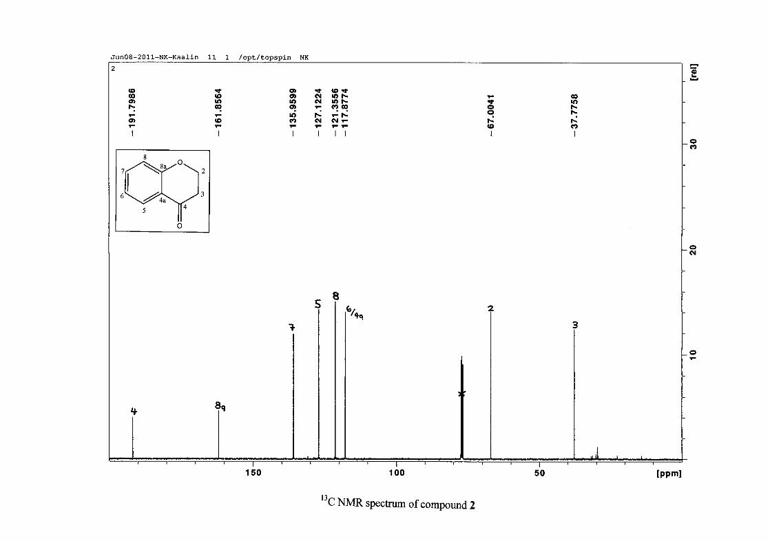
1.72 Hz) (Figure 11). The 13C NMR resonance of the carbonyl group appears at δC 191.80,
indicating the conversion from the acid to the cyclised chromanone in that electron donation
from the hydroxy group no longer occurs in the chromanone resulting in the carbonyl
resonance being more deshielded and closer to that of a pure ketone resonance. The
oxygenated aromatic C-O resonance occurs at δC 161.86 and the other aromatic carbon
resonances occur between δC 117.88 and δC 135.96 (C-4a, C-5-8). The methylene group
closest to the oxygen, C-2 occurs at δC 67.00 and that close to the carbonyl group appears at
δC 37.78.
2.1.2 Synthesis of the 3-benzylidene-4-chromanones
The condensation of 4-chromanone with the aromatic aldehyde is achieved with piperidine as
a base, which abstracts the most acidic proton at the alpha carbon (C-3) resulting in the
formation of an anion, followed by an aldol addition to the aldehyde forming a β-hydroxy
carbonyl compound. The extensive conjugation in the molecule drives the elimination of
water from this intermediate without the addition of acid to form a highly conjugated
molecule, the 3-benzylidene-4-chromanone (Scheme 7).
The reaction of 4-chromanone with various substituted benzaldehydes to give the
homoisoflavonoids were carried out under reflux at 80-90 °C. Reaction temperatures were
monitored and kept below 90 °C, due to exocyclic to endocyclic bond migration which may
occur at ~ 150 °C (Jacquot et al., 2012). Exocyclic to endocyclic bond migration, however
28
did occur at 80-90 °C with the electron withdrawing nitro groups resulting in the 3-benzyl
homoisoflavonoid rather than the 3-benzylidene homoisoflavonoid (Valkonen et al., 2012).
All homoisoflavonoids synthesised were obtained in good yields of between 50-80%. For the
deactivating groups, chloro and fluoro substituents, higher yields were obtained for the meta
substituted than the para substituted compounds. For the activating groups, the hydroxy and
methoxy group, a higher yield was obtained for the para substituted product. In the case of
all disubstituted benzaldehydes, the product yields were lower than that of the mono-
substituted benzaldehydes. For the hydroxy substituents, separation of these compounds
from the reaction mixture was problematic. In the extraction process with ethyl acetate, a
clear distinction could not be made between the phases and although careful care was taken
to recover the amount of ethyl acetate used, some of the product could have been sacrificed in
this procedure. This may account for the lower yields with the hydroxy groups as opposed to
the other substituents.
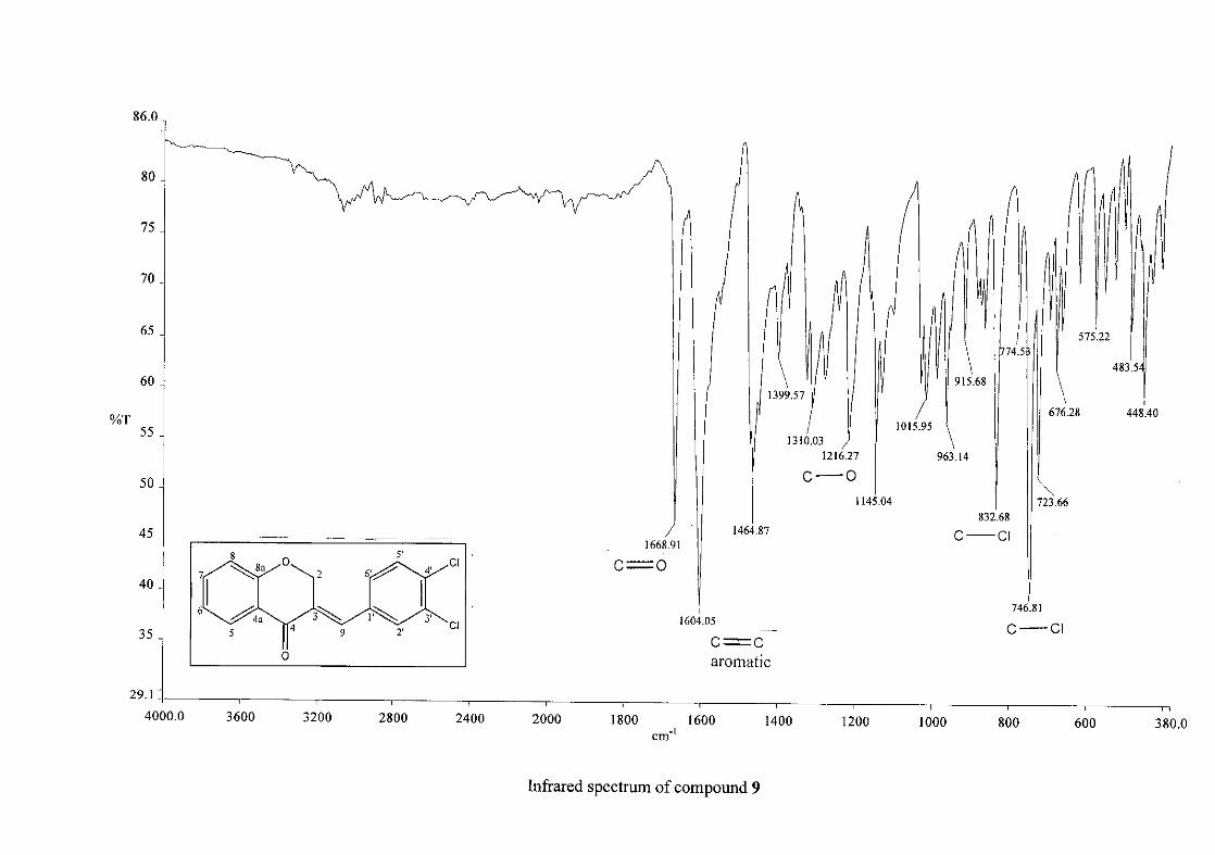
2.1.3 Structural elucidation of homoisoflavonoids (3-17)
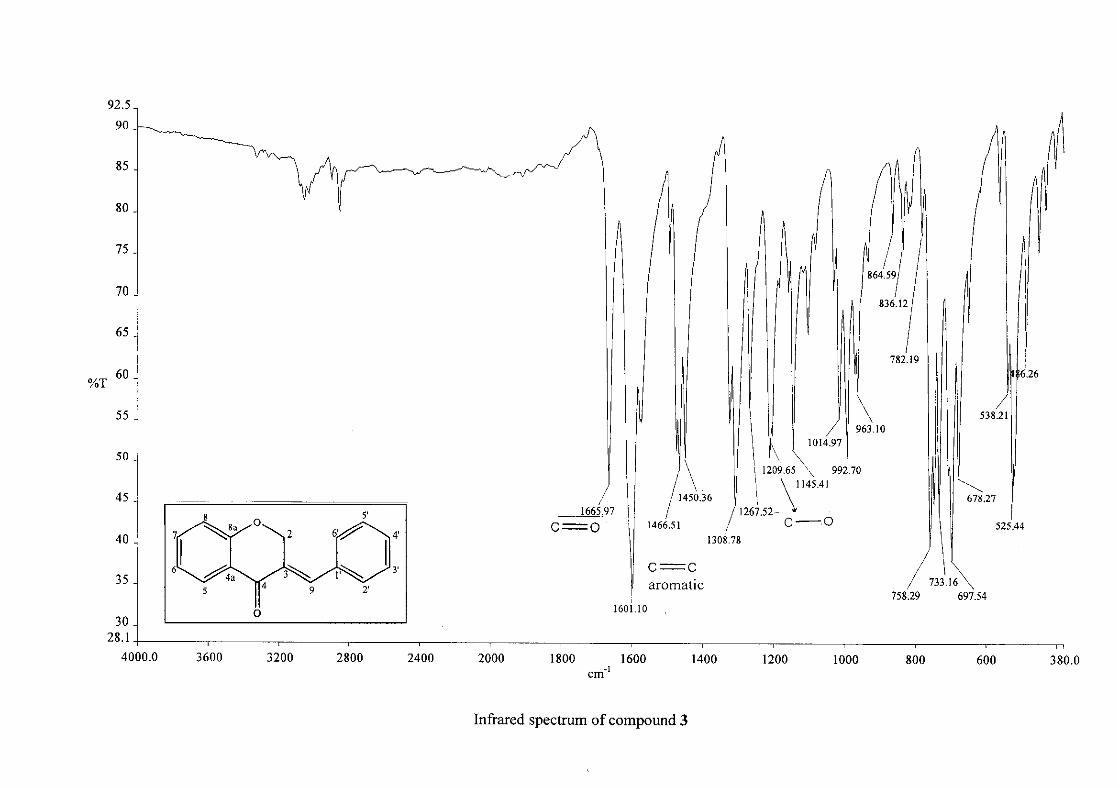
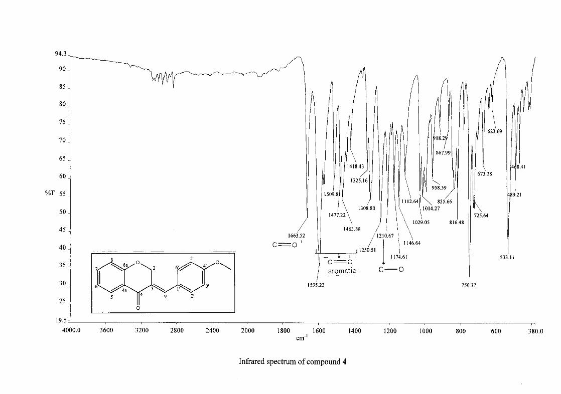
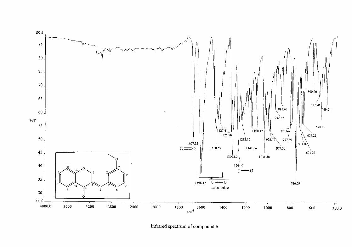
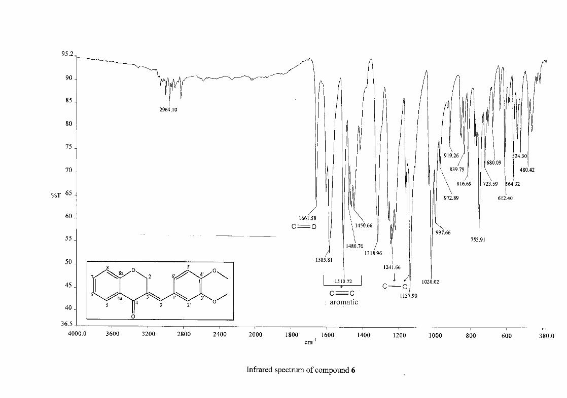
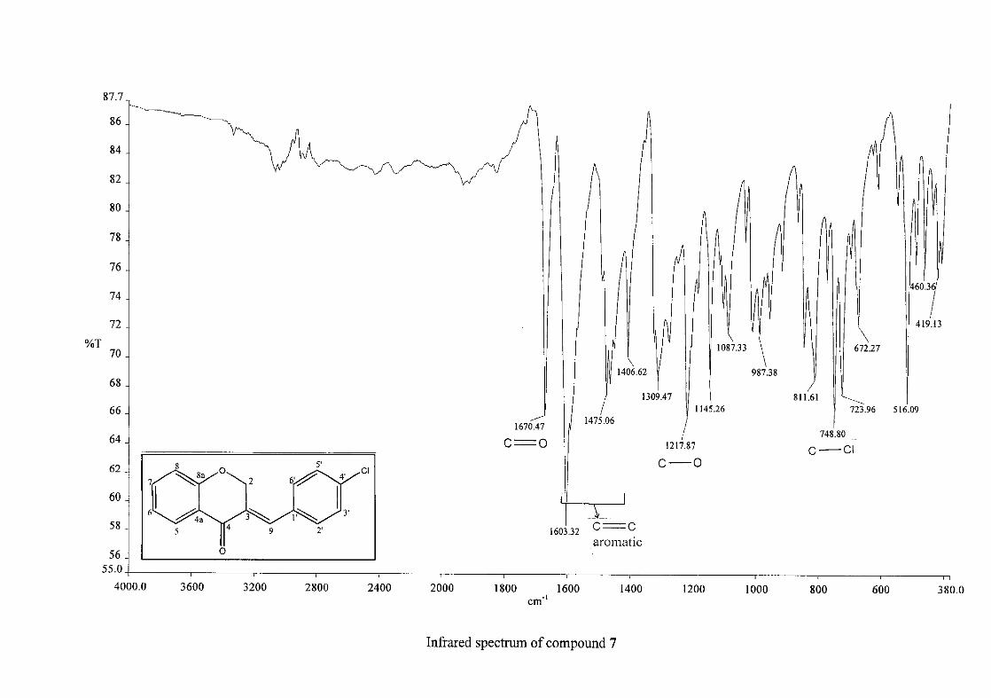
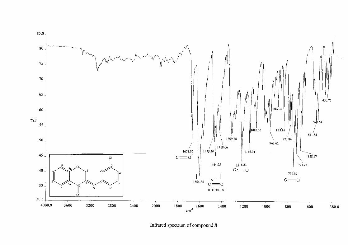
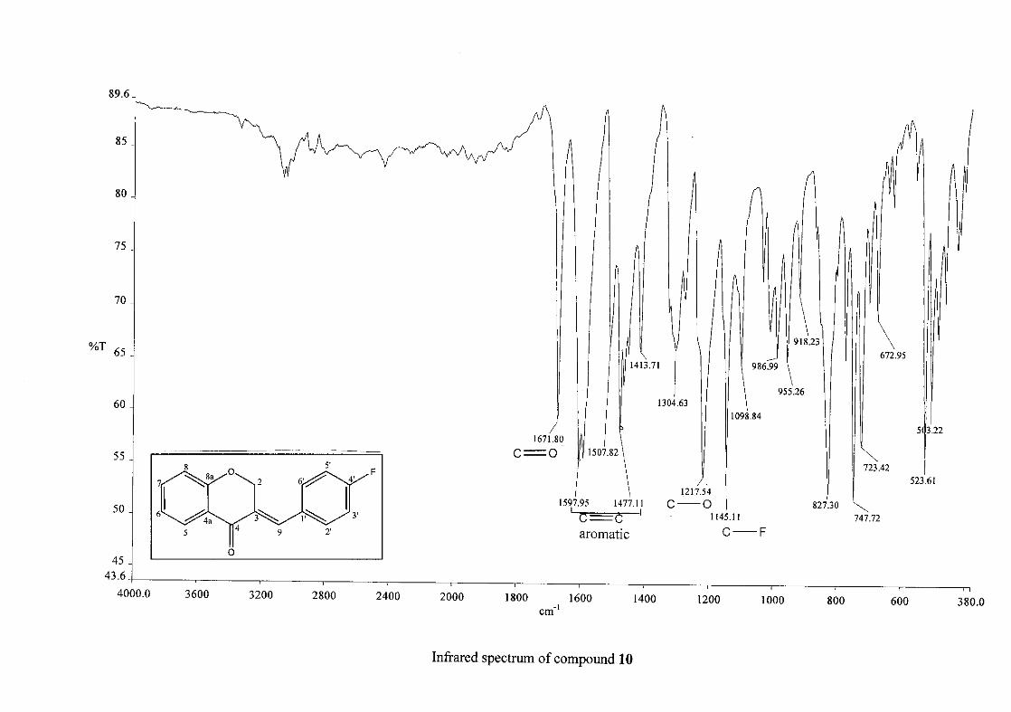
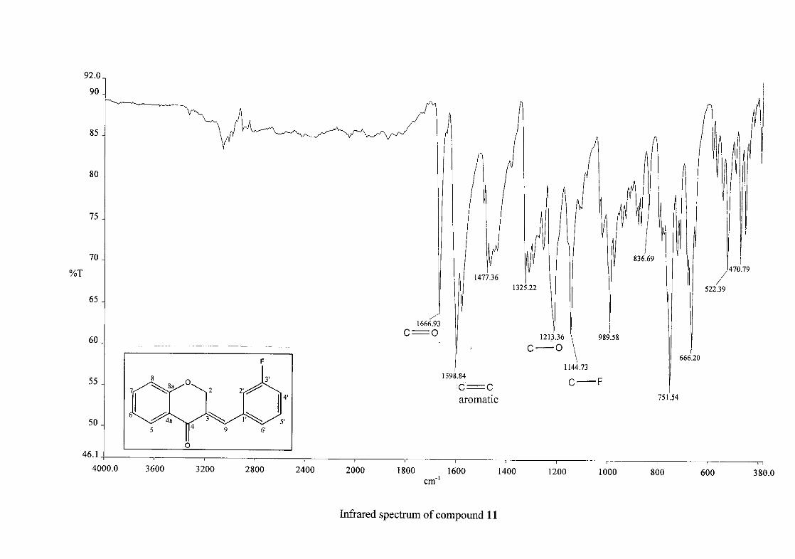
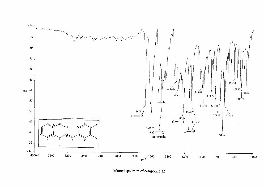
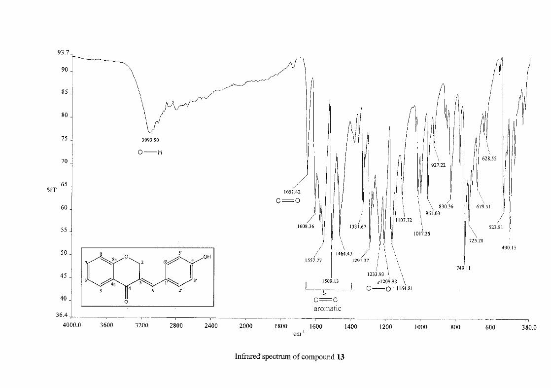
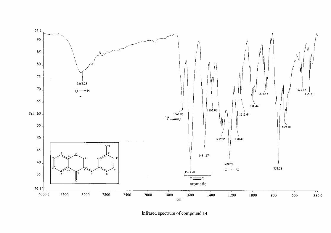
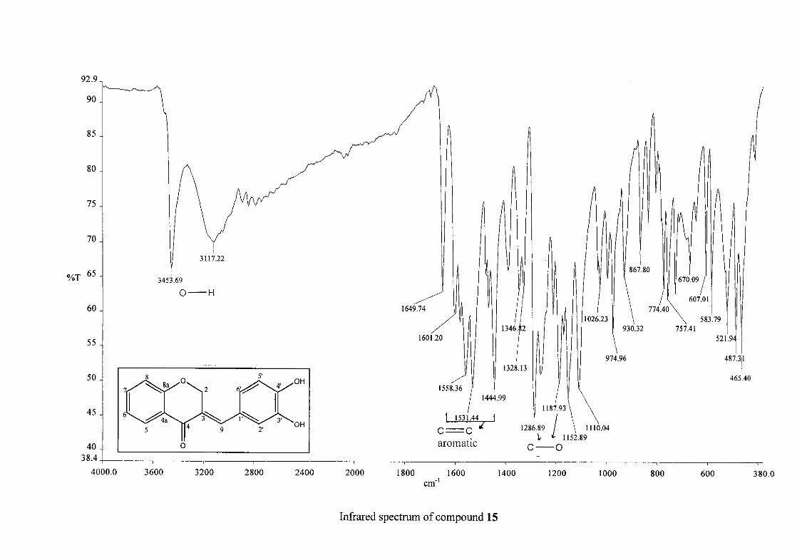
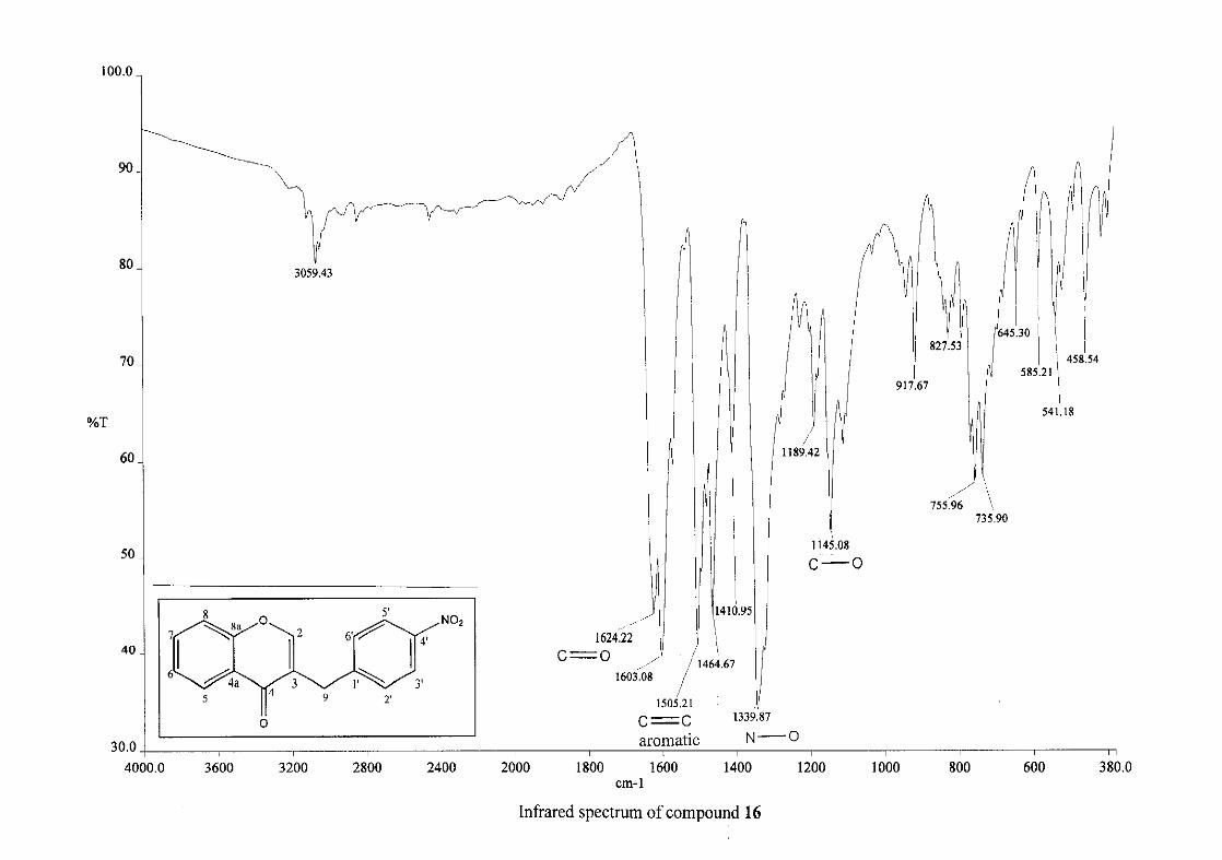
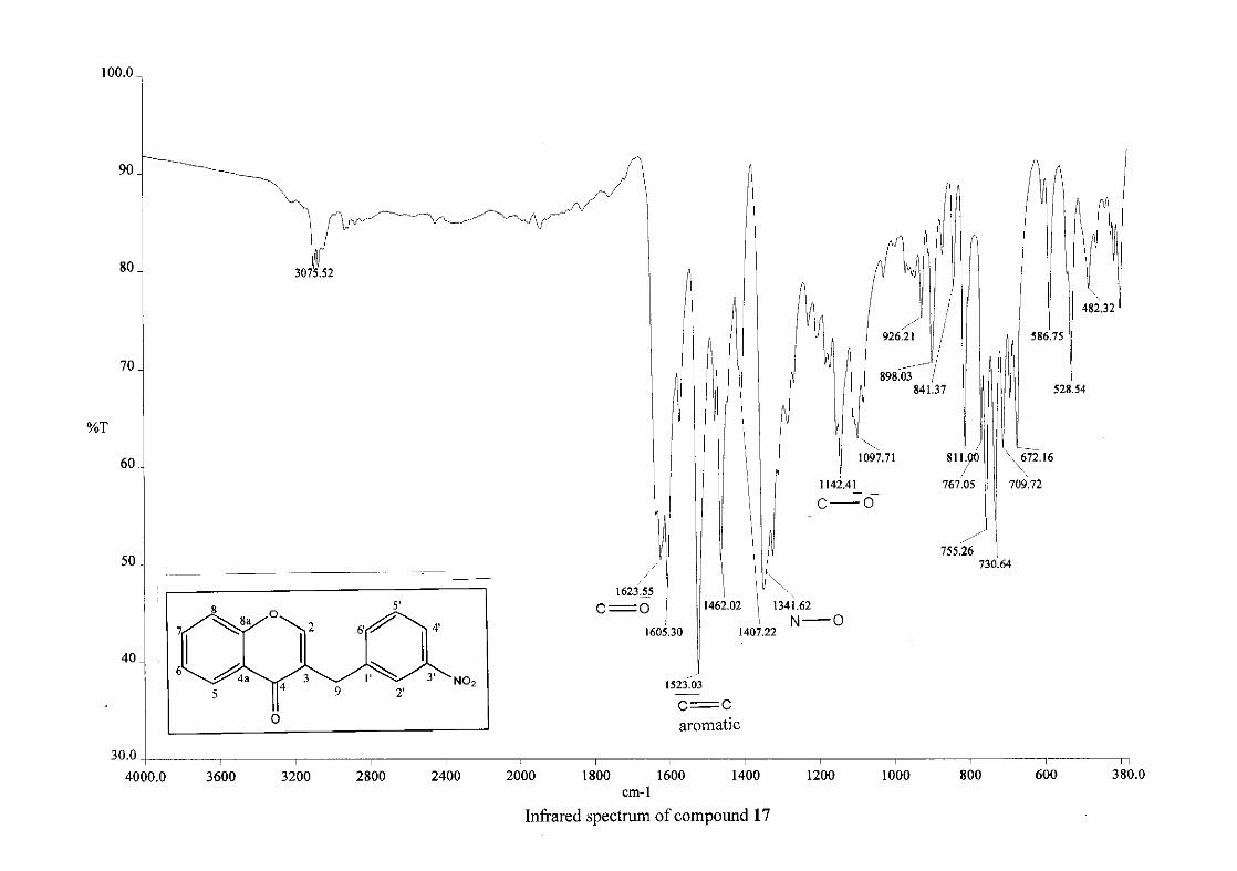
Infrared spectroscopy was used to confirm the presence of functional groups within the
homoisoflavonoids synthesised. The infrared spectrum of 3 showed a sharp peak at 1665
cm-1 which is attributed to the carbonyl group (C=O, C-4). The low frequency of the
absorption is indicative of the conjugation in the molecules resulting in greater single bond
character and lower wavenumbers. The peaks at 1466 and 1450 cm-1 are as a result
symmetrical stretching of the aromatic alkene (C=C) groups. The asymmetrical stretching
peaks for the aromatic C=C bonds are observed at 1308 and 1267 cm-1. The ether group (C-
O-C) stretching frequency is observed at 1209 cm-1.
29
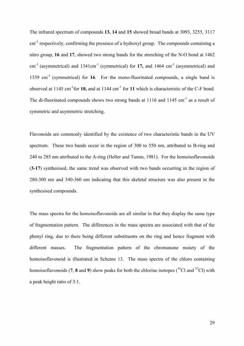
The infrared spectrum of compounds 13, 14 and 15 showed broad bands at 3093, 3255, 3117
cm-1 respectively, confirming the presence of a hydroxyl group. The compounds containing a
nitro group, 16 and 17, showed two strong bands for the stretching of the N-O bond at 1462
cm-1 (asymmetrical) and 1341cm-1 (symmetrical) for 17, and 1464 cm-1 (asymmetrical) and
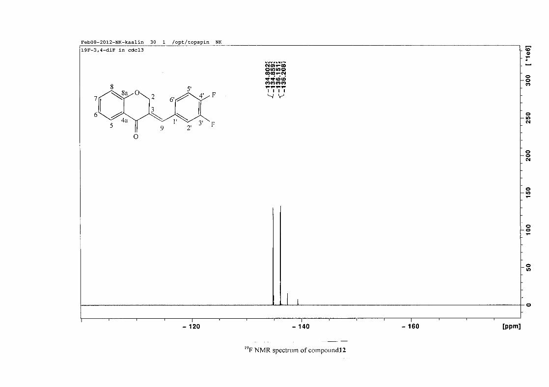
1339 cm-1 (symmetrical) for 16. For the mono-fluorinated compounds, a single band is
observed at 1145 cm-1for 10, and at 1144 cm-1 for 11 which is characteristic of the C-F bond.
The di-fluorinated compounds shows two strong bands at 1116 and 1145 cm-1 as a result of
symmetric and asymmetric stretching.
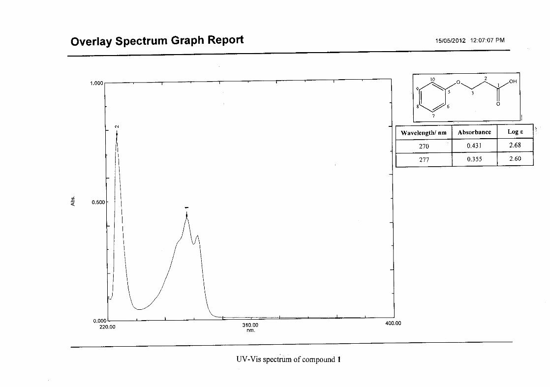
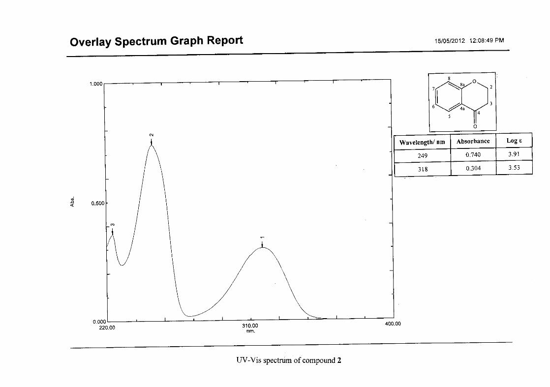
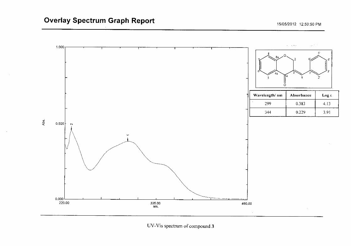
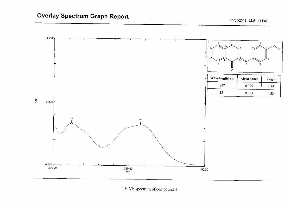
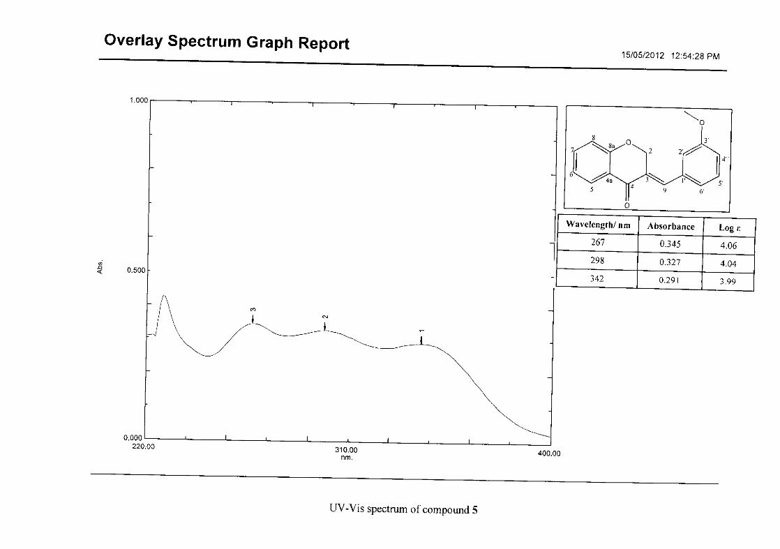
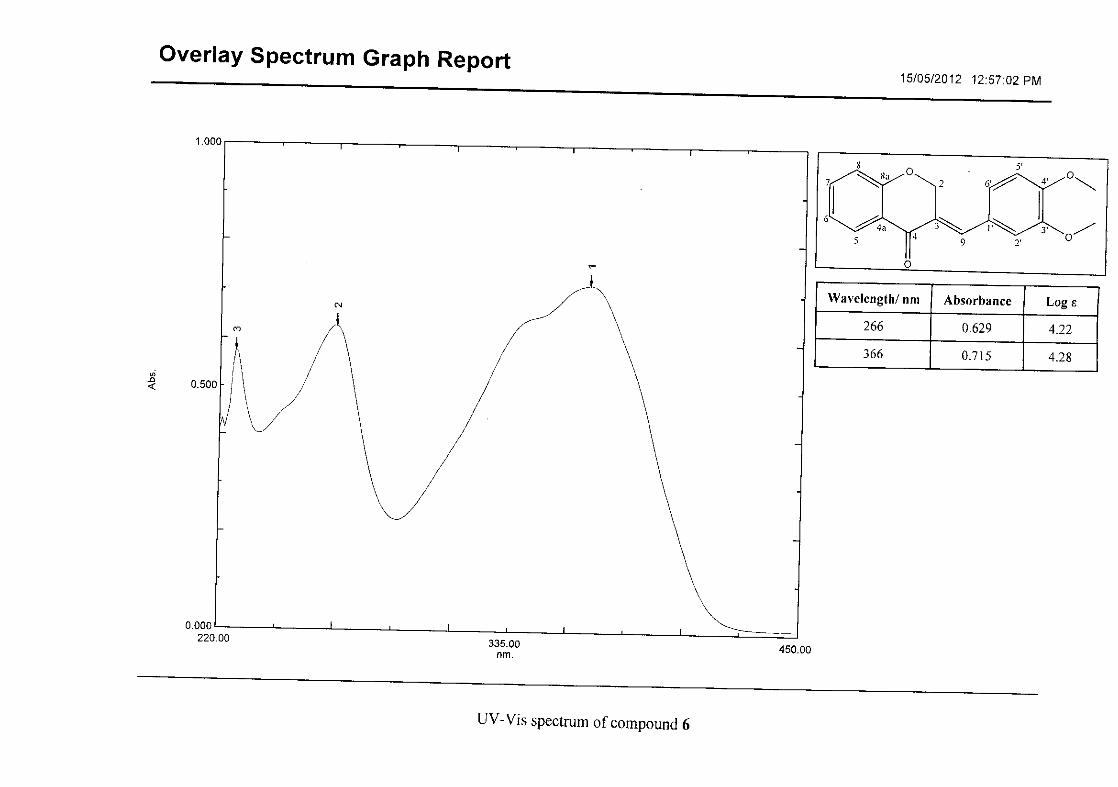
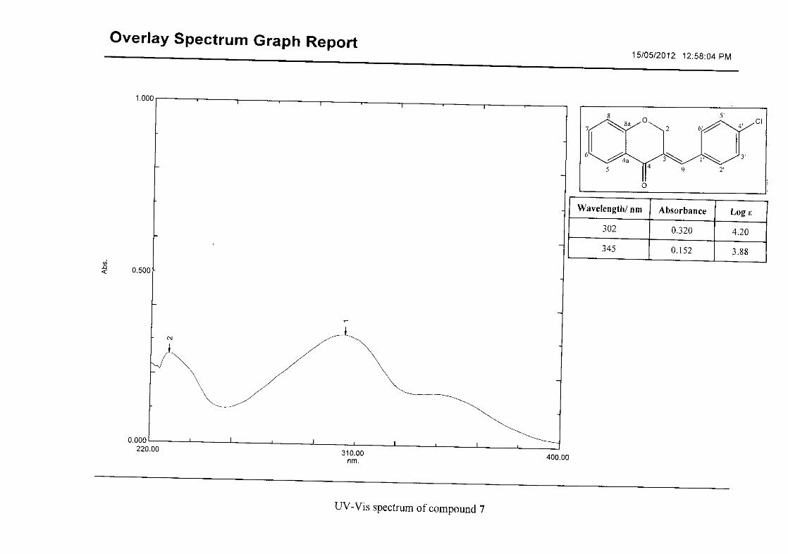
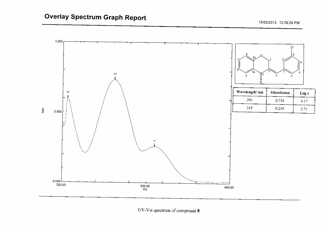
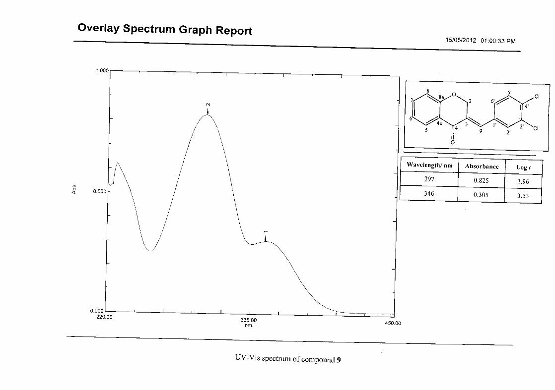
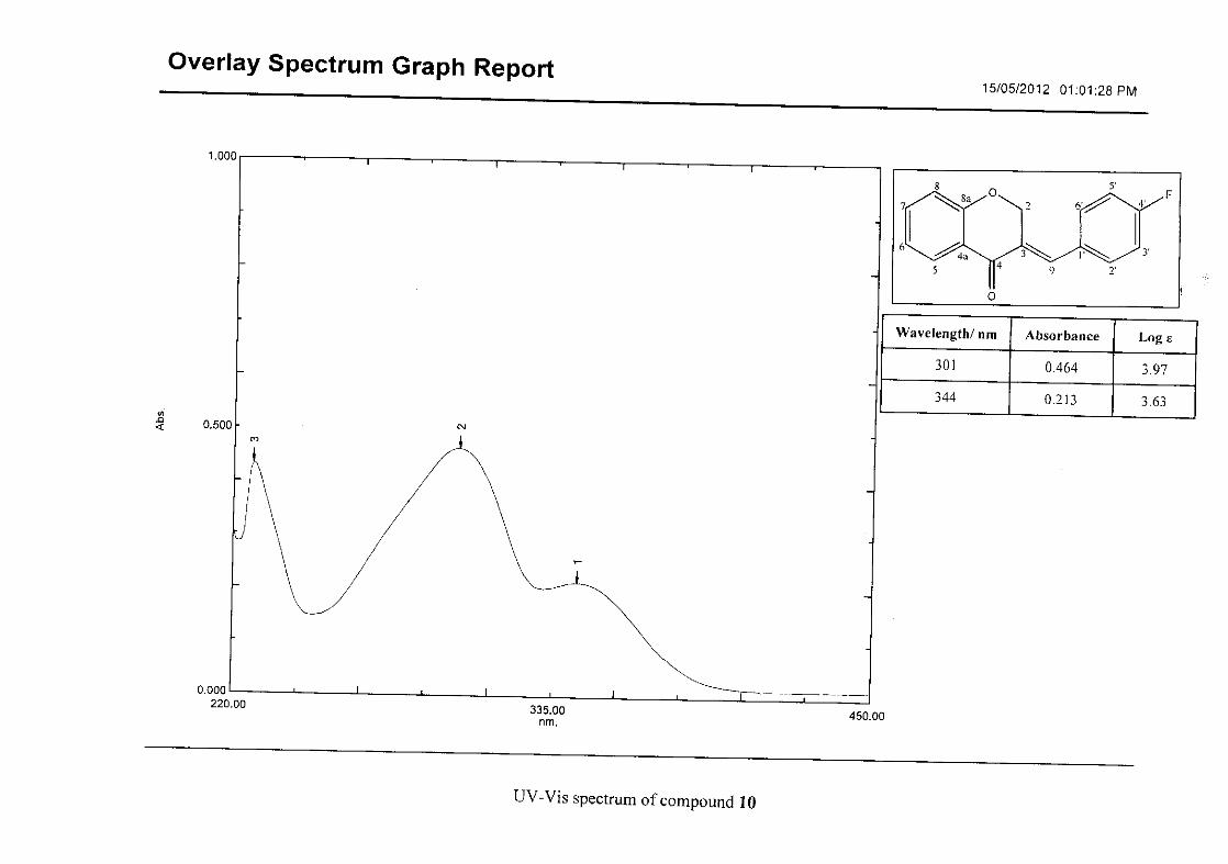
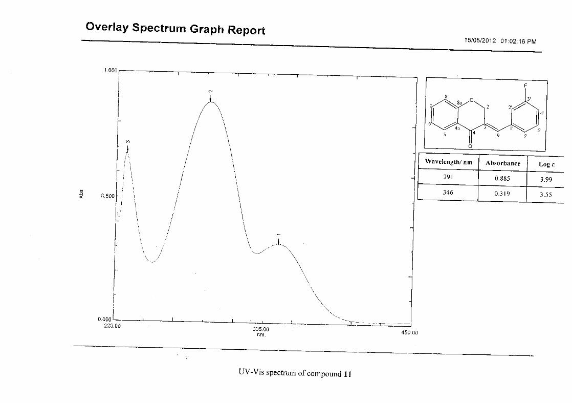
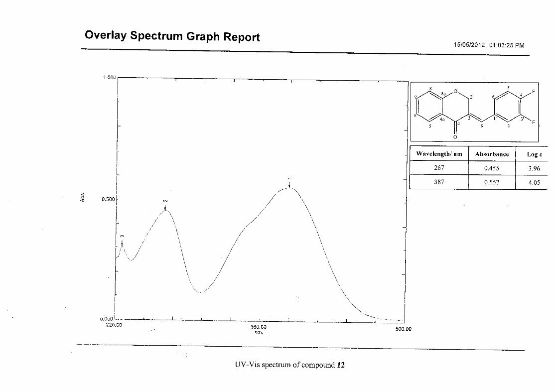
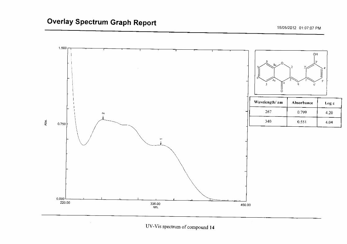
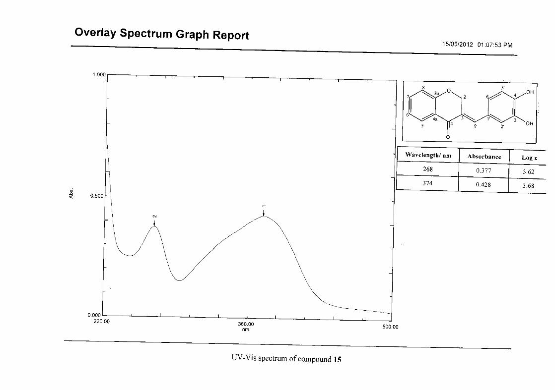
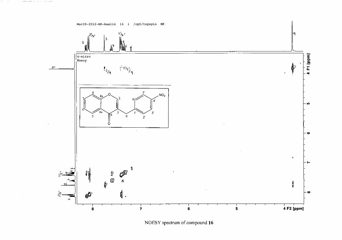
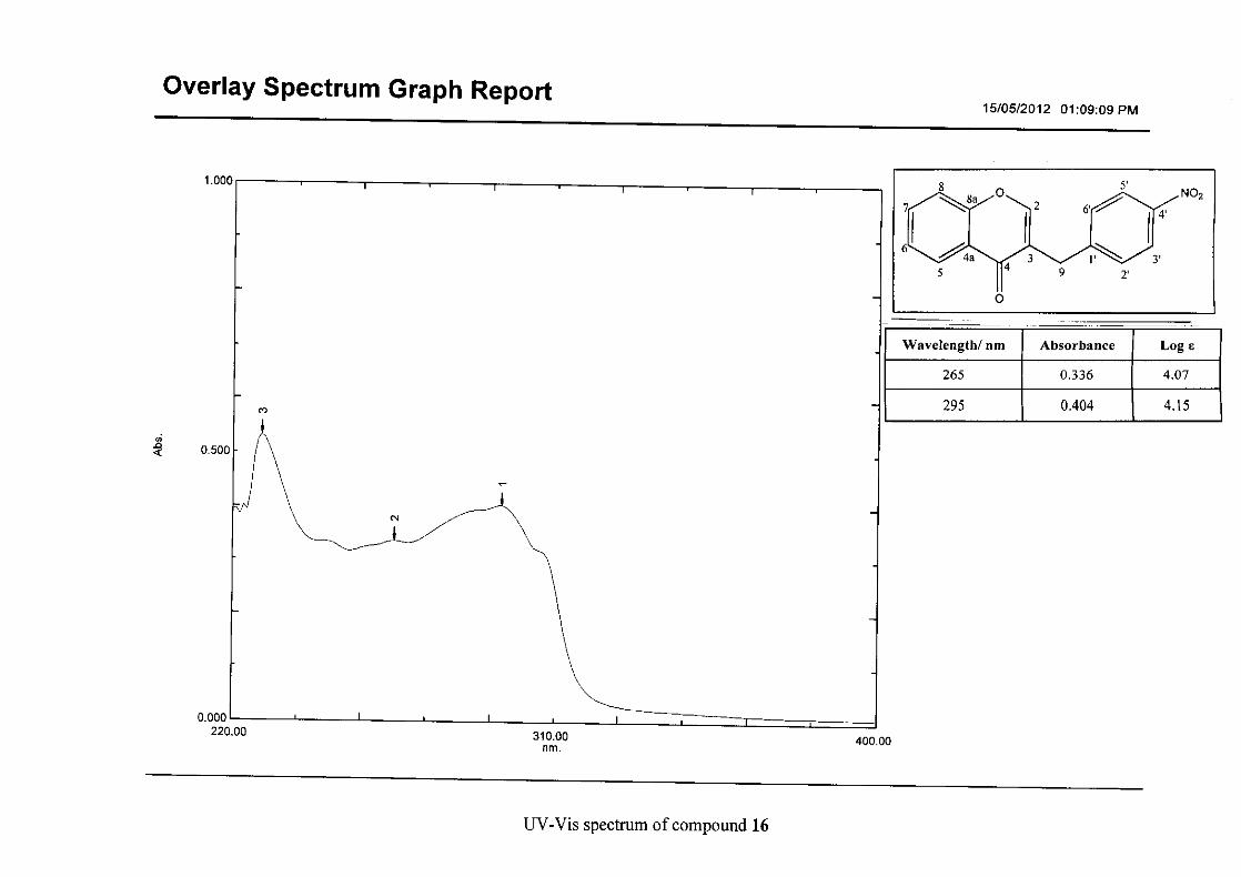
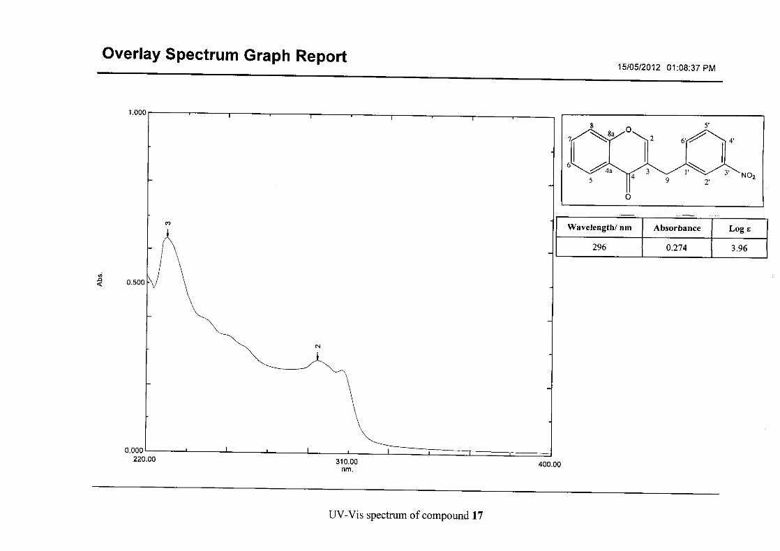
Flavonoids are commonly identified by the existence of two characteristic bands in the UV
spectrum. These two bands occur in the region of 300 to 550 nm, attributed to B-ring and
240 to 285 nm attributed to the A-ring (Heller and Tamm, 1981). For the homoisoflavonoids
(3-17) synthesised, the same trend was observed with two bands occurring in the region of
280-300 nm and 340-360 nm indicating that this skeletal structure was also present in the
synthesised compounds.
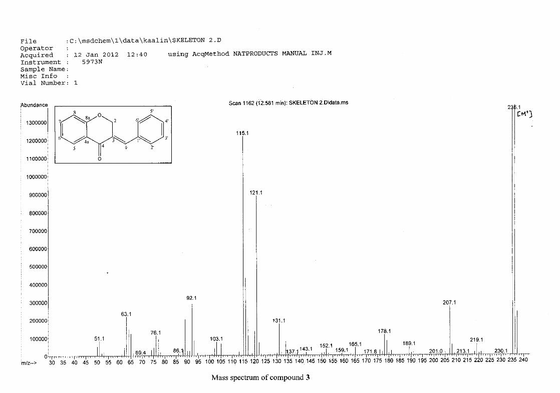
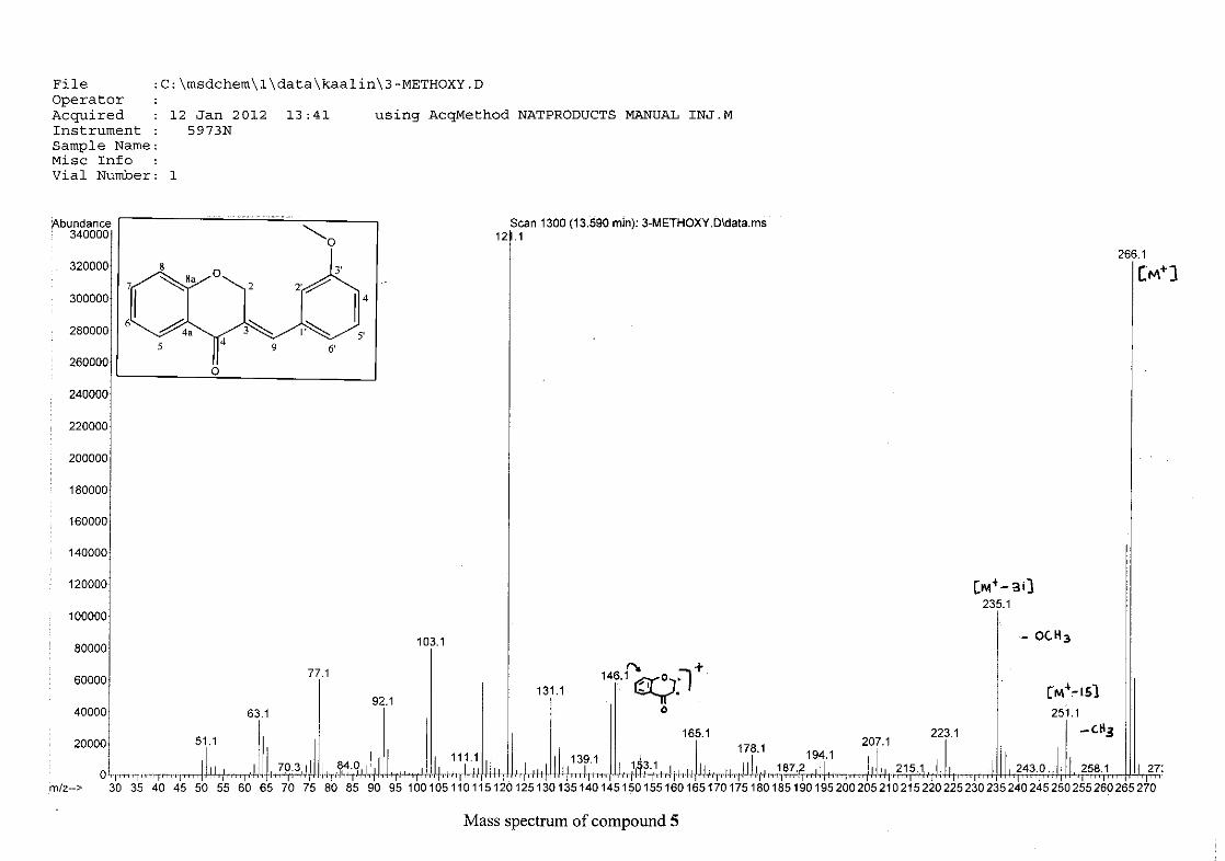
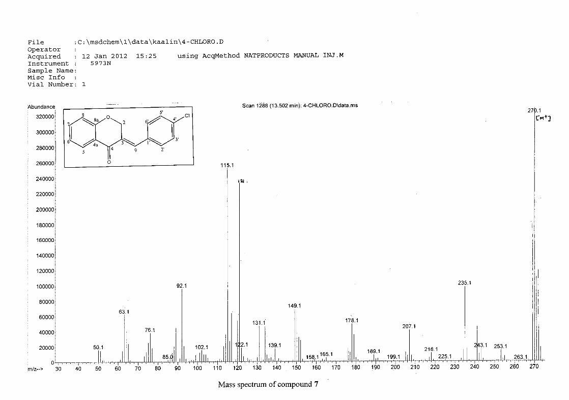
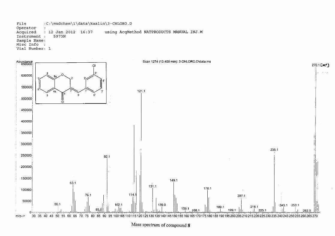
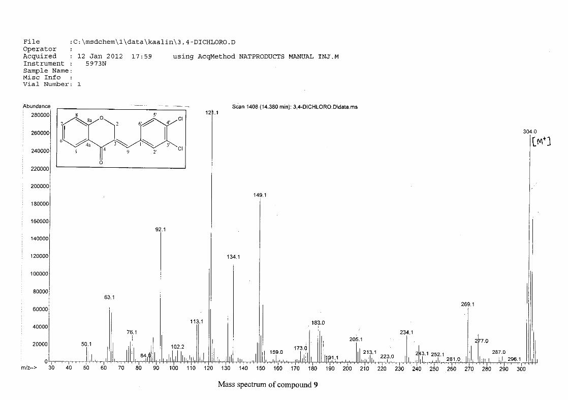
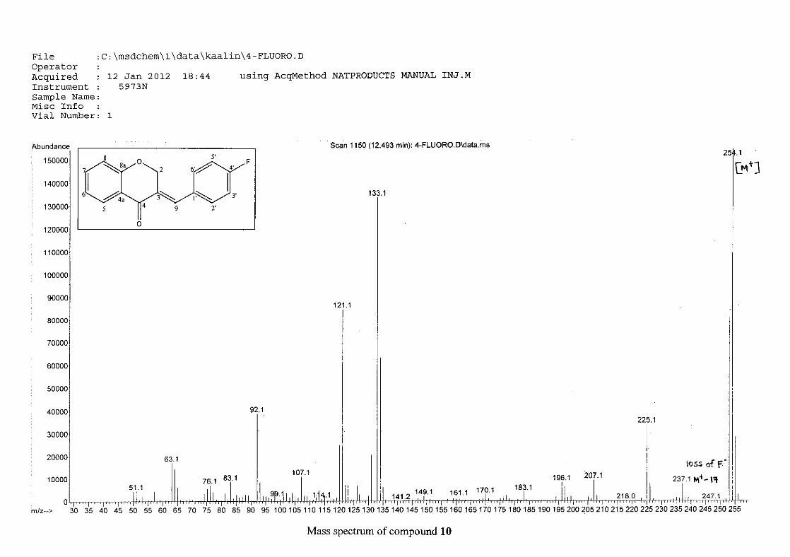
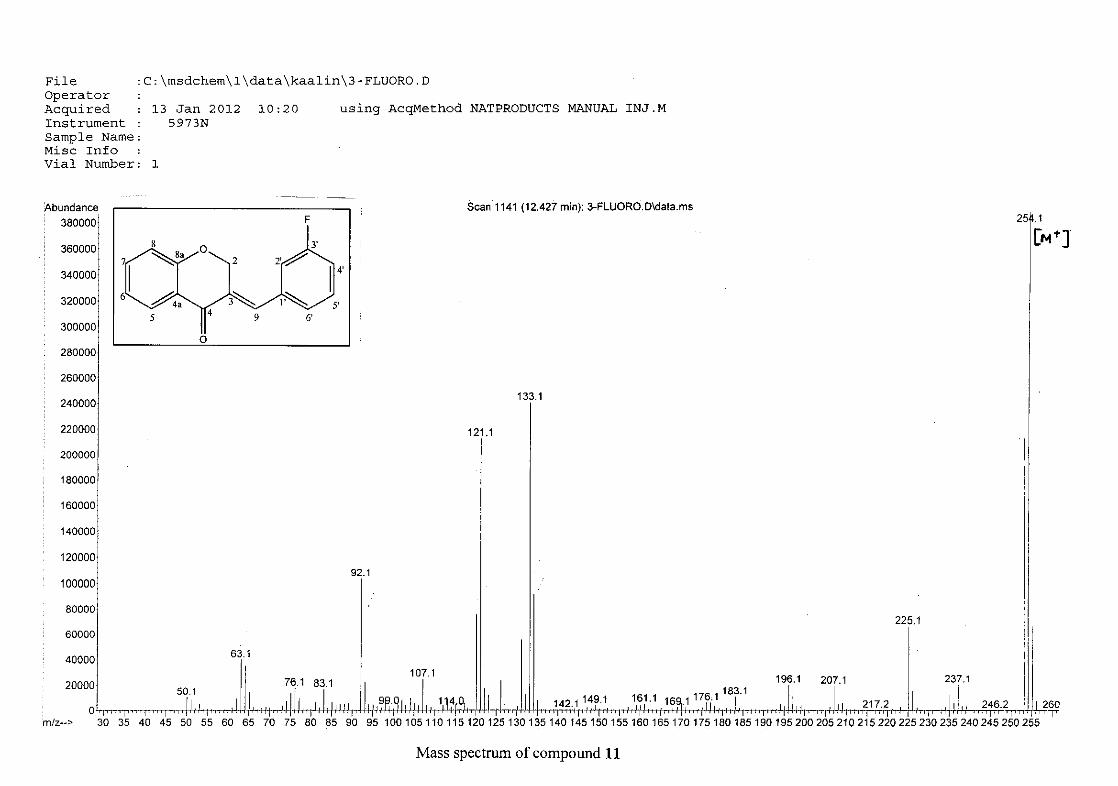
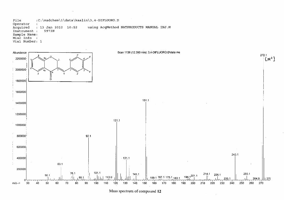
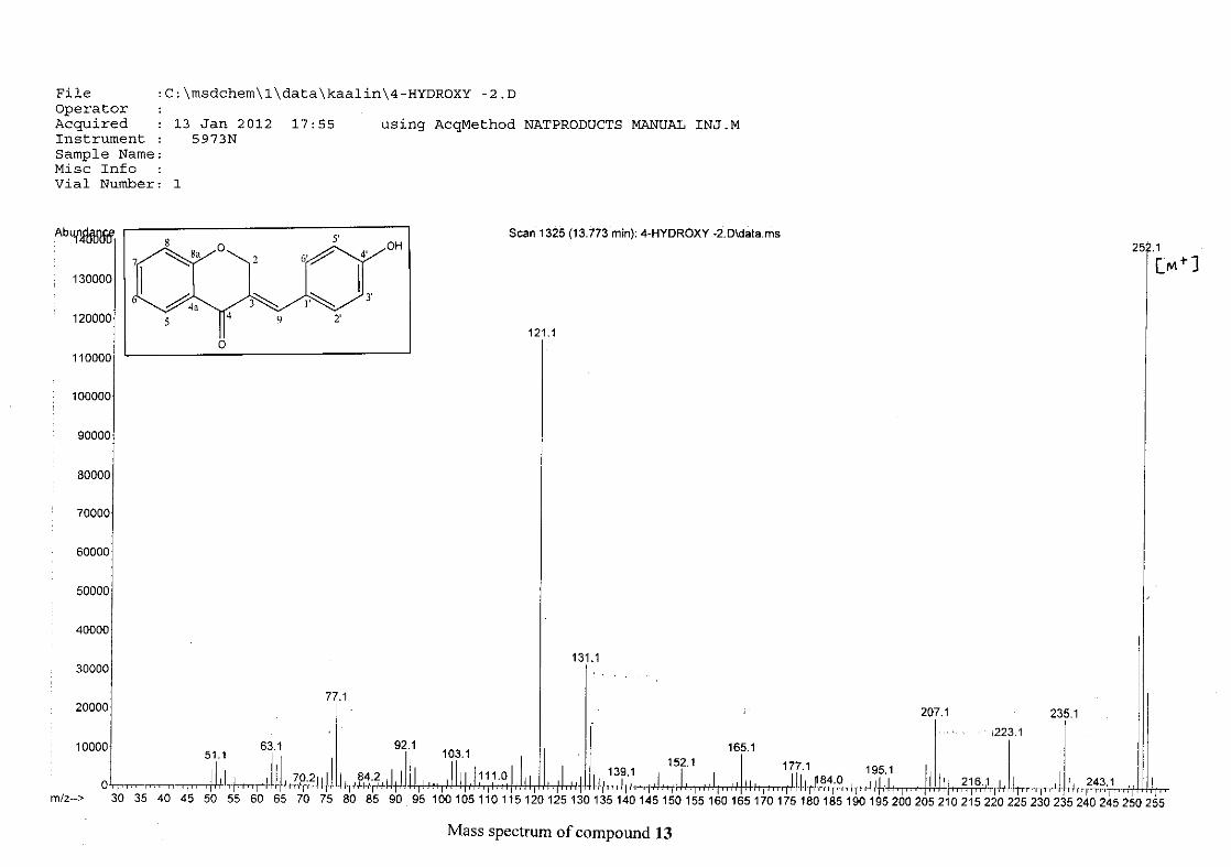
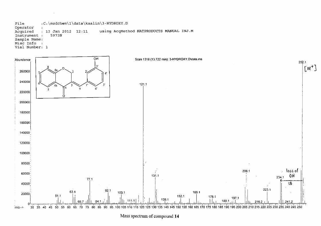
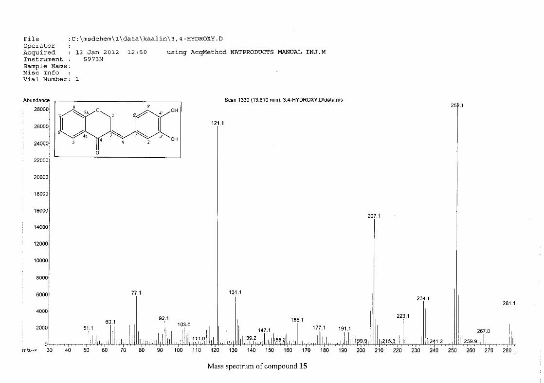
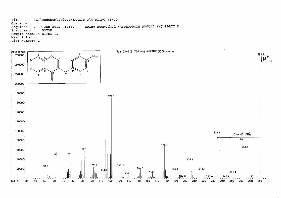
The mass spectra for the homoisoflavonoids are all similar in that they display the same type
of fragmentation pattern. The differences in the mass spectra are associated with that of the
phenyl ring, due to there being different substituents on the ring and hence fragment with
different masses. The fragmentation pattern of the chromanone moiety of the
homoisoflavonoid is illustrated in Scheme 13. The mass spectra of the chloro containing
homoisoflavonoids (7, 8 and 9) show peaks for both the chlorine isotopes (35Cl and 37Cl) with
a peak height ratio of 3:1.
30
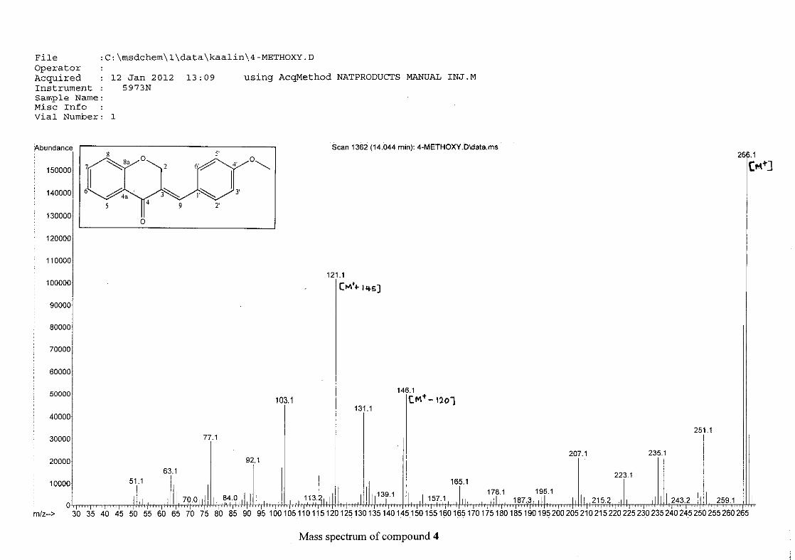
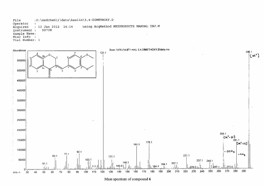
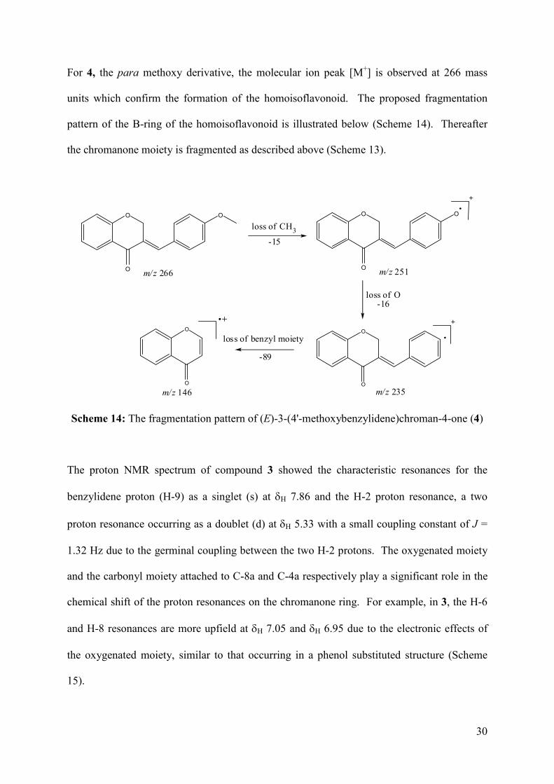
For 4, the para methoxy derivative, the molecular ion peak [M+] is observed at 266 mass
units which confirm the formation of the homoisoflavonoid. The proposed fragmentation
pattern of the B-ring of the homoisoflavonoid is illustrated below (Scheme 14). Thereafter
the chromanone moiety is fragmented as described above (Scheme 13).
m/z 266
O
O
O O
O
O
loss of CH3
-15
m/z 251
O
O
m/z 235
loss of O-16
O
O
m/z 146
loss of benzyl moiety
-89
Scheme 14: The fragmentation pattern of (E)-3-(4'-methoxybenzylidene)chroman-4-one (4)
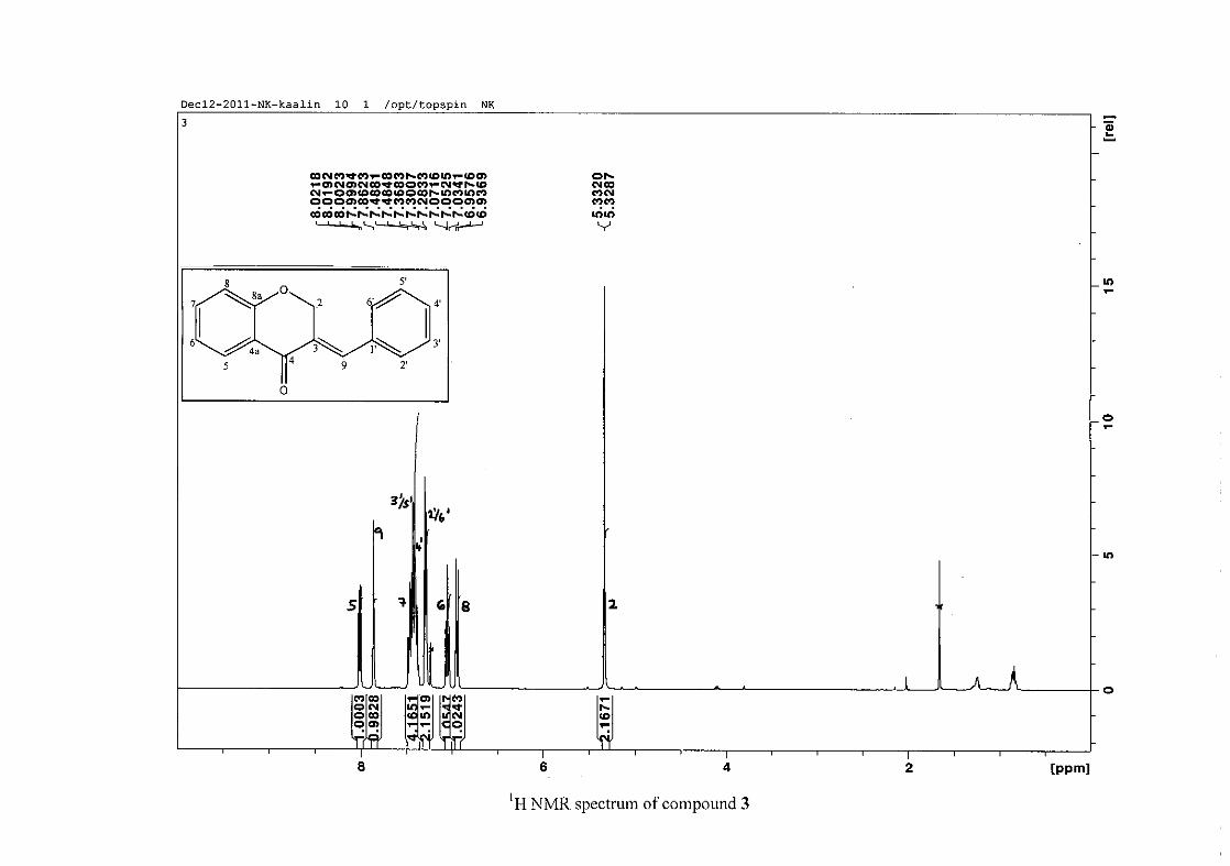
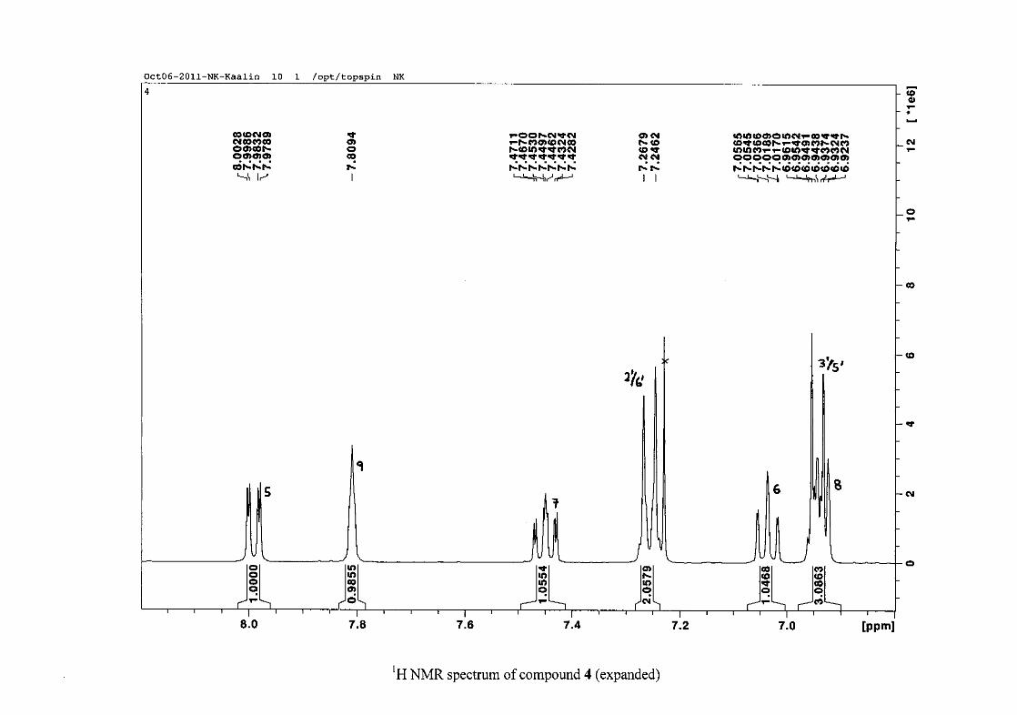
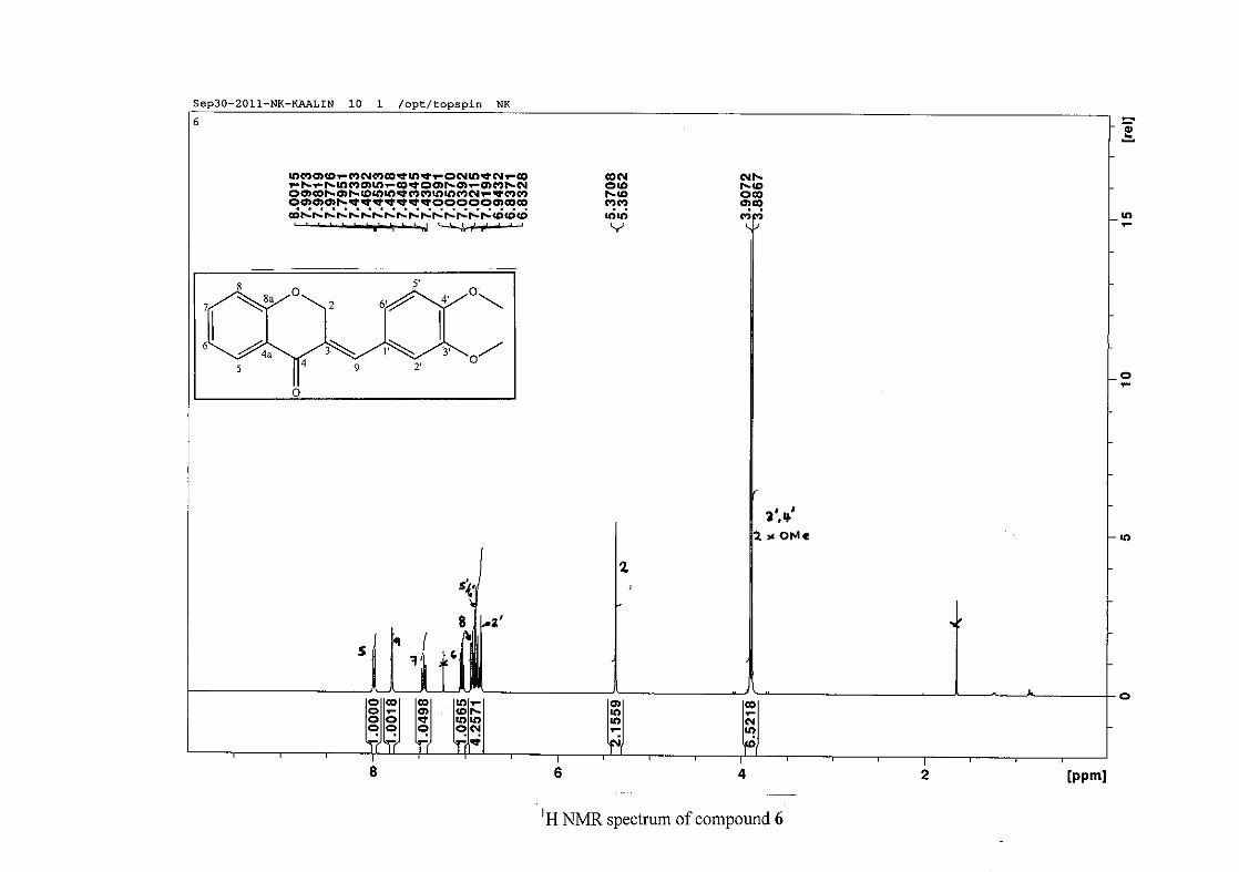
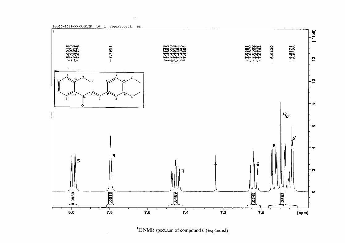
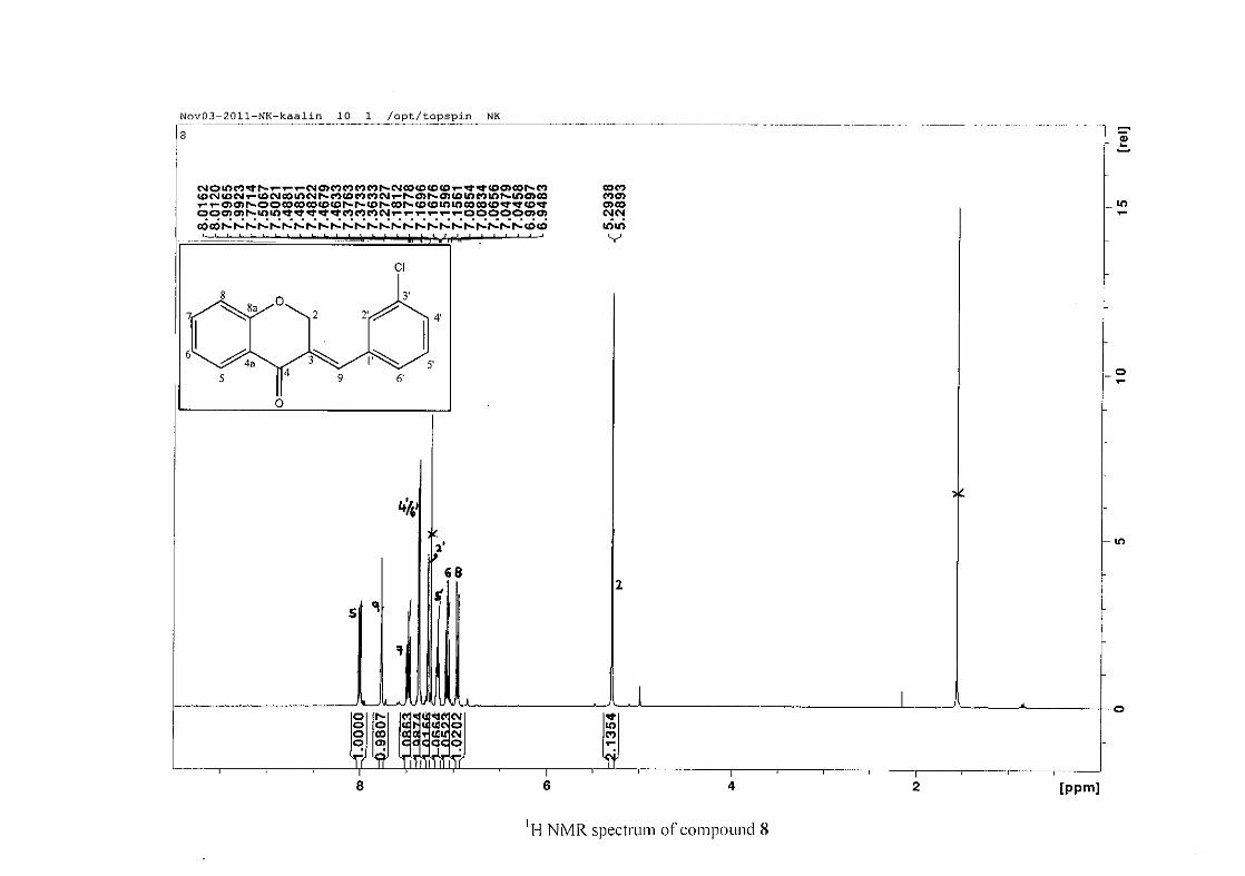
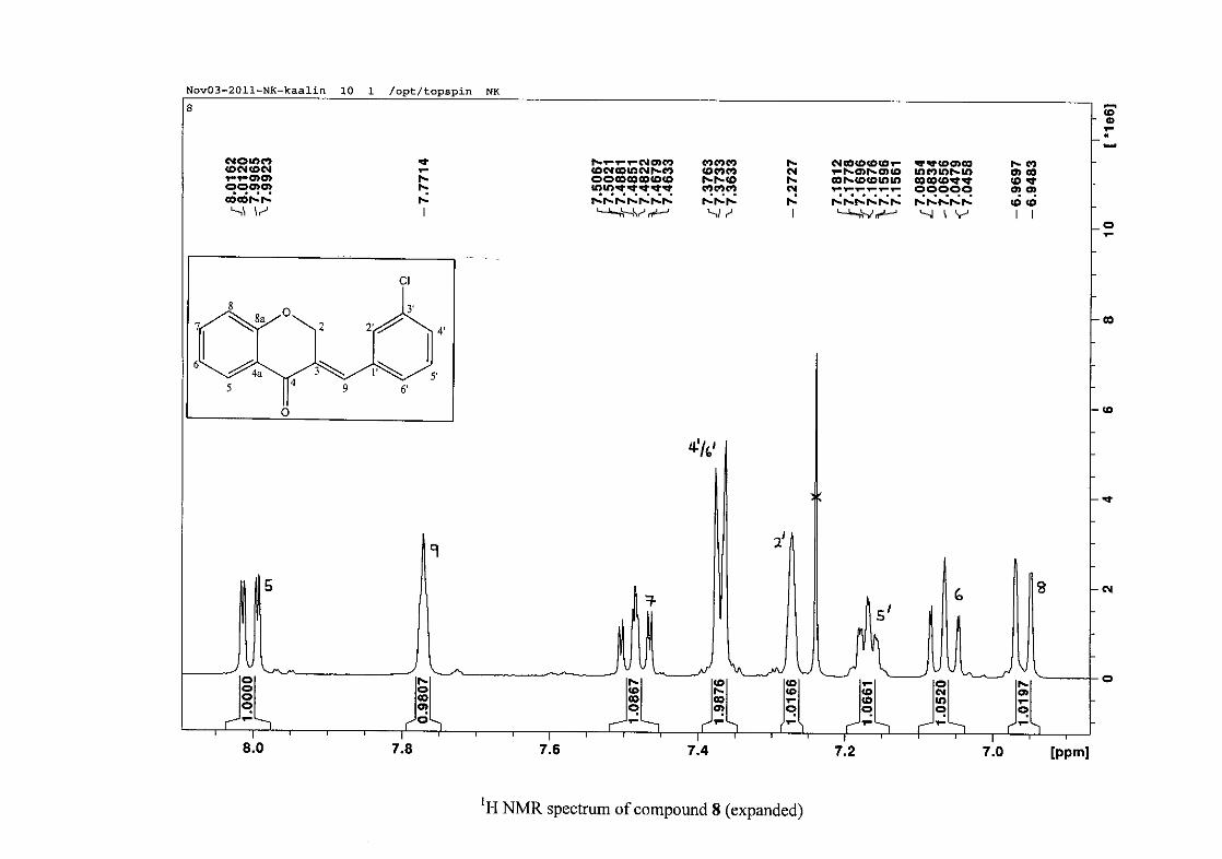
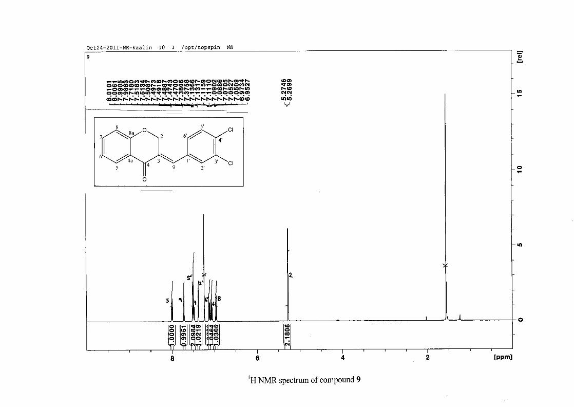
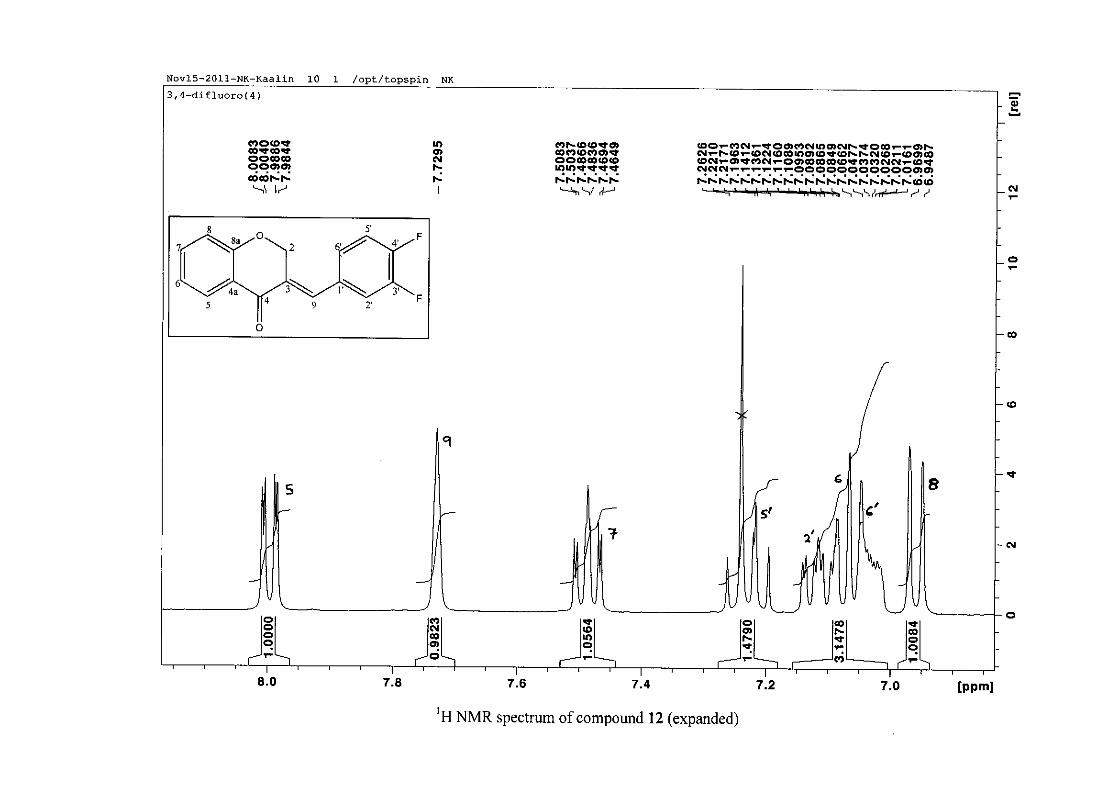
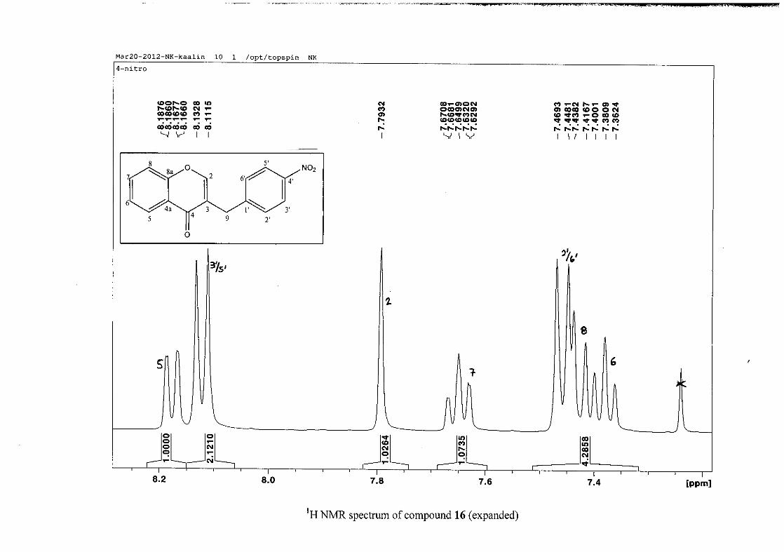
The proton NMR spectrum of compound 3 showed the characteristic resonances for the
benzylidene proton (H-9) as a singlet (s) at δH 7.86 and the H-2 proton resonance, a two
proton resonance occurring as a doublet (d) at δH 5.33 with a small coupling constant of J =
1.32 Hz due to the germinal coupling between the two H-2 protons. The oxygenated moiety
and the carbonyl moiety attached to C-8a and C-4a respectively play a significant role in the
chemical shift of the proton resonances on the chromanone ring. For example, in 3, the H-6
and H-8 resonances are more upfield at δH 7.05 and δH 6.95 due to the electronic effects of
the oxygenated moiety, similar to that occurring in a phenol substituted structure (Scheme
15).
31
OH O H
H
O H
H
O H
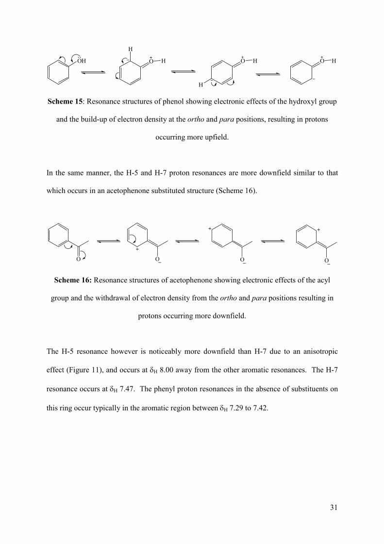
Scheme 15: Resonance structures of phenol showing electronic effects of the hydroxyl group
and the build-up of electron density at the ortho and para positions, resulting in protons
occurring more upfield.
In the same manner, the H-5 and H-7 proton resonances are more downfield similar to that
which occurs in an acetophenone substituted structure (Scheme 16).
O O O O
Scheme 16: Resonance structures of acetophenone showing electronic effects of the acyl
group and the withdrawal of electron density from the ortho and para positions resulting in
protons occurring more downfield.
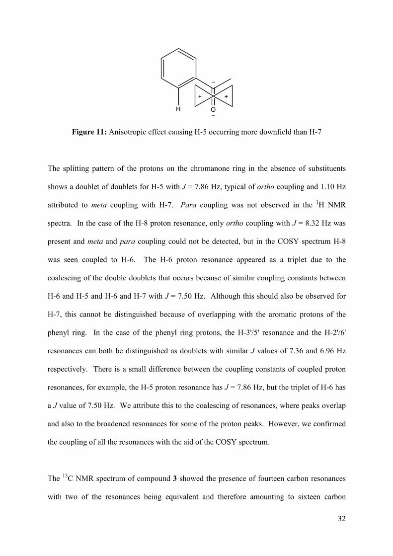
The H-5 resonance however is noticeably more downfield than H-7 due to an anisotropic
effect (Figure 11), and occurs at δH 8.00 away from the other aromatic resonances. The H-7
resonance occurs at δH 7.47. The phenyl proton resonances in the absence of substituents on
this ring occur typically in the aromatic region between δH 7.29 to 7.42.
32
OH
Figure 11: Anisotropic effect causing H-5 occurring more downfield than H-7
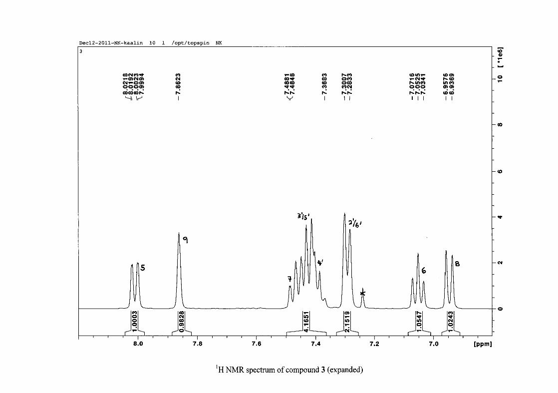
The splitting pattern of the protons on the chromanone ring in the absence of substituents
shows a doublet of doublets for H-5 with J = 7.86 Hz, typical of ortho coupling and 1.10 Hz
attributed to meta coupling with H-7. Para coupling was not observed in the 1H NMR
spectra. In the case of the H-8 proton resonance, only ortho coupling with J = 8.32 Hz was
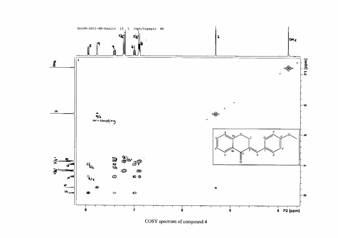
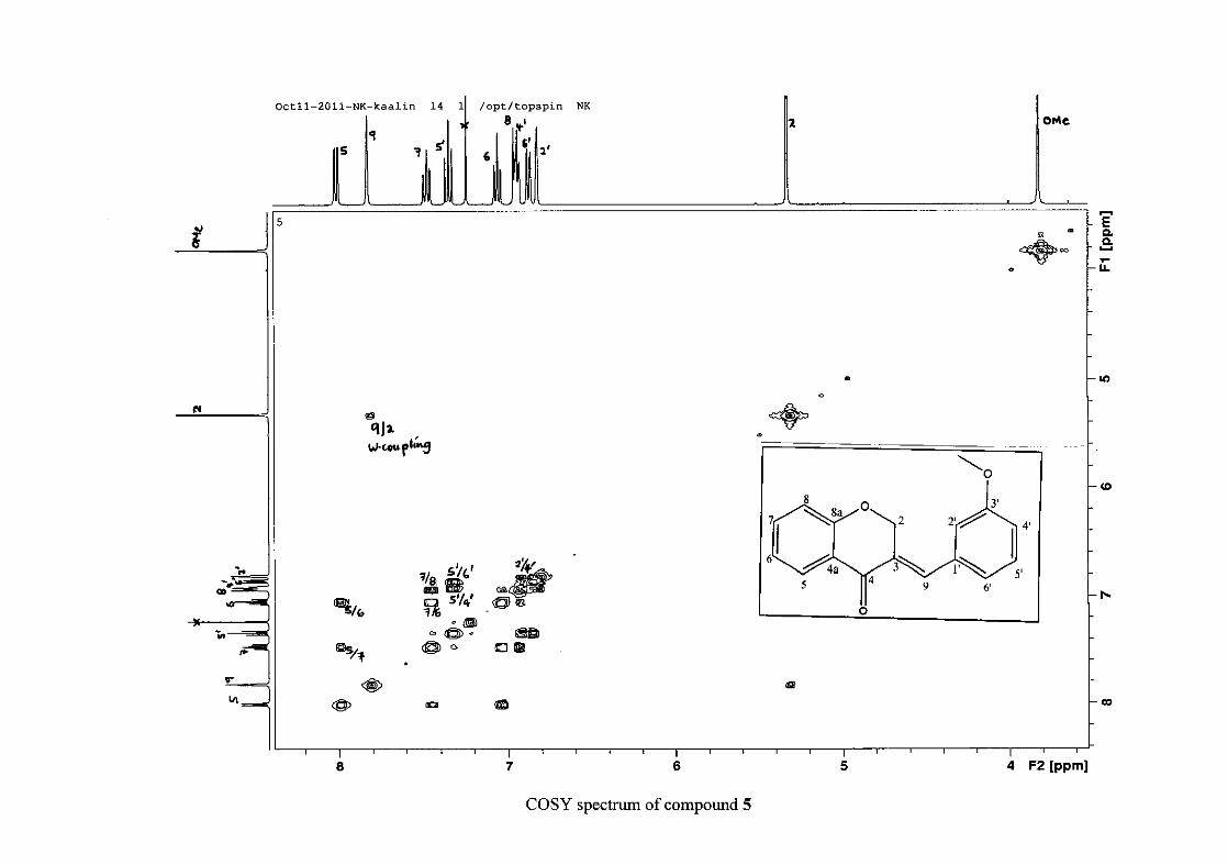
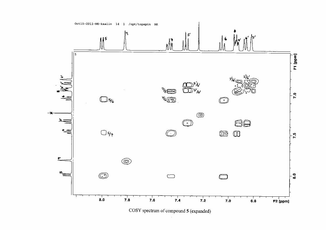
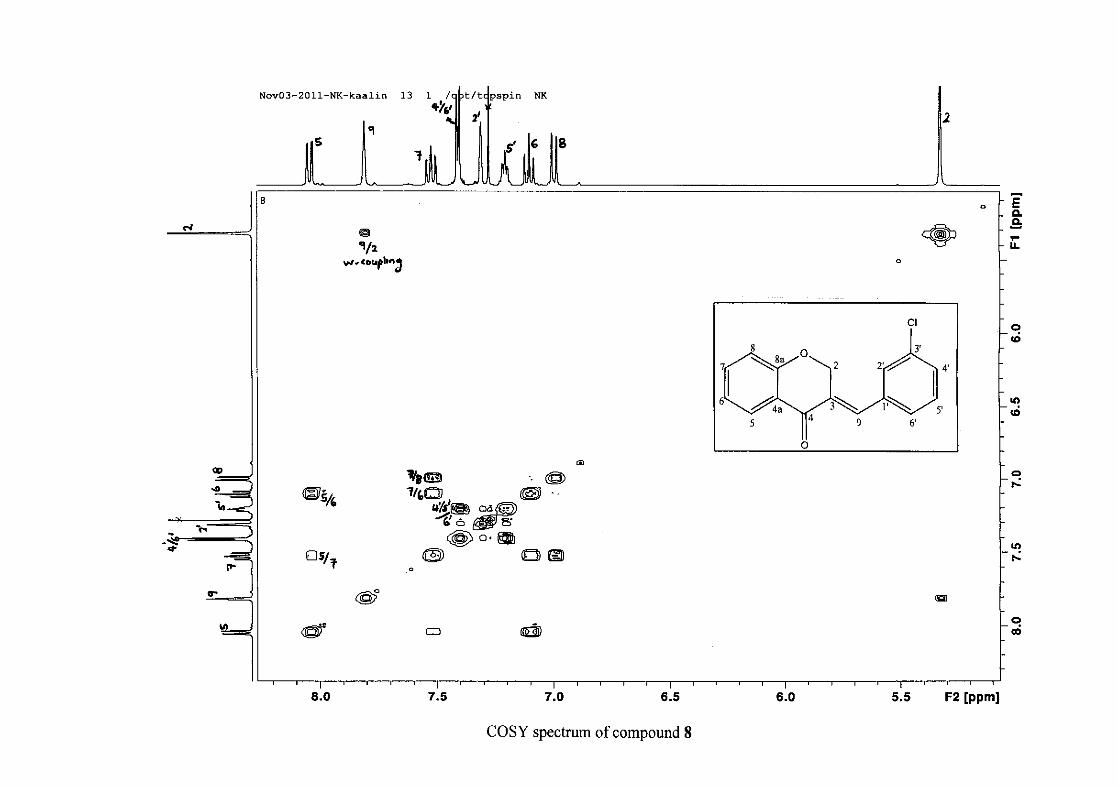
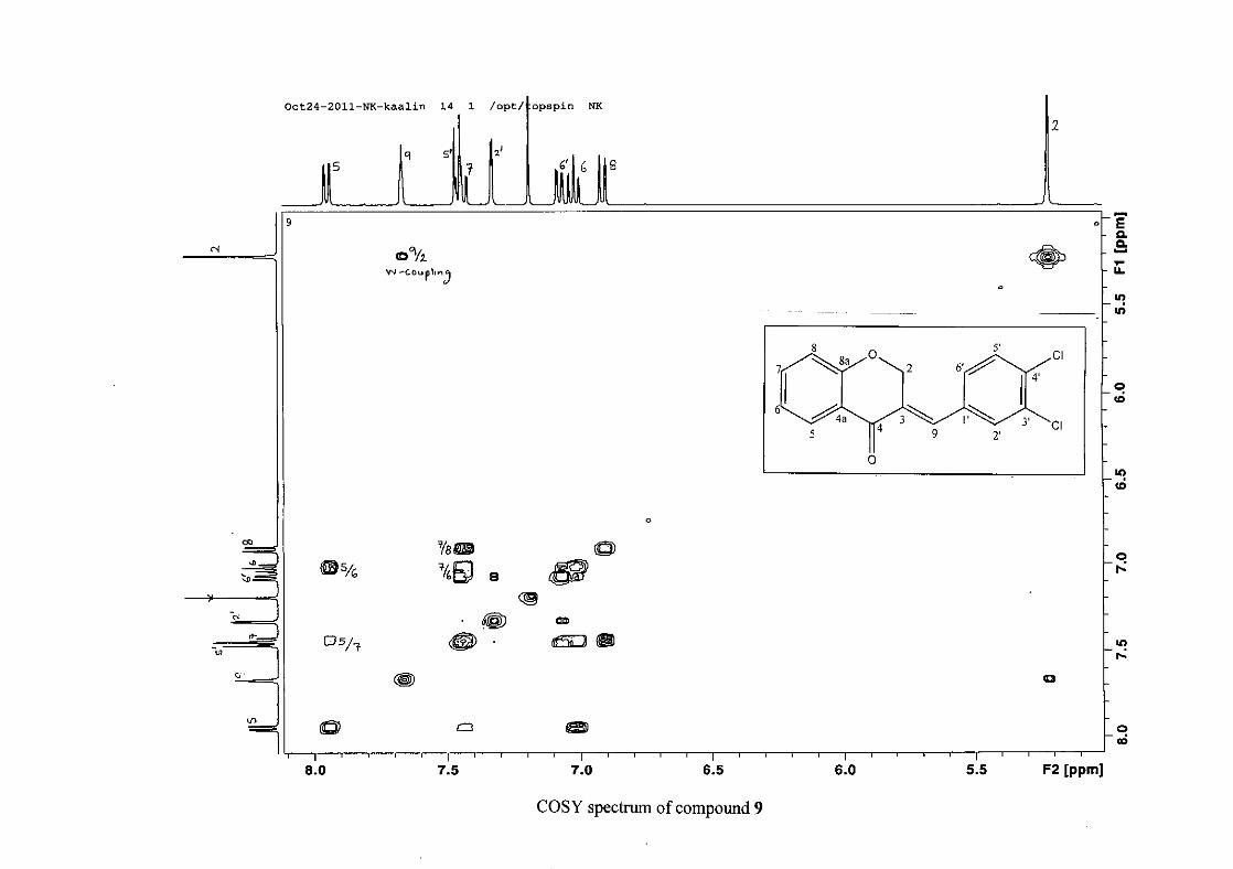
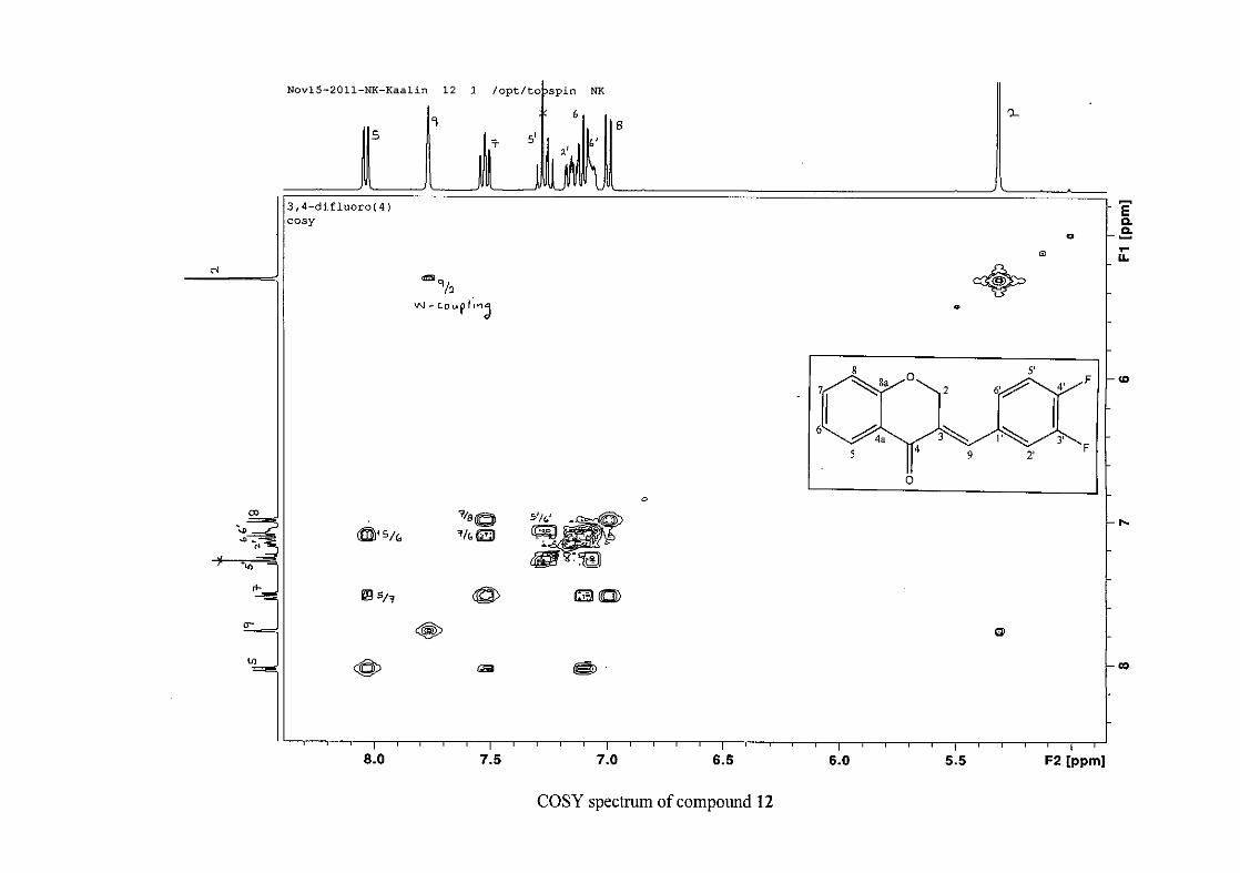
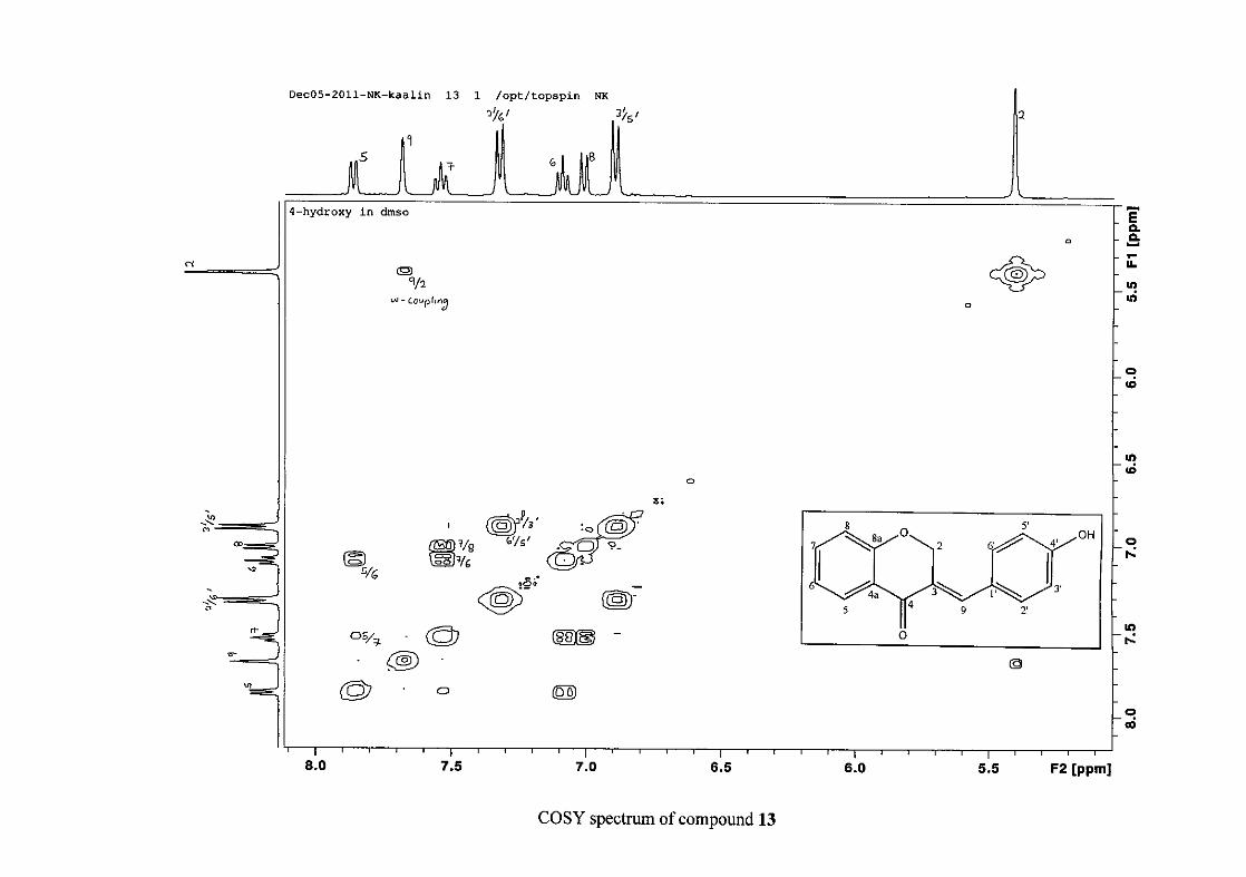
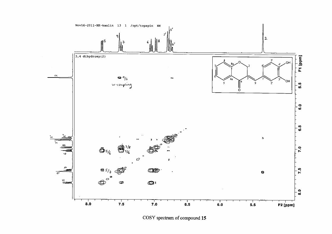
present and meta and para coupling could not be detected, but in the COSY spectrum H-8
was seen coupled to H-6. The H-6 proton resonance appeared as a triplet due to the
coalescing of the double doublets that occurs because of similar coupling constants between
H-6 and H-5 and H-6 and H-7 with J = 7.50 Hz. Although this should also be observed for
H-7, this cannot be distinguished because of overlapping with the aromatic protons of the
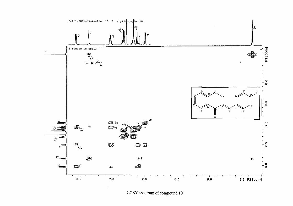
phenyl ring. In the case of the phenyl ring protons, the H-3'/5' resonance and the H-2'/6'
resonances can both be distinguished as doublets with similar J values of 7.36 and 6.96 Hz
respectively. There is a small difference between the coupling constants of coupled proton
resonances, for example, the H-5 proton resonance has J = 7.86 Hz, but the triplet of H-6 has
a J value of 7.50 Hz. We attribute this to the coalescing of resonances, where peaks overlap
and also to the broadened resonances for some of the proton peaks. However, we confirmed
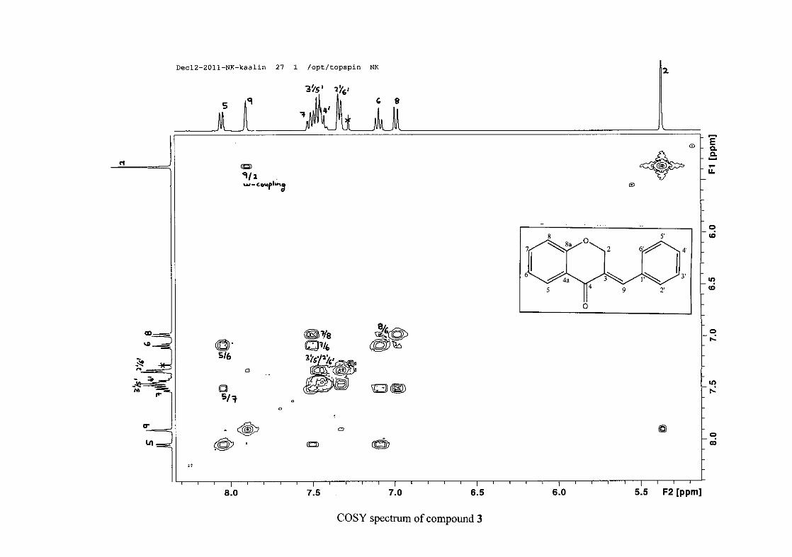
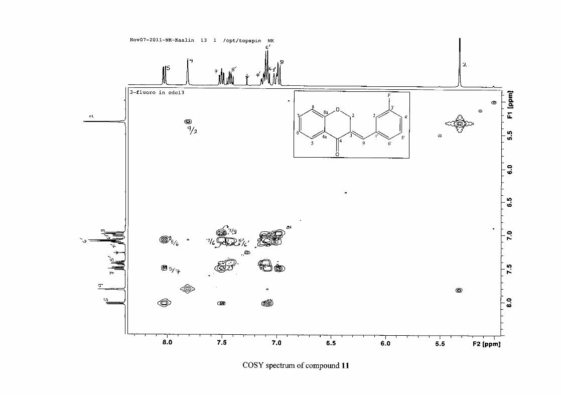
the coupling of all the resonances with the aid of the COSY spectrum.
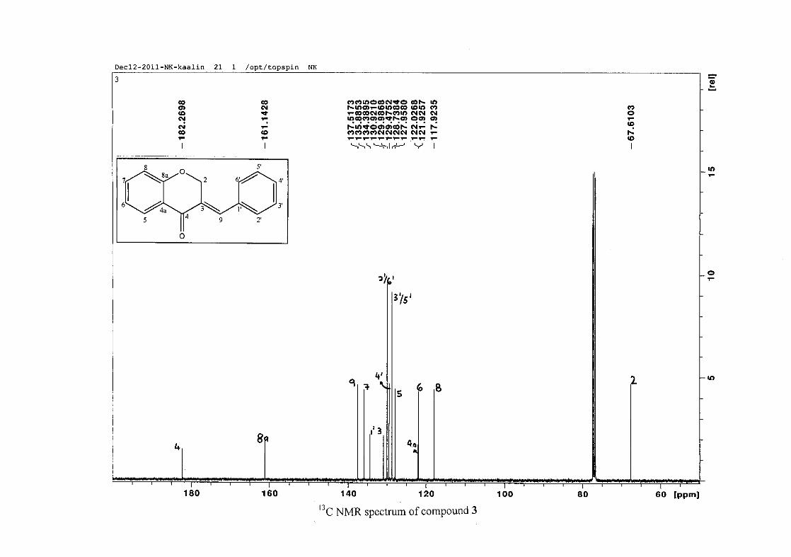
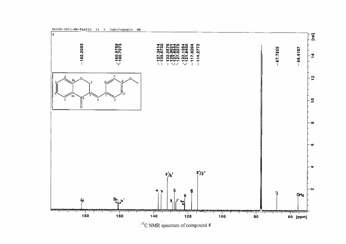
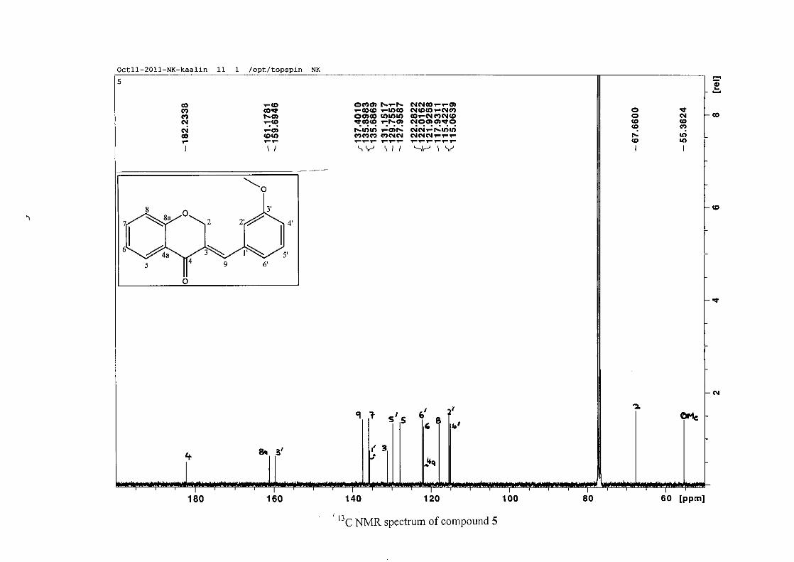
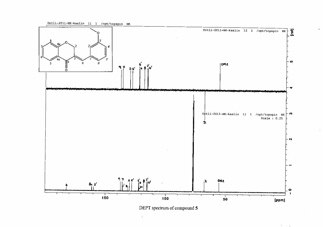
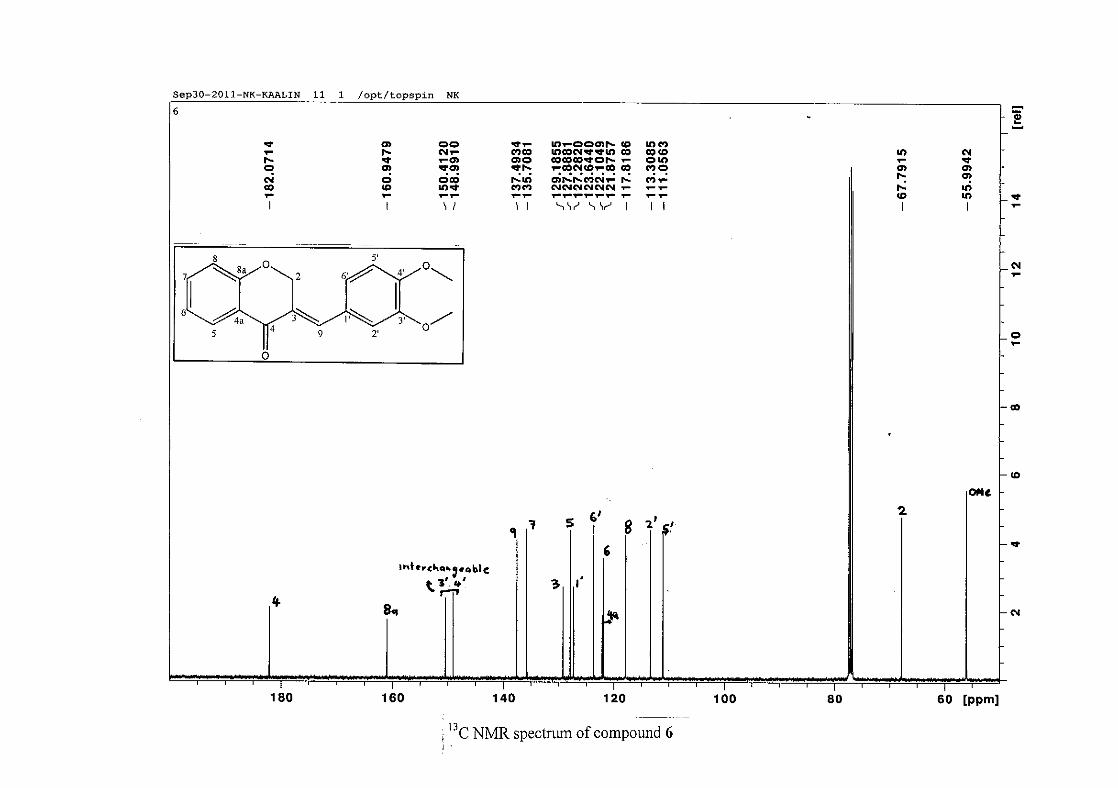
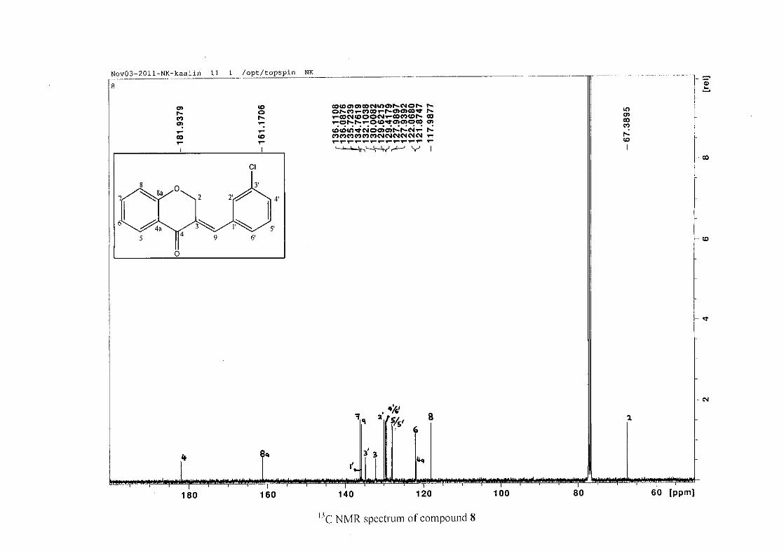
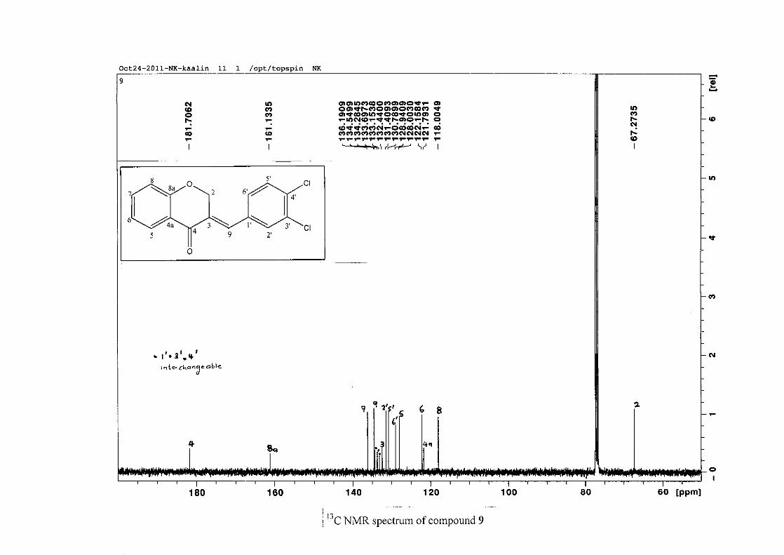
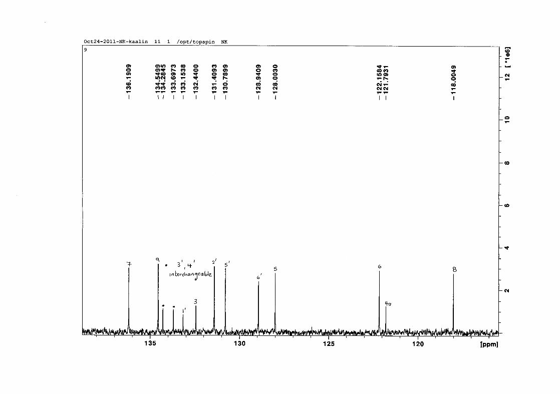
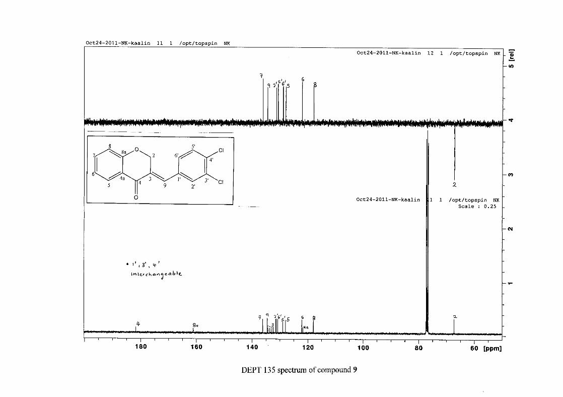
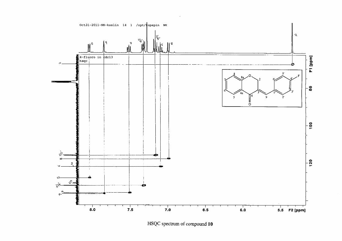
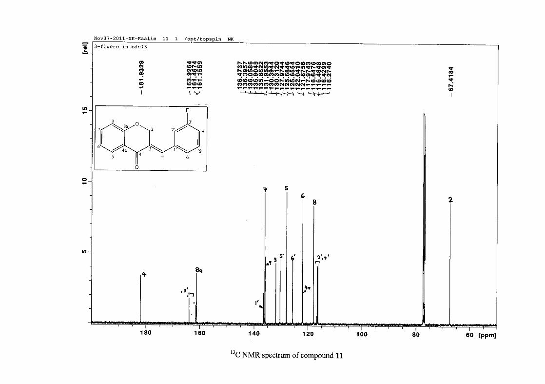
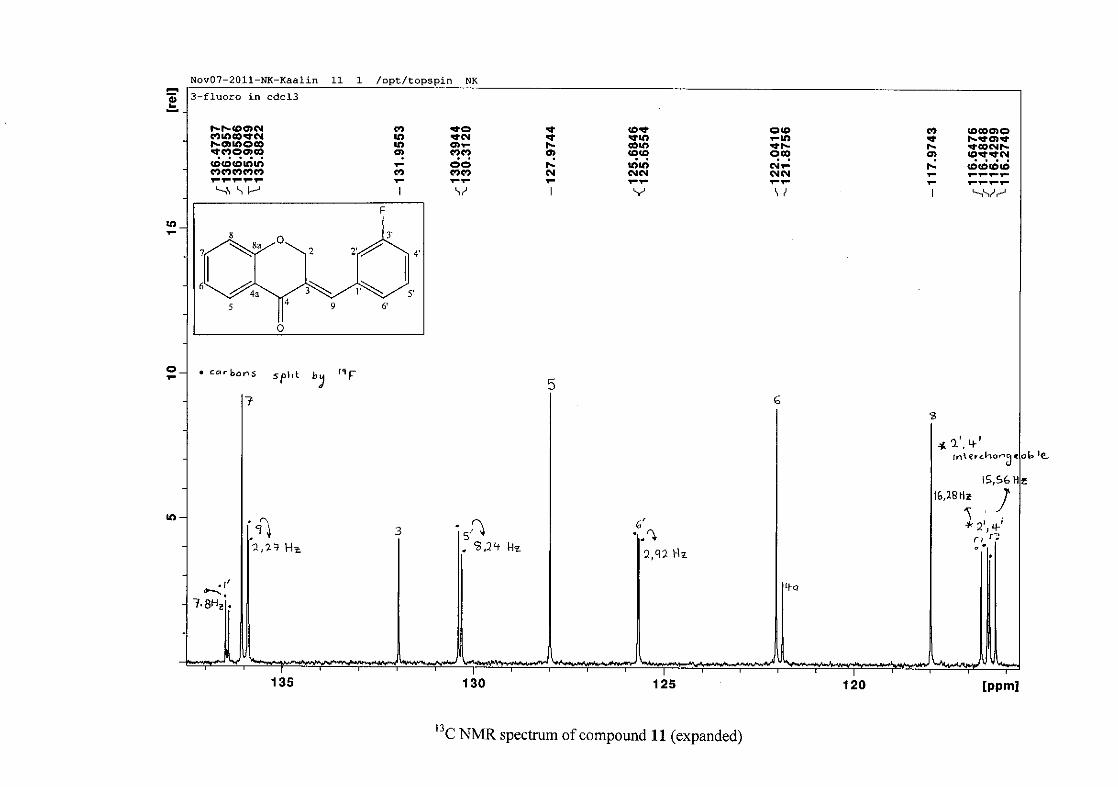
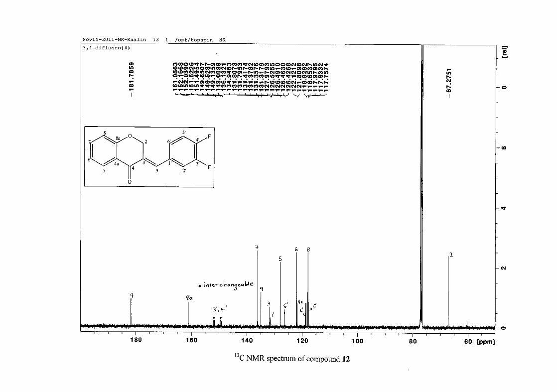
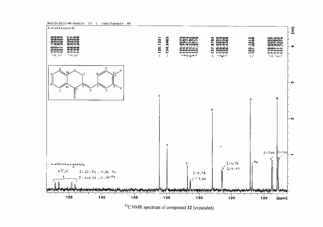
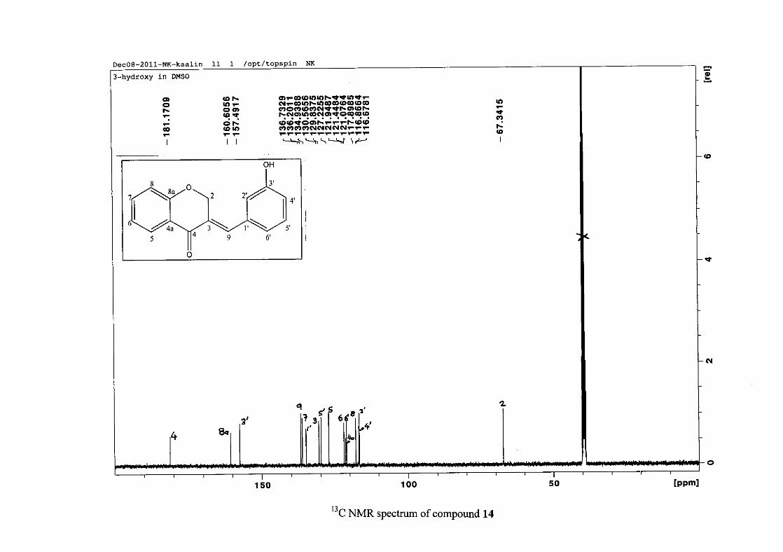
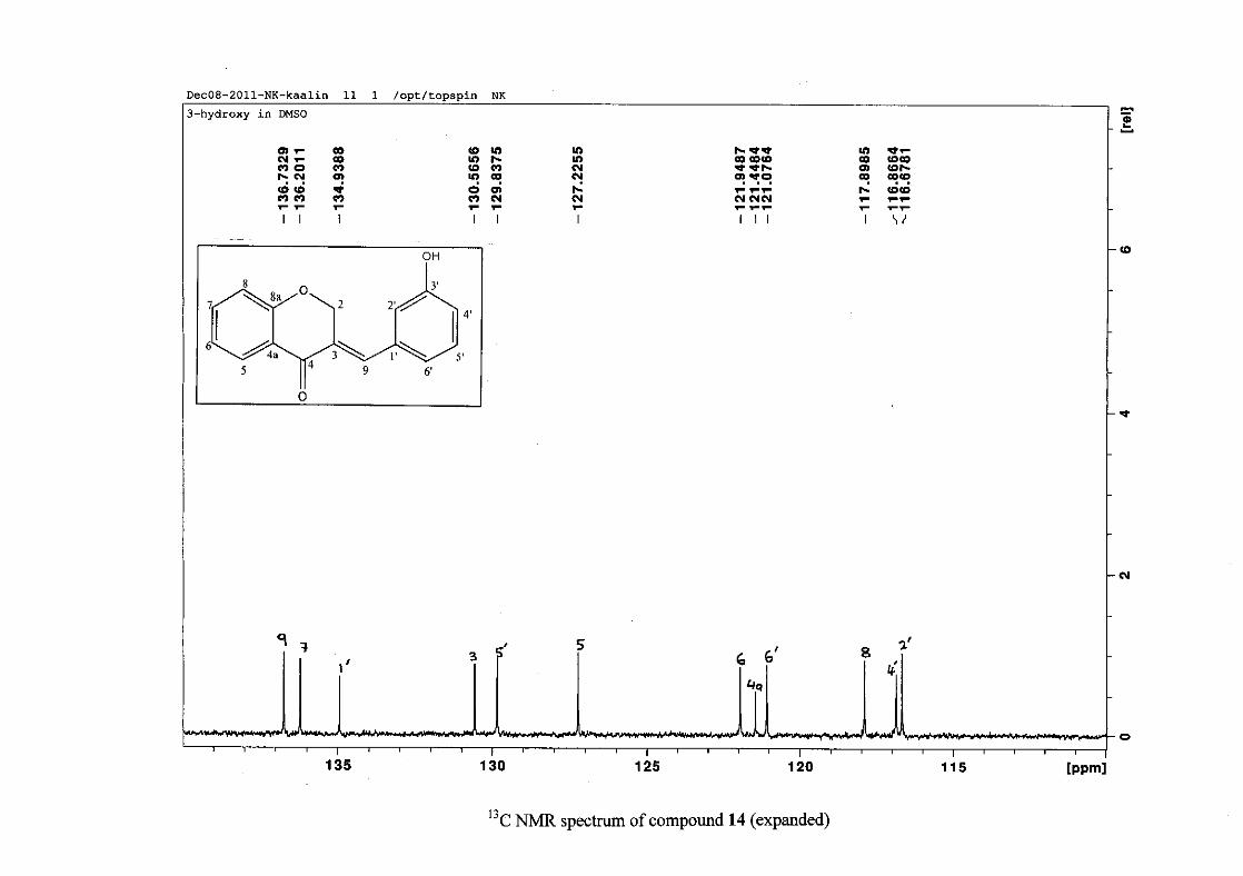
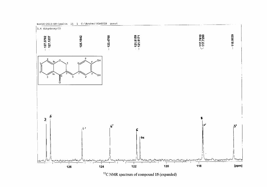
The 13C NMR spectrum of compound 3 showed the presence of fourteen carbon resonances
with two of the resonances being equivalent and therefore amounting to sixteen carbon
33
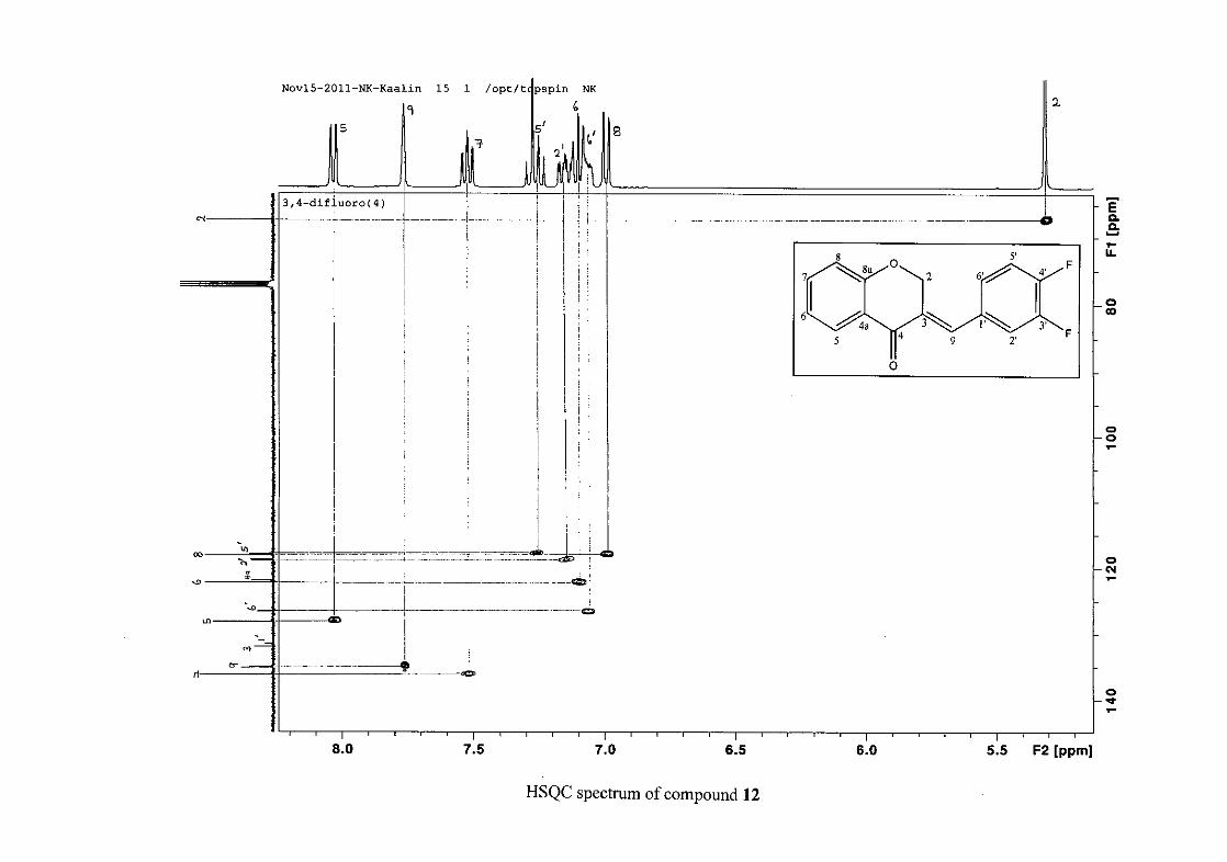
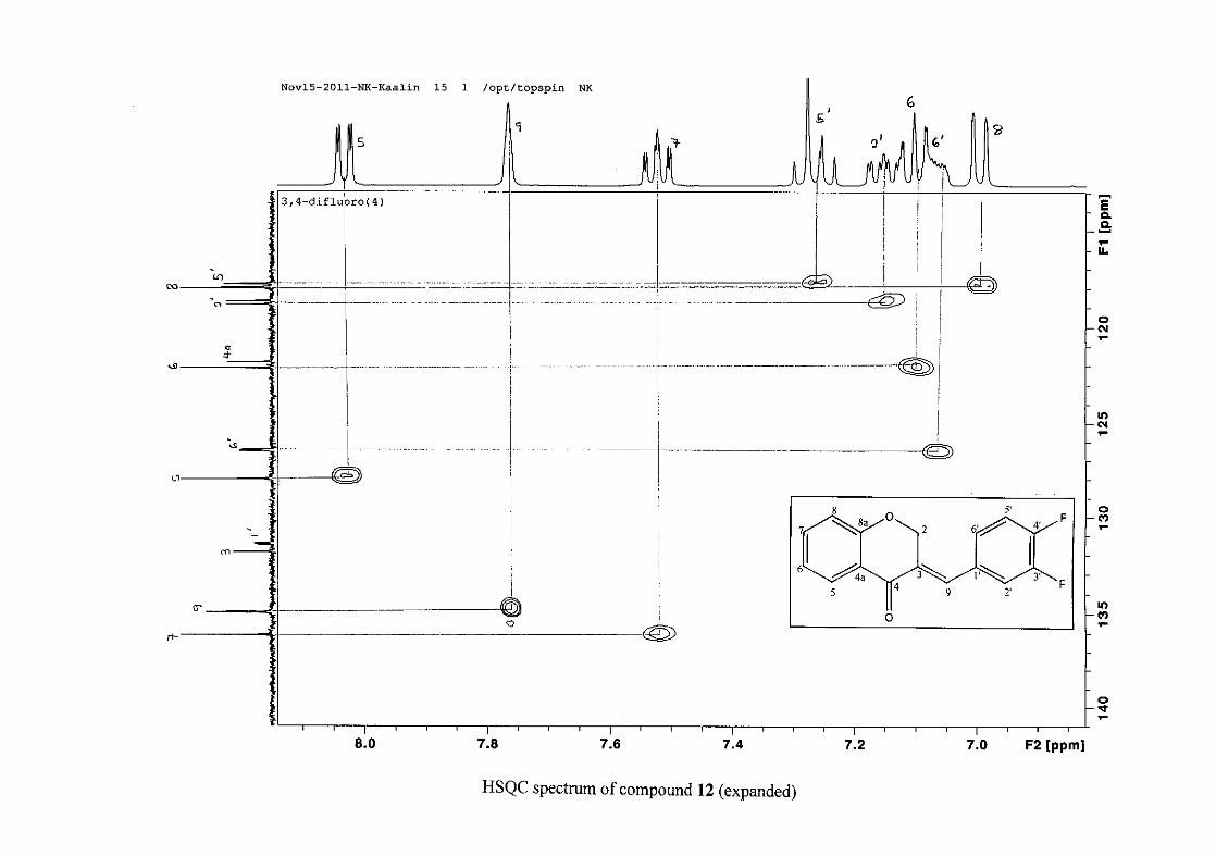
resonances, which confirms the presence of a homoisoflavonoid skeleton. The oxygenated
aliphatic carbon resonance of C-2 occurs at δC 67.61 typical for C-2, with that of C-4, the
carbonyl resonance occurring at δC 182.27 and the oxygenated aromatic carbon of C-8a
occurring at δC 161.14 typical for these resonances in benzylidene homoisoflavonoids
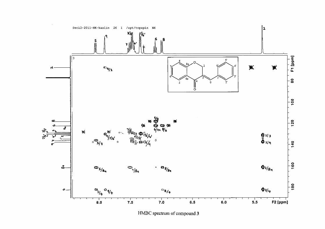
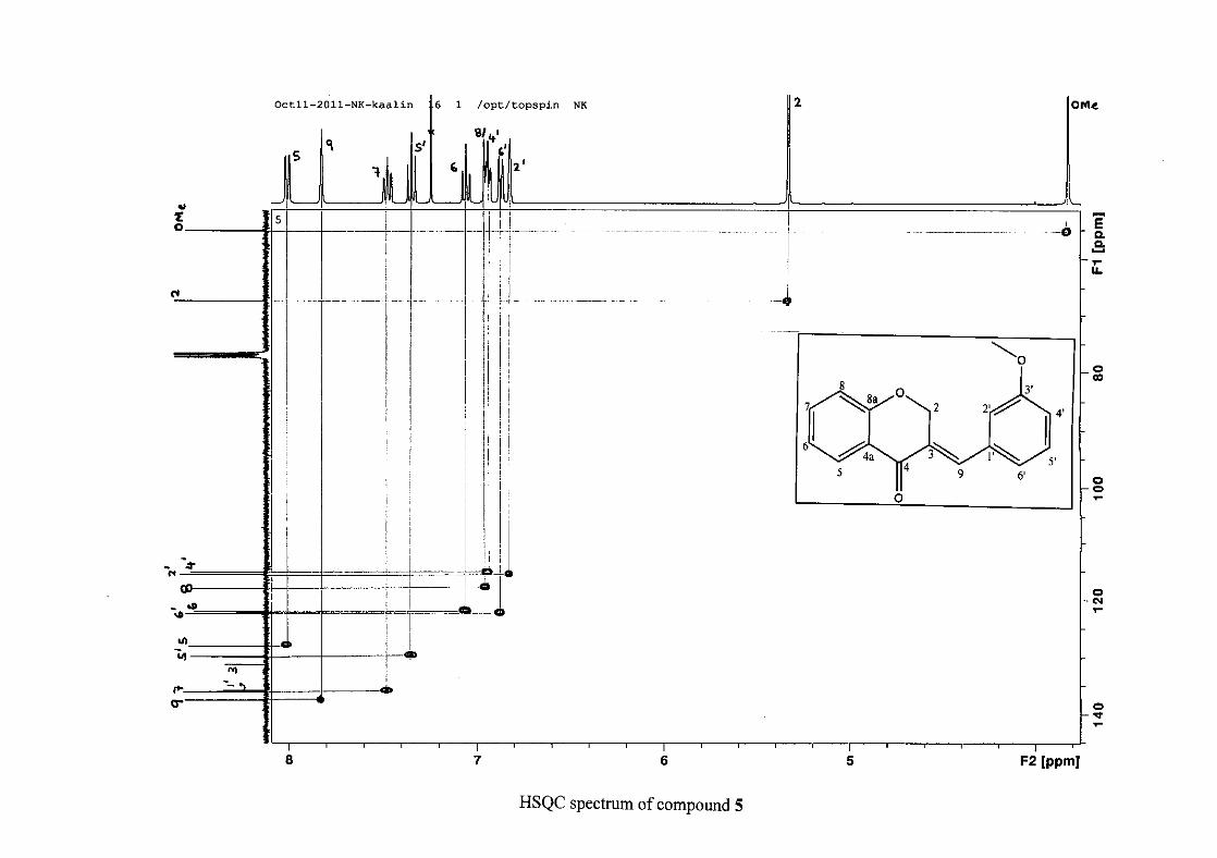
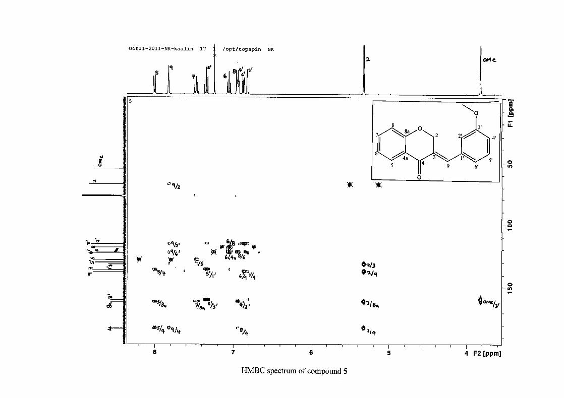
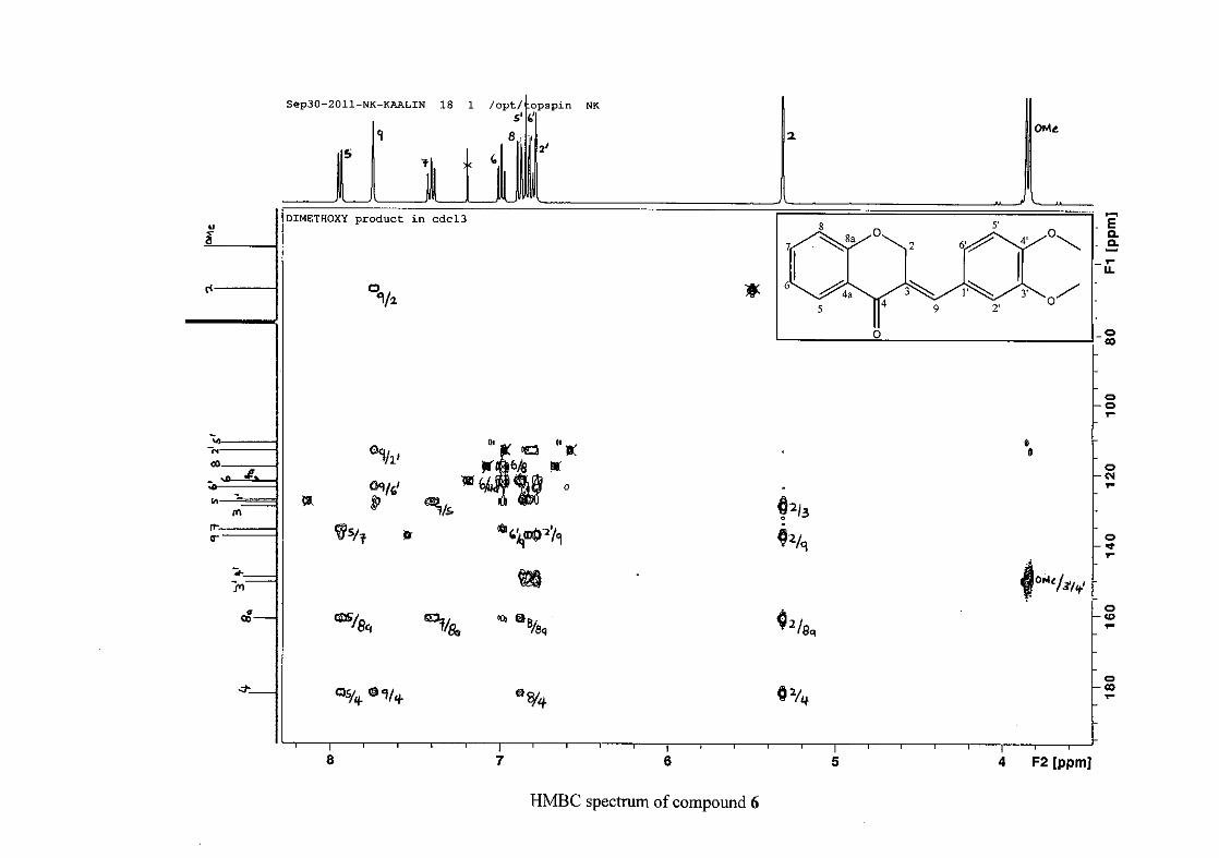
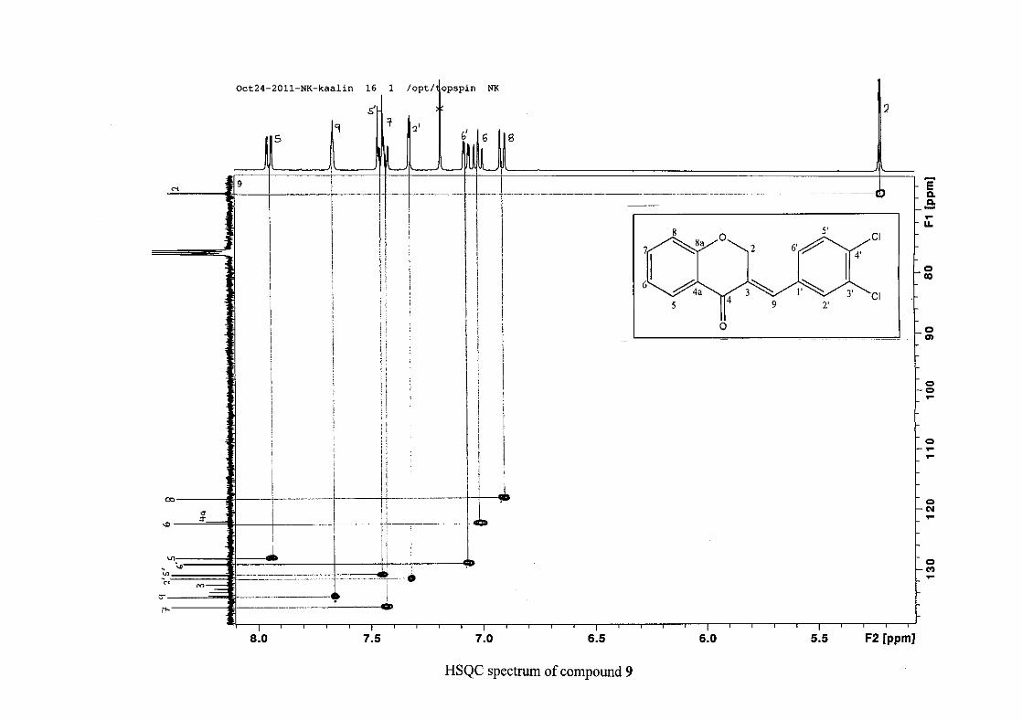
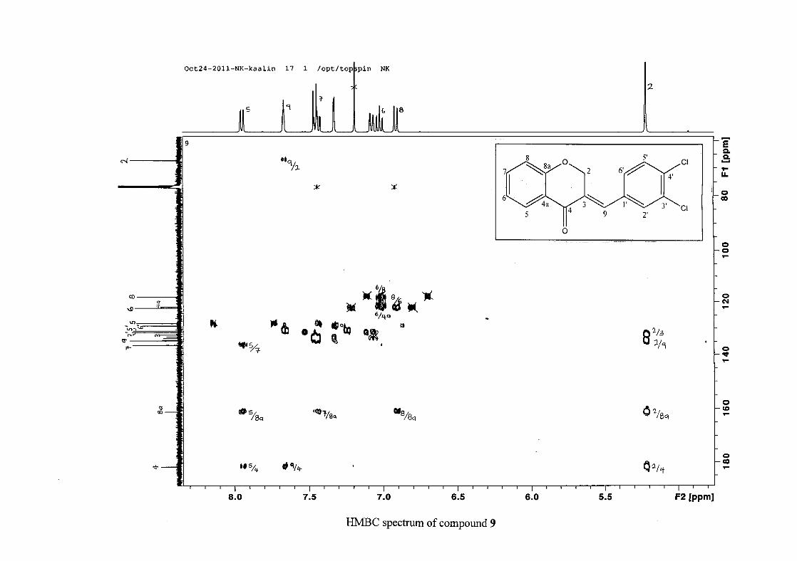
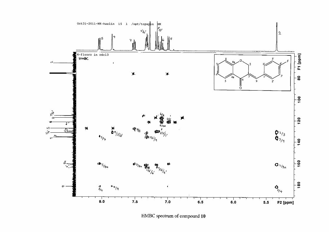
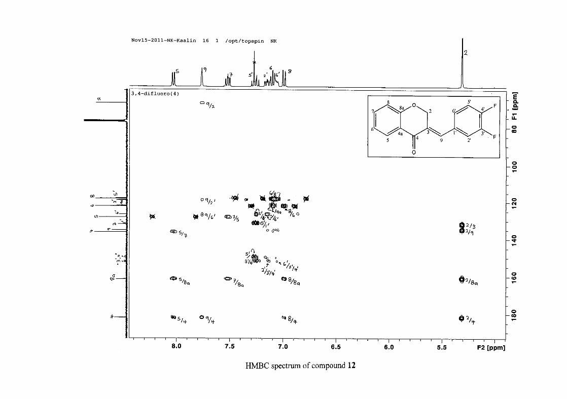
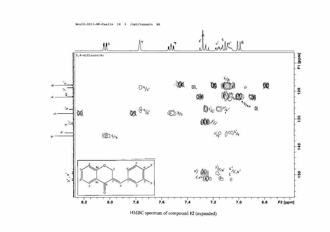
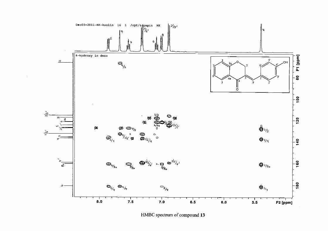
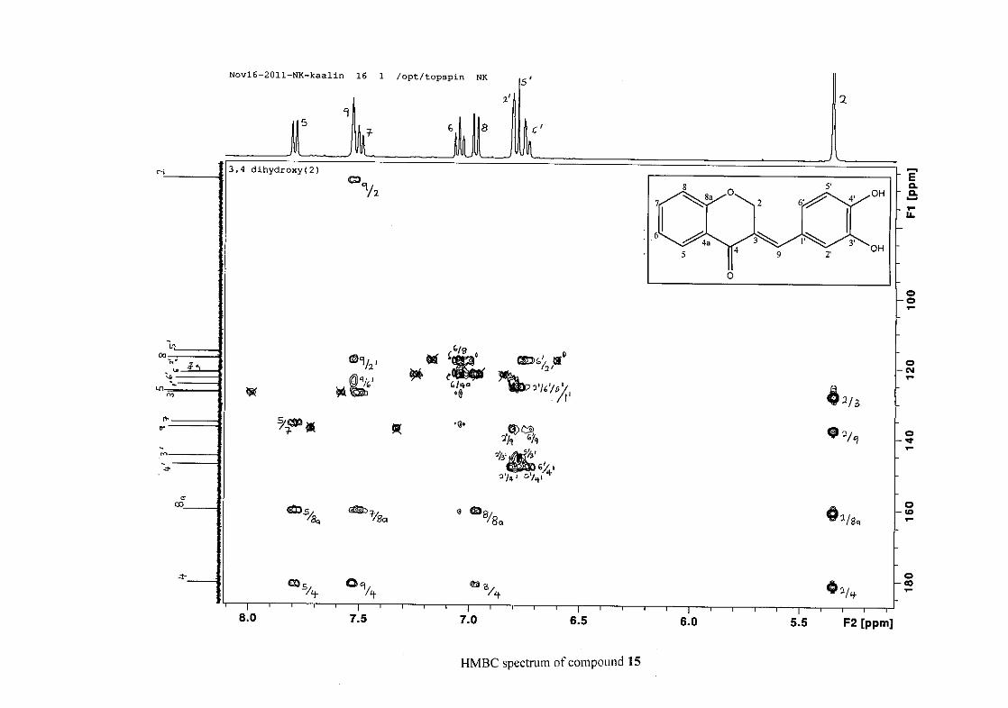
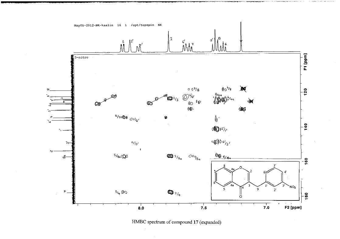
(Jacquot et al., 2012). This was confirmed by the presence of HMBC correlations between
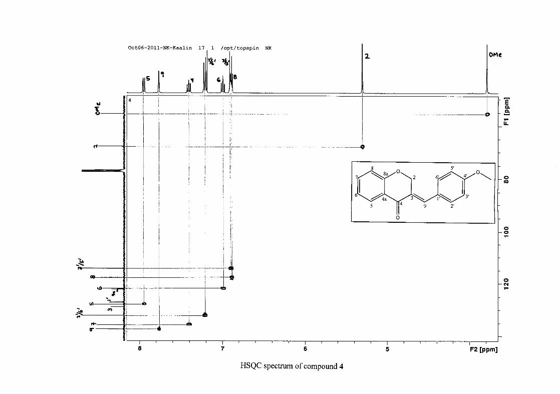
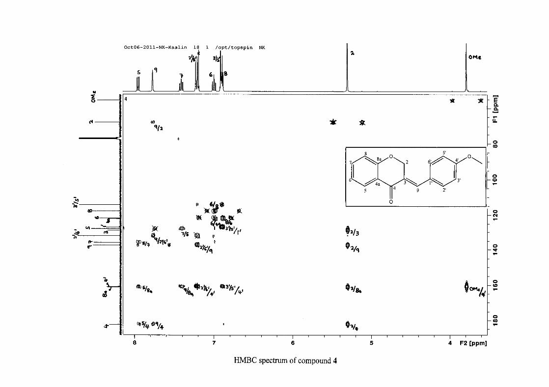
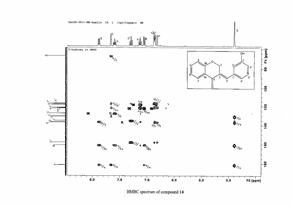
C-4 and H-5, C-2 and H-9 and C-8a with H-2, H-5 and H-8.
The C-6, C-8 and C-4a are the most shielded of all the aromatic resonances due to the
electronic effects explained above (Scheme 15 and Scheme 16). The remaining two aromatic
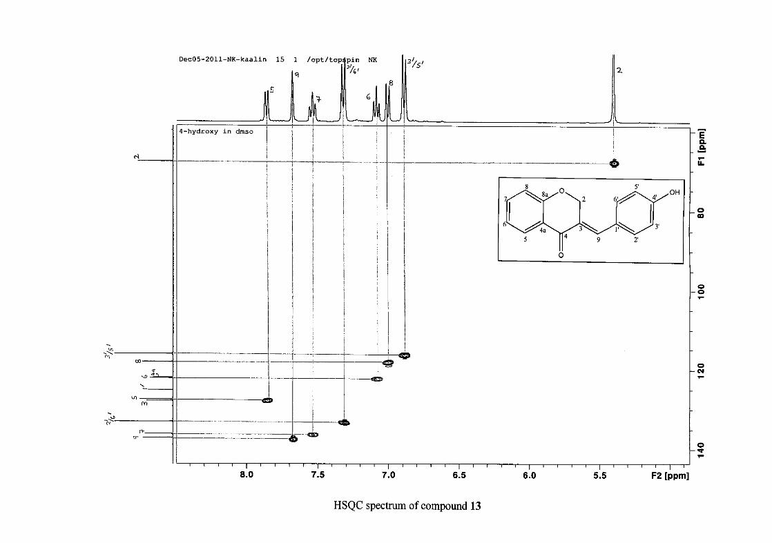
carbon resonances on the chromanone ring, C-5 and C-7 occur at δC 127.96 and 135.89
respectively. The resonances of C-5 to C-8 were determined by their corresponding proton
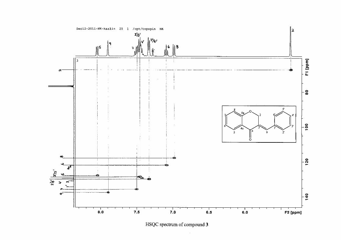
resonances in the HSQC spectrum. The equivalent phenyl carbon resonances of C-2'/6'
occurs slightly more downfield at δC 129.99 compared to the C-3'/5' resonance of δC 128.74.
This was confirmed by a HMBC correlation between C-2'/6' and H-9. The C-4' carbon
resonance lies in between these two resonances at δC 129.48. The C-1' carbon resonance was
assigned at δC 134.39 due to a HMBC correlation with H-3'/5'. The remaining olefinic
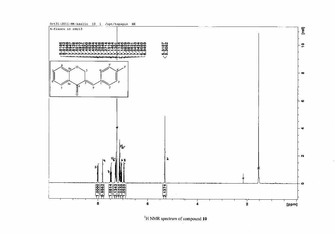
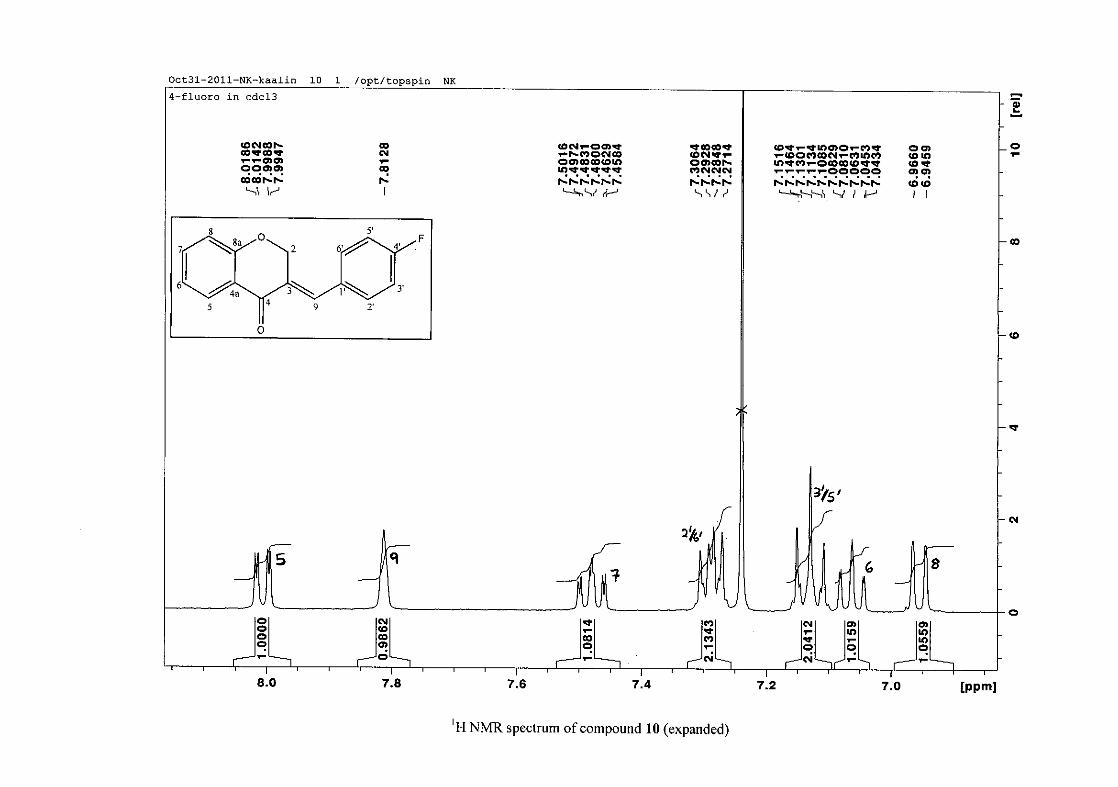
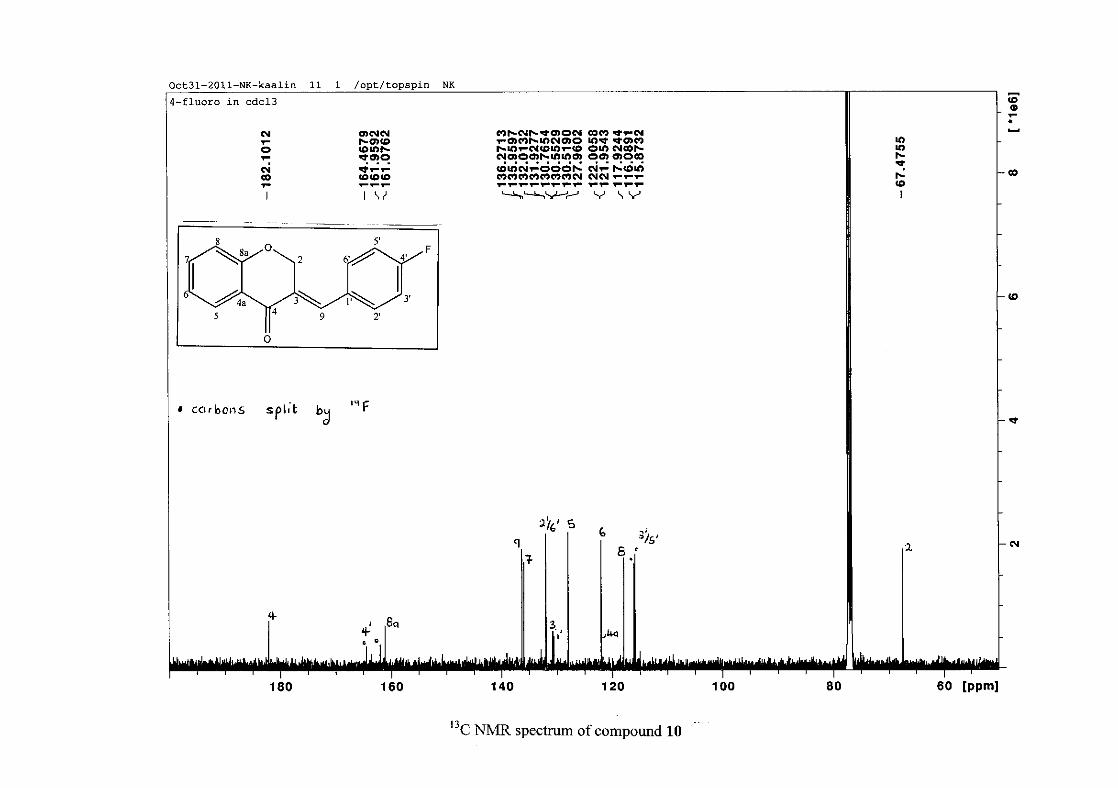
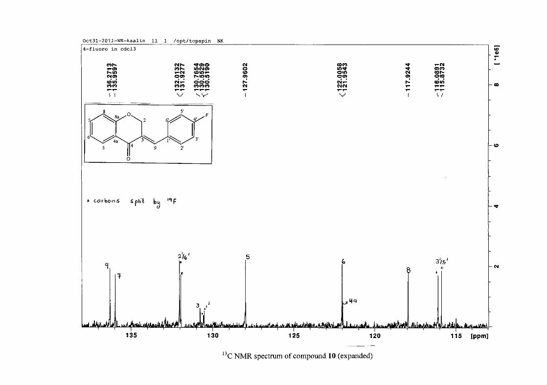
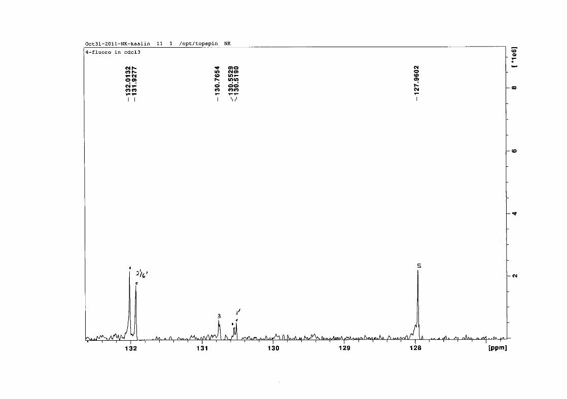
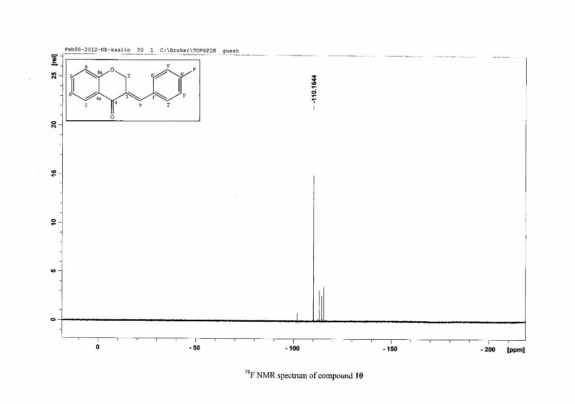
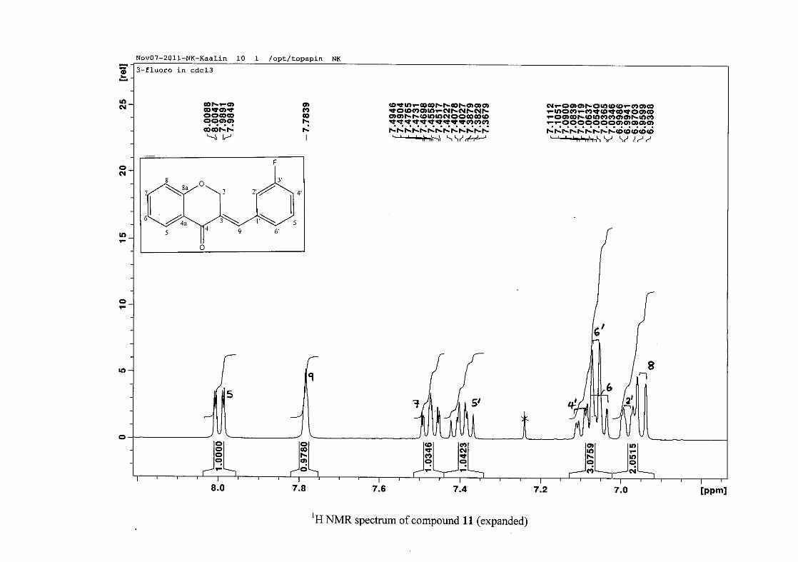
carbon resonance of C-3 was assigned to δC 130.92 because of a HMBC correlation to H-2.
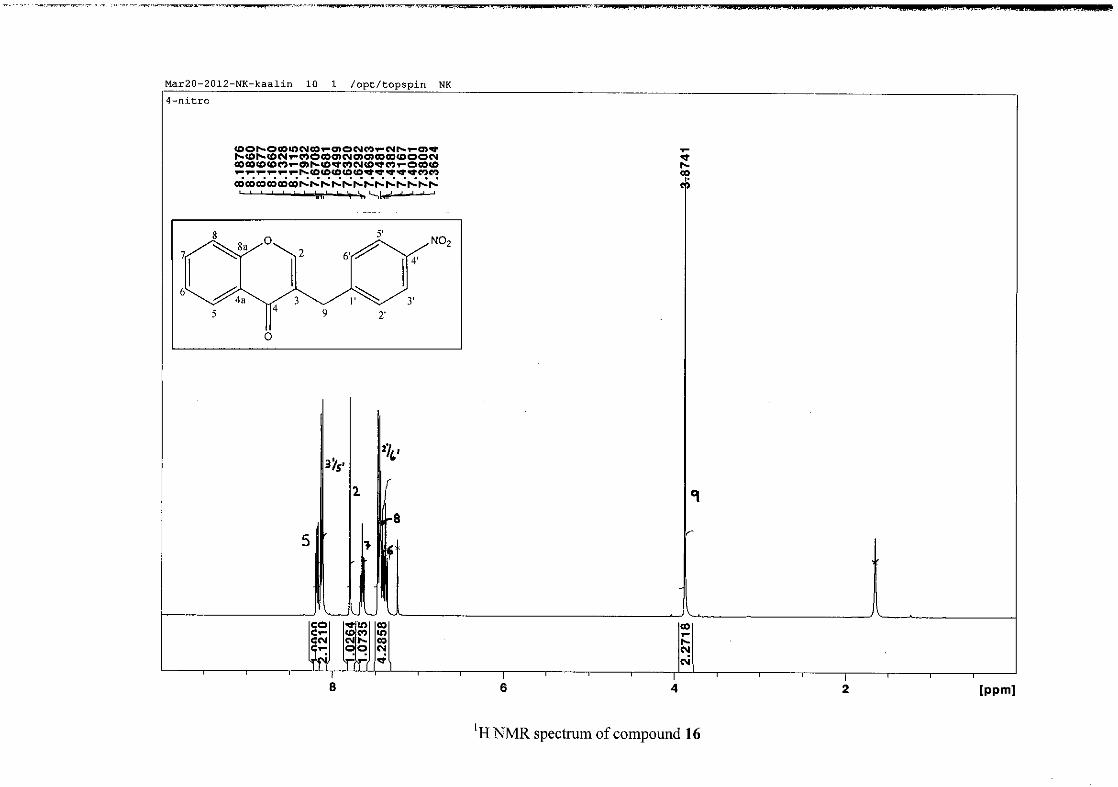
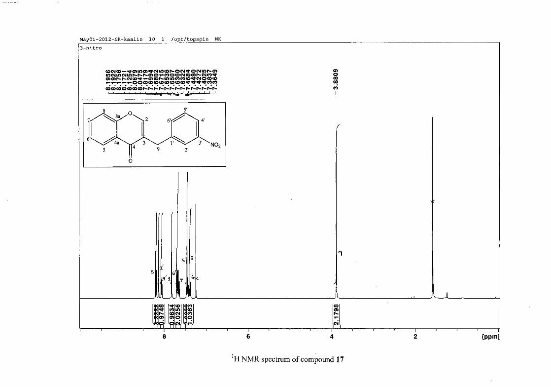
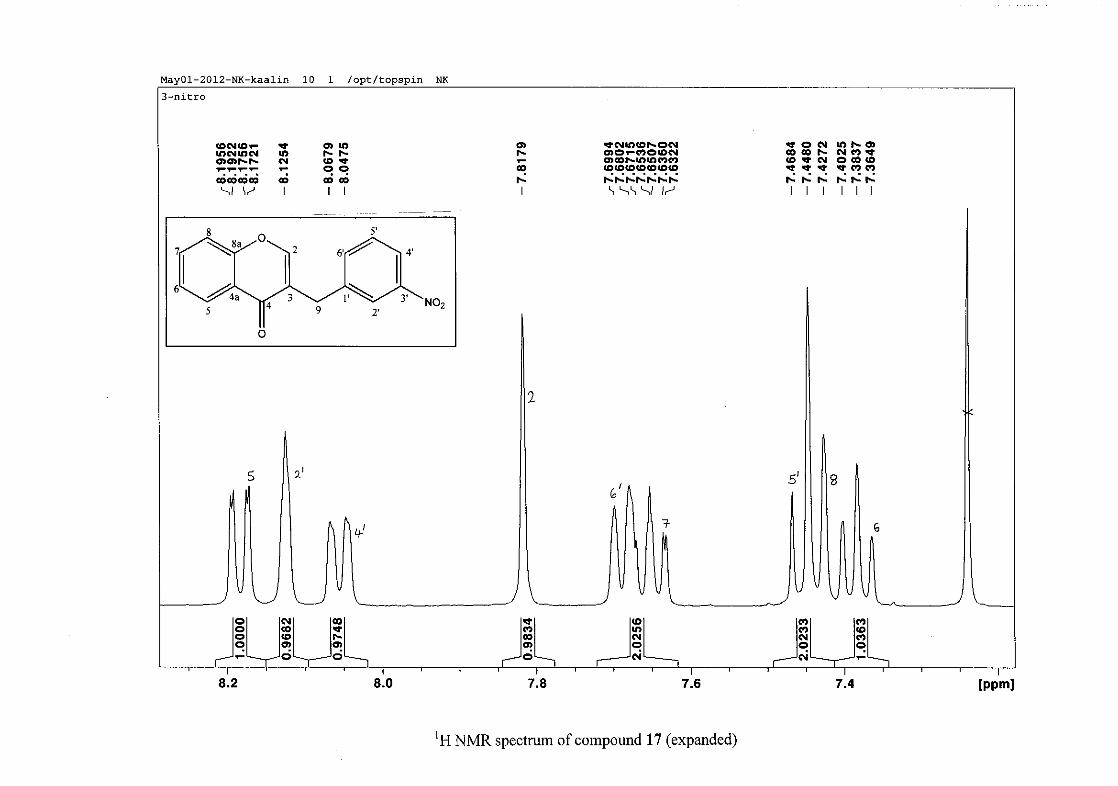
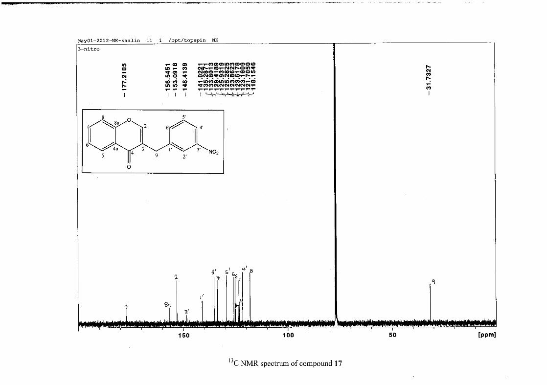
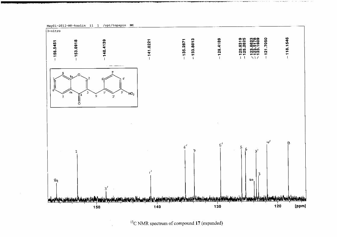
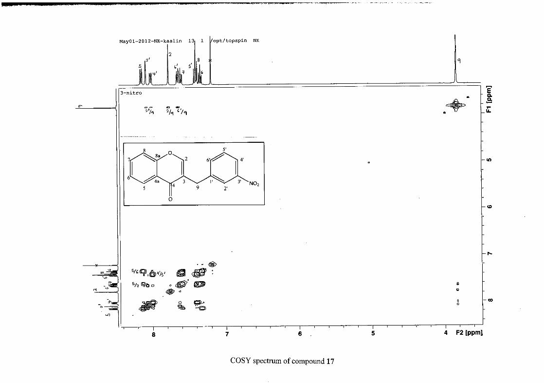
The 1H and 13C NMR resonances compare well with that in the literature (Jacquot et al.,
2012).
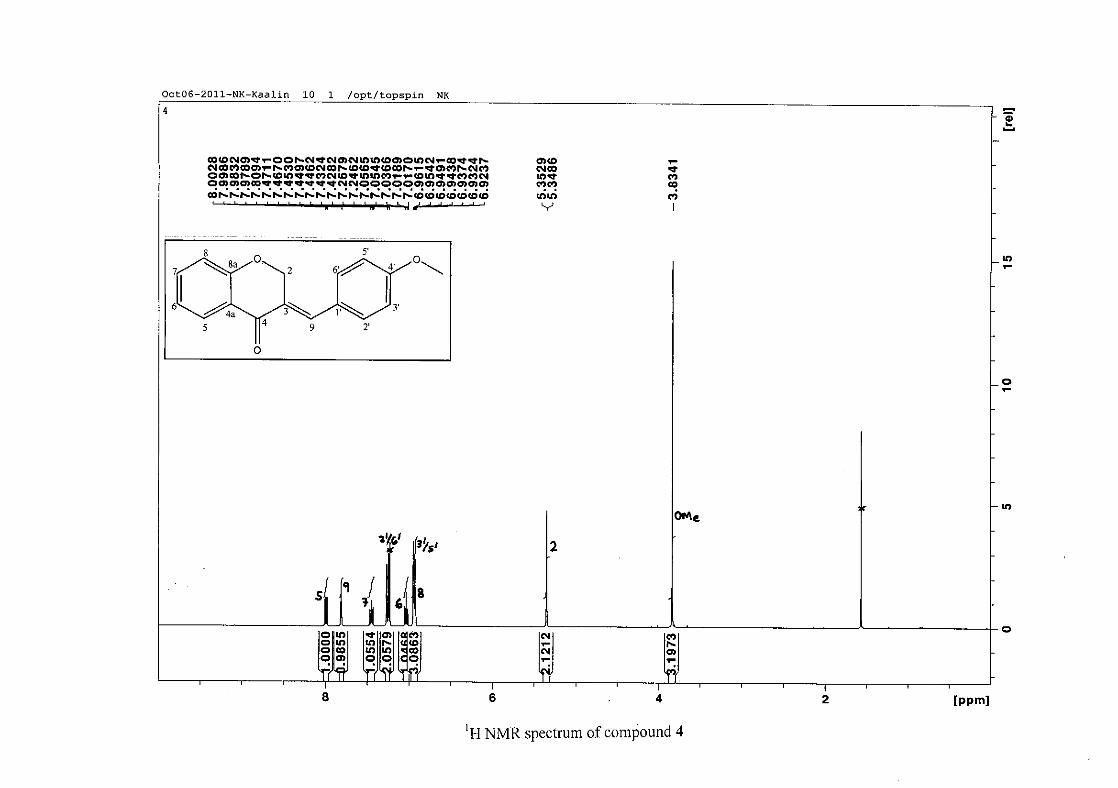
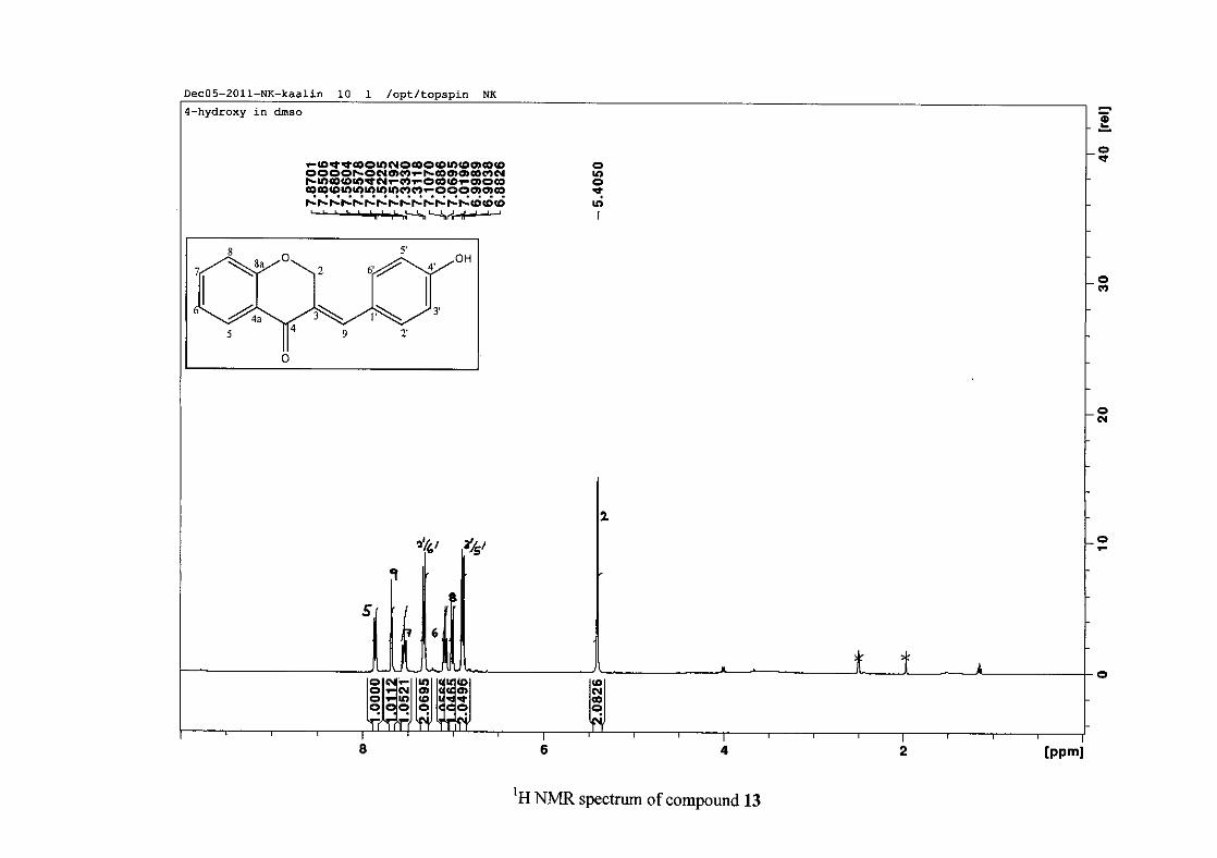
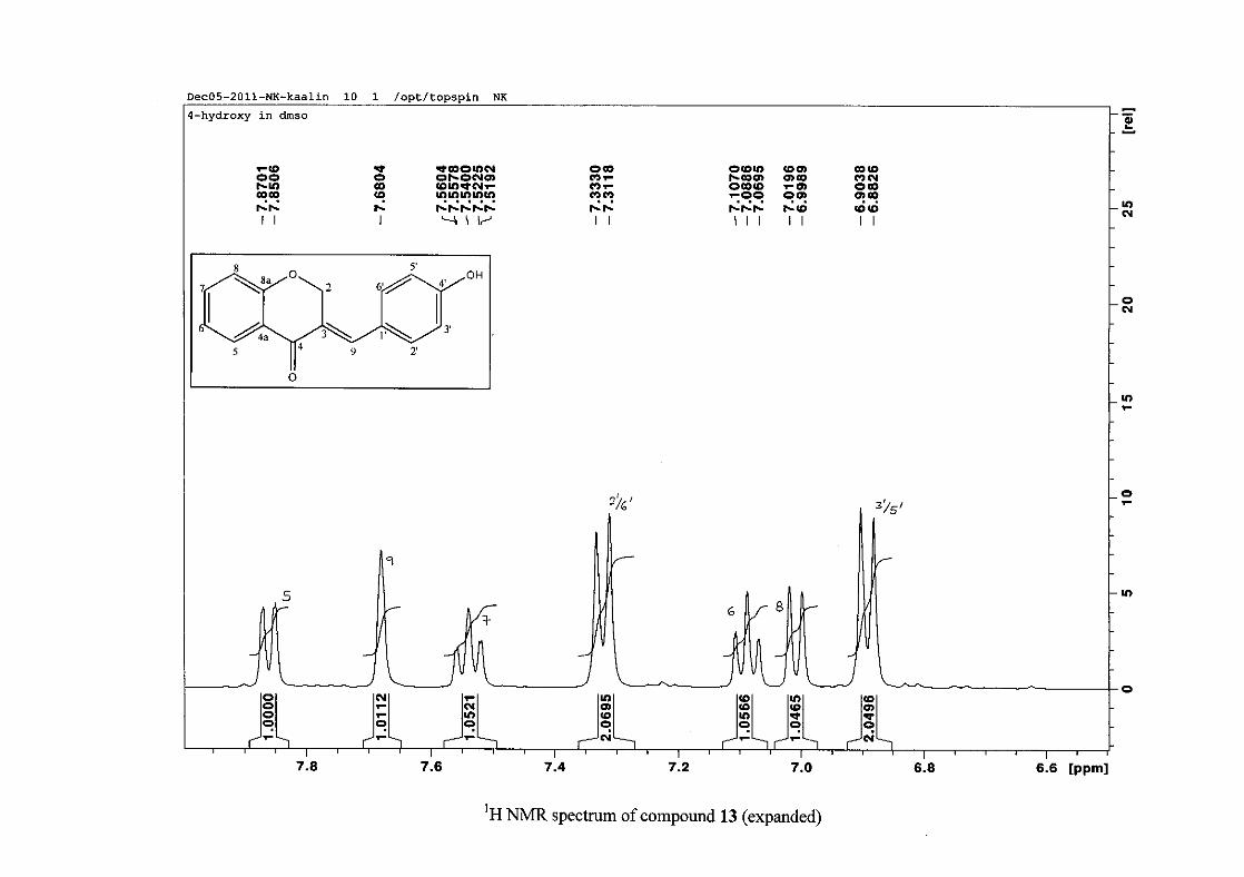
2.1.4 Structural elucidation of the para substituted derivatives (except 4'-fluoro)
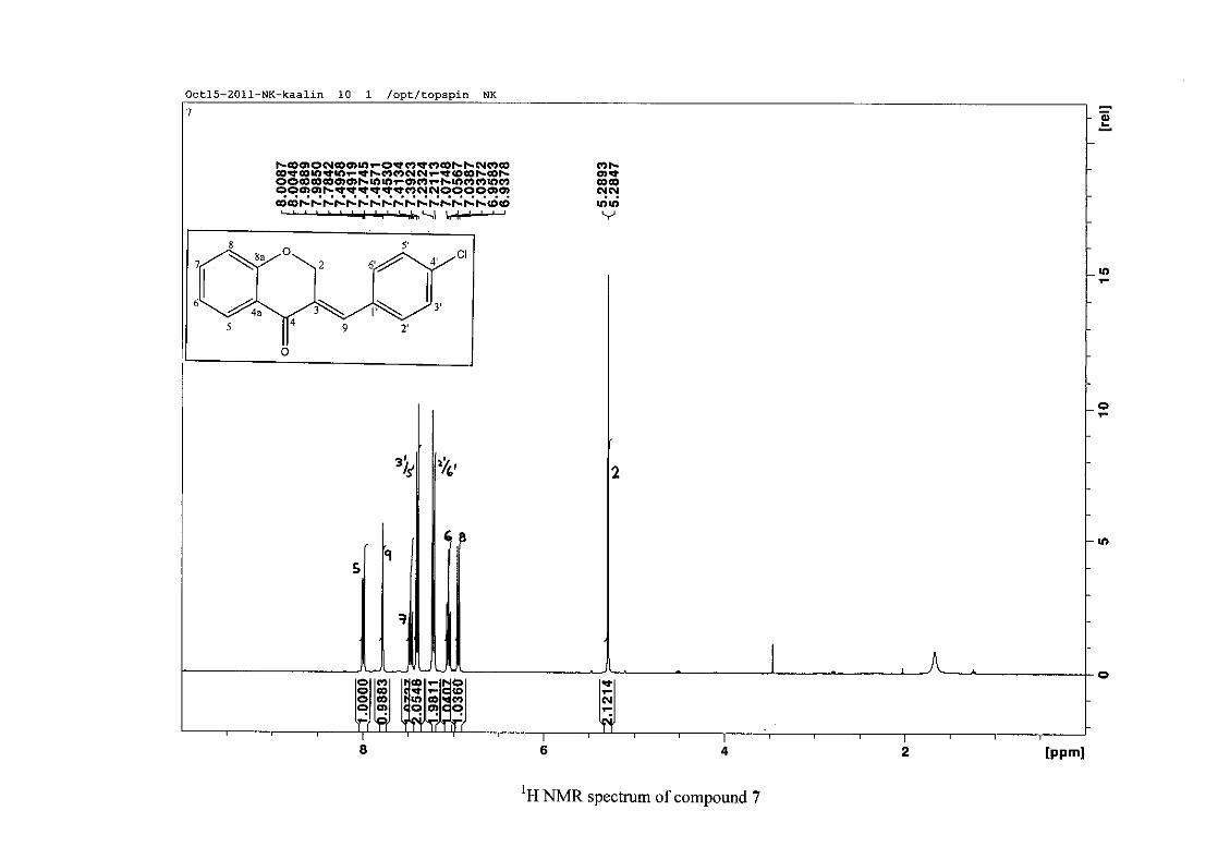
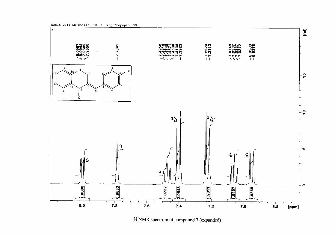
For the para substituted B-ring benzylidene homoisoflavonoids (4, 7 and 13), excluding the
para fluoro substituted compound (10), the 1H NMR spectrum showed the ortho coupled
proton resonances of H-2'/6' and H-3'/5' as doublets with coupling constants between 8.4 and
34
8.8 Hz for the three compounds. These are located at δH 7.26 and 6.96 for the methoxy
derivative (4), δH 7.32 and 6.89 for the hydroxy derivative (13) and δH 7.22 and 7.40 for the
chloro derivative (7) for the H-2'/6' and H-3'/5' resonances respectively. For compounds 4
and 13, with an activating electron donating methoxy or hydroxy group at the para position,
the H-3'/5' resonance is more shielded due to electron donation and build-up of electron
density at these carbon atoms. In contrast, the para chloro derivative had the H-3'/5' proton
resonances more deshielded. Even though the chloro group has lone pairs and is capable of
electron donation toward the ring, the inductive effects of the deactivating chloro group is
responsible for this effect. In the unsubstituted benzylidene homoisoflavonoid (3), the H-7
and H-3'/5' resonances overlapped at δH 7.47 but when a substituent was placed at the para
position of this ring, causing the H-3'/5' resonance to be shifted, the H-7 resonance can now
be clearly seen as a triplet of doublets (td) in the chloro (7) and hydroxy derivatives (13) with
J7,8 and J6,7 being the same at approximately 7.7 Hz. In 4, a double double doublet (ddd) was
seen with J7,8 being slightly larger than J6,7. Both coupling constants were in the region of
8.0 Hz. The J5,7 coupling constant was seen to be approximately 1.70 Hz. A singlet
resonance for the methoxy group was seen at δH 3.83 in 4.
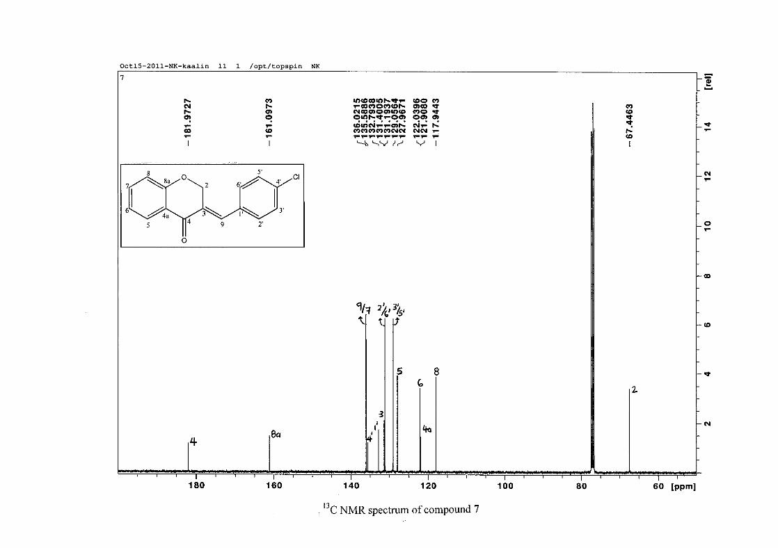
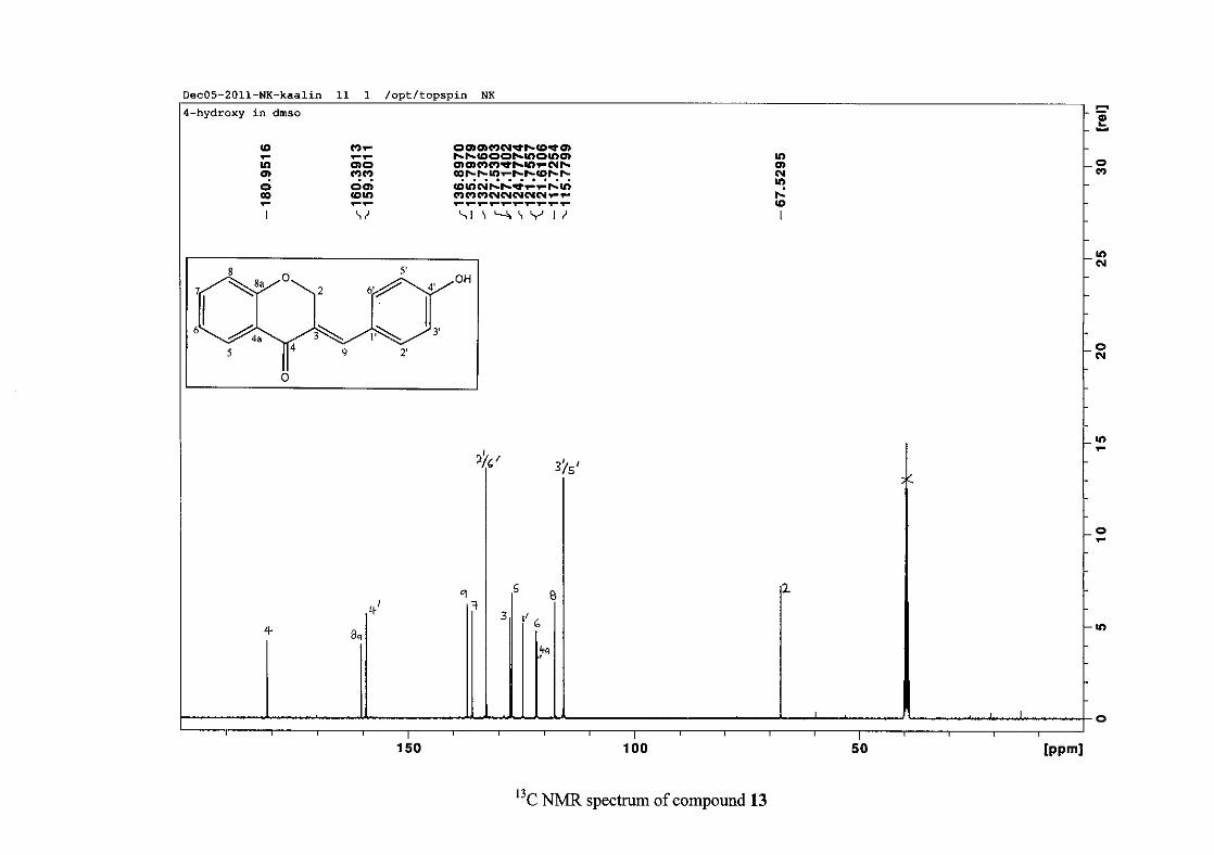
For 4 and 13, the C-4' resonance shifted more downfield in the region of C-8a at
approximately δC 160 because of the oxygenated substituent at this position. In 7, the para
chloro derivative, C-4' occurred more in the region of C-7 and C-9 at δC 135.59 due to the
chloro group being less electronegative than the oxygenated groups. The 1H and 13C NMR
data of 4, 7 and 13 compare well with that in the literature (Jacquot et al., 2012; Valkonen et
al., 2012).
35
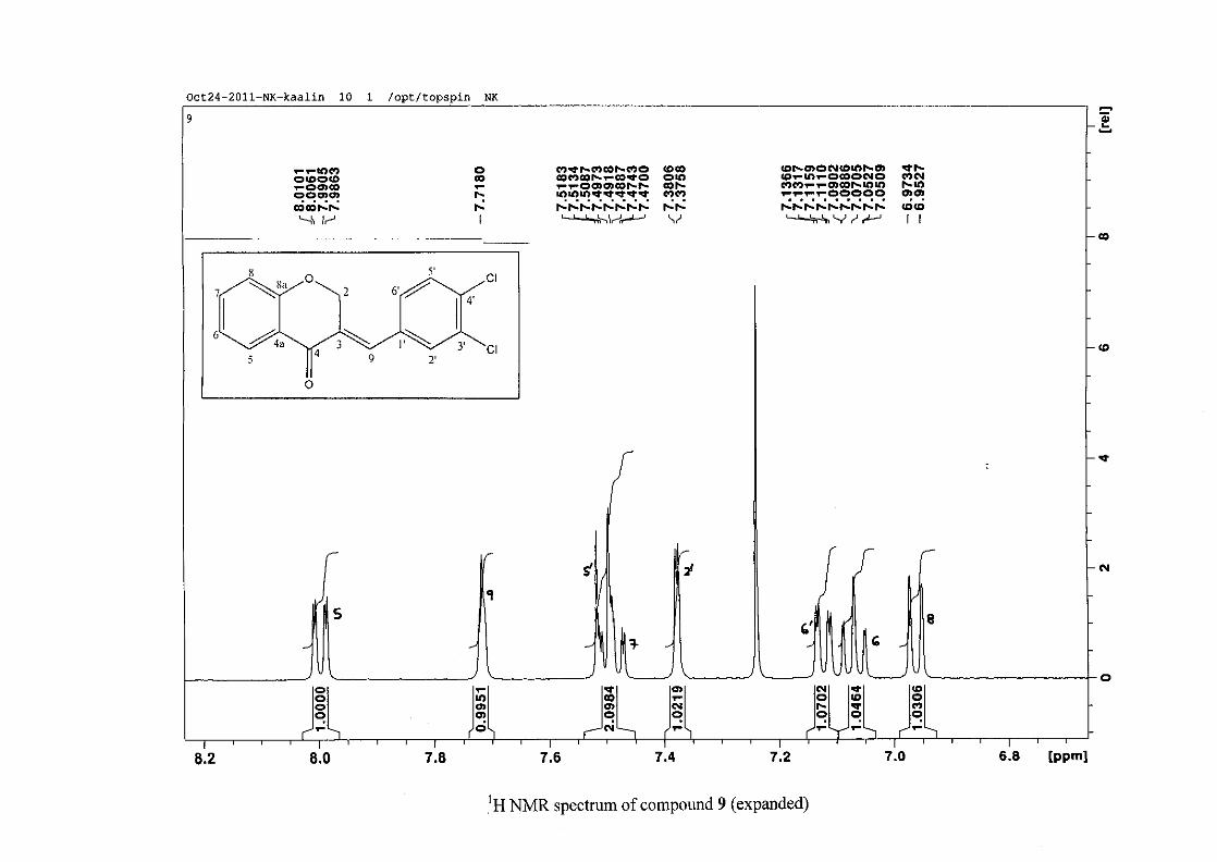
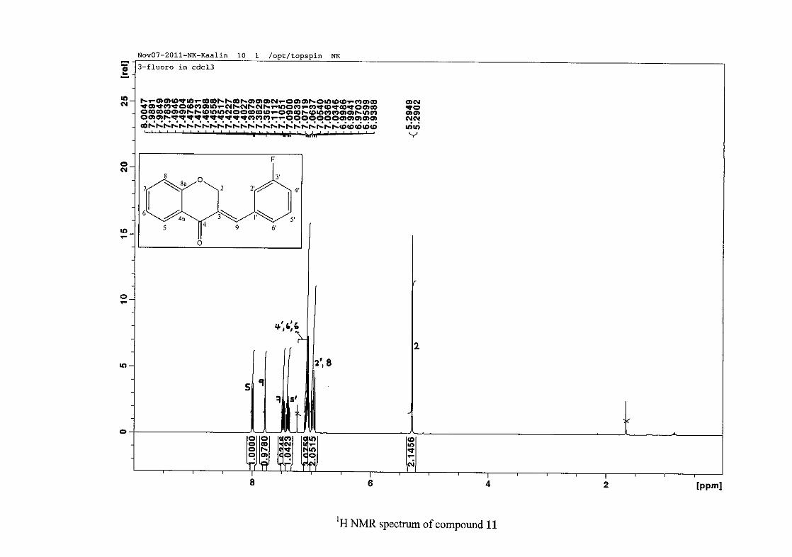
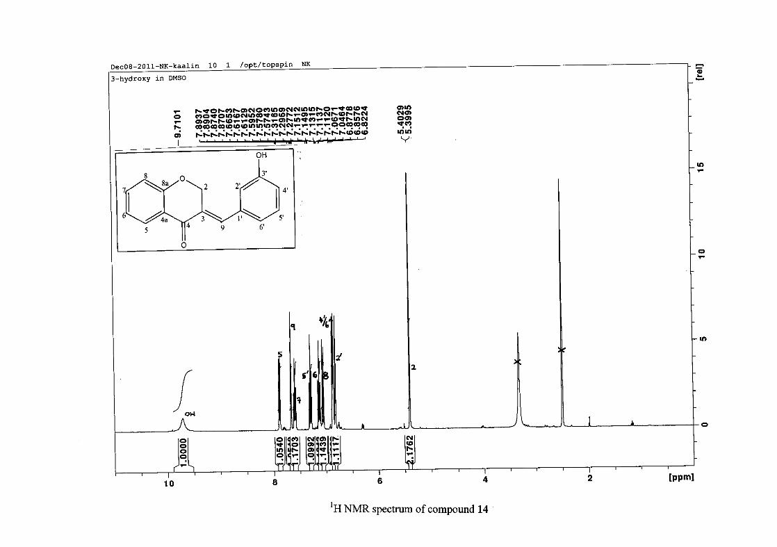
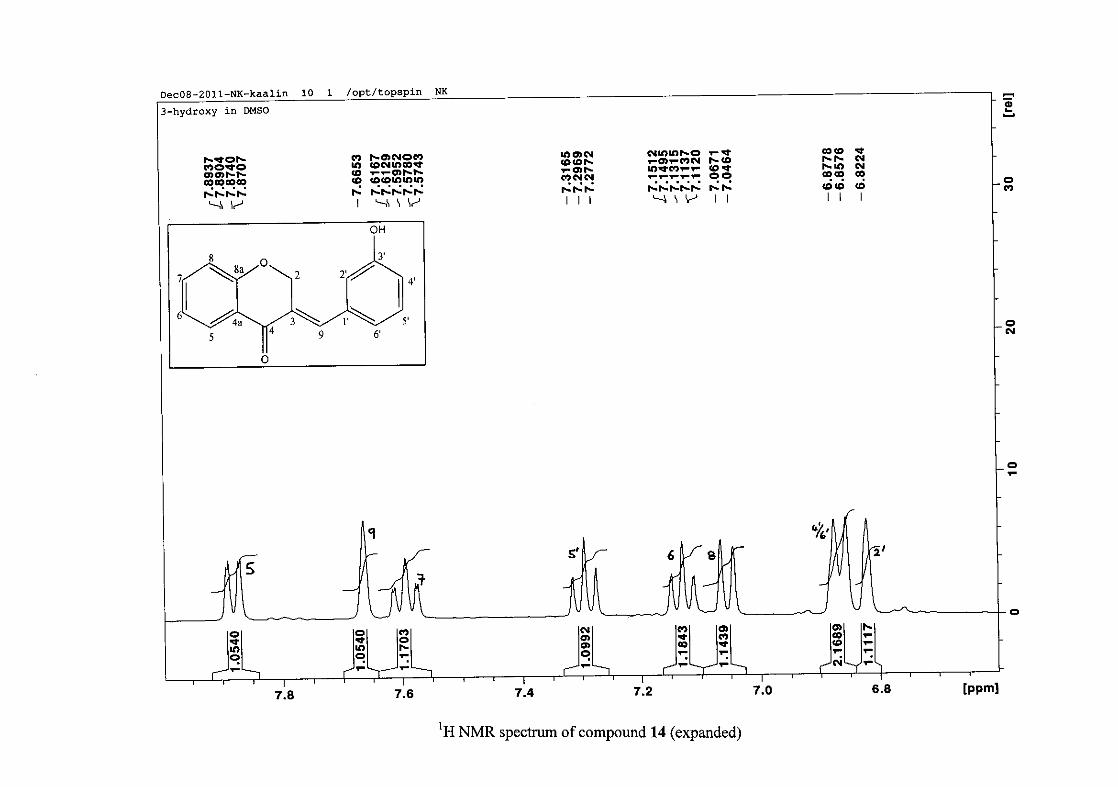
2.1.5 Structural elucidation of the meta substituted derivatives (except 3'-fluoro)
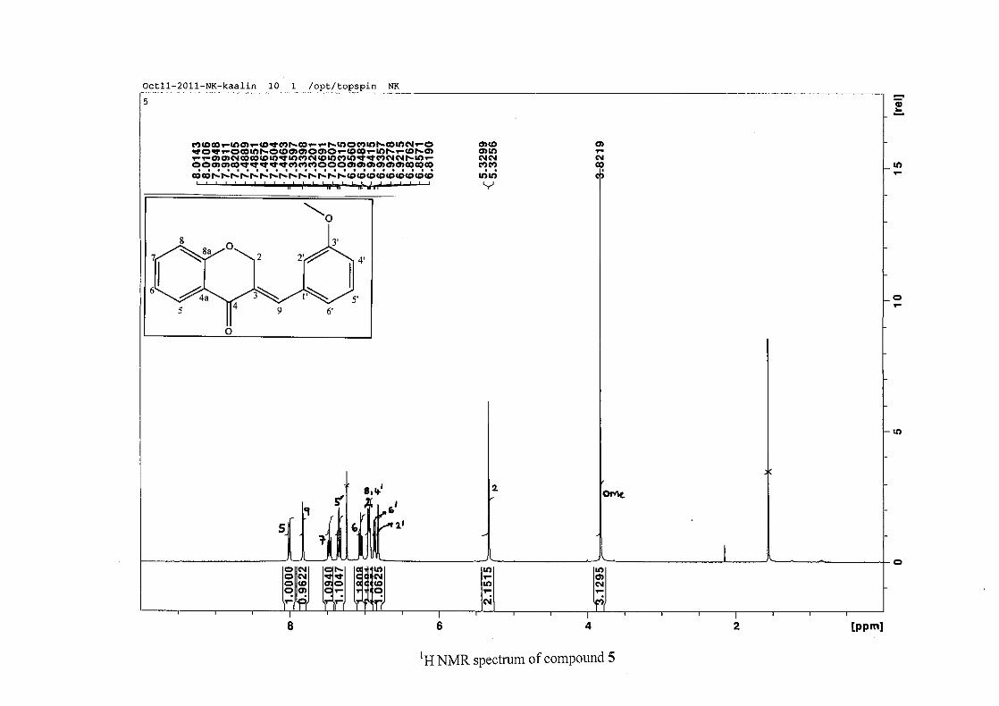
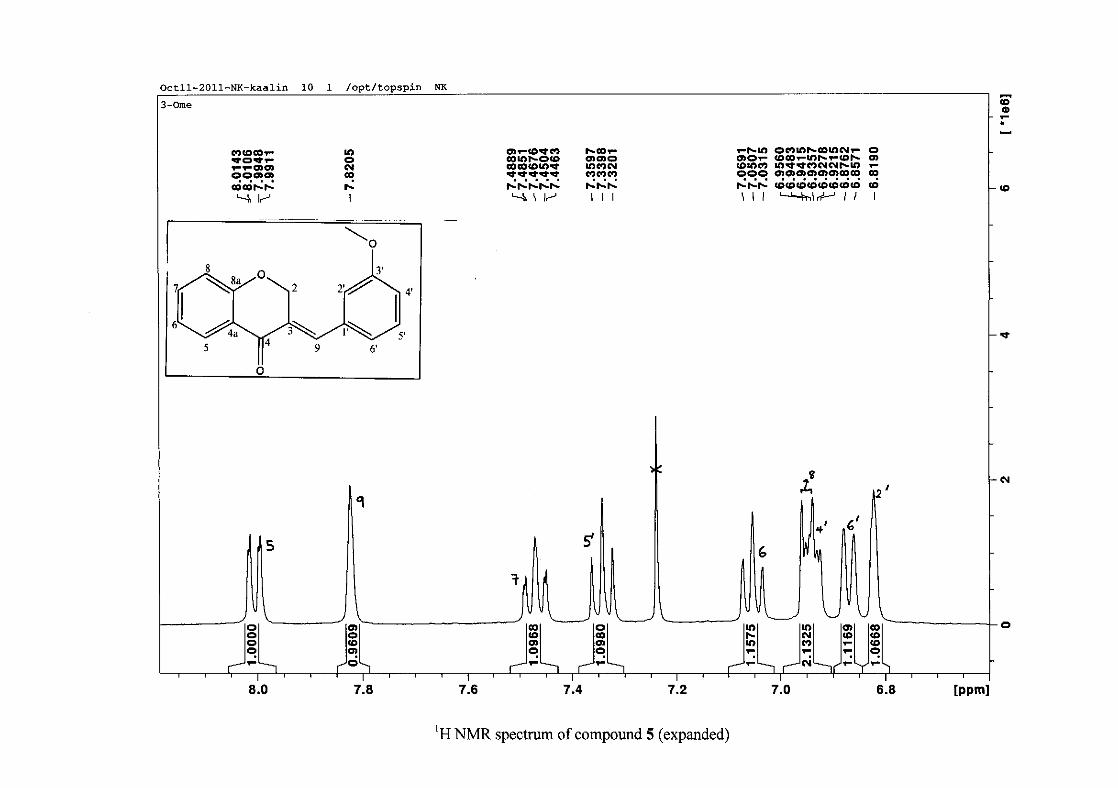
For the meta substituted compounds (5, 8 and 14, excluding the fluorinated compound 11),
all the NMR spectra were similar with some subtle changes brought about by the different
substituted groups. For 5, the 3-(3'-methoxybenzylidene)chroman-4-one, the proton
resonances for the aromatic ring and the chromanone ring are well resolved. The H-5
resonance appears as a double doublet at δH 8.00 (J = 7.82, 1.46 Hz), deshielded due to
hydrogen bonding as described above (Figure 11), the H-9 resonance appears as a singlet at
δH 7.82 and the H-7 and H-6 resonances occurred at δH 7.47 (td, J = 7.72, 1.60 Hz) and δH
7.05 (t, J = 7.52 Hz) respectively. The H-8 and H-4' resonances overlap at δH 6.94, however
the doublet resonance of H-8 could be distinguished and its coupling constant was
determined to be J = 8.12 Hz. The multiplicity of the H-4' resonance could not be determined
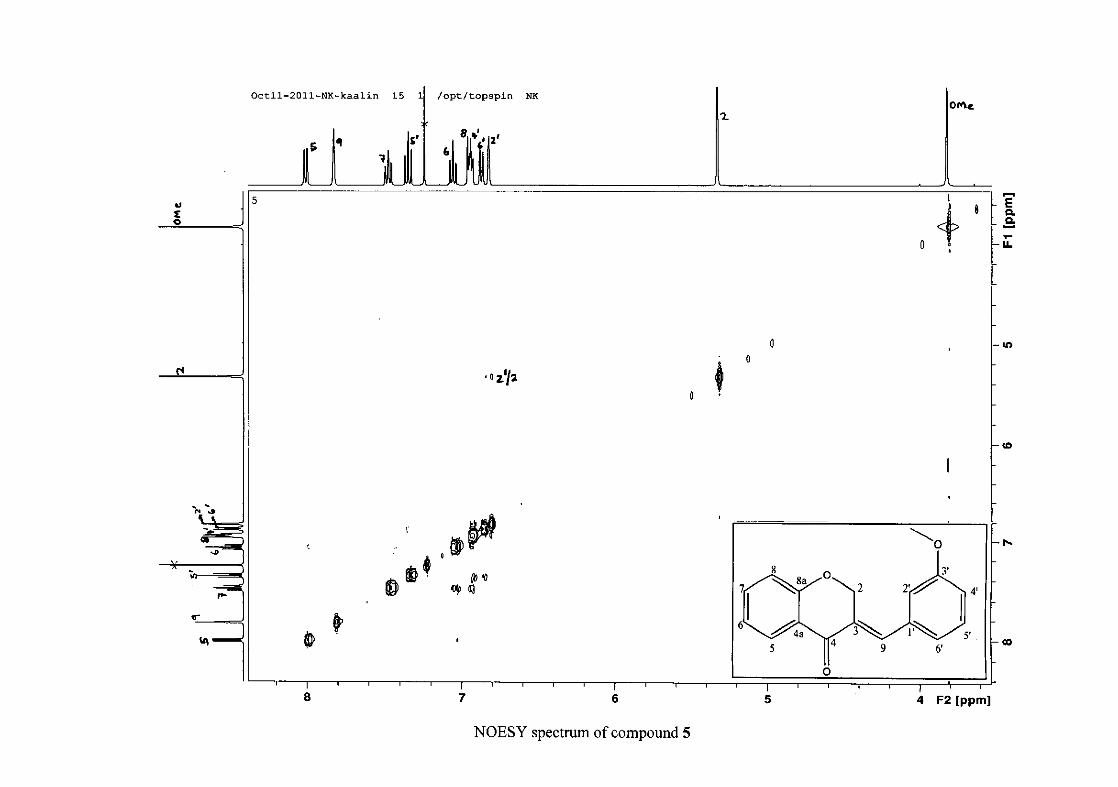
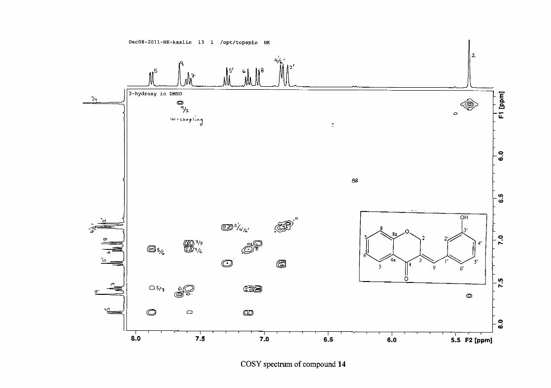
due to overlap with H-8. Due to the meta substitution on the phenyl ring, the H-2' resonance
should show meta coupling with either H-6' or H-4', but this was not observed. Rather, a
broad singlet for this resonance was observed at δH 6.82, however coupling in the COSY
spectrum was observed between this resonance and that of H-4' and the H-6' resonance which
appears as a doublet at δH 6.87 (J = 7.60 Hz). The H-5' resonance appears as a triplet at δH
7.34 (J = 7.92 Hz). HMBC correlations between H-5 and the carbonyl resonance, C-4 at δC
182.23 and H-8 and C-4 confirm these assignments. The H-2 resonance appears at δH 5.33,
which was confirmed by a HMBC correlation to C-4. The methoxy resonance was seen at δH
3.82 as an intense singlet resonance in the 1H NMR spectrum.
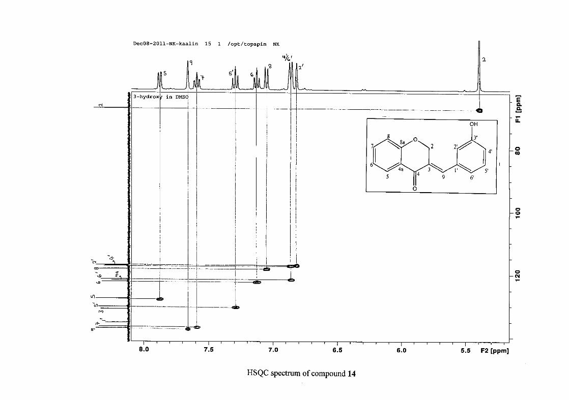
All the protonated carbon resonances were identified from their corresponding proton
resonances using the HSQC spectrum. There were five carbon resonances beside the
carbonyl resonance, which were non-protonated. These occurred at δC 122.02, 131.15,
135.69, 159.69, and 161.18 and were identified using HMBC correlations. The C-8a
36
resonance was identified at δC 161.18 by a HMBC correlation to H-2 and the other
oxygenated carbon resonance at δC 159.60 was then attributed to C-3'. This was corroborated
by HMBC correlations between C-3' and both H-4' and H-5'. The resonance at δC 131.15
shows a correlation to H-2 and was therefore attributed to C-3, and the resonance at δC
135.69 shows a correlation to H-5' and was attributed to C-1'. The remaining resonance at δC
122.02 showed a correlation to H-6 and was therefore assigned to C-4a.
The 1H NMR spectrum of 14, the 3'-hydroxy derivative was very similar to 5, but had the
notable difference in that the H-4' resonance which overlapped with H-8 in 5, was now seen
overlapping with the H-6' resonance and could be seen as a two proton doublet resonance for
H-4'/6' at δH 6.86 with J = 8.08 Hz. This allowed the H-8 resonance to be clearly seen as a
doublet at δH 7.06 with J = 8.28 Hz. The rest of the resonances have similar splitting patterns
and chemical shifts to 5. There was an added hydroxyl proton resonance at δH 9.71 for 14.
The carbon resonances in both 5 and 14 were similar. In 14, the resonances for C-4' and C-6'
could not be unequivocally assigned from their HMBC correlations and we have based their
assignments on those made for 5.
In the meta chloro derivative 8, all the proton resonances for the chromanone ring, H-2 and
H-5 to H-9 were all similar to 5. Changes were observed for the H-2', and H-4' to H-6'
resonances due to a now weakly deactivating group. The H-2' resonance shifted more
downfield from δH 6.82 in 5 to δH 7.27 in 8, probably by the electron withdrawal inductive
effects of the chloro group. The same can be seen for the H-4' and H-6' resonances which
now shifted more downfield to δH 7.37 and occur as overlapping resonances. It is observed
that the H-5' resonance for the chloro compound is shifted slightly more upfield from 7.34 in
37
5 to 7.17 in 8, probably due to a greater resonance effect by the chloro group, donating more
electron density to the meta position.
In the 13C NMR spectrum of 8, there was a noticeable shift of the C-2', C-3', C-4' and C-6'
resonances. The C-2', C-4' and C-6' resonances were all shifted more downfield to δC 130.00,
129.42 and 129.62 in 8, from 115.42, 115.06 and 122.28 in 5. The C-3' resonance shifted
more upfield to δC 134.76 in 8, from 159.69 in 5, due to the weaker electronegativity of the
chloro group as opposed to oxygen, resulting in less electron withdrawal by induction from
the chloro group.
2.1.6 Structural elucidation of the 3',4'-disubstituted derivatives (except 3',4'-difluoro)
The proton NMR resonances for the chromanone ring (including that of H-9) of compounds
6, 9 and 15 were similar to that described for 5, 8 and 14 above with regard to chemical shift
and splitting patterns. The carbon resonances of C-2 to C-9 including C-4a and C-8a of the
chromanone moiety were also similar to 5, 8 and 14.
With regard to the phenyl group, the 3',4'-substitution in 15 resulted in H-2', H-5' and H-6'
having the expected splitting patterns of doublets for H-2' and H-5' and a double doublet for
H-6' at δH 6.81 (J = 2.04 Hz), δH 6.79 (J = 9.16 Hz) and 6.73 (J = 9.14, 1.88 Hz) respectively.
Their corresponding carbon resonances occurred at δC 117.76 (C-2'), 115.85 (C-5') and
123.47 (C-6'). These assignments were confirmed by HMBC correlations between H-9 and
C-2' and C-6'. The two resonances at δC 145.38 and 147.87 were attributed to C-3' and C-4'
respectively. They were assigned as such because of a HMBC correlation between C-4' and
all the protons on the phenyl ring, H-2', H-5' and H-6', whereas C-3' showed a HMBC
correlation to H-2' and H-5' only. The extra HMBC correlation between C-4' and H-6'
38
allowed the unambiguous assignment of these two carbon resonances (C-3' and C-4'). The C-
1' carbon resonance was identified at δC 125.18 due to a HMBC correlation between this
carbon resonance and H-2', H-6' and H-5'.
For the dimethoxy compound 6, the resonances in the 1H NMR spectrum are slightly
different to that of 15 due to solvent effects, 15 being acquired in deuterated DMSO and 6
being acquired in deuterated CDCl3. This resulted in H-5' and H-6' overlapping at δH 6.88-
6.90 and H-2' being a doublet at δH 6.83 (J = 1.72 Hz). The C-5' and C-6' resonances were
difficult to identify using the HSQC spectrum because of the overlap of H-5' and H-6', but in
comparison to 15 they were made at δC 123.64 (C-6') and δC 111.06 (C-5'). This was
confirmed by a HMBC correlation between H-9 and C-6'. The H-2' resonance occurred as a
doublet at δH 6.83 (J = 1.72 Hz) with a corresponding carbon resonance at δC 113.31. Two
methoxy proton resonances can be seen in the 1H NMR spectrum at δH 3.89 and 3.91 as two
intense singlets, both corresponding to the overlapping carbon resonance at δC 55.99. The
aromatic C-O resonances occurred at δC 148.99 and 150.41 as for 15 above.
In the disubstituted chloro compound 9, the proton resonances for H-2', H-5' and H-6' all kept
their splitting patterns as that for above, but all these resonances shifted more downfield in
comparison to 6 and 15. The H-5' resonance now occurred as a doublet at δH 7.50 (J = 8.40
Hz), H-2' as a doublet at δH 7.38 (J = 1.92 Hz) and H-6' as a double doublet at δH 7.12 (J =
8.28, 1.96 Hz). This shift downfield was probably due to the greater inductive effect of
chlorine versus its electron donating effect. In 6 and 15, the oxygenated groups probably had
a greater electron donating effect than an inductive effect. This is consistent with the chloro
group being deactivating and the hydroxy and methoxy groups being activating. Due to the
chloro substitution at C-3' and C-4', these resonances shifted more upfield to δC 133.70 and
39
134.28. Similar to the proton resonances, the corresponding carbon resonances, C-2', C-5'
and C-6' shifted more downfield to δC 131.41, 130.79 and 128.94 respectively.
2.1.7 Structural elucidation of the fluorine containing compounds (10, 11 and 12)
As above, the proton and carbon resonances for the chromanone ring did not change and
therefore a discussion for this part of the molecule will not be repeated. The only changes
that occurred were in that of the phenyl ring. In compound 10, the para-fluorinated
compound, the H-2'/6' and H-3'/5' proton resonances, which appeared as a pair of doublets in
7 (the para-chlorinated compound) at δH 7.22 and 7.40 now appeared as a doublet of doublets
with J = 8.60 and 5.40 Hz at δH 7.28 (H-2'/6'), and a triplet with J= 8.62 Hz at δH 7.13 (H-
3'/5'). The H-3'/5' resonance was most affected by the substitution with fluorine in terms of
chemical shift, resulting in the resonance being shifted from δH 7.40 in 7 to δH 7.13 in 10.
The H-2'/6' resonance did not shift notably from δH 7.22 to δH 7.28. However, the splitting
patterns of the two resonances in the fluoro compound were markedly different to that of the
chloro compound. The H-2'/6' resonance was split into a double doublet by the fluorine with
JHF = 5.40 Hz and the proton ortho to it with J = 8.60 Hz. The H-3'/5' resonance being closer
to the fluorine substituent experienced the same coupling constant for both JHF and the ortho
coupled protons JH-2'/6', H-3'/5' with J = 8.62 Hz. These resonances were distinguished by the H-
2'/6' resonance showing a HMBC correlation with C-9 at δC 136.27 and the C-3'/5' resonance
a HMBC correlation to C-1' at δC 130.52.
In the 13C NMR spectrum, all the carbon resonances of the phenyl ring were seen coupled to
fluorine and appeared as doublets. The ipso carbon (C-4') directly bonded to the fluorine
atom appeared at δC 163.21 and had a coupling constant of J = 250.87 Hz. Care must be
40
taken when making these assignments so as not to confuse this resonance for two separate
carbon resonances. This resonance was confirmed by correlations to both the H-2'/6' and H-
3'/5'. Also present in the 13C NMR spectrum were resonances at δC 115.98 as a doublet with
J = 21.59 Hz, typical of ortho coupled carbon resonances and δC 131.97 (J = 8.55 Hz) typical
for meta coupling and were assigned to C-3'/5' and C-2'/6' respectively. The C-2'/6' carbon
resonance was coupled to H-9 in the HMBC spectrum as expected. The remaining doublet at
δC 130.52 (J = 2.97 Hz) was assigned to C-1' and the fact that this was a doublet was used to
distinguish between the C-1' and C-3 carbon resonances which were located in the same
region of the 13C NMR spectrum.
In compound 11, the 3'-fluoro derivative, the fluorine couples to the H-2', H-4' and H-5'
protons. Both the H-4' and H-5' proton resonances appear as triplets of doublets, however
they can be distinguished by their coupling constants. The H-4' resonance at δH 7.09
experiences JHF and JHH coupling with similar coupling constants, hence the triplet with J =
8.44 Hz and the meta coupling (J = 2.40 Hz) results in the triplet being further split into a
triplet of doublets. The H-5' resonance at δH 7.39 shows a larger second coupling (J = 5.96
Hz) since this is due to the H-F coupling, the triplet (J = 8.00 Hz) being due to the coupling
between the two protons ortho to it, H-4' and H-6'. The H-2' proton resonance, ortho to the
fluorine atom, appears as a doublet with J = 9.52 Hz at δH 6.98. Similar to 10, all the carbon
resonances of the phenyl ring were doublets displaying ipso (δC 162.70, J = 245.90 Hz, C-3'),
ortho (δC 116.35, J = 15.56 Hz, C-4'; δC 116.57, J = 16.28 Hz, C-2'), meta (δC 130.31, J =
8.24 Hz, C-5', δC 136.40, J = 7.80, C-1'), and para (δC 125.66, J = 2.92 Hz, C-6') coupling.
Since the C-9 carbon atom is now only four bonds away from fluorine, this carbon resonance
is also coupled by fluorine with a coupling constant of J = 2.27 Hz. HMBC correlations
41
between C-9 and H-2' and H-6' as well as between C-1' and H-5' are also present confirming
the assignments of these carbon resonances.
In the 1H NMR spectrum of 12, the 3',4'-difluoro derivative, the H-5' ortho coupled proton
appears as a triplet of doublets at δH 7.22 (J= 9.88 and 8.28 Hz), the triplet being due to the
same coupling constant between H-5' and the ortho fluorine and H-5' and H-6', which was
split further into a triplet of doublets by the other meta fluorine atom. The H-2' resonance
was split into a ddd because of coupling to both the ortho (J = 10.08 Hz) and meta (J =7.52
Hz) fluorine atoms as well as the meta proton (J = 2.04 Hz, H-6'). The H-6' proton resonance
overlaps with the multiplet of resonances at δH 7.04. HMBC correlations between H-2', H-6'
and C-9 as well as C-1' and H-5' confirm the assignments of these resonances.
The carbon resonances of the phenyl moiety are once again all split by the fluorine atoms.
However in this instance, double doublets are experienced by all but the ortho coupled C-2'
and C-5' resonances which appear as doublets at δC 118.74 (J = 17.66 Hz) and δC 117.85 (J =
17.69 Hz). This is due to there being two fluorine atoms present on the phenyl ring. The
other four resonances are present at δC 150.25 (J = 248.55 and 12.80 Hz, C-3'), δC 150.78 (J
= 261.53 and 12.86 Hz, C-4'), δC 131.40 (J = 5.98 and 3.80 Hz, C-1') and δC 126.48 (J = 6.25
and 3.47 Hz, C-6'). HMBC correlations are also seen for H-9 with C-2' and C-6' confirming
the assignment of these carbon resonances.
2.1.8 Structural elucidation of the nitro containing compounds (16 and 17)
In comparison to the other compounds previously described, there were changes to the
chromanone ring proton resonances in the nitro derivatives 16 and 17 indicating that some
kind of conversion must have taken place. Of particular importance was the HMBC
42
correlation between the proton resonance at δH 7.45 (J = 8.48 Hz), attributed to the H-2'/6'
resonance and the methylene carbon resonance at δC 31.89 in 16. This was not encountered
for the other molecules. This now meant that the methylene group, which occurred at C-2 in
the other molecules, was now present at C-9 in 16. This change was accompanied by a
change in chemical shift of the methylene carbon resonance from approximately δC 67 to δC
31.89 in 16, which also indicated that the methylene carbon was no longer situated next to the
oxygen atom. There was however no change in the olefinic methine resonance at δH 7.79
when compared to the other compounds, but the C-2 resonance was now more deshielded at
δC 153.14 in comparison to the C-9 resonance in the other compounds. This was due to being
situated close to the oxygen atom on the chromanone ring. This meant that an exo-endo bond
migration had occurred in 16 and 17 due to the highly electron withdrawing nitro group. This
was reported to occur previously (Valkonen et al., 2012).
For the H-5 to H-8 proton resonances in 16, all the resonances were shifted downfield in
comparison to the 4-chloro derivative 7, between 0.15 to 0.50 Hz. However, these
resonances retained their splitting patterns, being present as a doublet of doublets at δH 8.18
(J = 7.98, 0.66 Hz, H-5), δH 7.65, a triplet of doublets (J = 8.32, 1.10 Hz, H-7), δH 7.41, a
doublet (J = 8.60 Hz, H-8) and δH 7.38 as a triplet (J = 7.54 Hz, H-6). For the carbon
resonances in 16 there is not much difference in chemical shift of the carbon resonances C-4a
through to C-8a with the largest shift being experienced by C-8a, which is only a 5 ppm shift
upfield from δC 161 in 7 to δC 156 in 16. A 5 ppm shift was also experienced by the carbonyl
carbon resonance from δC 182 in 7 to δC 177 in 16.
In the phenyl ring of 16, due to the electron withdrawing nitro group, a downfield shift was
experienced by both the H-3'/5' and H-2'/6' resonances by 0.72 Hz and 0.23 Hz respectively,
43
H-3'/5' now occurring at δH 8.12 (J = 8.56 Hz) and H-2'/6' at δH 7.45 (J = 8.48 Hz) in
comparison to the para-chloro compound 7. In comparison to 7, the carbon resonances of the
phenyl ring C-1' through to C-6' were all shifted upfield by approximately 2 to 11 ppm in 16
at δC 123.06 (C-1'), 129.65 (C-2'/6'), 123.79 (C-3'/5') and 146.76 (C-4'). In 7, there was
conjugation between the phenyl ring with the C-3 (9) double bond, delocalising the electrons
amongst more carbon atoms, hence the more downfield chemical shift. In 16, due to the
double bond migrating to the ∆2 position, this delocalisation does not occur and the electrons
are localised to the phenyl group, hence more upfield chemical shifts are experienced.
In 17, there are now four separate resonances for the phenyl group protons as opposed to only
two in 16. This was due to the nitro group now being at the 3' position. The H-2' resonance
occurred as a broad singlet at δH 8.13, the H-4' and H-6' proton resonances occurred as
doublets at δH 8.06 (J = 8.16 Hz) and δH 7.69 (J = 7.68 Hz) and the H-5' resonance occurred
at δH 7.45 as a triplet with J being 8.16 Hz. The other proton resonances of 17, the
chromanone proton resonances as well as H-2 and H-9 were the same as that for 16 as were
the carbon resonances of C-2 to C-9. There were however changes in chemical shifts for the
carbon resonances on the phenyl ring. The C-3' carbon to which the nitro group was attached
had a resonance of δC 148.41 and the carbon resonances meta to the nitro group, C-1' and C-5'
resonated at δC 141.02 and δC 129.42 respectively, while the ortho positioned carbon atoms
(C-2' and C-4') resonated at δC 123.52 and δC 121.71. This is consistent with the ortho and
para carbon resonances being more shielded and the meta resonances being more deshielded
due to electron withdrawal by resonance by the nitro group.
44
2.2 Bioactivity of the synthesised homoisoflavonoids
The synthesised homoisoflavonoids were subjected to antibacterial tests using the disc
diffusion assay, and antioxidant testing by the DPPH radical scavenging and the ferric
reducing antioxidant power assay. The results are reported and discussed in relation to the
chemical structures of the homoisoflavonoids.
2.2.1 Antioxidant activity of the synthesised homoisoflavonoids
The three hydroxylated homoisoflavonoids (13-15) were tested for their antioxidant activity
by the DPPH radical scavenging assay and the FRAP assay as hydroxylated flavonoids are
commonly known to be antioxidants. Ascorbic acid, a common antioxidant, served as the
positive control for these experiments. A sample, containing all required reagents except for
the antioxidant was used as a blank.
2.2.1.1 DPPH radical scavenging assay
The principles for this assay are discussed in chapter 1 (1.4.1.1), and the methodology
described in chapter 3 (3.2.1.1).
Polyhydroxylated compounds are known to have good antioxidant activity. Compounds 13,
14 and 15 are hydroxylated compounds and were therefore tested for their antioxidant
activity. As expected, compound 15, which has two hydroxy groups at the 3' and 4' positions,
showed strong antioxidant activity, comparable to that of ascorbic acid, and compounds 13
(the 4'-hydroxy) and 14 (the 3'-hydroxy) which have one hydroxy group showed weak
antioxidant activity.
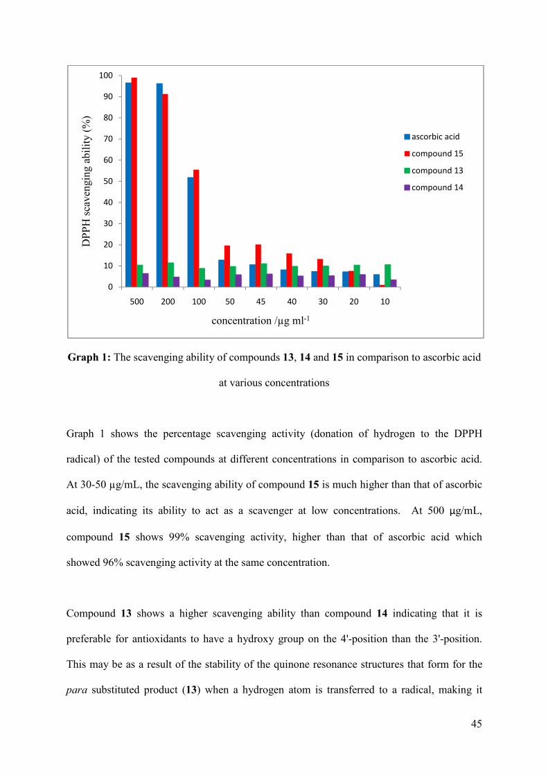
45
Graph 1: The scavenging ability of compounds 13, 14 and 15 in comparison to ascorbic acid
at various concentrations
Graph 1 shows the percentage scavenging activity (donation of hydrogen to the DPPH
radical) of the tested compounds at different concentrations in comparison to ascorbic acid.
At 30-50 µg/mL, the scavenging ability of compound 15 is much higher than that of ascorbic
acid, indicating its ability to act as a scavenger at low concentrations. At 500 µg/mL,
compound 15 shows 99% scavenging activity, higher than that of ascorbic acid which
showed 96% scavenging activity at the same concentration.
Compound 13 shows a higher scavenging ability than compound 14 indicating that it is
preferable for antioxidants to have a hydroxy group on the 4'-position than the 3'-position.
This may be as a result of the stability of the quinone resonance structures that form for the
para substituted product (13) when a hydrogen atom is transferred to a radical, making it
0
10
20
30
40
50
60
70
80
90
100
500 200 100 50 45 40 30 20 10
DP
PH
sca
veng
ing
abil
ity
(%)
concentration /µg ml-1
ascorbic acid
compound 15
compound 13
compound 14
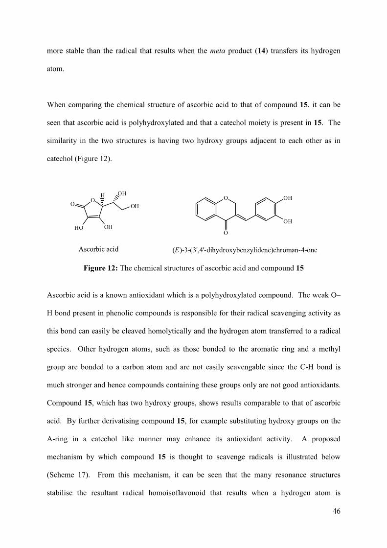
46
more stable than the radical that results when the meta product (14) transfers its hydrogen
atom.
When comparing the chemical structure of ascorbic acid to that of compound 15, it can be
seen that ascorbic acid is polyhydroxylated and that a catechol moiety is present in 15. The
similarity in the two structures is having two hydroxy groups adjacent to each other as in
catechol (Figure 12).
O
HO OH
O OH
H OHO
O
OH
OH
Ascorbic acid (E)-3-(3',4'-dihydroxybenzylidene)chroman-4-one
Figure 12: The chemical structures of ascorbic acid and compound 15
Ascorbic acid is a known antioxidant which is a polyhydroxylated compound. The weak O–
H bond present in phenolic compounds is responsible for their radical scavenging activity as
this bond can easily be cleaved homolytically and the hydrogen atom transferred to a radical
species. Other hydrogen atoms, such as those bonded to the aromatic ring and a methyl
group are bonded to a carbon atom and are not easily scavengable since the C-H bond is
much stronger and hence compounds containing these groups only are not good antioxidants.
Compound 15, which has two hydroxy groups, shows results comparable to that of ascorbic
acid. By further derivatising compound 15, for example substituting hydroxy groups on the
A-ring in a catechol like manner may enhance its antioxidant activity. A proposed
mechanism by which compound 15 is thought to scavenge radicals is illustrated below
(Scheme 17). From this mechanism, it can be seen that the many resonance structures
stabilise the resultant radical homoisoflavonoid that results when a hydrogen atom is
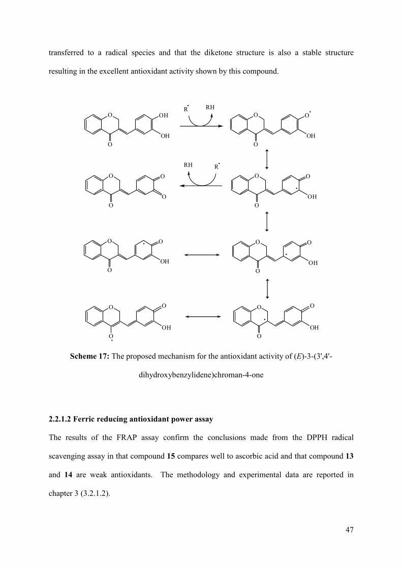
47
transferred to a radical species and that the diketone structure is also a stable structure
resulting in the excellent antioxidant activity shown by this compound.
O
O
OH
OH
R RHO
O
O
OH
O
O
O
OH
O
O
O
O
RRH
O
O
O
OH
O
O
O
O
O
O
OH
OH
O
O
O
OH
Scheme 17: The proposed mechanism for the antioxidant activity of (E)-3-(3',4'-
dihydroxybenzylidene)chroman-4-one
2.2.1.2 Ferric reducing antioxidant power assay
The results of the FRAP assay confirm the conclusions made from the DPPH radical
scavenging assay in that compound 15 compares well to ascorbic acid and that compound 13
and 14 are weak antioxidants. The methodology and experimental data are reported in
chapter 3 (3.2.1.2).
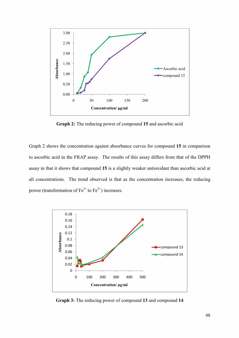
48
Graph 2: The reducing power of compound 15 and ascorbic acid
Graph 2 shows the concentration against absorbance curves for compound 15 in comparison
to ascorbic acid in the FRAP assay. The results of this assay differs from that of the DPPH
assay in that it shows that compound 15 is a slightly weaker antioxidant than ascorbic acid at
all concentrations. The trend observed is that as the concentration increases, the reducing
power (transformation of Fe3+ to Fe2+) increases.
Graph 3: The reducing power of compound 13 and compound 14
0.00
0.50
1.00
1.50
2.00
2.50
3.00
0 50 100 150 200
Ab
sorb
an
ce
Concentration/ µg/ml
Ascorbic acid
compound 15
0
0.02
0.04
0.06
0.08
0.1
0.12
0.14
0.16
0.18
0 100 200 300 400 500
Ab
sorb
an
ce
Concentration/ µg/ml
compound 13
compound 14
49
Graph 3 shows, by the low absorbance values as compared to those in graph 2 for ascorbic
acid and 15 that compounds 13 and 14 are weak antioxidants at low concentrations. At
concentrations higher than 200 µg/mL, a sharp increase in its antioxidant activity is observed
but its activity is still nowhere near that of 15 and ascorbic acid. The para hydroxy derivative
(13) shows slightly better antioxidant activity than the meta hydroxy derivative (14), which is
consistent with the results from the DPPH assay. The reducing power of the tested
compounds in decreasing order was found to be: ascorbic acid > compound 15 > compound
13 > compound 14.
2.2.2 Antibacterial activity of the synthesised homoisoflavonoids
The antibacterial activities of the homoisoflavonoids synthesised were tested against ten gram
positive and six gram negative bacterial strains using the Kirby-Bauer disk-diffusion method.
In general, the homoisoflavonoids tested were more active against the gram positive than the
gram negative bacteria. No antibacterial activity was shown against the following gram
negative strains of bacteria: Salmonella arizonae (ATCC 13314), Escherichia coli (ATCC
35219), Pseudomonas aeruginosa (ATCC 27853 and ATCC 35037), and Klebsiella
pneumoniae (ATCC 70063). This is because gram negative bacteria have an extra
membrane, known as the outer membrane which is made up of lipopolysaccharides and
protein, which is difficult to diffuse or penetrate through. Therefore gram negative bacteria
are more difficult to destroy. Since the homoisoflavonoids show good antibacterial activity
against gram positive bacteria, they may be tested for antituberculosis activity and against
nosocomial pathogens.
50
These homoisoflavonoids show the best inhibitory activity against the bacterial strain,
Staphylococcus aureus, therefore two strains of Staphylococcus aureus were used in the
antibacterial tests, Staphylococcus aureus (ATCC 29212) and Staphylococcus aureus (ATCC
43300).
The para substituted derivatives showed weaker activity than the meta substituted derivatives
except for the para fluoro derivative against Staphylococcus saprophyticus (ATCC 35552).
The para methoxy (4), chloro (7) and nitro (16) derivatives showed no activity against the
bacterial strains tested.
Compound 14, the meta substituted hydroxylated homoisoflavonoid, showed the broadest
range of antibacterial activity. It was the only homoisoflavonoid to show mild activity
against the gram negative strain, Escherichia coli (ATCC 29522). Compound 14 was
strongly active against Staphylococcus aureus (ATCC 29212 and ATCC 43300) and was
moderately active against Staphylococcus saprophyticus (ATCC 35552), Staphylococcus
scuiri (ATCC 29062), Staphylococcus xylosus (ATCC 35033) and Streptococcus pyogenes
(ATCC 19615).
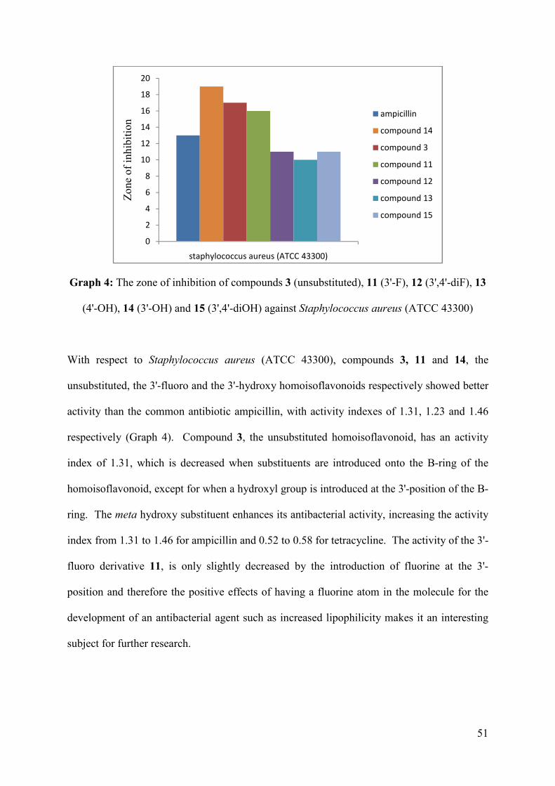
51
Graph 4: The zone of inhibition of compounds 3 (unsubstituted), 11 (3'-F), 12 (3',4'-diF), 13
(4'-OH), 14 (3'-OH) and 15 (3',4'-diOH) against Staphylococcus aureus (ATCC 43300)
With respect to Staphylococcus aureus (ATCC 43300), compounds 3, 11 and 14, the
unsubstituted, the 3'-fluoro and the 3'-hydroxy homoisoflavonoids respectively showed better
activity than the common antibiotic ampicillin, with activity indexes of 1.31, 1.23 and 1.46
respectively (Graph 4). Compound 3, the unsubstituted homoisoflavonoid, has an activity
index of 1.31, which is decreased when substituents are introduced onto the B-ring of the
homoisoflavonoid, except for when a hydroxyl group is introduced at the 3'-position of the B-
ring. The meta hydroxy substituent enhances its antibacterial activity, increasing the activity
index from 1.31 to 1.46 for ampicillin and 0.52 to 0.58 for tetracycline. The activity of the 3'-
fluoro derivative 11, is only slightly decreased by the introduction of fluorine at the 3'-
position and therefore the positive effects of having a fluorine atom in the molecule for the
development of an antibacterial agent such as increased lipophilicity makes it an interesting
subject for further research.
0
2
4
6
8
10
12
14
16
18
20
staphylococcus aureus (ATCC 43300)
Zon
e of
inhi
biti
onampicillin
compound 14
compound 3
compound 11
compound 12
compound 13
compound 15
52
By comparing the activity indices of the fluorinated compounds 10, 11 and 12 against
Staphylococcus aureus (ATCC 43300), it can be seen that the most favourable substitution
pattern for antibacterial activity is the meta substitution with activity indices of 1.23 (AMP)
and 0.48 (TET). The para substituted fluoro derivative showed no activity. The
disubstituted fluorinated derivative, 12, showed a decrease in activity when compared to the
meta fluoro derivative. By introducing an additional fluorine atom on the 4'-position of the
meta substituted derivative, the activity is decreased from 1.23 to 0.85. This may be due to
the mechanism by which the antibacterial agent works. Substitution at the 4'-position may
inhibit binding to the active site of enzymes required for normal functional of the bacterial
species. This explains why the activity decreases when the homoisoflavonoid is
disubstituted. In contrast, substitution at the 3'-position with a molecule or group capable of
hydrogen bonding, such as fluorine (11) or the hydroxy group (14) may be perfect for binding
to enzymes responsible for the functioning of bacterial cells. Once bound to these enzymes,
they may alter their function and hence lead either to cell death of the bacterial species or
inhibit replication of the bacterial species.
Staphylococcus aureus (ATCC 43300) is resistant to both methicillin and oxacillin
(http://www.straininfo.net/strains/54914/). Such strains of bacteria are referred to as
methicillin resistant Staphylococcus aureus (MRSA). Compound 14 may therefore serve as a
useful antibacterial agent against Staphylococcus aureus (ATCC 43300). MRSA is one of
the most dangerous bacterial infections that occur in the commercial health care sector
(hospitals), as these bacterial infections cannot be easily destroyed. As compound 14, shows
good antibacterial activity against Staphylococcus aureus (ATCC 43300), it may be applied
to combat such infections. Derivatisation of compound 14, by varying the substitution on the
A-ring can be conducted in order to develop better antibacterial agents.
53
CHAPTER 3 EXPERIMENTAL
This chapter includes the experimental techniques employed to synthesise and characterise
the thirteen benzylidene and two benzyl homoisoflavonoids. Characteristic data, such as the
1H and 13C NMR, UV, IR and MS data are reported in the chapter. New crystal structures for
seven of the homoisoflavonoids were obtained. The methodology used to determine the
antioxidant and antibacterial activities of the test compounds are also included here.
3.1 Chemistry
The experimental procedure for the synthesis of the intermediates 3-phenoxypropanoic acid
(1) and 4-chromanone (2) as well as the homoisoflavonoids (3-17) are included here. The
instrument details and parameters used for the characterisation techniques employed are also
reported.
3.1.1 General experimental procedures
The reagents and chemicals used in this study were purchased from Sigma Aldrich via
Capital Lab, South Africa and were reagent grade. All organic solvents were redistilled and
dried according to standard procedures. The melting points were recorded on an Ernst Leitz
Wetziar micro-hot stage melting point apparatus. IR spectra were recorded on a Perkin
Elmer Spectrum 100 FTIR spectrometer with universal ATR sampling accessory. UV
spectra were obtained on a Varian Cary UV-VIS Spectrophotometer in dichloromethane (1-
12, 16-17) and methanol (13-15). For GC-MS analyses, the samples were analysed on an
Agilent GC–MSD apparatus equipped with DB-5SIL MS (30 m x 0.25 mm i.d., 0.25 µm film
thickness) fused-silica capillary column. Helium (at 2 mL/min) was used as a carrier gas.
The MS was operated in the EI mode at 70 eV. The NMR spectra were recorded using a
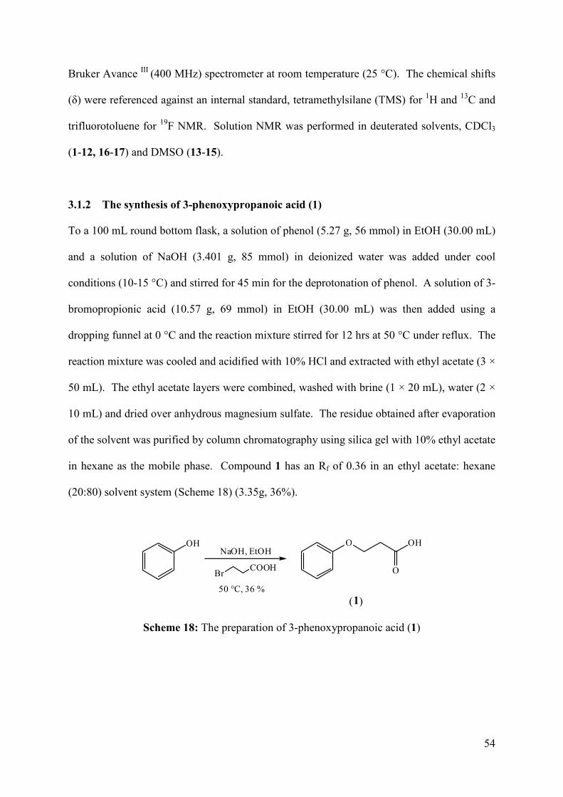
54
Bruker Avance III (400 MHz) spectrometer at room temperature (25 °C). The chemical shifts
(δ) were referenced against an internal standard, tetramethylsilane (TMS) for 1H and 13C and
trifluorotoluene for 19F NMR. Solution NMR was performed in deuterated solvents, CDCl3
(1-12, 16-17) and DMSO (13-15).
3.1.2 The synthesis of 3-phenoxypropanoic acid (1)
To a 100 mL round bottom flask, a solution of phenol (5.27 g, 56 mmol) in EtOH (30.00 mL)
and a solution of NaOH (3.401 g, 85 mmol) in deionized water was added under cool
conditions (10-15 °C) and stirred for 45 min for the deprotonation of phenol. A solution of 3-
bromopropionic acid (10.57 g, 69 mmol) in EtOH (30.00 mL) was then added using a
dropping funnel at 0 °C and the reaction mixture stirred for 12 hrs at 50 °C under reflux. The
reaction mixture was cooled and acidified with 10% HCl and extracted with ethyl acetate (3 ×
50 mL). The ethyl acetate layers were combined, washed with brine (1 × 20 mL), water (2 ×
10 mL) and dried over anhydrous magnesium sulfate. The residue obtained after evaporation
of the solvent was purified by column chromatography using silica gel with 10% ethyl acetate
in hexane as the mobile phase. Compound 1 has an Rf of 0.36 in an ethyl acetate: hexane
(20:80) solvent system (Scheme 18) (3.35g, 36%).
OH O OH
O
(1)
NaOH, EtOH
50 °C, 36 %
BrCOOH
Scheme 18: The preparation of 3-phenoxypropanoic acid (1)
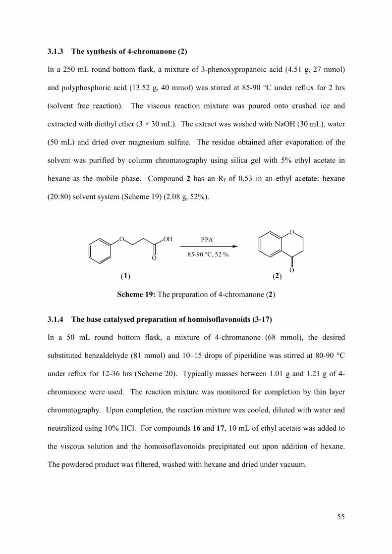
55
3.1.3 The synthesis of 4-chromanone (2)
In a 250 mL round bottom flask, a mixture of 3-phenoxypropanoic acid (4.51 g, 27 mmol)
and polyphosphoric acid (13.52 g, 40 mmol) was stirred at 85-90 °C under reflux for 2 hrs
(solvent free reaction). The viscous reaction mixture was poured onto crushed ice and
extracted with diethyl ether (3 × 30 mL). The extract was washed with NaOH (30 mL), water
(50 mL) and dried over magnesium sulfate. The residue obtained after evaporation of the
solvent was purified by column chromatography using silica gel with 5% ethyl acetate in
hexane as the mobile phase. Compound 2 has an Rf of 0.53 in an ethyl acetate: hexane
(20:80) solvent system (Scheme 19) (2.08 g, 52%).
O OH
O
O
O
PPA
85-90 °C, 52 %
(1) (2)
Scheme 19: The preparation of 4-chromanone (2)
3.1.4 The base catalysed preparation of homoisoflavonoids (3-17)
In a 50 mL round bottom flask, a mixture of 4-chromanone (68 mmol), the desired
substituted benzaldehyde (81 mmol) and 10–15 drops of piperidine was stirred at 80-90 °C
under reflux for 12-36 hrs (Scheme 20). Typically masses between 1.01 g and 1.21 g of 4-
chromanone were used. The reaction mixture was monitored for completion by thin layer
chromatography. Upon completion, the reaction mixture was cooled, diluted with water and
neutralized using 10% HCl. For compounds 16 and 17, 10 mL of ethyl acetate was added to
the viscous solution and the homoisoflavonoids precipitated out upon addition of hexane.
The powdered product was filtered, washed with hexane and dried under vacuum.
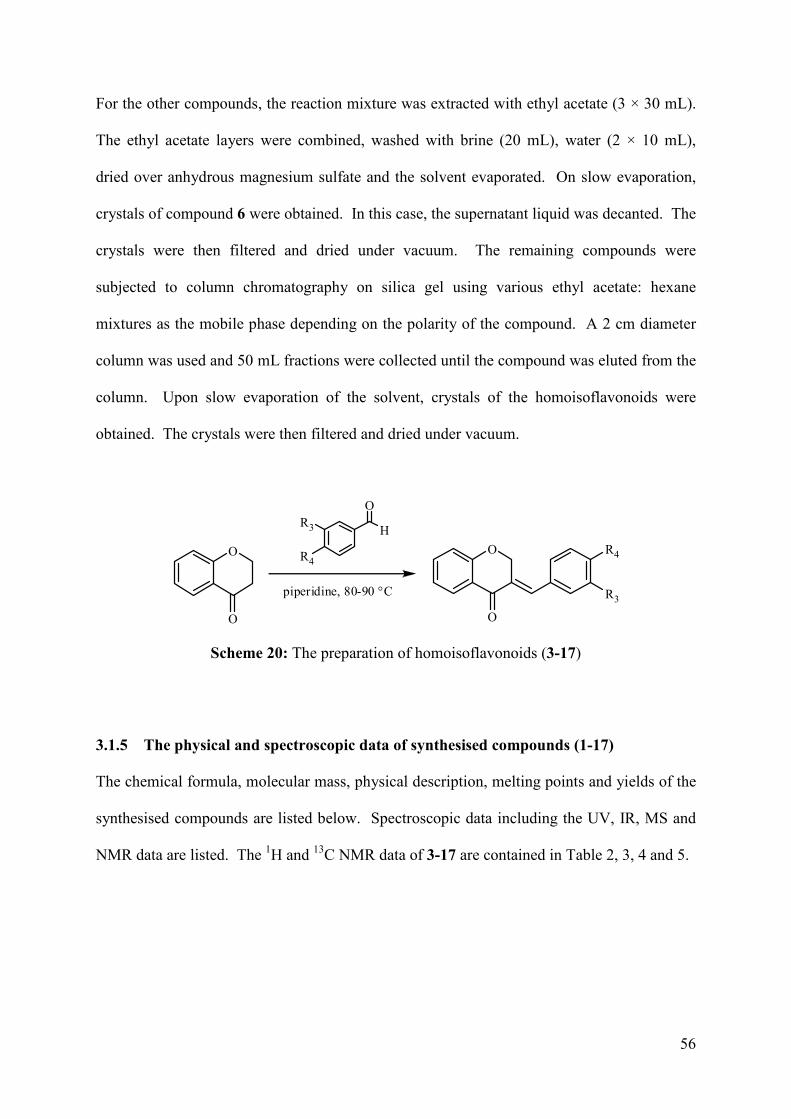
56
For the other compounds, the reaction mixture was extracted with ethyl acetate (3 × 30 mL).
The ethyl acetate layers were combined, washed with brine (20 mL), water (2 × 10 mL),
dried over anhydrous magnesium sulfate and the solvent evaporated. On slow evaporation,
crystals of compound 6 were obtained. In this case, the supernatant liquid was decanted. The
crystals were then filtered and dried under vacuum. The remaining compounds were
subjected to column chromatography on silica gel using various ethyl acetate: hexane
mixtures as the mobile phase depending on the polarity of the compound. A 2 cm diameter
column was used and 50 mL fractions were collected until the compound was eluted from the
column. Upon slow evaporation of the solvent, crystals of the homoisoflavonoids were
obtained. The crystals were then filtered and dried under vacuum.
O
O
O
O
R4
R3
H
R4
R3
O
piperidine, 80-90 °C
Scheme 20: The preparation of homoisoflavonoids (3-17)
3.1.5 The physical and spectroscopic data of synthesised compounds (1-17)
The chemical formula, molecular mass, physical description, melting points and yields of the
synthesised compounds are listed below. Spectroscopic data including the UV, IR, MS and
NMR data are listed. The 1H and 13C NMR data of 3-17 are contained in Table 2, 3, 4 and 5.
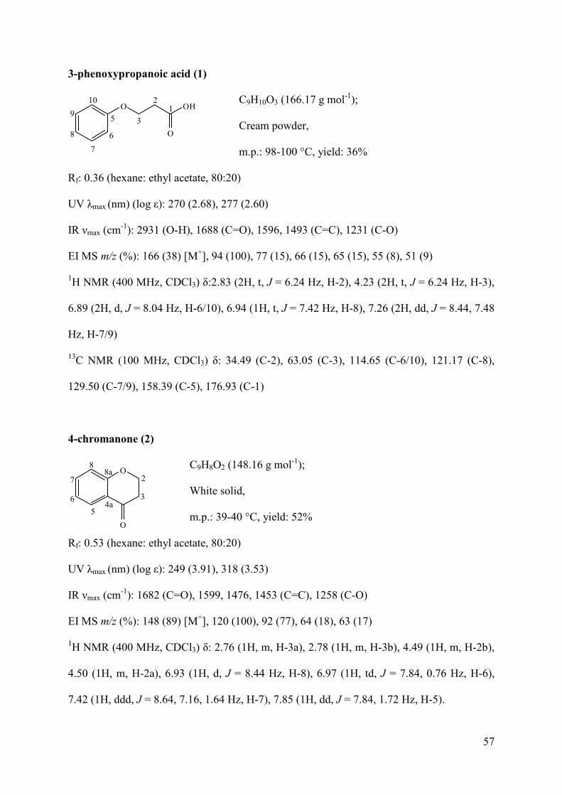
57
3-phenoxypropanoic acid (1)
C9H10O3 (166.17 g mol-1);
Cream powder,
m.p.: 98-100 °C, yield: 36%
Rf: 0.36 (hexane: ethyl acetate, 80:20)
UV λmax (nm) (log ε): 270 (2.68), 277 (2.60)
IR νmax (cm-1): 2931 (O-H), 1688 (C=O), 1596, 1493 (C=C), 1231 (C-O)
EI MS m/z (%): 166 (38) [M+], 94 (100), 77 (15), 66 (15), 65 (15), 55 (8), 51 (9)
1H NMR (400 MHz, CDCl3) δ:2.83 (2H, t, J = 6.24 Hz, H-2), 4.23 (2H, t, J = 6.24 Hz, H-3),
6.89 (2H, d, J = 8.04 Hz, H-6/10), 6.94 (1H, t, J = 7.42 Hz, H-8), 7.26 (2H, dd, J = 8.44, 7.48
Hz, H-7/9)
13C NMR (100 MHz, CDCl3) δ: 34.49 (C-2), 63.05 (C-3), 114.65 (C-6/10), 121.17 (C-8),
129.50 (C-7/9), 158.39 (C-5), 176.93 (C-1)
4-chromanone (2)
C9H8O2 (148.16 g mol-1);
White solid,
m.p.: 39-40 °C, yield: 52%
Rf: 0.53 (hexane: ethyl acetate, 80:20)
UV λmax (nm) (log ε): 249 (3.91), 318 (3.53)
IR νmax (cm-1): 1682 (C=O), 1599, 1476, 1453 (C=C), 1258 (C-O)
EI MS m/z (%): 148 (89) [M+], 120 (100), 92 (77), 64 (18), 63 (17)
1H NMR (400 MHz, CDCl3) δ: 2.76 (1H, m, H-3a), 2.78 (1H, m, H-3b), 4.49 (1H, m, H-2b),
4.50 (1H, m, H-2a), 6.93 (1H, d, J = 8.44 Hz, H-8), 6.97 (1H, td, J = 7.84, 0.76 Hz, H-6),
7.42 (1H, ddd, J = 8.64, 7.16, 1.64 Hz, H-7), 7.85 (1H, dd, J = 7.84, 1.72 Hz, H-5).
O OH
O
12
35
6
7
8
9
10
O
O
4a
8a2
3
5
6
7
8
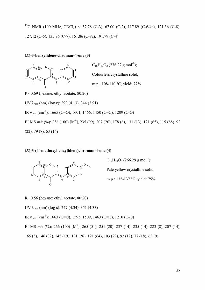
58
13C NMR (100 MHz, CDCl3) δ: 37.78 (C-3), 67.00 (C-2), 117.89 (C-6/4a), 121.36 (C-8),
127.12 (C-5), 135.96 (C-7), 161.86 (C-8a), 191.79 (C-4)
(E)-3-benzylidene-chroman-4-one (3)
C16H12O2 (236.27 g mol-1);
Colourless crystalline solid,
m.p.: 108-110 °C, yield: 77%
Rf: 0.69 (hexane: ethyl acetate, 80:20)
UV λmax (nm) (log ε): 299 (4.13), 344 (3.91)
IR νmax (cm-1): 1665 (C=O), 1601, 1466, 1450 (C=C), 1209 (C-O)
EI MS m/z (%): 236 (100) [M+], 235 (99), 207 (20), 178 (8), 131 (13), 121 (65), 115 (88), 92
(22), 79 (8), 63 (16)
(E)-3-(4'-methoxybenzylidene)chroman-4-one (4)
C17H14O3 (266.29 g mol-1);
Pale yellow crystalline solid,
m.p.: 135-137 °C, yield: 75%
Rf: 0.56 (hexane: ethyl acetate, 80:20)
UV λmax (nm) (log ε): 247 (4.34), 351 (4.33)
IR νmax (cm-1): 1663 (C=O), 1595, 1509, 1463 (C=C), 1210 (C-O)
EI MS m/z (%): 266 (100) [M+], 265 (51), 251 (20), 237 (14), 235 (14), 223 (8), 207 (14),
165 (5), 146 (32), 145 (19), 131 (26), 121 (64), 103 (29), 92 (12), 77 (18), 63 (9)
O
O
4a
8a2
3
5
6
7
8
91'
2'
3'
4'
5'
6'
O
O
4a
8a 2
3
5
6
7
8
91'
2'
3'
4'
5'
6'O
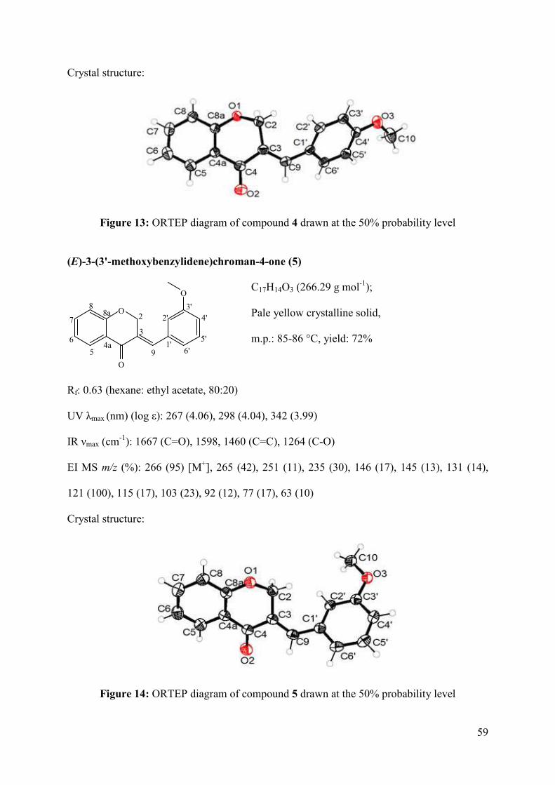
59
Crystal structure:
Figure 13: ORTEP diagram of compound 4 drawn at the 50% probability level
(E)-3-(3'-methoxybenzylidene)chroman-4-one (5)
C17H14O3 (266.29 g mol-1);
Pale yellow crystalline solid,
m.p.: 85-86 °C, yield: 72%
Rf: 0.63 (hexane: ethyl acetate, 80:20)
UV λmax (nm) (log ε): 267 (4.06), 298 (4.04), 342 (3.99)
IR νmax (cm-1): 1667 (C=O), 1598, 1460 (C=C), 1264 (C-O)
EI MS m/z (%): 266 (95) [M+], 265 (42), 251 (11), 235 (30), 146 (17), 145 (13), 131 (14),
121 (100), 115 (17), 103 (23), 92 (12), 77 (17), 63 (10)
Crystal structure:
Figure 14: ORTEP diagram of compound 5 drawn at the 50% probability level
O
O
4a
8a 2
3
5
6
7
8
91'
2'3'
4'
5'
6'
O
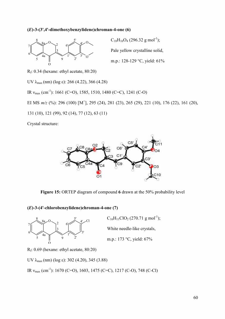
60
(E)-3-(3',4'-dimethoxybenzylidene)chroman-4-one (6)
C18H16O4 (296.32 g mol-1);
Pale yellow crystalline solid,
m.p.: 128-129 °C, yield: 61%
Rf: 0.34 (hexane: ethyl acetate, 80:20)
UV λmax (nm) (log ε): 266 (4.22), 366 (4.28)
IR νmax (cm-1): 1661 (C=O), 1585, 1510, 1480 (C=C), 1241 (C-O)
EI MS m/z (%): 296 (100) [M+], 295 (24), 281 (23), 265 (29), 221 (10), 176 (22), 161 (20),
131 (10), 121 (99), 92 (14), 77 (12), 63 (11)
Crystal structure:
Figure 15: ORTEP diagram of compound 6 drawn at the 50% probability level
(E)-3-(4'-chlorobenzylidene)chroman-4-one (7)
C16H11ClO2 (270.71 g mol-1);
White needle-like crystals,
m.p.: 173 °C, yield: 67%
Rf: 0.69 (hexane: ethyl acetate, 80:20)
UV λmax (nm) (log ε): 302 (4.20), 345 (3.88)
IR νmax (cm-1): 1670 (C=O), 1603, 1475 (C=C), 1217 (C-O), 748 (C-Cl)
O
O
4a
8a 2
3
5
6
7
8
91'
2'3'
4'5'
6'O
O
O
O
4a
8a2
3
5
6
7
8
91'
2'
3'
4'5'
6'Cl
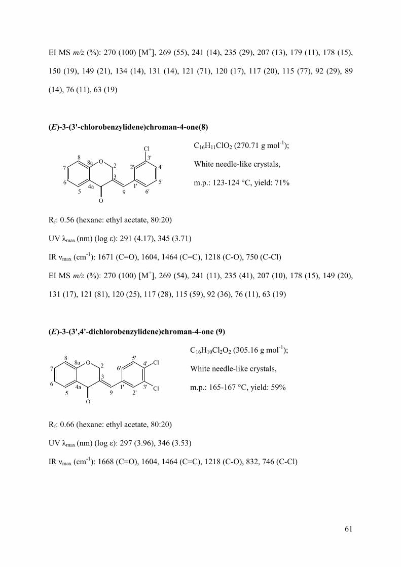
61
EI MS m/z (%): 270 (100) [M+], 269 (55), 241 (14), 235 (29), 207 (13), 179 (11), 178 (15),
150 (19), 149 (21), 134 (14), 131 (14), 121 (71), 120 (17), 117 (20), 115 (77), 92 (29), 89
(14), 76 (11), 63 (19)
(E)-3-(3'-chlorobenzylidene)chroman-4-one(8)
C16H11ClO2 (270.71 g mol-1);
White needle-like crystals,
m.p.: 123-124 °C, yield: 71%
Rf: 0.56 (hexane: ethyl acetate, 80:20)
UV λmax (nm) (log ε): 291 (4.17), 345 (3.71)
IR νmax (cm-1): 1671 (C=O), 1604, 1464 (C=C), 1218 (C-O), 750 (C-Cl)
EI MS m/z (%): 270 (100) [M+], 269 (54), 241 (11), 235 (41), 207 (10), 178 (15), 149 (20),
131 (17), 121 (81), 120 (25), 117 (28), 115 (59), 92 (36), 76 (11), 63 (19)
(E)-3-(3',4'-dichlorobenzylidene)chroman-4-one (9)
C16H10Cl2O2 (305.16 g mol-1);
White needle-like crystals,
m.p.: 165-167 °C, yield: 59%
Rf: 0.66 (hexane: ethyl acetate, 80:20)
UV λmax (nm) (log ε): 297 (3.96), 346 (3.53)
IR νmax (cm-1): 1668 (C=O), 1604, 1464 (C=C), 1218 (C-O), 832, 746 (C-Cl)
O
O
4a
8a 2
3
5
6
7
8
91'
2'
3'
4'
5'
6'
Cl
O
O
4a
8a 2
3
5
6
7
8
91'
2'3'
4'5'
6'Cl
Cl
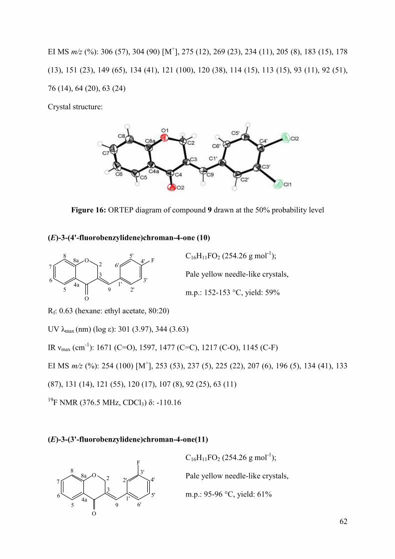
62
EI MS m/z (%): 306 (57), 304 (90) [M+], 275 (12), 269 (23), 234 (11), 205 (8), 183 (15), 178
(13), 151 (23), 149 (65), 134 (41), 121 (100), 120 (38), 114 (15), 113 (15), 93 (11), 92 (51),
76 (14), 64 (20), 63 (24)
Crystal structure:
Figure 16: ORTEP diagram of compound 9 drawn at the 50% probability level
(E)-3-(4'-fluorobenzylidene)chroman-4-one (10)
C16H11FO2 (254.26 g mol-1);
Pale yellow needle-like crystals,
m.p.: 152-153 °C, yield: 59%
Rf: 0.63 (hexane: ethyl acetate, 80:20)
UV λmax (nm) (log ε): 301 (3.97), 344 (3.63)
IR νmax (cm-1): 1671 (C=O), 1597, 1477 (C=C), 1217 (C-O), 1145 (C-F)
EI MS m/z (%): 254 (100) [M+], 253 (53), 237 (5), 225 (22), 207 (6), 196 (5), 134 (41), 133
(87), 131 (14), 121 (55), 120 (17), 107 (8), 92 (25), 63 (11)
19F NMR (376.5 MHz, CDCl3) δ: -110.16
(E)-3-(3'-fluorobenzylidene)chroman-4-one(11)
C16H11FO2 (254.26 g mol-1);
Pale yellow needle-like crystals,
m.p.: 95-96 °C, yield: 61%
O
O
4a
8a2
3
5
6
7
8
91'
2'
3'
4'5'
6'F
O
O
4a
8a 2
3
5
6
7
8
91'
2'3'
4'
5'
6'
F
63
Rf: 0.66 (hexane: ethyl acetate, 80:20)
UV λmax (nm) (log ε): 291 (3.99), 346 (3.55)
IR νmax (cm-1): 1666 (C=O), 1598, 1477 (C=C), 1213 (C-O), 1144 (C-F)
EI MS m/z (%): 254 (100) [M+], 253 (55), 237 (5), 225 (17), 134 (23), 133 (62), 121 (55),
120 (20), 92 (26), 63 (14)
19F NMR (376.5 MHz, CDCl3) δ: 112.15
(E)-3-(3',4'-difluorobenzylidene)chroman-4-one (12)
C16H10F2O2 (272.25 g mol-1);
Pale yellow needle-like crystals,
m.p.: 138-139 °C, yield: 50%
Rf: 0.69 (hexane: ethyl acetate, 80:20)
UV λmax (nm) (log ε): 267 (3.96), 387 (4.05)
IR νmax (cm-1): 1672 (C=O), 1603, 1477 (C=C), 1217 (C-O), 1116, 1145 (C-F)
EI MS m/z (%): 272 (100) [M+], 271 (29), 243 (19), 152 (19), 151 (62), 134 (10), 131 (16),
121 (46), 120 (27), 92 (34), 63 (11)
19F NMR (376.5 MHz, CDCl3) δ: -136.2 (d, J = 21.65 Hz), -134.83 (d, J = 21.46 Hz)
(E)-3-(4'-hydroxybenzylidene)chroman-4-one (13)
C16H12O3 (252.26 g mol-1);
Yellow powder,
m.p.: 222-224 °C, yield: 54%
Rf: 0.22 (hexane: ethyl acetate, 80:20)
UV λmax (nm) (log ε): 331 (3.98), 359 (3.97)
O
O
4a
8a 2
3
5
6
7
8
91'
2'3'
4'5'
6'F
F
O
O
4a
8a2
3
5
6
7
8
91'
2'
3'
4'5'
6'OH
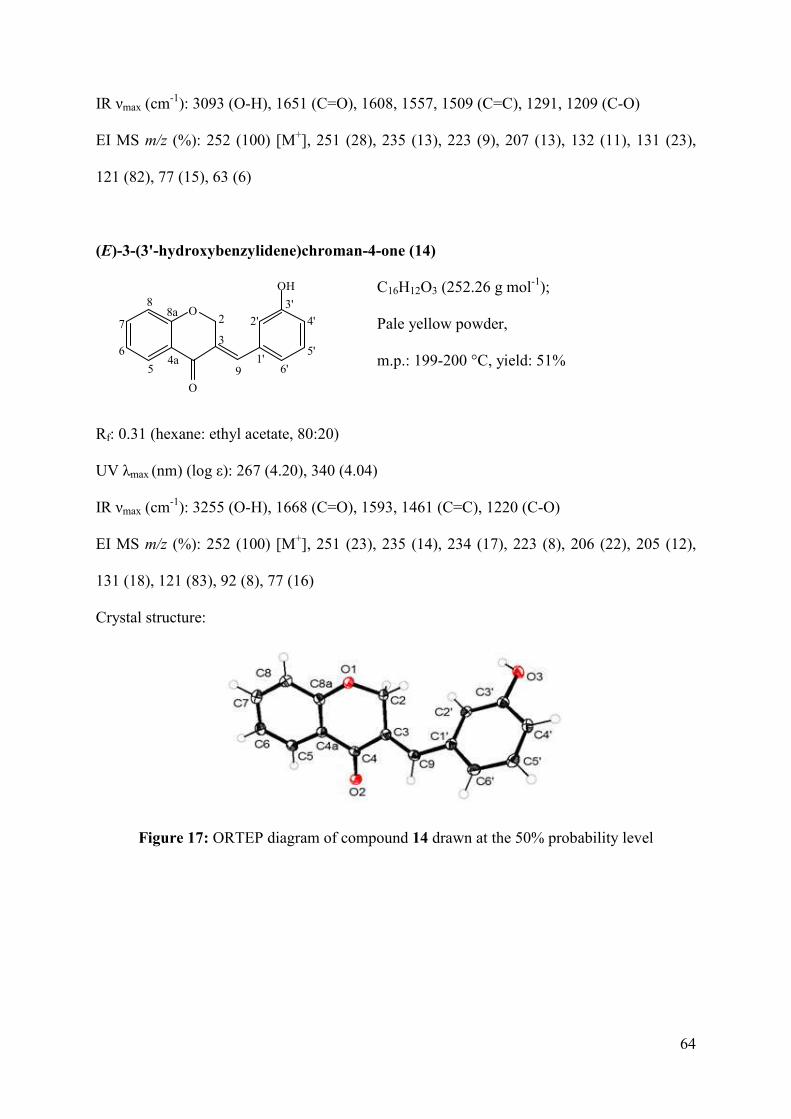
64
IR νmax (cm-1): 3093 (O-H), 1651 (C=O), 1608, 1557, 1509 (C=C), 1291, 1209 (C-O)
EI MS m/z (%): 252 (100) [M+], 251 (28), 235 (13), 223 (9), 207 (13), 132 (11), 131 (23),
121 (82), 77 (15), 63 (6)
(E)-3-(3'-hydroxybenzylidene)chroman-4-one (14)
C16H12O3 (252.26 g mol-1);
Pale yellow powder,
m.p.: 199-200 °C, yield: 51%
Rf: 0.31 (hexane: ethyl acetate, 80:20)
UV λmax (nm) (log ε): 267 (4.20), 340 (4.04)
IR νmax (cm-1): 3255 (O-H), 1668 (C=O), 1593, 1461 (C=C), 1220 (C-O)
EI MS m/z (%): 252 (100) [M+], 251 (23), 235 (14), 234 (17), 223 (8), 206 (22), 205 (12),
131 (18), 121 (83), 92 (8), 77 (16)
Crystal structure:
Figure 17: ORTEP diagram of compound 14 drawn at the 50% probability level
O
O
4a
8a2
3
5
6
7
8
91'
2'
3'
4'
5'
6'
OH
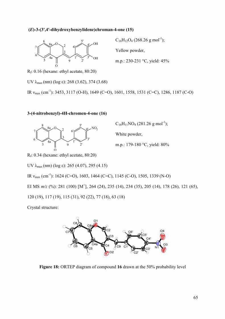
65
(E)-3-(3',4'-dihydroxybenzylidene)chroman-4-one (15)
C16H12O4 (268.26 g mol-1);
Yellow powder,
m.p.: 230-231 °C, yield: 45%
Rf: 0.16 (hexane: ethyl acetate, 80:20)
UV λmax (nm) (log ε): 268 (3.62), 374 (3.68)
IR νmax (cm-1): 3453, 3117 (O-H), 1649 (C=O), 1601, 1558, 1531 (C=C), 1286, 1187 (C-O)
3-(4-nitrobenzyl)-4H-chromen-4-one (16)
C16H11NO4 (281.26 g mol-1);
White powder,
m.p.: 179-180 °C, yield: 80%
Rf: 0.34 (hexane: ethyl acetate, 80:20)
UV λmax (nm) (log ε): 265 (4.07), 295 (4.15)
IR νmax (cm-1): 1624 (C=O), 1603, 1464 (C=C), 1145 (C-O), 1505, 1339 (N-O)
EI MS m/z (%): 281 (100) [M+], 264 (24), 235 (14), 234 (35), 205 (14), 178 (26), 121 (65),
120 (19), 117 (19), 115 (31), 92 (22), 77 (18), 63 (18)
Crystal structure:
Figure 18: ORTEP diagram of compound 16 drawn at the 50% probability level
O
O
4a
8a 2
3
5
6
7
8
91'
2'3'
4'5'
6'OH
OH
O
O
4a
8a 2
3
5
6
7
8
91'
2'
3'
4'5'
6'NO2
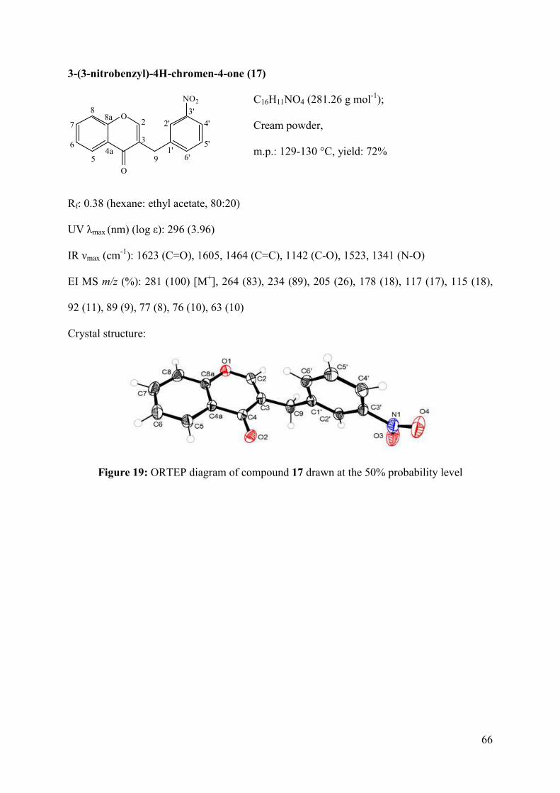
66
3-(3-nitrobenzyl)-4H-chromen-4-one (17)
C16H11NO4 (281.26 g mol-1);
Cream powder,
m.p.: 129-130 °C, yield: 72%
Rf: 0.38 (hexane: ethyl acetate, 80:20)
UV λmax (nm) (log ε): 296 (3.96)
IR νmax (cm-1): 1623 (C=O), 1605, 1464 (C=C), 1142 (C-O), 1523, 1341 (N-O)
EI MS m/z (%): 281 (100) [M+], 264 (83), 234 (89), 205 (26), 178 (18), 117 (17), 115 (18),
92 (11), 89 (9), 77 (8), 76 (10), 63 (10)
Crystal structure:
Figure 19: ORTEP diagram of compound 17 drawn at the 50% probability level
O
O
4a
8a2
3
5
6
7
8
91'
2'
3'
4'
5'
6'
NO2
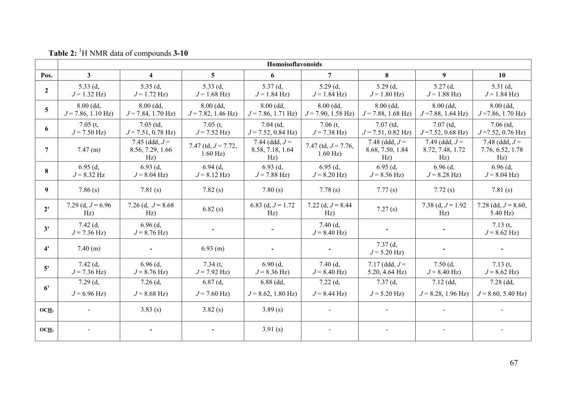
67
Table 2: 1H NMR data of compounds 3-10
Homoisoflavonoids
Pos. 3 4 5 6 7 8 9 10
2 5.33 (d,
J = 1.32 Hz) 5.35 (d,
J = 1.72 Hz) 5.33 (d,
J = 1.68 Hz) 5.37 (d,
J = 1.84 Hz) 5.29 (d,
J = 1.84 Hz) 5.29 (d,
J = 1.80 Hz) 5.27 (d,
J = 1.88 Hz) 5.31 (d,
J = 1.84 Hz)
5 8.00 (dd,
J = 7.86, 1.10 Hz) 8.00 (dd,
J = 7.84, 1.70 Hz) 8.00 (dd,
J = 7.82, 1.46 Hz) 8.00 (dd,
J = 7.86, 1.71 Hz) 8.00 (dd,
J = 7.90, 1.58 Hz) 8.00 (dd,
J = 7.88, 1.68 Hz) 8.00 (dd,
J =7.88, 1.64 Hz) 8.00 (dd,
J =7.86, 1.70 Hz)
6 7.05 (t,
J = 7.50 Hz) 7.05 (td,
J = 7.51, 0.78 Hz) 7.05 (t,
J = 7.52 Hz) 7.04 (td,
J = 7.52, 0.84 Hz) 7.06 (t,
J = 7.38 Hz) 7.07 (td,
J = 7.51, 0.82 Hz) 7.07 (td,
J =7.52, 0.68 Hz) 7.06 (td,
J =7.52, 0.76 Hz)
7 7.47 (m) 7.45 (ddd, J =
8.56, 7.29, 1.66 Hz)
7.47 (td, J = 7.72, 1.60 Hz)
7.44 (ddd, J = 8.58, 7.18, 1.64
Hz)
7.47 (td, J = 7.76, 1.60 Hz)
7.48 (ddd, J = 8.68, 7.50, 1.84
Hz)
7.49 (ddd, J = 8.72, 7.48, 1.72
Hz)
7.48 (ddd, J = 7.76, 6.52, 1.78
Hz)
8 6.95 (d,
J = 8.32 Hz
6.93 (d, J = 8.04 Hz)
6.94 (d, J = 8.12 Hz)
6.93 (d, J = 7.88 Hz)
6.95 (d, J = 8.20 Hz)
6.95 (d, J = 8.56 Hz)
6.96 (d, J = 8.28 Hz)
6.96 (d, J = 8.04 Hz)
9 7.86 (s) 7.81 (s) 7.82 (s) 7.80 (s) 7.78 (s) 7.77 (s) 7.72 (s) 7.81 (s)
2' 7.29 (d, J = 6.96
Hz) 7.26 (d, J = 8.68
Hz) 6.82 (s)
6.83 (d, J = 1.72 Hz)
7.22 (d, J = 8.44 Hz)
7.27 (s) 7.38 (d, J = 1.92
Hz) 7.28 (dd, J = 8.60,
5.40 Hz)
3' 7.42 (d,
J = 7.36 Hz) 6.96 (d,
J = 8.76 Hz) - -
7.40 (d, J = 8.40 Hz)
- - 7.13 (t,
J = 8.62 Hz)
4' 7.40 (m) - 6.93 (m) - - 7.37 (d,
J = 5.20 Hz) - -
5' 7.42 (d,
J = 7.36 Hz) 6.96 (d,
J = 8.76 Hz) 7.34 (t,
J = 7.92 Hz) 6.90 (d,
J = 8.36 Hz) 7.40 (d,
J = 8.40 Hz) 7.17 (ddd, J = 5.20, 4.64 Hz)
7.50 (d, J = 8.40 Hz)
7.13 (t, J = 8.62 Hz)
6' 7.29 (d,
J = 6.96 Hz)
7.26 (d,
J = 8.68 Hz)
6.87 (d,
J = 7.60 Hz)
6.88 (dd,
J = 8.62, 1.80 Hz)
7.22 (d,
J = 8.44 Hz)
7.37 (d,
J = 5.20 Hz)
7.12 (dd,
J = 8.28, 1.96 Hz)
7.28 (dd,
J = 8.60, 5.40 Hz)
OCH3 - 3.83 (s) 3.82 (s) 3.89 (s) - - - -
OCH3 - - - 3.91 (s) - - - -
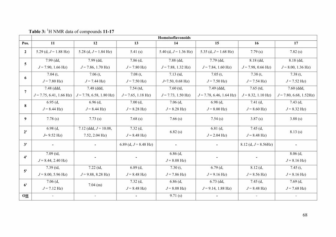
68
Table 3: 1H NMR data of compounds 11-17
Homoisoflavonoids
Pos. 11 12 13 14 15 16 17
2 5.29 (d, J = 1.88 Hz) 5.28 (d, J = 1.84 Hz) 5.41 (s) 5.40 (d, J = 1.36 Hz) 5.35 (d, J = 1.68 Hz) 7.79 (s) 7.82 (s)
5 7.99 (dd,
J = 7.90, 1.66 Hz)
7.99 (dd,
J = 7.86, 1.70 Hz)
7.86 (d,
J = 7.80 Hz)
7.88 (dd,
J = 7.88, 1.32 Hz)
7.79 (dd,
J = 7.84, 1.60 Hz)
8.18 (dd,
J = 7.98, 0.66 Hz)
8.18 (dd,
J = 8.00, 1.36 Hz)
6 7.04 (t,
J = 7.80 Hz)
7.06 (t,
J = 7.44 Hz)
7.08 (t,
J = 7.50 Hz)
7.13 (td,
J=7.50, 0.68 Hz)
7.05 (t,
J = 7.50 Hz)
7.38 (t,
J = 7.54 Hz)
7.38 (t,
J = 7.52 Hz)
7 7.48 (ddd,
J = 7.75, 6.41, 1.66 Hz)
7.48 (ddd,
J = 7.78, 6.58, 1.80 Hz)
7.54 (td,
J = 7.65, 1.18 Hz)
7.60 (td,
J = 7.73, 1.50 Hz)
7.49 (ddd,
J = 7.78, 6.46, 1.64 Hz)
7.65 (td,
J = 8.32, 1.10 Hz)
7.60 (ddd,
J = 7.80, 6.68, 1.52Hz)
8 6.95 (d,
J = 8.44 Hz)
6.96 (d,
J = 8.44 Hz)
7.00 (d,
J = 8.28 Hz)
7.06 (d,
J = 8.28 Hz)
6.98 (d,
J = 8.00 Hz)
7.41 (d,
J = 8.60 Hz)
7.43 (d,
J = 8.32 Hz)
9 7.78 (s) 7.73 (s) 7.68 (s) 7.66 (s) 7.54 (s) 3.87 (s) 3.88 (s)
2' 6.98 (d,
J= 9.52 Hz)
7.12 (ddd, J = 10.08,
7.52, 2.04 Hz)
7.32 (d,
J = 8.48 Hz) 6.82 (s)
6.81 (d,
J = 2.04 Hz)
7.45 (d,
J = 8.48 Hz) 8.13 (s)
3' - - 6.89 (d, J = 8.48 Hz) - - 8.12 (d, J = 8.56Hz) -
4' 7.09 (td,
J = 8.44, 2.40 Hz) - -
6.86 (d,
J = 8.08 Hz) - -
8.06 (d,
J = 8.16 Hz)
5' 7.39 (td,
J = 8.00, 5.96 Hz)
7.22 (td,
J = 9.88, 8.28 Hz)
6.89 (d,
J = 8.48 Hz)
7.30 (t,
J = 7.86 Hz)
6.79 (d,
J = 9.16 Hz)
8.12 (d,
J = 8.56 Hz)
7.45 (t,
J = 8.16 Hz)
6' 7.06 (d,
J = 7.12 Hz) 7.04 (m)
7.32 (d,
J = 8.48 Hz)
6.86 (d,
J = 8.08 Hz)
6.73 (dd,
J = 9.14, 1.88 Hz)
7.45 (d,
J = 8.48 Hz)
7.69 (d,
J = 7.68 Hz)
OH - - - 9.71 (s) - - -
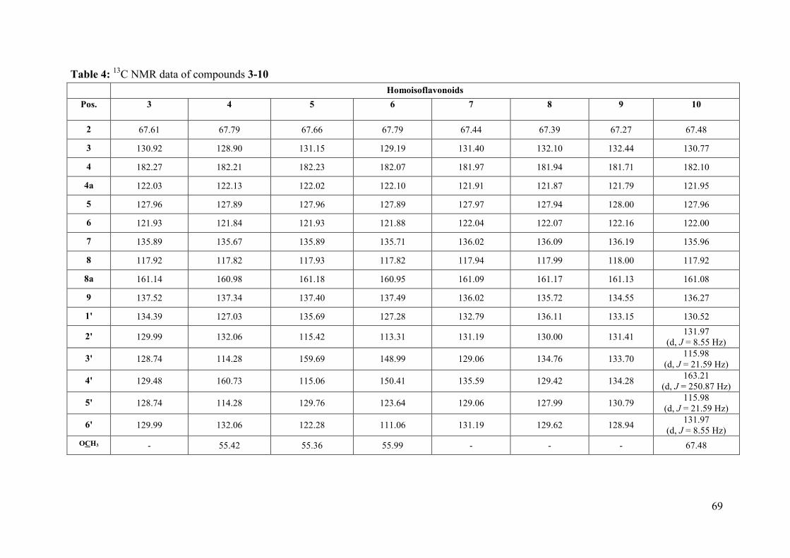
69
Table 4: 13C NMR data of compounds 3-10
Homoisoflavonoids
Pos. 3 4 5 6 7 8 9 10
2 67.61 67.79 67.66 67.79 67.44 67.39 67.27 67.48
3 130.92 128.90 131.15 129.19 131.40 132.10 132.44 130.77
4 182.27 182.21 182.23 182.07 181.97 181.94 181.71 182.10
4a 122.03 122.13 122.02 122.10 121.91 121.87 121.79 121.95
5 127.96 127.89 127.96 127.89 127.97 127.94 128.00 127.96
6 121.93 121.84 121.93 121.88 122.04 122.07 122.16 122.00
7 135.89 135.67 135.89 135.71 136.02 136.09 136.19 135.96
8 117.92 117.82 117.93 117.82 117.94 117.99 118.00 117.92
8a 161.14 160.98 161.18 160.95 161.09 161.17 161.13 161.08
9 137.52 137.34 137.40 137.49 136.02 135.72 134.55 136.27
1' 134.39 127.03 135.69 127.28 132.79 136.11 133.15 130.52
2' 129.99 132.06 115.42 113.31 131.19 130.00 131.41 131.97
(d, J = 8.55 Hz)
3' 128.74 114.28 159.69 148.99 129.06 134.76 133.70 115.98
(d, J = 21.59 Hz)
4' 129.48 160.73 115.06 150.41 135.59 129.42 134.28 163.21
(d, J = 250.87 Hz)
5' 128.74 114.28 129.76 123.64 129.06 127.99 130.79 115.98
(d, J = 21.59 Hz)
6' 129.99 132.06 122.28 111.06 131.19 129.62 128.94 131.97
(d, J = 8.55 Hz) OCH3 - 55.42 55.36 55.99 - - - 67.48
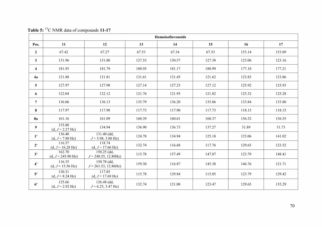
70
Table 5: 13C NMR data of compounds 11-17
Homoisoflavonoids
Pos. 11 12 13 14 15 16 17
2 67.42 67.27 67.53 67.34 67.53 153.14 153.09
3 131.96 131.80 127.53 130.57 127.38 123.06 123.16
4 181.93 181.79 180.95 181.17 180.99 177.18 177.21
4a 121.88 121.81 121.61 121.45 121.62 123.83 123.86
5 127.97 127.98 127.14 127.23 127.12 125.92 125.93
6 122.04 122.12 121.76 121.95 121.82 125.32 125.28
7 136.06 136.13 135.79 136.20 135.86 133.84 133.80
8 117.97 117.98 117.73 117.90 117.73 118.15 118.15
8a 161.16 161.09 160.39 160.61 160.37 156.52 156.55
9 135.88
(d, J = 2.27 Hz) 134.94 136.90 136.73 137.27 31.89 31.73
1' 136.40
(d, J = 7.80 Hz) 131.40 (dd,
J = 5.98, 3.80 Hz) 124.78 134.94 125.18 123.06 141.02
2' 116.57
(d, J = 16.28 Hz) 118.74
(d, J = 17.66 Hz) 132.74 116.68 117.76 129.65 123.52
3' 162.70
(d, J = 245.90 Hz) 150.25 (dd,
J = 248.55, 12.80Hz) 115.78 157.49 147.87 123.79 148.41
4' 116.35
(d, J = 15.56 Hz) 150.78 (dd,
J = 261.53, 12.86Hz) 159.30 116.87 145.38 146.76 121.71
5' 130.31
(d, J = 8.24 Hz) 117.85
(d, J = 17.69 Hz) 115.78 129.84 115.85 123.79 129.42
6' 125.66
(d, J = 2.92 Hz) 126.48 (dd,
J = 6.25, 3.47 Hz) 132.74 121.08 123.47 129.65 135.29
71
3.2 Biochemistry
The experimental techniques employed to determine the antioxidant and antibacterial
activities of the homoisoflavonoids are stated below as well as the experimental data.
3.2.1 Antioxidant activity of the homoisoflavonoids synthesised
The antioxidant activities of the homoisoflavonoids were determined using two common
simple methods, i.e. the DPPH radical scavenging assay and the ferric reducing antioxidant
power assay.
3.2.1.1 DPPH radical scavenging assay
The DPPH scavenging activity of homoisoflavonoids was determined according to the
modified method by (Murthy et al., 2012). The free radical scavenging activity was
determined spectrophotometrically using a stable free radical, 1,1-diphenyl-2-picrylhydrazyl
(DPPH). Stock solutions of each compound were prepared by dissolving 10 mg of the
compound in 10 mL of methanol (1000 µg/mL). The stock solutions were used to prepare a
series of eight concentrations (500, 200, 100, 50, 40, 30, 20, 10 µg/mL). A solution of DPPH
was prepared by dissolving 1.97 mg of DPPH in 50 mL of methanol (0.1 mM) and protected
from light by covering the volumetric flask with aluminum foil. An aliquot of each dilution
of the compound (150 µl) was mixed with methanolic solution of DPPH (2850 µl) in glass
test tubes. The mixtures were shaken vigorously and set in a dark cupboard at ambient
temperature for 30 min. The absorbance was measured at 517 nm against methanol as a
blank. All measurements were done in triplicate and the average absorbance was used. The
percent scavenging activity of the compounds were calculated using the following formula:
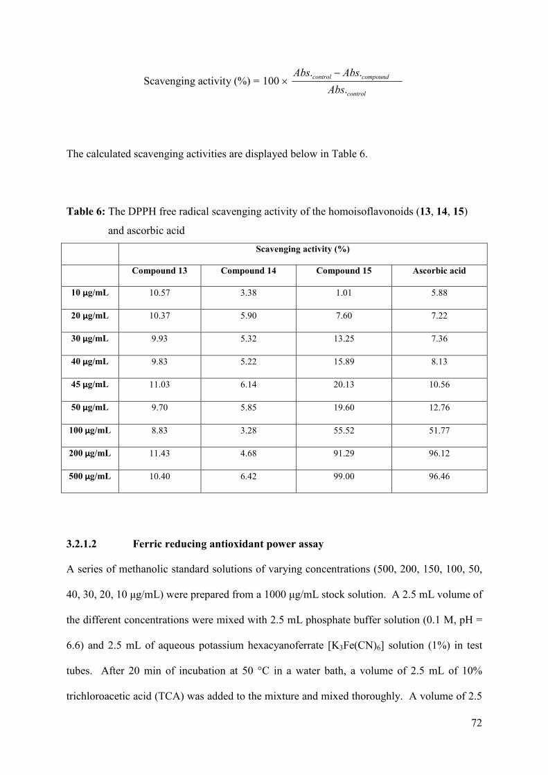
72
Scavenging activity (%) = control
compoundcontrol
Abs
AbsAbs
.
..100
−×
The calculated scavenging activities are displayed below in Table 6.
Table 6: The DPPH free radical scavenging activity of the homoisoflavonoids (13, 14, 15)
and ascorbic acid
Scavenging activity (%)
Compound 13 Compound 14 Compound 15 Ascorbic acid
10 µg/mL 10.57 3.38 1.01 5.88
20 µg/mL 10.37 5.90 7.60 7.22
30 µg/mL 9.93 5.32 13.25 7.36
40 µg/mL 9.83 5.22 15.89 8.13
45 µg/mL 11.03 6.14 20.13 10.56
50 µg/mL 9.70 5.85 19.60 12.76
100 µg/mL 8.83 3.28 55.52 51.77
200 µg/mL 11.43 4.68 91.29 96.12
500 µg/mL 10.40 6.42 99.00 96.46
3.2.1.2 Ferric reducing antioxidant power assay
A series of methanolic standard solutions of varying concentrations (500, 200, 150, 100, 50,
40, 30, 20, 10 µg/mL) were prepared from a 1000 µg/mL stock solution. A 2.5 mL volume of
the different concentrations were mixed with 2.5 mL phosphate buffer solution (0.1 M, pH =
6.6) and 2.5 mL of aqueous potassium hexacyanoferrate [K3Fe(CN)6] solution (1%) in test
tubes. After 20 min of incubation at 50 °C in a water bath, a volume of 2.5 mL of 10%
trichloroacetic acid (TCA) was added to the mixture and mixed thoroughly. A volume of 2.5
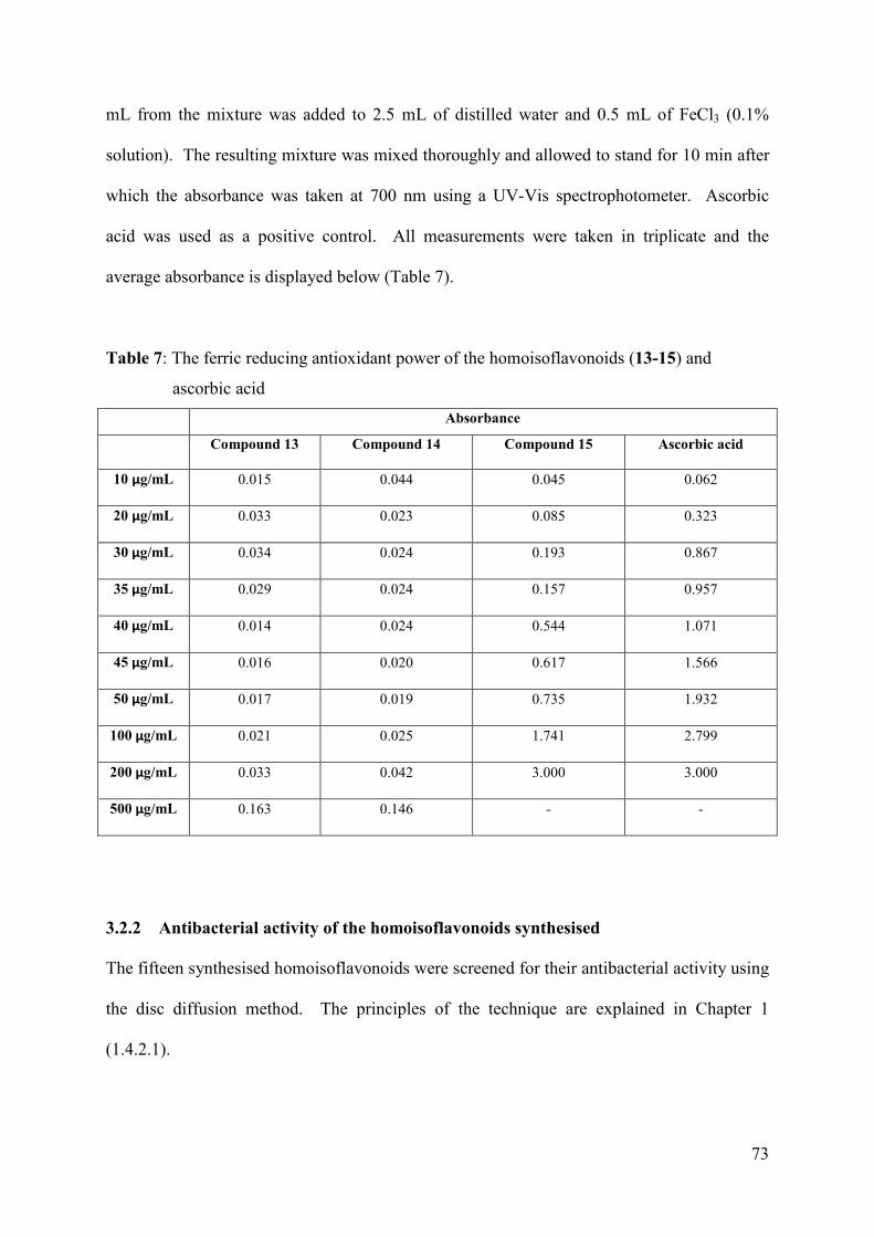
73
mL from the mixture was added to 2.5 mL of distilled water and 0.5 mL of FeCl3 (0.1%
solution). The resulting mixture was mixed thoroughly and allowed to stand for 10 min after
which the absorbance was taken at 700 nm using a UV-Vis spectrophotometer. Ascorbic
acid was used as a positive control. All measurements were taken in triplicate and the
average absorbance is displayed below (Table 7).
Table 7: The ferric reducing antioxidant power of the homoisoflavonoids (13-15) and
ascorbic acid
Absorbance
Compound 13 Compound 14 Compound 15 Ascorbic acid
10 µg/mL 0.015 0.044 0.045 0.062
20 µg/mL 0.033 0.023 0.085 0.323
30 µg/mL 0.034 0.024 0.193 0.867
35 µg/mL 0.029 0.024 0.157 0.957
40 µg/mL 0.014 0.024 0.544 1.071
45 µg/mL 0.016 0.020 0.617 1.566
50 µg/mL 0.017 0.019 0.735 1.932
100 µg/mL 0.021 0.025 1.741 2.799
200 µg/mL 0.033 0.042 3.000 3.000
500 µg/mL 0.163 0.146 - -
3.2.2 Antibacterial activity of the homoisoflavonoids synthesised
The fifteen synthesised homoisoflavonoids were screened for their antibacterial activity using
the disc diffusion method. The principles of the technique are explained in Chapter 1
(1.4.2.1).
74
3.2.2.1 Disc diffusion antimicrobial susceptibility testing
The antibacterial activities of the synthesised homoisoflavonoids were determined using the
Kirby-Bauer disk-diffusion method. The homoisoflavonoids synthesised were tested against
ten gram positive and six gram negative bacteria (Table 8). Stock solutions (5 mg/mL) of
each homoisoflavonoid were prepared by dissolving compounds in 1 mL of DMSO. Blank
discs (5 mm; MAST, UK) were impregnated with 50, 100 and 200 µg/mL of each
homoisoflavonoid and allowed to dry. Bacterial isolates were grown overnight on TSA agar
plates and the turbidity of cell suspensions were adjusted equivalent to that of a 0.5
McFarland standard. These were used to inoculate Mueller-Hinton (MH) agar plates by
streaking swabs over the entire agar surface followed by the application of the respective
homoisoflavonoid discs. Plates were then incubated for 21 hrs at 30 °C. Testing was done in
duplicate and tetracycline (TET) and ampicillin (AMP) discs were used as standard
antimicrobial agent controls. The negative control was 5 µl of DMSO (100%). Zone
diameters were measured physically and averaged. Activity indices of each compound were
calculated by comparing zones of inhibition obtained with each of the compounds with those
obtained with the standard antimicrobial agents, tetracycline and ampicillin. The following
equation was used:
Activity index (AI) = agent ialantimicrob standard with (mm)diameter Inhibition
compound with test(mm)diameter Inhibition
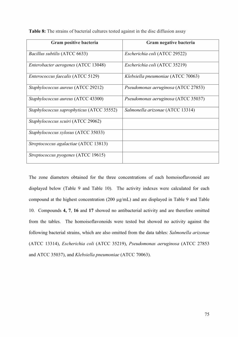
75
Table 8: The strains of bacterial cultures tested against in the disc diffusion assay
Gram positive bacteria Gram negative bacteria
Bacillus subtilis (ATCC 6633) Escherichia coli (ATCC 29522)
Enterobacter aerogenes (ATCC 13048) Escherichia coli (ATCC 35219)
Enterococcus faecalis (ATCC 5129) Klebsiella pneumoniae (ATCC 70063)
Staphylococcus aureus (ATCC 29212) Pseudomonas aeruginosa (ATCC 27853)
Staphylococcus aureus (ATCC 43300) Pseudomonas aeruginosa (ATCC 35037)
Staphylococcus saprophyticus (ATCC 35552) Salmonella arizonae (ATCC 13314)
Staphylococcus scuiri (ATCC 29062)
Staphylococcus xylosus (ATCC 35033)
Streptococcus agalactiae (ATCC 13813)
Streptococcus pyogenes (ATCC 19615)
The zone diameters obtained for the three concentrations of each homoisoflavonoid are
displayed below (Table 9 and Table 10). The activity indexes were calculated for each
compound at the highest concentration (200 µg/mL) and are displayed in Table 9 and Table
10. Compounds 4, 7, 16 and 17 showed no antibacterial activity and are therefore omitted
from the tables. The homoisoflavonoids were tested but showed no activity against the
following bacterial strains, which are also omitted from the data tables: Salmonella arizonae
(ATCC 13314), Escherichia coli (ATCC 35219), Pseudomonas aeruginosa (ATCC 27853
and ATCC 35037), and Klebsiella pneumoniae (ATCC 70063).
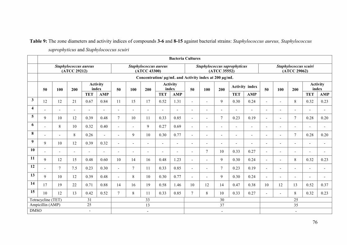
76
Table 9: The zone diameters and activity indices of compounds 3-6 and 8-15 against bacterial strains: Staphylococcus aureus, Staphylococcus
saprophyticus and Staphylococcus scuiri
Bacteria Cultures
Staphylococcus aureus
(ATCC 29212)
Staphylococcus aureus
(ATCC 43300)
Staphylococcus saprophyticus
(ATCC 35552)
Staphylococcus scuiri
(ATCC 29062)
Concentration/ µg/mL and Activity index at 200 µg/mL
50 100 200
Activity
index 50 100 200
Activity
index 50 100 200 Activity index
50 100 200
Activity
index
TET AMP TET AMP TET AMP TET AMP
3 12 12 21 0.67 0.84 11 15 17 0.52 1.31 - - 9 0.30 0.24 - - 8 0.32 0.23
4 - - - - - - - - - - - - - - - - - - - -
5 9 10 12 0.39 0.48 7 10 11 0.33 0.85 - - 7 0.23 0.19 - - 7 0.28 0.20
6 - 8 10 0.32 0.40 - - 9 0.27 0.69 - - - - - - - - - -
8 - - 8 0.26 - - 9 10 0.30 0.77 - - - - - - - 7 0.28 0.20
9 9 10 12 0.39 0.32 - - - - - - - - - - - - - - -
10 - - - - - - - - - - - 7 10 0.33 0.27 - - - - -
11 9 12 15 0.48 0.60 10 14 16 0.48 1.23 - - 9 0.30 0.24 - - 8 0.32 0.23
12 - 7 7.5 0.23 0.30 - 7 11 0.33 0.85 - - 7 0.23 0.19 - - - - -
13 9 10 12 0.39 0.48 - 8 10 0.30 0.77 - - 9 0.30 0.24 - - - - -
14 17 19 22 0.71 0.88 14 16 19 0.58 1.46 10 12 14 0.47 0.38 10 12 13 0.52 0.37
15 10 12 13 0.42 0.52 7 8 11 0.33 0.85 7 8 10 0.33 0.27 - - 8 0.32 0.23
Tetracycline (TET) 31 33 30 25 Ampicillin (AMP) 25 13 37 35 DMSO - - - -
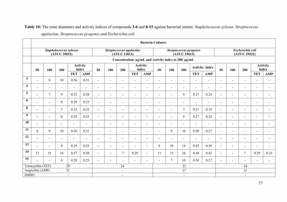
77
Table 10: The zone diameters and activity indices of compounds 3-6 and 8-15 against bacterial strains: Staphylococcus xylosus, Streptococcus
agalactiae, Streptococcus pyogenes and Escherichia coli
Bacteria Cultures
Staphylococcus xylosus
(ATCC 35033)
Streptococcus agalactiae
(ATCC 13813)
Streptococcus pyogenes
(ATCC 19615)
Escherichia coli
(ATCC 29522)
Concentration/ µg/mL and Activity index at 200/ µg/mL
50 100 200
Activity
index 50 100 200
Activity
index 50 100 200 Activity index
50 100 200
Activity
index
TET AMP TET AMP TET AMP TET AMP
3 - 8 10 0.36 0.31 - - - - - - - - - - - - - - -
4 - - - - - - - - - - - - - - - - - - - -
5 - 7 9 0.32 0.28 - - - - - - - 9 0.27 0.24 - - - - -
6 - - 8 0.29 0.25 - - - - - - - - - - - - - - -
8 - - 7 0.25 0.22 - - - - - - - 7 0.21 0.19 - - - - -
9 - - 8 0.29 0.25 - - - - - - - 9 0.27 0.24 - - - - -
10 - - - - - - - - - - - - - - - - - - - -
11 8 9 10 0.36 0.31 - - - - - - 9 10 0.30 0.27 - - - - -
12 - - - - - - - - - - - - - - - - - - - -
13 - - 8 0.29 0.25 - - - - - 9 10 14 0.42 0.38 - - - - -
14 13 15 16 0.57 0.50 - - 7 0.29 - 11 15 16 0.48 0.43 - - 7 0.29 0.33
15 - - 8 0.29 0.25 - - - - - - 7 10 0.30 0.27 - - - - -
Tetracycline (TET) 28 24 33 24 Ampicillin (AMP) 32 - 37 21 DMSO - - - -
78
CHAPTER 4 CONCLUSION
A series of fifteen homoisoflavonoids, three of the 3-benzylidene and two of the 3-benzyl
type, were synthesised in a three step reaction. The solvent free base-cataylsed aldol
condensation of 4-chromanone and substituted benzaldehydes in the presence of piperidine
resulted in the formation of the homoisoflavonoids in good yields of between 50 and 90%.
Substitution of the phenyl ring was varied at the 3'-position, 4'-position and 3',4'-positions
with methoxy, hydroxy, chloro, fluoro and nitro groups. Compounds containing the electron
withdrawing nitro groups resulted in the formation of the 3-benzyl-4-chromanone rather than
the desired 3-benzylidene-4-chromanone. The synthesised compounds were fully
characterised by 1H, 13C and 19F NMR, IR and UV spectroscopy and EI-MS. Crystal
structures of seven homoisoflavonoids were also obtained and reported for the first time in
this work.
The antioxidant testing of the homoisoflavonoids, using the DPPH radical scavenging assay
and the ferric reducing antioxidant power assay, showed that the polyhydroxylated
compounds have good antioxidant activity due to the fact that the hydrogen on an O-H group
is scavengable. Compound 15, a disubstituted hydroxyl compound, showed good antioxidant
activity comparable to that of ascorbic acid. This was attributed to the fact compound 15 has
a catechol moiety. Mono-substituted hydroxy containing homoisoflavonoids (13 and 14)
showed weak antioxidant activity compared to compound 15. Further derivatisation of
compound 15, by substituting a catechol moiety on the A-ring of the homoisoflavonoid, may
result in increased antioxidant activity. Compound 15 is therefore an interesting target
molecule to derivatise in the pursuit of good antioxidants.
79
The synthesised homoisoflavonoids were also subjected to antibacterial testing which showed
that homoisoflavonoids are more active against gram positive than gram negative bacteria.
The synthesised homoisoflavonoids showed good antibacterial activity against a methicillin
resistant strain of bacteria, Staphylococcus aureus (ATCC 43300). Compounds 3, 11 and 14
showed better antibacterial activity than the common antibiotic ampicillin. Compound 14,
the meta hydroxy homoisoflavonoid, showed the highest activity index of 1.46.
Derivatisation of compound 14 with respect to the A-ring can be conducted and a structure-
activity relationship determined.
80
REFERENCES
Abegaz, B. M., Mutanyatta-Comar, J., Nindi, M., 2007. Naturally occurring
homoisoflavonoids: Phytochemistry, biological activities and synthesis. Natural
Product Communications 2, 475-498.
Adinolfi, M., Lanzetta, R., Laonigro, G., Parrilli, M., Breitmaier, E., 1986. 1H and 13C
chemical shift assignments of homoisoflavanones. Magnetic Resonance in Chemistry
24, 663-666.
Basavaiah, D., Bakthadoss, M., 1998. A new protocol for the syntheses of (E)-3-
benzylidenechroman-4-ones: a simple synthesis of the methyl ether of bonducellin.
Chemical Communications 16, 1639-1640 .
Bhandari, P., Crombie, L., Daniels, P., Holden, I., Van Bruggen, N., Whiting, D. A., 1992.
Biosynthesis of the A/B/C/D-ring system of the rotenoid amorphigenin by Amorpha
fruticosa seedlings. Journal of the Chemical Society, Perkin Transactions 1, 839-849.
Böhler, P., Tamm, C., 1967. The homo-isoflavones, a new class of natural product. Isolation
and structure of eucomin and eucomol. Tetrahedron Letters 8, 3479-3483.
Cheng, X.-M., Huang, Z.-T., Zheng, Q.-Y., 2011. Topochemical photodimerization of (E)-3-
benzylidene-4-chromanone derivatives from β-type structures directed by halogen
groups. Tetrahedron 67.
Clayden, J., Greeves, N., Warren, S., Wothers, P., 2001. Organic Chemistry. Oxford
Univeristy Press, United States.
Conti, C., Desideri, N., 2009. Synthesis and antirhinovirus activity of new 3-benzyl chromene
and chroman derivatives. Bioorganic & Medicinal Chemistry 17, 3720-3727.
81
Das, B., Thirupathi, P., Ravikanth, B., Kumar, R. A., Sarma, A. V. S., Basha, S. J., 2009.
Isolation, Synthesis, and Bioactivity of Homoisoflavonoids from Caesalpinia
pulcherrima. Chemical Pharmaceutical Bulletin. 57, 1139-1141.
Desideri, N., Bolasco, A., Fioravanti, R., Proietti Monaco, L., Orallo, F., Yáñez, M., Ortuso,
F., Alcaro, S., 2011. Homoisoflavonoids: Natural scaffolds with potent and selective
monoamine oxidase-B inhibition properties. Journal of Medicinal Chemistry 54,
2155-2164.
Dewick, P. M., 1975. Biosynthesis of the 3-Benzylchroman-4-one Eucomin in Eucomis
bicolor. Phytochemistry 14, 983-988.
du Toit, K., Drewes, S. E., Bodenstein, J., 2010. The chemical structures, plant origins,
ethnobotany and biological activities of homoisoflavanones. Natural Product
Research 24, 457-490.
du Toit, K., Elgorashi, E. E., Malan, S. F., Mulholland, D. A., Drewes, S. E., Van Staden, J.,
2007. Antibacterial activity and QSAR of homoisoflavanones isolated from six
Hyacinthaceae species. South African Journal of Botany 73, 236-241.
Erel, O., 2004. A novel automated direct measurement method for total antioxidant capacity
using a new generation, more stable ABTS radical cation. Clinical Biochemistry 37,
277-285.
Evans, D., Lockhart, I. M., 1966. Reaction of aromatic aldehydes and nitroso-compounds
with 4-chromanones. Journal of the Chemical Society C: Organic, 711-712.
Farkas, L., Gottsegen, A., Nógrádi, M., 1968. The synthesis of eucomin and (±)-eucomol.
Tetrahedron Letters 38, 4099-4100.
Farkas, L., Gottsegen, Á., Nógrádi, M., Strelisky, J., 1971. Synthesis of homoisoflavanones—
II : Constituents of Eucomis autumn alis and E. Punctata. Tetrahedron 27, 5049-5054.
82
Foroumadi, A., Samzadeh-Kermani, A., Emami, S., Dehghan, G., Sorkhi, M., Arabsorkhi, F.,
Heidari, M. R., Abdollahi, M., Shafiee, A., 2007. Synthesis and antioxidant properties
of substituted 3-benzylidene-7-alkoxychroman-4-ones. Bioorganic & Medicinal
Chemistry Letters 17, 6764-6769.
Hahlbrock, K., Grisebach, H., 1975. Biosynthesis of Flavonoids :The Flavonoids. Chapman
and Hall London.
Heller, W., Tamm, C., 1981. Homoisoflavanones and biogenetically related compounds.
Fortschritte der Chemie organischer Naturstoffe, 40, 105-152.
Hung, T. M., Thu, C. V., Dat, N. T., Ryoo, S.-W., Lee, J. H., Kim, J. C., Na, M., Jung, H.-J.,
Bae, K., Min, B. S., 2010. Bioorganic & Medicinal Chemistry Letters 20, 2412-2416.
Jacquot, Y., Byrne, C., Xicluna, A., Leclercq, G., 2012. Synthesis, structure, and estrogenic
activity of 2- and 3-substituted 2,3-dihydro-4H-1-benzopyran-4-ones. Medical
Chemistry Research 22, 681-691.
Kirkiacharian, B. S., Gomis, M., Tongo, H. G., Mahuteau, J., Brion, J. D., 1984. Organic
Magnetic Resonance. 22, 106.
Koorbanally, C., Crouch, N. R., Mulholland, D. A., 2006. The phytochemistry and
ethnobotany of the southern African genus Eucomis (Hyacinthaceae:
Hyacinthoideae). Phytochemistry: Advances in Research 2, 69-85.
Lévai, A., 2004. Synthesis of exocyclic α,β-unsaturated ketones. ARKIVOC vii, 15-33.
Lévai, A., Schág, B., 1979. Synthesis of 3-Benzylidenechroman-4-one and -1-thiochroman-4-
ones. Pharmazie 34, 749.
Lin, L.-G., Xie, H., Li, H.-L., Tong, L.-J., Tang, C.-P., Ke, C.-Q., Liu, Q.-F., Lin, L.-P.,
Geng, M.-Y., Jiang, H., Zhao, W.-M., Ding, J., Ye, Y., 2008. Naturally Occurring
Homoisoflavonoids Function as Potent Protein Tyrosine Kinase Inhibitors by c-Src-
Based High-Throughput Screening. Journal of Medicinal Chemistry 51, 4419-4429.
83
Lin, Y., Zhu, D., Qi, J., Qin, M., Yu, B., 2010. Characterization of homoisoflavonoids in
different cultivation regions of Ophiopogon japonicus and related antioxidant activity.
Journal of Pharmaceutical and Biomedical Analysis 52, 757-762.
Mann, J., Davidson, R. S., Hobbs, J. B., Banthorpe, D. V., Harborne, J. B., 1994. Natural
Products: Their Chemistry and Biological Significance. Longman Scientific &
Technical.
Miadoková, E., Mašterová, I., Vlčková, V., Dúhová, V., Tóth, J., 2002. Antimutagenic
potential of homoisoflavonoids from Muscari racemosum. Journal of
Ethnopharmacology 81, 381-386.
Murthy, P. S., Manjunatha, M. R., Sulochannama, G., Naidu, M. M., 2012. Extraction,
Characterization and Bioactivity of Coffee Anthocyanins. European Journal of
Biological Sciences 4, 13-19.
Osman, A. M., Wong, K. K. Y., Fernyhough, A., 2006. ABTS radical-driven oxidation of
polyphenols: Isolation and structural elucidation of covalent adducts. Biochemical and
Biophysical Research Communications 346, 321-329.
Perjési, P., Das, U., De Clercq, E., Balzarini, J., Kawase, M., Sakagami, H., Stables, J. P.,
Lorand, T., Rozmer, Z., Dimmock, J. R., 2008. Design, synthesis and antiproliferative
activity of some 3-benzylidene-2,3-dihydro-1-benzopyran-4-ones which display
selective toxicity for malignant cells. European Journal of Medicinal Chemistry 43,
839-845.
Rao, V. M., Damu, G. L. V., Sudhakar, D., Siddaiah, V., Rao, C. V., 2008. New efficient
synthesis and bioactivity of homoisoflavonoids. ARKIVOC xi 285-294.
Shaikh, M., Petzold, K., Kruger, H., Toit, K. d., 2011a. Synthesis and NMR elucidation of
homoisoflavanone analogues. Structural Chemistry 22, 161-166.
84
Shaikh, M. M., Kruger, H. G., Bodenstein, J., Smith, P., du Toit, K., 2011b. Anti-
inflammatory activities of selected synthetic homoisoflavanones. Natural Product
Research 26, 1473-1482.
Shankar, T., Gandhidasan, R., Venkataraman, S., 2012. Synthesis and characterization and
antiinflammatory and antibacterial evaluation of 3-Arylidene-7-methoxychroman-4-
ones. Indian Journal of Chemistry-Section B, 1202-1207
Shyam, K. R., K, M., Kumar, G. M., 2012. Preparation, Characterization and Antioxidant
Activities of Gallic Acid-Phospholipids Complex. International Journal of Research in
Pharmacy and Science 2, 138-148.
Siddaiah, V., Maheswara, M., Venkata Rao, C., Venkateswarlu, S., Subbaraju, G. V., 2007.
Synthesis, structural revision, and antioxidant activities of antimutagenic
homoisoflavonoids from Hoffmanosseggia intricata. Bioorganic & Medicinal
Chemistry Letters 17, 1288-1290.
Siddaiah, V., Rao, C. V., Venkateswarlu, S., Krishnaraju, A. V., Subbaraju, G. V., 2006.
Synthesis, stereochemical assignments, and biological activities of
homoisoflavonoids. Bioorganic & Medicinal Chemistry 14, 2545-2551.
Tait, S., Salvati, A. L., Desideri, N., Fiore, L., 2006. Antiviral activity of substituted
homoisoflavonoids on enteroviruses. Antiviral Research 72, 252-255.
Thaipong, K., Boonprakob, U., Crosby, K., Cisneros-Zevallos, L., Hawkins Byrne, D., 2006.
Comparison of ABTS, DPPH, FRAP, and ORAC assays for estimating antioxidant
activity from guava fruit extracts. Journal of Food Composition and Analysis 19, 669-
675.
Valgas, C., Souza, S. M. d., Smânia, E. F. A., Jr, A. S., 2007. Screening methods to
determine antibacterial activity of natural products, Brazilian Journal of Microbiology
38, 369-380.
85
Valkonen, A., Laihia, K., Kolehmainen, E., Kauppinen, R., Perjési, P., 2012. Structural
studies of seven homoisoflavonoids, six thiohomoisoflavonoids, and four structurally
related compounds. Structural Chemistry 23, 209-217.
Yan, J., Sun, L.-R., Zhou, Z.-Y., Chen, Y.-C., Zhang, W.-M., Dai, H.-F., Tan, J.-W., 2012.
Homoisoflavonoids from the medicinal plant Portulaca oleracea. Phytochemistry 80,
37-41.
Zhang, L., Zhang, W.-G., Kang, J., Bao, K., Dai, Y., Yao, X.-S., 2008. Synthesis of (±)
homoisoflavanone and corresponding homoisoflavane. Journal of Asian Natural
Products Research 10, 909-913.