syncams: from synaptic adhesion to synapse formation
TRANSCRIPT

SynCAMs: From Synaptic Adhesionto Synapse Formation
Thomas Biederer
Department of Molecular Biophysics and Biochemistry
Yale University

Synaptogenesis is Key to the Developing Brain
Spacek and Harris (1998)J Comp Neurol. 393:58-68.

Molecular Complexes of the Synaptic Cleft
● extensive connections along the cleft form a highly connected structure
Lucic et al. (2005) Structure 13:423-34.
● dimensions of pre- and postsynaptic specialization are tightly correlated● width of synaptic cleft is even
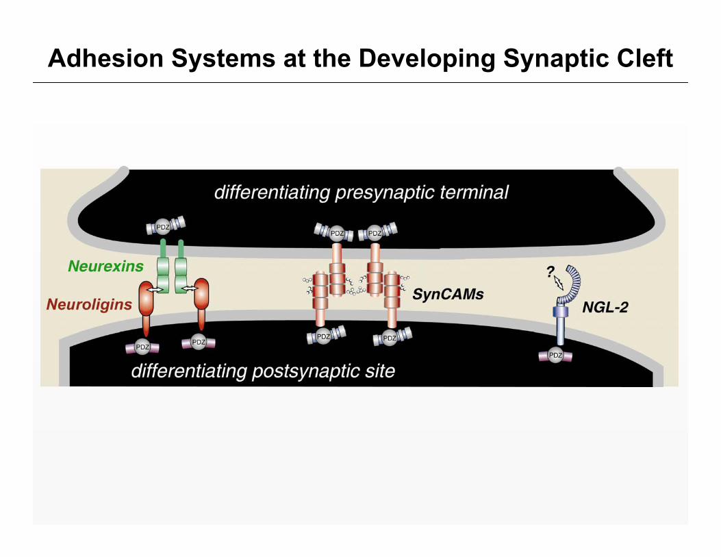
Adhesion Systems at the Developing Synaptic Cleft

SynCAM 1 Mediates Homophilic Adhesion
red: SynCAM 1-cherrygreen: soluble GFP
Massimiliano Stagi
heterologous expression of SynCAM 1 in COS cells

SynCAM 1 Recruits Scaffolding Molecules to Sites of Homophilic Adhesion
heterologousexpression in COS cells
Massimiliano Stagi
green:GFP-CASK
green:GFP-CASK
red:SynCAM 1-cherry
overlay

SynCAM 1 Potentiates Excitatory Transmission in Hippocampal Neurons
Yildirim Sara and Ege Kavalali

Induction of Synaptic Specializations in Co-Cultures
epithelial-like HEK293cells expressing bothSynCAM 1 and ECFPare seeded atophippocampal neurons
after 1-2 days in vitro,the co-cultures are analyzed for the formation of specializations containing presynaptic markers on the surface of the HEK293 cells

SynCAM 1 Induces Presynaptic Specializations
red synaptophysin

SynCAM 1 and Neuroligin Induce Presynaptic Terminals with Functional SV Recycling

Reconstitution of Synaptic Transmission with SynCAM 1 and GluR2
Yildirim Sara and Ege Kavalali

Sequence Alignment of SynCAM Family Members

All Four SynCAM Family Members are Transcribedin the Developing and Adult Brain
real-timeRT-PCRstandardized to actin
Mike Akins

SynCAM Family Expression in Brain
in situ hybridizations at P15:
Mike Akins
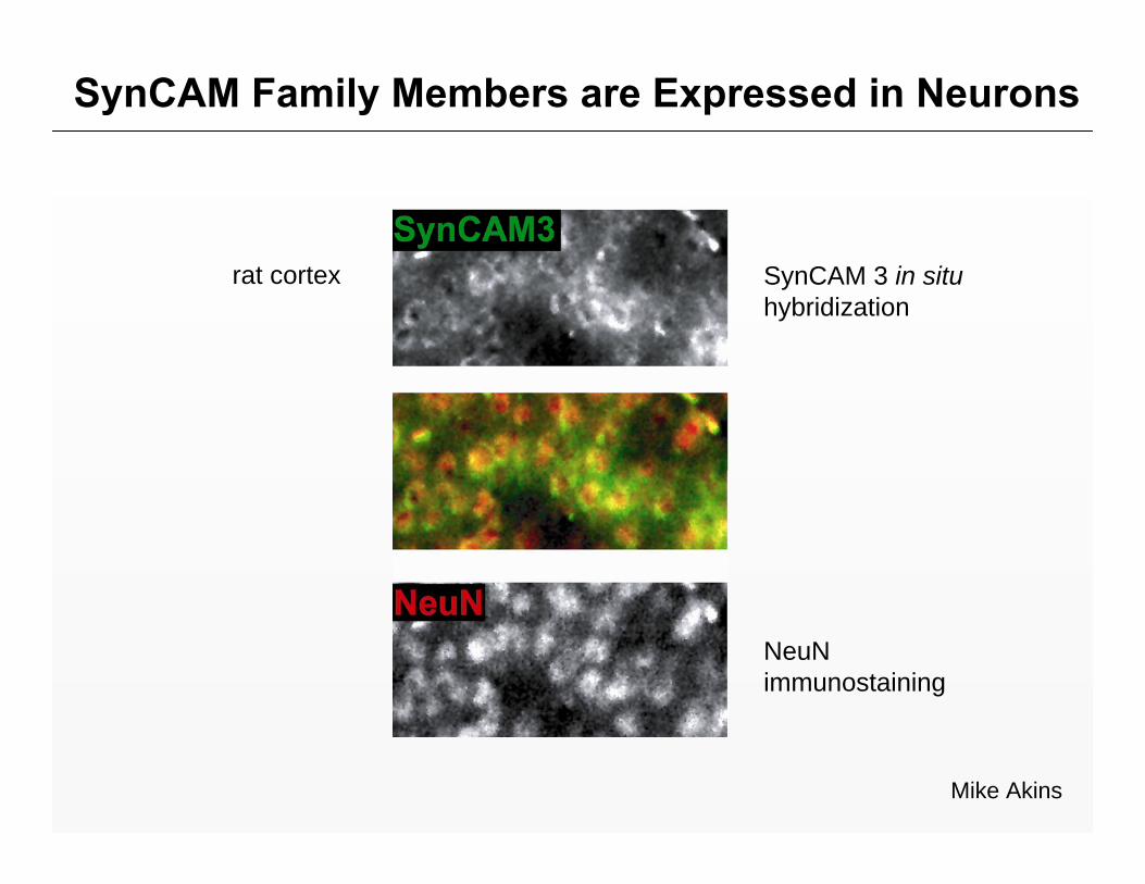
SynCAM Family Members are Expressed in Neurons
Mike Akins
SynCAM 3 in situ hybridization
NeuNimmunostaining
rat cortex

SynCAM Antibody Specificity
Adam Fogel
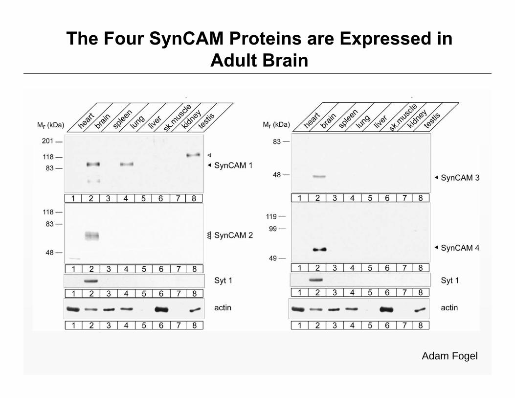
The Four SynCAM Proteins are Expressed in Adult Brain
Adam Fogel

The Developmental Profile of SynCAM Proteins Correlates with Synaptogenesis
Adam Fogel
rat brain preparations:

SynCAMs Fractionate asSynaptic Plasma Membrane Proteins
rat forebrain P9

SynCAM Proteins are Prominent Components ofSynaptic Plasma Membranes
Adam Fogel
SynCAM 1 1/180
SynCAM 2 1/300
SynCAM 3 1/1200
fraction of total SPM protein in P15 forebrain

SynCAM Proteins Can Function as HomophilicAdhesion Molecules
SynCAM 1, 2and 3 interact homophilically
no evidence for string homophilic SynCAM 4 interactions
SynCAM-ECD coated beads
control beads
Mike Akins

SynCAM Proteins Can Engage in SpecificHeterophilic Interactions
Mike Akins

SynCAM Proteins Can Engage in SpecificHeterophilic Interactions
Adam Fogel

Distinct SynCAM Expression in Hippocampus
in situ hybridizations at P15:
Mike Akins

SynCAM 4 Induces Presynaptic Specializations
Mike Akins
**p < 0.01

Synaptic Adhesion and Synaptogenesis: A Model

Acknowledgements
Mike AkinsMassimiliano StagiAdam FogelElissa RobbinsLisa ThomasYuling Lei
Ege T. KavalaliYildirim Sara
Thomas C. Südhof
Funding Support:NIH/NIDA RO1 DA018928, March of Dimes Foundation and The Brain Tumor Society